Embed Size (px)
Citation preview
�������� ����� ��
The core promoter: at the heart of gene expression
Yehuda M. Danino, Dan Even, Diana Ideses, Tamar Juven-Gershon
PII: S1874-9399(15)00090-5DOI: doi: 10.1016/j.bbagrm.2015.04.003Reference: BBAGRM 874
To appear in: BBA - Gene Regulatory Mechanisms
Received date: 17 February 2015Revised date: 19 April 2015Accepted date: 23 April 2015
Please cite this article as: Yehuda M. Danino, Dan Even, Diana Ideses, Tamar Juven-Gershon, The core promoter: at the heart of gene expression, BBA - Gene RegulatoryMechanisms (2015), doi: 10.1016/j.bbagrm.2015.04.003
This is a PDF file of an unedited manuscript that has been accepted for publication.As a service to our customers we are providing this early version of the manuscript.The manuscript will undergo copyediting, typesetting, and review of the resulting proofbefore it is published in its final form. Please note that during the production processerrors may be discovered which could affect the content, and all legal disclaimers thatapply to the journal pertain.

ACC
EPTE
D M
ANU
SCR
IPT
ACCEPTED MANUSCRIPT
The core promoter: at the heart of gene expression
Yehuda M. Danino1, Dan Even1, Diana Ideses1 and Tamar Juven-Gershon1*
1The Mina and Everard Goodman Faculty of Life Sciences, Bar-Ilan University,
Ramat Gan 5290002, Israel
Running title: The core promoter: a central player in gene expression
Key words: core promoter; RNA Pol II transcription; core promoter elements/motifs;
enhancer-promoter specificity; core promoter preferential activation; gene expression
The authors declare that there are no potential conflicts of interest.
* To whom correspondence should be addressed. Tel: +972-3-531-8244; Fax: +972-
3-738-4058; Email: [email protected]

ACC
EPTE
D M
ANU
SCR
IPT
ACCEPTED MANUSCRIPT
2
ABSTRACT
The identities of different cells and tissues in multicellular organisms are determined
by tightly controlled transcriptional programs that enable accurate gene expression.
The mechanisms that regulate gene expression comprise diverse multiplayer
molecular circuits of multiple dedicated components. The RNA polymerase II (Pol II)
core promoter establishes the center of this spatiotemporally orchestrated molecular
machine. Here, we discuss transcription initiation, diversity in core promoter
composition, interactions of the basal transcription machinery with the core promoter,
enhancer-promoter specificity, core promoter-preferential activation, enhancer RNAs,
Pol II pausing, transcription termination, Pol II recycling and translation. We further
discuss recent findings indicating that promoters and enhancers share similar
features and may not substantially differ from each other, as previously assumed.
Taken together, we review a broad spectrum of studies that highlight the importance
of the core promoter and its pivotal role in the regulation of metazoan gene
expression and suggest future research directions and challenges.

ACC
EPTE
D M
ANU
SCR
IPT
ACCEPTED MANUSCRIPT
3
Introduction
Appropriate temporal and spatial gene expression is a highly complex process
underlying the fate and function of different cells and tissues. The regulation of this
process is composed of multiple levels and orchestrated molecular events [1-3]. A
central event in the regulation of eukaryotic gene expression is the initiation of
transcription. The initiation of transcription of protein-coding genes and distinct non-
coding RNAs occurs following the recruitment of RNA polymerase II (Pol II) to the
core promoter region by the basal transcription machinery [4].
The core promoter is generally defined as the minimal DNA sequence that directs
accurate initiation of transcription. The core promoter sequence encompasses the
transcription start site (TSS), typically referred to as the +1 position [5, 6].
Examination of the distribution of TSSs reveals that there are multiple modes of
transcription initiation (Fig. 1A). Distinct molecular players can open the chromatin
structure at the core promoter region and thus facilitate initiation of transcription.
Interestingly, active promoters are associated with specific chromatin signatures.
These include: nucleosome-depleted regions (NDR) or reduced nucleosome
occupancy over the promoters, DNaseI hypersensitive sites (DHS) and the
enrichment of specific histone modifications, such as di- and tri-methylation of H3K4
and acetylation of H3K4 and H3K27 (Fig. 1B) [7, 8]. Notably, both human and yeast
nucleosomes that are upstream and downstream of the TSSs of multiple genes have
been shown to correlate with the occupancy of the histone variant H2A.Z (termed
Htz1 in yeast) (reviewed in [9]).
In the past, it was assumed that the core promoter is a generic entity that functions in
a universal manner. Nowadays however, the growing convention is that the unique

ACC
EPTE
D M
ANU
SCR
IPT
ACCEPTED MANUSCRIPT
4
properties of a given promoter are a function of its architecture and core promoter
motifs composition (Fig. 1C and D) [5, 6, 10, 11].
The core promoter, which is often referred to as “the gateway to transcription”, is a
central component in the initiation of transcription [12, 13]. Research in the past
decade has enhanced our understanding of the fundamental roles that the core
promoter plays in the initiation of transcription, as well as in the regulation of
additional aspects of gene expression. Insights are gained from studies of specific
genes and gene networks [13-15], as well as from genome-wide studies [11, 16]
utilizing methodologies such as PEAT [17], 5' RACE [18], CAGE [19], FAIRE-seq
[20], ChIP-seq [21], Gro-seq [22], and RNA-seq [23], and key projects and consortia
(e.g. modENCODE [24], ENCODE [25] and FANTOM5 [26]), which developed
following the implementation of some of the above methods. Accordingly, core
promoters can be studied at different resolutions: from genomic architecture,
transcription co-regulators and sequence-specific transcription factors (Fig. 2A),
through basal transcription factors (Fig. 2B and C) and DNA sequence motifs (Fig.
2C). Importantly, the different experimental strategies complement each other and
together, provide the elaborate view of core promoters. Here, we review the current
state of knowledge relevant to the contribution of the core promoter to multiple
aspects of gene expression, and discuss future directions and challenges in the field.
1. Diversity in the transcription initiation landscape
1.1. Multiple modes of transcription initiation
The core promoter is best known for its role in directing proper transcription initiation
at the TSS. Two modes of transcription initiation, focused and dispersed, were

ACC
EPTE
D M
ANU
SCR
IPT
ACCEPTED MANUSCRIPT
5
previously noted in metazoan (Fig. 1A) (reviewed in [6, 11]). Focused (also termed
“sharp peak”) promoters contain a single predominant TSS or a few TSSs within a
narrow region of several nucleotides [10]. Focused promoters encompass
approximately between -40 to +40 nucleotides relative to the TSS (referred to as the
+1 position). Focused transcription initiation is associated with spatiotemporally
regulated tissue specific genes [27] and with canonical core promoter elements that
have a positional bias, such as the TATA box, Initiator, MTE and DPE [28] (Fig. 1C).
Dispersed (also termed “broad”) promoters contain multiple weak start sites that
spread over 50 to 100 nucleotides at the promoter region ([10, 11] and refs therein).
Dispersed transcription initiation is associated with constitutive or housekeeping
genes. Vertebrate dispersed promoters often contain CpG islands and Sp1 and NF-Y
sites [6, 10, 29] whereas Drosophila core promoters often contain elements that have
weaker positional biases (as compared to the focused promoters), but frequently co-
occur in a specific order and orientation: Ohler 1, DNA replication element (DRE),
Ohler 6 and Ohler 7 [28, 30] (Fig. 1D). Although the focused promoter architecture
exists in all the organisms and is the predominant initiation mode in simpler
organisms, the dispersed mode is more common in higher eukaryotes [10, 27]. For
example, over 70% of vertebrate promoters are dispersed [29, 31-33]. From a
teleological standpoint, the associations of sharp TSSs with regulated genes and of
broad TSSs patterns with constitutively expressed genes are rather intuitive. It would
be easier to achieve a more precise control of gene expression from focused TSSs,
as compared with dispersed promoters of housekeeping genes, which would be
constitutively transcribed with minimal variation of gene expression by usage of
multiple start sites [10].
1.2. Focused versus Dispersed initiation patterns - recent studies, new insights

ACC
EPTE
D M
ANU
SCR
IPT
ACCEPTED MANUSCRIPT
6
Despite the abovementioned distinction between the two modes of transcription
initiation, classification of transcription initiation landscapes is not so straightforward.
Functional experiments and genome-wide studies using advanced technologies imply
that there are multiple ways to classify promoters. Thus, the boundaries between
these two major types of promoters are sometimes unclear [6, 34]. With respect to the
“focused vs. dispersed” sub-classifications mentioned above, a mixed promoter (also
termed “broad with peak”; [17]), an additional promoter type, was revealed. This
promoter type exhibits a dispersed initiation pattern with a single strong transcription
start site [6, 35] (Fig. 1A). Several studies classified mammalian promoters using
alternative criteria [27, 29, 33]. The Ren Lab classified active promoters based on
genome-wide ChIP experiments for TFIID and Pol II, as well as H3Ac and H3K4me,
regardless of focused or dispersed initiation patterns [33]. Bajic et. al. [29] define four
promoter types, based on distribution of dinucleotides over the promoter regions,
CpG Islands and TATA boxes. Moreover, Carninci et. al. [27] classified promoters into
four groups based on CAGE analysis: single peak, broad shape peak,
bimodal/multimodal peak and broad with dominant peak. These studies also
challenge the “focused vs. dispersed” classification, as some mouse and human
promoters contain both CpG Islands and TATA boxes. A recent comprehensive
review [11], which compared genome-wide studies in human and Drosophila,
presented another sub-classification of three major types of promoters termed Type I,
Type II and Type III. Type I promoters contain TATA boxes and focused TSSs, lack
CpG islands and are associated with tissue-specific expression in adult tissues. Type
II promoters contain CpG islands and dispersed TSSs. In mammals, type II promoters
lack TATA boxes, and in Drosophila they contain DRE, Ohler 1 or Ohler 6 motifs.
Genes belonging to this group are associated with broad expression throughout the

ACC
EPTE
D M
ANU
SCR
IPT
ACCEPTED MANUSCRIPT
7
organism's life. Type III promoters are associated with developmentally regulated
genes, which in Drosophila contain combinations of Initiator and DPE motifs. In
mammals, type III promoters contain large CpG islands.
Taken together, the transcriptional initiation landscape is more complex than the
simple classification of two types of promoters.
1.3. Bidirectional and divergent transcription
Another manifestation of the complexity of transcription initiation is the phenomenon
of bidirectional transcription. Bidirectional transcription, which presents two closely
spaced transcription initiation events (within less than 1kb) of head-to-head Pol II
transcripts in both sense and anti-sense orientations, was originally defined for
adjacent head-to-head oriented pairs of protein-coding genes [36]. The relatively
short region that contains the opposite-oriented initiations and separates between
these genes, is often called a “bidirectional promoter” [37]. Experimental and
computational studies have characterized many features of bidirectional promoters. In
general, it is shown that 10%-22% of the genes in mammals are organized in this
manner [38]. Moreover, the bidirectionality was shown to be controlled in a cell-type
specific manner, and these pairs of genes are coordinately regulated ([38] and refs
therein). Hence, bidirectional promoters might have evolved to facilitate the regulation
of transcription of different genes at the same time, and might consist of two separate,
yet dependent, core promoters. Additionally, a computational analysis supports an
evolutionary role for bidirectional promoters in the emergence of novel species-
specific transcripts [39]. Bioinformatics analysis of the distribution of common core
promoter elements (BREu, TATA box, Inr and DPE) and CpG islands at bidirectional
versus unidirectional promoters, demonstrated that while the BREu is enriched at
ACC
EPTE
D M
ANU
SCR
IPT
ACCEPTED MANUSCRIPT
8
bidirectional promoters, the Inr and DPE elements are similarly detected at both
promoter types [40]. The TATA box is rare in general, but is enriched in bidirectional
promoters of histone genes. Moreover, it was shown that the CpG islands and Sp1
binding sites are common features of most of the bidirectional promoters, compared
to unidirectional promoters [41]. Other studies focused on overrepresented binding-
sites of different transcription factors, and in some cases - on their influence on the
expression of two opposite genes regulated by a bidirectional promoter [38, 42].
Interestingly, another manifestation of bidirectional transcription involving non-
coding RNAs (ncRNAs) was recently characterized. Multiple classes of ncRNAs were
identified in different organisms (reviewed in [43]). One of these classes is promoter-
associated ncRNAs. During the years, classes of promoter-associated non-coding
transcripts were discovered in bacteria, yeast, Drosophila, mouse, human and plants
([43-45] and refs therein). Four studies, published back-to-back in 2008, described
new classes of promoter-associated ncRNAs in humans and mice [22, 46-49]. These
ncRNAs were generally divided into two classes, termed TSS-associated RNAs
(TSSa-RNAs) [48] and promoter upstream transcripts (PROMPTs) [47] or upstream
antisense RNAs (uaRNAs) [50], which share many features. They are short, present
at low abundance and are associated with CpG islands and active-promoter-related
histone marks (H3K4me3, H3ac), but not with elongation-related histone marks
(H3K36me3, H3K79me3).
Non-coding antisense RNAs derived from bidirectional promoters have very short
half-lives and are barely detectable. Two recent studies have shown that an
asymmetric distribution of polyadenylation signals and U1 snRNP-binding sites
surrounding TSSs control transcript stability [50-52]. Notably, bidirectional initiation is
also a feature of enhancer RNAs (eRNA; see section 7) [53, 54].

ACC
EPTE
D M
ANU
SCR
IPT
ACCEPTED MANUSCRIPT
9
The Lis lab has demonstrated that nearly 80% of active genes have bidirectional
promoters, suggesting that bidirectional initiation is a general feature of mammalian
genomes [22, 55]. Hence, these divergent ncRNAs may be regarded as markers for
active promoters of protein-coding genes [22, 46-48, 56]. Duttke et al. have recently
analyzed transcription from human promoters in HeLa cells and have classified
promoters into three types: unidirectional promoters, divergent promoters (containing
an annotated gene in the forward direction and no annotated gene in the reverse
direction) and bidirectional promoters (containing annotated genes in both directions)
[57]. Surprisingly, they discovered that about half of human active promoters are
intrinsically unidirectional. Moreover, the divergent transcripts result from their own
reverse-oriented core promoters. The authors suggest that divergent transcription is
not an inherent property of the transcription process, but a consequence of the
presence of both forward and reverse-directed promoters. This suggestion is in line
with the two occupancy peaks observed for each TBP and Pol II by the Lis lab [55].
The Lis lab observed tight spacing (estimated 110 bp) between the forward and
reverse-directed promoters [55], whereas the Ohler & Kadonaga labs, observed
variable, however larger, spacing between the two [57]. It remains to be determined
whether the difference between these findings results from the differences between
the different cell lines used or from the analysis methodology.
Despite the impressive discoveries related to bidirectional transcription in the last
few years (which highlight the complexity of gene expression), the functional role of
short non-coding antisense RNAs still remains elusive. From this point onwards, we
only refer to the comprehensively studied focused and dispersed core promoter
types.
ACC
EPTE
D M
ANU
SCR
IPT
ACCEPTED MANUSCRIPT
10
2. Core promoter elements: the combinatorial code of precise transcription
initiation
The Pol II core promoter is composed of short DNA sequences that are referred to as
core promoter elements or motifs. The majority of core promoter motifs serve as
binding sites for components of the basal transcription machinery, in particular TFIID,
which is composed of TATA box-binding protein (TBP) and TBP-associated factors
(TAFs), and TFIIB [4, 58, 59].
The basal transcription machinery recruits Pol II to the core promoter that directs
the initiation of transcription [4, 6, 10, 60-62]. Nevertheless, there are no universal
core promoter elements, and diverse core promoter compositions have been reported
[6, 63]. In this section, we will briefly discuss the majority of core promoter elements
(schematically depicted in Fig. 1C and D), which have been analyzed in Drosophila
and mammals, with particular emphasis on their variety and the relations between
them.
2.1. The precisely positioned core promoter elements are common in the focused
promoters
Early studies from the Chambon lab described the existence of a putative element at
the TSS [64]. The function of the initiator (Inr) as a transcriptional element that
encompasses the +1 TSS was articulated by Smale and Baltimore [65]. The Inr is
probably the most prevalent core promoter motif in focused core promoters [66-68]. It
is mainly bound by the TAF1 and TAF2 subunits of TFIID [69-72]. The mammalian Inr
consensus sequence is YYA+1NWYY (IUPAC nomenclature) [73], and the Drosophila
consensus is TCA+1KTY [71, 74]. Inr-like sequences were also identified in
Saccharomyces cerevisiae [75]. Computational analyses of promoters argue that the

ACC
EPTE
D M
ANU
SCR
IPT
ACCEPTED MANUSCRIPT
11
Inr consensus is only YR (-1, +1 positions) in humans [11, 27, 76] or TCA+1GTY for
Drosophila [66, 68]. The A nucleotide (or R in the YR consensus) is generally
designated as the +1 position, even when transcription does not initiate at this
specific nucleotide. This critical convention is instrumental, because functional
downstream elements are completely dependent on the presence of an Inr and the
precise spacing from it [6, 10, 13].
Notably, a strict version of the mammalian initiator (sINR), which is present in 1.5%
of human genes and enriched in TATA-less promoters of specific functional
categories, was defined as CCA+1TYTT, with conserved sequences flanking the motif
[77]. The sINR motif functions in cooperation with Sp1 and can replace the
conventional Inr, but not vice versa. Similarly to the canonical Inr element, sINR is
bound by TAF1 and its function depends on it [77]. The YY1 transcription factor binds
sINR, but this binding is dispensable for sINR function [77].
In addition to these versions of the Inr, a few core elements that encompass the
transcription start site were identified. The polypyrimidine initiator motif (TCT), which
was originally identified in mouse, is conserved from Drosophila to humans [14, 78-
80]. The TCT has a consensus sequence of YYC+1TTTYY in Drosophila and
YC+1TYTYY in humans, in which C is the +1 TSS. Although the Inr consensus
resembles the TCT consensus, the TCT motif cannot substitute for an Inr to initiate
transcription [14]. The TCT overlaps with a motif that was previously identified in
humans, termed 5'-terminal oligopyrimidine tract (5'-TOP) (reviewed in [81]), which is
functionally distinct from it [14]. Both the TCT and the 5‟-TOP elements are enriched
and are functional in the transcription of ribosomal protein genes and proteins
involved in the regulation of translation [14, 78].

ACC
EPTE
D M
ANU
SCR
IPT
ACCEPTED MANUSCRIPT
12
Two additional core promoter motifs that are located around TSSs were originally
identified in the hepatitis B virus X gene promoter, which contains two TSSs. The X
gene core promoter element 1 (XCPE1) drives Pol II transcription from the first TSS
of the X gene promoter as well as from other human promoters, when accompanied
by co-activator sites. XCPE1 is found in ~1% of the human genes (particularly TATA-
less genes) and its consensus sequence DSGYGGRAS+1M spans positions -8 to +2
relative to the TSS [82]. Unlike XCPE1, The X gene core promoter element 2
(XCPE2) is sufficient to drive Pol II transcription by itself. The XCPE2 directs
transcription from the second TSS of the X gene mRNA, but it also drives
transcription from additional human promoters, in a TAF-free manner. Its consensus
sequence VCYCRTTRCM+1Y spans positions -9 to +2 relative to the TSS [83].
There are core promoter elements that are located upstream of the TSS. The
TATA box motif is the first core promoter motif to be identified [84]. Although the
TATA box was previously considered to be a universal element, it is presently
estimated that only 8%-30% of metazoan core promoters [27, 33, 60, 68, 85] and
20%-46% yeast promoters [62, 86, 87] are TATA-dependent. The TATA box motif is
also present in plants [88, 89], however the majority of Arabidopsis promoters are
TATA-less [90]. The TATA box is bound by the TBP subunit of TFIID ([5, 6, 63] and
refs therein). Both the TATA box element and the TBP are conserved from archae to
humans [10, 91]. The consensus sequence of the TATA box is TATAWAAR, where
the 5' T is usually located at -30 or -31 relative to the TSS in metazoans (or at -120 to
-40 in yeast). A wide range of sequences can functionally replace the yeast TATA box
for in vivo transcriptional activity [92]. Notably, transcription initiation in yeast, which
occurs at variable and greater distances downstream of core promoter elements such
as the TATA box, has been suggested to occur via Pol II scanning, where a PIC
ACC
EPTE
D M
ANU
SCR
IPT
ACCEPTED MANUSCRIPT
13
assembles at the TATA box and Pol II translocates downstream, searching the DNA
for suitable start sites (reviewed in [93]). Interestingly, the TATA box has recently
been shown to contribute to high rates of transcription re-initiation of human
microRNAs, resulting in reduced microRNA levels [94]. The authors suggested that
the lower synthesis rates directed by the TATA box result from Pol II crowding at
pause sites, thus increasing the chance for premature termination.
The TFIIB recognition elements (BRE), which are bound by the TFIIB basal
transcription factor, are located immediately upstream or downstream of the TATA
box, respectively [95-97]. TFIIB contacts these two elements by two independent
DNA-recognition motifs within its core domain [96]. The consensus of the upstream
BRE (BREu) is SSRCGCC [97], and the consensus of the downstream BRE (BREd) is
RTDKKKK [95]. The TFIIB and the BRE elements are conserved from archae to
humans [6, 96]. Both BREu and BREd act in conjunction with the TATA box [6, 10]. A
bioinformatics analysis using the EPD database showed that 25% of the eukaryotic
core promoters contain a potential BREu [85]. Surprisingly, this study revealed that the
BREu is more prevalent in TATA-less promoters (28.1%) than in TATA-containing
promoters (11.8%). Both elements exert positive as well as negative effects on basal
transcription and on activated transcription in a manner that is context-dependent [95,
97-99].
In addition to the abovementioned upstream elements there are core promoter
elements that are located downstream of the TSS. The downstream core promoter
element (DPE), which was discovered as a TFIID recognition site that is downstream
of the Inr, is precisely located at +28 to +33 relative to the A+1 of the Inr, with a
functional range set of DSWYVY [100-102]. In addition to this functional range set,
the guanine at +24 was shown to contribute to DPE function [102]. The DPE is

ACC
EPTE
D M
ANU
SCR
IPT
ACCEPTED MANUSCRIPT
14
prevalent in developmental gene networks [11, 15, 99, 103]. Importantly, a recent
study provides in vivo evidence that expression driven by the homeotic Antennapedia
P2 promoter during Drosophila embryogenesis is dependent on the DPE [103]. The
motif ten element (MTE) was identified as an overrepresented core promoter
sequence, which is located immediately upstream of the DPE, encompassing
positions +18 to +29 relative to the A+1 of the Inr [68]. As positions +28 to +29 overlap
the DPE, the MTE consensus sequence was defined for positions +18 to +27
(CSARCSSAAC) [104]. Although the majority of the MTE-containing promoters
contain a DPE, the MTE motif functions independently of the DPE [104, 105]. Both
the MTE and DPE serve as recognition sites for TFIID and appear to be in close
proximity to TAF6 and TAF9 [101, 105]. Interestingly, TAF1 and TAF2 may also
contribute to the recognition of downstream core promoter elements [70]. Using
single-nucleotide substitution analysis, the MTE and DPE together were found to
consist of three functional sub-regions: positions 18-22, 27-29 and 30-33 downstream
to the A+1 of the Inr. The bridge configuration, which includes the first and the third
functional sub-regions (bridge I, positions 18-22 with favored nucleotides CSARC;
bridge II, positions 30-33 with favored nucleotides WYVY), was shown to be a
naturally rare but functional core promoter element [105]. Both the MTE and DPE are
conserved from Drosophila to humans [6, 100, 101, 104-108]. The MTE, DPE and
Bridge motifs are exclusively dependent on the presence of a functional Inr, and are
enriched in TATA-less promoters. However, co-occurrence of putative TATA, Inr and
DPE motifs was observed in a small fraction of Drosophila genes [15, 85].
An additional downstream element was identified and characterized in the human
adult β-globin promoter. This element, termed downstream core element (DCE), was
detected by scanning mutagenesis of the +10 to +45 in the promoter region. The DCE

ACC
EPTE
D M
ANU
SCR
IPT
ACCEPTED MANUSCRIPT
15
is composed of three sub-elements, located at positions +6 to +11 (necessary motif
CTTC), +16 to +21 (necessary motif CTGT), and +30 to +34 (necessary motif AGC)
relative to the TSS. The DCE is distinct from the MTE, DPE and Bridge downstream
elements, as the DCE is recognized and bound by TAF1 [109] and not by TAF6 or
TAF9 [101, 105]. Moreover, unlike the DPE, the DCE is frequently found in TATA
box-containing promoters [109, 110]. Table 1 summarizes the precisely positioned
core promoter elements found in focused promoters and the proteins that bind them.
2.2. Core promoter elements with weak positional biases in dispersed promoters
Even though the vast majority of core promoter elements are precisely located in
focused promoters, there are still a few variably located motifs that were also
identified in dispersed promoters. These variably located elements, like some of the
precisely located elements discussed above, are associated with specific gene
groups.
As mentioned, there are sequence motifs such as the DNA-replicated-related
element (DRE) and Ohler 1, 6 and 7 motifs, which were detected by a computational
analysis as commonly expressed in dispersed promoters of Drosophila genes with
maternally inherited transcripts [28]. The consensus sequences of the DRE, Ohler 1,
6 and 7 motifs are WATCGATW, YGGTCACACTR, KTYRGTATWTTT and
KNNCAKCNCTRNY, respectively [68]. The DRE is a target of the DNA replication-
related-element binding factor (DREF). DREF, which was discovered in Drosophila
and was later found to have orthologues in many other species (including humans), is
involved in transcriptional regulation of proliferation-related genes [111]. A motif 1
binding protein (M1BP) has recently been identified and the enrichment of Motif 1 and

ACC
EPTE
D M
ANU
SCR
IPT
ACCEPTED MANUSCRIPT
16
M1BP was implicated in cytoskeletal organization, mitotic cell cycle and metabolism
[112].
2.3. The interplay between core promoter elements
With the notion that there are no universal core promoter elements and that core
promoter elements are a very important feature of regulation of gene expression,
many studies examined the combinations between core promoter elements such as:
Inr, TATA box, BREu, BREd, MTE and DPE, and their effects on the transcriptional
output. For example, the BRE elements were originally characterized as functional
elements with conjugation to TATA box. In this context, both the BREu and the BREd
either increase or decrease the levels of basal transcription [95, 97, 98, 113]. Notably,
the addition of a BREu element to a core promoter of a Caudal target gene has a
differential effect on transcription in a TATA box- or DPE- context [99]. The TATA box
and the Inr cooperate, in certain cases, as synergistic elements [114]. An antagonistic
behavior was demonstrated between TBP, which activates TATA transcription and
inhibits DPE transcription, and NC2 and Mot1, which activate DPE transcription by
inhibiting the function of TBP [115].
The functionality of the DPE, MTE and Bridge elements is, by definition, dependent
on their precise location relative to the Inr [100, 101, 104, 105]. Synergy was
observed between the MTE and DPE, as well as between the MTE and TATA box
[104]. Based on these relationships, a synthetic core promoter, termed super core
promoter (SCP), containing a TATA box, Inr, MTE and DPE was designed.
Remarkably, the SCP is stronger than any of the natural core promoters examined
[116].
Collectively, these findings indicate that the levels of gene expression can be
modulated by the core promoter composition. Such modulation is directly achieved by

ACC
EPTE
D M
ANU
SCR
IPT
ACCEPTED MANUSCRIPT
17
the impact of the combinations of core promoter elements on the architecture of the
basal transcription machinery, which provides an additional level of transcriptional
regulation. The core promoter may have diversified during evolution so that each
element may work with the other, depending on the context and organism. Hence,
simple categorization may disregard the complexity of gene expression.
3. Functional and structural insights regarding the role of the core promoter
in the assembly of the Pol II transcription machinery
In this section, we describe the assembly of the basal transcription machinery
components (primarily based on the analysis of TATA-dependent promoters) and
their distinct roles in specific cellular contexts.
3.1. Terminology change: from “general” to “basal” transcription machinery
Classic biochemical studies performed over 30 years ago using the TATA box-
containing adenovirus major late promoter identified the general transcription factors
(GTFs) as accessory factors for accurate Pol II transcription initiation [117, 118]. The
GTFs were named TFIIA, TFIIB, TFIID, TFIIE, TFIIF and TFIIH, based on the protein
fractions they purified in (reviewed in [4]) . These components, together with Pol II,
were necessary and sufficient for basal transcription of the adenovirus major late
promoter. They assemble into the preinitiation complex (PIC) by protein-protein
interactions and by mediating core promoter recognition (Fig. 2B).
In the past, it was generally accepted that the PIC composition of GTFs does not
vary between promoters with different core promoter architecture, and the PIC is
nucleated by the binding of the TBP subunit of TFIID, which binds the TATA box [119]
(reviewed in [4, 31]). Traditionally, this simple model has been considered “general”.

ACC
EPTE
D M
ANU
SCR
IPT
ACCEPTED MANUSCRIPT
18
However, due to the diversity in core promoter composition and the realization that
the known GTFs are insufficient to transcribe DPE-containing promoters [120], it is
suggested that the GTFs do not function in a “general” manner, and different
compositions of PIC exist. Indeed, the non-ubiquitous expression pattern of certain
TAFs imply that they cannot be PIC components in every cell type [58]. Moreover,
many studies have presented the variability in PIC formation, specifically by the
molecular flexibility in TFIID composition. Hence, GTFs should be addressed as
“basal” rather than “general” transcription factors (also discussed in [58, 121-123]).
3.2. Compatibility between PIC components, related factors and core promoter
elements
Undoubtedly, the diverse assembly of the basal transcription factors, as well as the
diversity of core promoter elements, is a complex subject, both structurally and
functionally. Nevertheless, due to this complexity, the PIC, which is pivotal for core
promoter recognition ([58, 121, 124] and refs therein), can assemble at core
promoters with varying compositions and regulate Pol II transcription in different cells
and organisms. In agreement with that, requirements for a “match” between the PIC
and the core promoter have been observed in recent years.
This compatibility has mainly been reflected in studies addressing the flexibility and
modularity of TFIID subunits and the entire TFIID complex. Early footprinting assays
detected differential TFIID protection patterns with respect to the presence of a TATA
box and BRE in mammalian promoters [125, 126], and a DPE in Drosophila [101].
These studies and others [127] have demonstrated the important roles of TAFs in the
assembly of the PIC, and hence, in the transcription process. As mentioned earlier,
sub-modules of TFIID bind specific core promoter elements, e.g. TBP binds the TATA
box, TAF1/TAF2 bind the Inr, TAF1 binds the DCE and TAF6/9 bind the DPE and the

ACC
EPTE
D M
ANU
SCR
IPT
ACCEPTED MANUSCRIPT
19
MTE (Fig. 2C) [69-71, 100, 101, 104, 107, 109]. It is noteworthy that TAF4/TAF12 and
TAF4b/TAF12 sub-complexes can also bind core promoters [107], and are necessary
for transcription of a sub-group of genes, which are mostly associated with TATA box
and Inr motifs [128]. Interestingly, TAF1 contains two distinct enzymatic activities: an
acetyl-transferase and a kinase activity, which are important for regulating non-
overlapping, different gene sets in vivo [129], suggesting that different functional
modules of the PIC contribute to transcription of different target genes.
While TBP and TAF1 were initially considered the nucleating subunits of holo-
TFIID assembly [130], Wright et. al. [131] discovered that Drosophila TAF4
preferentially nucleates TFIID in TATA-less, DPE-containing promoters. This study
also uncovered a stable core-sub-complex, composed of TAF5 and the histone fold
domain (HFD)-containing TAF4, TAF6, TAF9 and TAF12. This core sub-complex is
associated with the peripheral subunits TAF1, TAF2, TAF11 and TBP. These core
TAFs are incorporated into TFIID in two copies, and are organized in five heterodimer
pairs with other HFD-containing TAFs (TAF3-TAF10, TAF6-TAF9, TAF4-TAF12,
TAF8-TAF10 and TAF11-TAF13) ([124] and refs therein). Recent structural analysis
of human TFIID demonstrated that these core TAFs exhibit two-fold symmetry [132].
Interestingly, incorporation of the TAF8-TAF10 pair breaks the symmetry and allows
the entry of the single copy TAFs and TBP into the structure, resulting in an
asymmetric holo-TFIID that can nucleate the PIC.

ACC
EPTE
D M
ANU
SCR
IPT
ACCEPTED MANUSCRIPT
20
Several TBP-free complexes have been characterized [127, 133, 134]. One of
them, the TBP-free TAF-containing complex (TFTC; also termed SAGA, STAGA and
PCAF), is capable of replacing the canonical TFIID at both TATA-less and TATA-
containing promoters in vitro [127]. The assembly of TAF-less TBP-containing
complexes (such as TBP-TFIIA-containing complexes) at specific core promoters,
which was somewhat surprising, has also been observed [135-137]. A TAF-free TBP-
containing PIC is important for transcription from HIV-1 LTR promoter [136].
Interestingly, a distinctive TBP-TAF complex, lacking TAF1, TAF4 and TAF10, is
involved in transcription of the U2 snRNA gene [138].
These findings add to a growing body of evidence implying that distinct core
promoters would be differentially recognized by PICs that contain TBP or are devoid
of it. Notably, TBP activates TATA-dependent transcription and represses DPE-
dependent transcription, whereas Mot1 and NC2 block TBP function and thus repress
TATA-dependent transcription and activate DPE-dependent transcription [115, 139].
Interestingly, Deng et. al. [140] demonstrated that NC2 acts positively at promoters
that lack functional BREs, while TFIIA recruitment, which is dependent on the
presence of BREs, reduces transcriptional activity. The association of BRE elements
with TATA boxes further supports these findings [85, 97]. Interestingly, the
architectural DNA-binding protein HMGA1 has been shown to interact with the
Mediator and activate transcription of mammalian promoters containing both a TATA
box and an Inr [141].
Remarkably, the Nogales lab used electron microscopy to visualize human TFIID
with promoter DNA, and discovered that TFIID exists in two structurally distinct
conformations (termed canonical and rearranged) [142]. The transition between the
two states is modulated by TFIIA, and the presence of TFIIA and promoter DNA

ACC
EPTE
D M
ANU
SCR
IPT
ACCEPTED MANUSCRIPT
21
facilitates the formation of the rearranged conformation [142]. Human TFIID is
comprised of three main structural lobes (termed lobe A, B and C) [142, 143]. Using
the super core promoter DNA [116], lobe C was shown to interact with downstream
elements (DPE and MTE), while lobe A interacts with the Inr and TATA box.
Three TBP-related factors (TRF1, TRF2 and TRF3) have been discovered in the
animal kingdom based on their homology to the C-terminal core domain of TBP,
which is essential for interaction with the TATA box (reviewed in [121-123, 144-146].
Unlike TRF1 and TRF3 (also termed TBP2 and TBPL2), TRF2 (also termed TLP,
TLF, TRP and TBPL1), is unable to recognize the TATA box, as the TATA-interacting
Phe residues of TBP are not conserved in TRF2 [147-149]. Drosophila TRF2
selectively regulates the TATA-less Histone H1 promoter, whereas TBP regulates the
TATA-containing core Histones genes [137, 150]. The Kadonaga lab has recently
discovered that TRF2, and not TBP, regulates transcription of ribosomal protein
genes that lack TATA box and contain functional TCT motifs [151]. Kedmi et. al. [152]
discovered that TRF2 preferentially functions as a core promoter regulator of DPE-
containing promoters. These findings and others have highlighted the involvement of
TRF2 in the regulation of diverse biological processes driven by distinct core
promoter compositions (reviewed in [123]). Taken together, promoter recognition by
multiple TAFs, TRFs, TBP-free or TBP-containing complexes, underscore a key
regulatory role for core promoters in transcription initiation, and may provide an
explanation for evolutionary changes affecting the PIC-promoter interface [153].
3.3. Different basal transcription factors promote distinct biological processes
The diversity in the components of the PIC, especially in TFIID subunits, establishes
distinct protein complexes that drive transcription of specific sets of genes (e.g. with

ACC
EPTE
D M
ANU
SCR
IPT
ACCEPTED MANUSCRIPT
22
cell type- or tissue-specific functions) (reviewed in [154]). The Wassarman lab has
shown that Drosophila TAF1 affects multiple developmental events in vivo [155], and
that Drosophila TAF6 is broadly required for cell growth and cell fate specification
[156]. Moreover, Drosophila TAF4 and TAF6 were shown to be required for
transcription of the snail and twist Dorsal-target genes in vivo [157]. Human TAF8
was implicated in differentiation of cultured 3T3-L1 preadipocytes to adipocytes [158].
Interestingly, the Drosophila TAF10 homologues TAF10 and TAF10b, are
differentially expressed during Drosophila embryogenesis [159]. Expression of mouse
TAF10 was later shown to be required for early mouse embryogenesis of the inner
cell mass, but not the trophoblast [160]. Remarkably, conditional knock out of mouse
TAF10 in embryonic and adult liver resulted in the dissociation of TFIID into individual
components [161]. Based on these findings, it was suggested that TFIID is not
required for the maintenance of ongoing transcription of hepatic genes. Rather, it is
involved in mechanism of postnatal silencing of hepatic genes [161]. Additional
studies reveal an important role for distinct TFIID complexes in regulating
pluripotency of embryonic stem cells [162, 163].
Multiple TAF paralogues have been implicated in different biological processes. A
retroposed homologue of human TAF1 (TAF1L) and TAF7L are expressed during
male germ-cells differentiation [164, 165]. Similarly to humans, TAF7L in mice is
required for spermatogenesis in cooperation with TRF2 [165-167]. TAF7L was
recently demonstrated to be an important regulator of white- as well as brown-
adipose tissue differentiation [168, 169]. TAF4b was originally identified as a cell-
type-specific TAF in a human B lymphocyte cell line [170]. Using knockout mice,
TAF4b was shown to be important for ovarian development and spermatogenesis
[171-174]. Remarkably, mouse TAF9L was recently shown to regulate neuronal gene

ACC
EPTE
D M
ANU
SCR
IPT
ACCEPTED MANUSCRIPT
23
expression in vivo [175]. Interestingly, tissue-specific TAF homologues of Drosophila
TAF4 (no hitter), TAF5 (cannonball), TAF6 (meiosis 1 arrest), TAF8 (spermatocyte
arrest) and TAF12 (ryan express) collaborate to control a testis-specific transcriptional
program [176].
TBP paralogues are involved in distinct biological processes, such as embryonic
development, differentiation and morphogenesis (reviewed in [121, 123, 145, 177]).
TRF2 regulates a subset of genes that differ from TBP-regulated genes. TRF2 is
essential for embryonic development of C. elegans, Drosophila, zebrafish and
Xenopus [121, 123, 145, 177]. It is highly conserved in evolution and is present in all
bilaterian organisms [147]. Since bilaterian organisms contain three germ layers
(endoderm, mesoderm and ectoderm) and more ancient animals only contain two
germ layers (endoderm and ectoderm), it is tempting to speculate that TRF2 may be
important for mesoderm formation. This suggestion is further supported by the fact
that the DPE motif is prevalent among Drosophila genes that are involved in
embryonic development [15, 99]. Mouse TRF2, unlike C. elegans, Drosophila,
zebrafish and Xenopus TRF2, is not required for embryonic development but is
essential for spermiogenesis [178, 179]. A separate study demonstrated that the
cleavage of TFIIA- precursor (into the and subunits of TFIIA) is necessary for
activation spermiogenic TRF2 target genes [180]. Drosophila trf2 is also required for
the response to the steroid hormone ecdysone during Drosophila metamorphosis
[181]. Hence, TRF2 drives multiple transcriptional programs [123].
Zebrafish TRF3 is important for initiation of hematopoiesis during embryonic
development [182, 183], however, both zebrafish and Xenopus TRF3 are mainly
expressed in oocytes and are essential for embryogenesis [184, 185]. Mouse TRF3,
which is exclusively expressed in oocytes, is essential for the differentiation of female

ACC
EPTE
D M
ANU
SCR
IPT
ACCEPTED MANUSCRIPT
24
germ cells but not for embryonic development [186].
These fascinating findings emphasize the motivation to investigate the regulation
of gene expression at the core promoter level. It is possible that there are core
promoter motifs that have not yet been discovered, and they might be bound by other
PIC components. Thus, the analysis of novel core promoter elements in multiple
organisms is likely to shed light on mechanistic aspects of transcriptional regulation.
4. Enhancer-promoter connectivity
Zooming out from the basal transcription resolution uncovers another facet of
regulation of gene expression, namely, enhancer-promoter interactions that regulate
the activation of specific genes in a precise spatio-temporal manner. Enhancers
contain DNA binding sites for sequence-specific transcription factors that in turn,
recruit co-activators and co-repressors and determine the overall activity of the
enhancers (reviewed in [187-194]). Originally, scientists searched for enhancers as
cis-regulatory elements that stimulate transcription levels from the nearest promoter,
irrespective of orientation. Enhancer-promoter pairs are commonly engaged by
enhancer's looping, which physically brings these regulatory elements into proximity,
through recruitment of multiple proteins (activators, co-activators, Mediator, cohesin
and the PIC). Studies in recent years, employing advanced global methodologies
such as chromatin conformation capture (3C), its derivatives (4C, 5C, Hi-C) and
ChIA-PET, have led to the discovery of both intrachromosomal and interchromosomal
physical contacts with promoters. While multiple enhancers can interact with multiple
promoters, specificity between certain enhancers and promoters has been observed.
The mechanisms that determine enhancer–promoter specificity are still poorly
understood, but they are thought to include biochemical compatibility, constraints

ACC
EPTE
D M
ANU
SCR
IPT
ACCEPTED MANUSCRIPT
25
imposed by the three-dimensional architecture of chromosomes, insulator elements,
and effects of local chromatin environment [194].
In the last twenty years, the compatibility of enhancer-promoter interactions has
mostly been studied in Drosophila. One of the early studies analyzing the
compatibility between enhancer-promoter pairs examined the expression of the
neighboring gooseberry (gsb) and gooseberry neuro (gsbn) genes [195]. Swapping
experiments revealed that although both enhancers (GsbE and GsbnE) are located
between the two TSSs of the two genes (and thus cross-activation could potentially
occur), the GsbE could only activate the gsb promoter, while the GsbnE could only
activate the gsbn promoter. Another study showed compatibility between the
decapentaplegic (dpp) promoter and its enhancer, which only activates the dpp gene,
but not other genes that are located closer to it [196]. Erythroid-specific long-range
interactions have been observed in vivo between the active murine β-globin gene and
the locus control region (LCR) [197]. These long-range interactions of the β-globin
gene were not observed in non-expressing brain cells. High-throughput imaging of
thousands of transparent transgenic zebrafish embryos (which were injected with
about two hundred combinations of enhancer-core promoter pairs driving the
expression of the GFP reporter gene), demonstrated the specificity of individual
enhancer-promoter interactions and underscored the importance of the core promoter
sequence in these interactions [198]. Taken together, these results demonstrate
distinct compatibilities of enhancers to their cognate promoters and the importance of
the core promoters in the regulation of enhancer-promoter interactions.
While a few studies in Drosophila demonstrated the involvement of proximal-
promoter elements in enhancer specificity [199, 200], there are multiple examples of
enhancer-promoter communications that are affected by specific core promoter

ACC
EPTE
D M
ANU
SCR
IPT
ACCEPTED MANUSCRIPT
26
elements. Promoter competition experiments revealed that both the AE1 enhancer
from the Drosophila Antennapedia gene complex and the IAB5 enhancer from the
Bithorax gene complex preferentially activate TATA-containing promoters when
challenged with linked TATA-less promoters [201]. Nevertheless, both enhancers
were able to activate transcription from a TATA-less promoter in reporters that lacked
a linked TATA-containing promoter [201]. Enhancer-promoter specificity was first
demonstrated in transgenic Drosophila sister lines that contain a DPE- or a TATA-
dependent reporter gene at precisely the same genomic position relative to the
enhancer [202]. Remarkably, this study identified enhancers that can discriminate
between core promoters that are dependent on a TATA or a DPE motif. Furthermore,
Caudal, a sequence-specific transcription and a key regulator of the Drosophila HOX
gene network, activates transcription with a preference for a DPE motif relative to the
TATA-box [99]. More recently, Zehavi et. al. [15] analyzed the Drosophila dorsal-
ventral developmental gene network that is regulated by the sequence-specific
transcription factor Dorsal, and discovered that the majority of Dorsal target genes
contain DPE sequence motifs. The DPE motif is functional in multiple Dorsal target
genes, as mutation of the DPE leads to a loss of transcriptional activity. Moreover, the
analysis of hybrid enhancer-promoter constructs of Dorsal targets reveals that the
core promoter plays a pivotal role in the transcriptional output [103].
High-throughput analyses of enhancers in diverse biological systems have led to a
wealth of information with regards to long-range enhancer-promoter interactions and
three-dimensional chromatin landscapes. We highlight several remarkable findings
below. First, most of the enhancer-promoter interaction loops of regulated genes are
distal, and are not localized at the nearest promoter as originally considered [203-
205]. Second, enhancer looping enables cooperative regulation of genes of the same
ACC
EPTE
D M
ANU
SCR
IPT
ACCEPTED MANUSCRIPT
27
biological process by organizing them in physical proximity [203, 205]. This may
indicate a similar core promoter composition among these gene networks or gene
clusters (as previously described for the Hox and dorsal-ventral developmental gene
regulatory networks [15, 99]).
A recently developed genome-wide screen termed STARR-seq (self-transcribing
active regulatory region sequencing) identified thousands of enhancers that could
activate transcription of a synthetic promoter containing four core promoter elements
in a single promoter - the TATA-box, Inr, MTE and DPE motifs [206]. Notably,
enhancers near ribosomal protein genes were under-represented among the
enhancers identified in this study, which could be due to the fact that the majority of
ribosomal protein gene promoters are regulated via the TCT core promoter element
[14, 194, 206].
Remarkably, both the Furlong lab analyzing enhancer three-dimensional contacts
during Drosophila embryogenesis, and the Ren lab analyzing long-range chromatin
interactions in human cells, discovered that the majority of enhancer interactions
remain unchanged during marked developmental transitions or activation following
gene induction, respectively [203, 207]. This “on-hold” enhancer-promoter
connections, may be preparing the cell for rapid activation of transcription. The
Furlong lab discovered that the pre-existing loops are associated with paused Pol II
and proposed a model where through transcription factor–enhancer occupancy, an
enhancer loops towards the promoter and polymerase is recruited, but paused in the
majority of cases (Pol II pausing is discussed below). They suggest that the
subsequent recruitment of transcription factor(s) or additional enhancers at preformed
enhancer-promoter interaction hubs could trigger activation by releasing Pol II
pausing [207]. Notably, enhancer–promoter interactions analyzed in these studies

ACC
EPTE
D M
ANU
SCR
IPT
ACCEPTED MANUSCRIPT
28
involve active promoters, with high enrichment for H3K27ac and H3K4me3, and
active enhancers, defined by H3K27ac, Pol II and H3K79me3, indicating similarities in
3D regulatory principles from flies to humans [203, 204, 207].
Strikingly, the Stark lab has recently demonstrated that distinct sets of enhancers
activate transcription with core promoter specificity using two types of Drosophila
cultured cells [208]. They used the core promoter of a ribosomal protein gene driven
by the TCT motif, as a representative of housekeeping promoters, and a synthetic
promoter (derived from the even skipped promoter), which contains four core
promoter elements in a single promoter - the TATA-box, Inr, MTE and DPE motifs, as
a representative of developmental promoters. Thousands of enhancers exhibit a
marked specificity to one of the two core promoters - the housekeeping promoter or
the developmental promoter. Interestingly, TSSs next to housekeeping enhancers
were enriched in Ohler motifs 1, 5, 6 and 7 (consistent with the ubiquitous expression
and housekeeping functions of these genes), whereas TSSs next to developmental
enhancers were enriched in TATA box, Inr, MTE and DPE motifs (which are
associated with cell-type-specific gene expression).
Taken together, these observations strengthen the concept that the core promoter
composition is not only a pivotal component in basal transcription and initiation, but
also an active regulator of transcription that is instrumental for activating
developmental and housekeeping gene regulatory programs via sequence-encoded
enhancer-promoter specificity.
5. Transcription initiation, Pol II recycling and steps in between: the
crosstalk between the core promoter and other modules in the
transcription cycle

ACC
EPTE
D M
ANU
SCR
IPT
ACCEPTED MANUSCRIPT
29
Apart from transcription initiation, Pol II-driven transcription cycle contains additional
steps: elongation and termination. These steps contain at least eight transition points
at which transcription is regulated by multiple dedicated factors, and each can be rate
limiting (reviewed in [209, 210]). Moreover, maturation of mRNA precursors occurs
co-transcriptionally [211]. Below, we briefly describe these highly regulated steps with
a focus on the direct or indirect role of the core promoter.
5.1. Timing and synchrony - Pol II pausing and productive elongation
Early elongation, following proper transcription initiation and preceding productive
elongation, contains two sequential steps: promoter-escape and promoter-proximal
pausing of Pol II. Pol II pausing is a highly regulated step, which is characterized by
accumulation of Pol II, typically at 20-60 nucleotides downstream of the TSS
(reviewed in [210, 212, 213]). The transition from initiation to early elongation is
regulated by multiple factors and phosphorylation events of the heptad repeats within
the C-terminal domain (CTD) of the largest subunit of Pol II. The CTD is mostly
unphosphorylated when Pol II is recruited to the promoter. Serine 5 (Ser5) of the CTD
is then phosphorylated by TFIIH, which causes destabilization of the interaction
between Pol II and other PIC components and thus, permits promoter escape and
early elongation. Following Ser5 phosphorylation, association of DRB sensitivity-
inducing factor (DSIF) and Negative elongation factor (NELF) complexes with the
phosphorylated Pol II leads to pausing at the promoter-proximal region [214]. Next,
positive transcription elongation factor b (P-TEFb) complex phosphorylates the Ser2
residue of the Ser5-phosphorylated CTD, and the DSIF and NELF factors. These
post-translational modifications result in productive elongation (reviewed in [210, 212,
213]).

ACC
EPTE
D M
ANU
SCR
IPT
ACCEPTED MANUSCRIPT
30
Pol II pausing was originally identified in Drosophila heat-shock and human c-myc
genes [215-218]. Although Pol II pausing was originally considered to be restricted to
a few specific genes, nowadays, the pausing of Pol II appears to be a common step
in the transcription process of multiple genes, and generally prevalent in metazoans
[22, 219-223]. Specifically, multiple genome-wide assays and studies in vitro and in
vivo, mostly in Drosophila, showed that the Pol II pausing has a role in facilitating
metazoan developmental control genes and genes that respond to environmental
stimuli ([224] and refs therein). Thus, Pol II pausing contributes to developmental
dynamics, along with designated transcription initiation programs [225, 226]. Notably,
Pol II pausing under normal growth conditions is very rare in C. elegans [227] and this
is consistent with the lack of a C. elegans NELF [214], which contributes to pausing in
other organisms. It was previously argued that Pol II pausing prepares genes for a
rapid and synchronous induction. Recent studies, however, suggest that paused Pol
II is not absolutely required for rapid gene induction, as genes in which Pol II is not
paused, can be induced just as quickly, and to even higher levels than paused genes
([213, 224] and refs therein). Promoters regulated by pausing possess a distinct
chromatin architecture that may facilitate the plasticity of gene expression in response
to signaling events [213]. Notably, paused Pol II complexes were recently shown to
be more stable than originally considered, and thus, pausing may serve as a time-
window to integrate regulatory signals [228]. There are two known sequence-specific
transcription factors that regulate pausing: the GAGA factor (GAF) [215, 216, 221,
229] and the more recently identified M1BP factor [112].
Pausing allows synchronous gene expression of developmentally regulated genes
following their induction during embryogenesis [224, 230-233]. Differences in
synchronicity are most likely due to the core promoter composition, as demonstrated
ACC
EPTE
D M
ANU
SCR
IPT
ACCEPTED MANUSCRIPT
31
by promoter-swapping experiments [231] and the relationship between Pol II pausing
and core promoter sequence during Drosophila development [230, 234].
The positive elongation factor P-TEFb controls NFκB target genes driven by TATA-
containing promoters, whereas the negative elongation factor DSIF controls weak
TATA and TATA-less genes [235]. Interestingly, Drosophila TATA-dependent
promoters are associated with a low degree of pausing [230, 234], suggesting that the
TATA box prevents Pol II pausing and promotes P-TEFb activity, leading to a more
productive elongation [235].
Remarkably, the Levine lab has shown that at least one fourth of paused
Drosophila promoters contain a shared sequence motif, the „„pause button‟‟ (PB),
whose consensus (KCGRWCG) [236] is similar to that of the DPE (DSWYVY) [10].
The PB motif is typically located between +25 and +35 (somewhat overlapping the
DPE, although it has a wider distribution with regards to its location relative to the
TSS). Over one-fifth of the paused Drosophila promoters are enriched for the DPE,
MTE and PB core promoter motifs, all of which are located close to the pause site
[236]. Notably, 75% of the genes in the dorsal-ventral network were identified as
paused genes [236]. Over two thirds of Dorsal target genes contain a DPE motif [15].
These correlations, in addition to the fact that PB and DPE are GC-rich and share the
'GGWC' sub-consensus, and that both motifs overlap with the paused Pol II (see
above), may indicate that the DPE, as opposed to the TATA box, could contribute to
Pol II pausing. The Adelman lab has later found out that both the DPE and PB
precisely align with the peak of Pol II pausing [222].
In addition, a current study indicates that whereas proximity of Pol II pausing to the
TSSs is correlated with focused initiation, pausing at dispersed promoters is located
more distally, and with a wider pattern [224, 237]. Moreover, it seems that in contrast
ACC
EPTE
D M
ANU
SCR
IPT
ACCEPTED MANUSCRIPT
32
to dispersed promoters, Pol II pausing at focused promoters is not dependent on
nucleosome regulation. When the core promoter elements are not located at optimal
position, or do not match the consensus sequence, pausing appears to be weaker
and located more downstream (+60 to +80) than its typical location. Thus, initiation
modes and core promoter architecture affect the strength and location of pausing
[237].
It is well known that enhancers play a major effect on activity and synchrony of
gene expression in development. Remarkably, Lagha et al. [231] used a promoter
swapping strategy and advanced imaging methods and discovered that promoters of
key developmental genes play a pivotal role in pausing, which in turn determines the
“time to synchrony”- the time it takes to achieve coordinated gene expression in over
50% of the nuclei in the developing Drosophila embryo. The authors demonstrate that
substitutions of paused promoters (e.g. tup), which show rapid and synchronous
activity, with non-paused promoters (such as pnr), result in slow and stochastic
activation of gene expression. Moreover, elements associated with pausing (e.g.
GAGA) influence the timing and synchrony of the gene expression. The synchronous
activation is essential for proper mesoderm invagination in the developing Drosophila
embryo. They provide evidence for a positive correlation between pausing, synchrony
and gene expression levels, which are necessary for morphogenesis. Hence, it is the
promoter, and not the enhancer, that determines the levels of paused Pol II and the
synchrony of gene activation [231, 232].
To summarize, these studies provide evidence regarding different aspects of
regulation of Pol II pausing via the core promoter. However, additional biochemical
studies are needed to elucidate the mechanisms underlying pausing.

ACC
EPTE
D M
ANU
SCR
IPT
ACCEPTED MANUSCRIPT
33
5.2. Termination, polyadenylation and recycling of Pol II - back to square one
The promoter and terminator modules define the boundaries of the transcribed region
of protein-coding genes. Transcription termination includes dephosphorylation of the
Pol II CTD, its disassociation from the 3'-end and cleavage of the pre-mRNA.
Furthermore, this highly regulated event is coupled with the 3'-end polyadenylation
processing [238]. Numerous factors in multi-subunit protein complexes and several
RNA elements mediate the termination/polyadenylation processes, including two
central complexes: cleavage and polyadenylation specificity factor (CPSF) and
cleavage stimulation factor (CstF) [239, 240]. Although several factors are shared, the
termination mechanism for metazoan replication-dependent core histone genes,
which are not polyadenylated, is different than the termination mechanism of
polyadenylated genes (reviewed in [239, 241, 242].
There are mutual links between transcription initiation and termination/
polyadenylation. It should be noted that although many studies were done using
yeast, we focus here on metazoan transcriptional termination. The CPSF complex
was first immunoprecipitated and co-purified with holo-TFIID from nuclear extracts of
human cell-lines almost twenty years ago [243]. The authors showed that CPSF is
recruited to the core promoter by TFIID and later dissociates from TFIID and
continues to be associated with the elongating Pol II and later with the polyA site.
Specifically, the CPSF-160 subunit mainly interacts with TAF5, TAF7 and TAF12, but
not with TAF1, TAF10 and TAF15 and minimally, if at all, with TBP. Overexpression
of TBP reduced polyadenylation of transcripts initiated from a TATA-containing
promoter, while both polyadenylated transcripts and non-polyadenylated transcripts
that initiated from a TATA-less promoter were unaffected [59, 243]. Furthermore, the
recruitment of CstF by TFIIB to the core promoter through PIC assembly was also

ACC
EPTE
D M
ANU
SCR
IPT
ACCEPTED MANUSCRIPT
34
demonstrated ([244] and refs therein). Thus, subunits of the main termination factors
CPSF and CstF are brought to the PIC and transferred to Pol II, which eventually
leads to transcription termination. Moreover, components of the core histone
termination machinery were also found associated with histone promoters ([239] and
refs therein). Nevertheless, it was previously observed that the
termination/polyadenylation machinery influences PIC assembly and the efficiency of
transcription re-initiation through Pol II recycling ([245] and refs therein). These
transcription initiation-termination/polyadenylation connections are mediated by two
different chromatin and genomic mechanisms: gene looping from 3'-end processing
sites to core promoters, which brings both modules into spatial and physical
proximity, and compartmentalization of genes into “gene factories” [3, 239, 246]. It is
noteworthy that these connections and couplings are conserved throughout
eukaryotes. In this regard, it is possible that the PIC assemblies and 3'-associated
machineries of the core histone genes are particularly specialized, as compared to
other protein-encoding genes [137, 239].
In a recent paper, Oktaba et al. [247] demonstrated that the promoters are involved
in the regulation of alternative cleavage and polyadenylation. The nuclear RNA-
binding protein embryonic lethal abnormal visual system (ELAV) is known to inhibit
the canonical polyadenylation processing at the 3' UTRs of genes, which causes to
Pol II read-through and 3' UTR extension, during the development of the nervous
system in Drosophila and vertebrates. The authors provide evidence that ELAV-
mediated 3' UTR extension is dependent on the promoter and Pol II pausing in the
developing Drosophila nervous system [247]. Using double-labeling assays and
swapping promoters experiments, they show that only reporter constructs that were
driven by promoters of known extended genes in vivo, produced extended transcripts

ACC
EPTE
D M
ANU
SCR
IPT
ACCEPTED MANUSCRIPT
35
in transgenic Drosophila embryos. Ectopic expression of ELAV in non-neural tissues
resulted in the induction of 3‟ UTR extension. Moreover, sequence analysis of 252
neural-specific transcripts with 3‟ UTR extensions revealed the enrichment of the
GAGA motif and Pol II pausing. Indeed, reduced 3' UTR extension levels were
observed in GAGA-binding protein Tritorax-like (Trl)-mutant Drosophila embryos.
ChIP-seq analysis revealed the enrichment of ELAV in promoter regions of extended
genes, as well as in 3' UTRs and introns. Thus, ELAV is selectively recruited to the 3'
UTRs of extended genes through paused Pol II promoters, perhaps via looping
between the promoters and the termination regions. Taken together, the above
studies strengthen the link between transcription initiation and termination and the
pivotal role of the promoter in this linkage.
6. Is the dogma really composed of sequential steps? – the transcription-
translation linkage
Traditionally, eukaryotic translation has been defined as a separate process that is
independent from transcription. However, the translation machinery depends on
mRNA-maturation processing, such as the m7G cap structure at the 5‟ UTR and its
associated protein complexes [248]. These complexes recruit the small ribosomal
subunit that in turn reaches the first codon, AUG, via a 5' UTR scanning mechanism
(reviewed in [249]). A common element for translation initiation is the Kozak element
(RCCAUGG), which contains the AUG [250, 251]. In addition to this well-defined
translational initiator, a distinguished element, Translation Initiator of Short 5' UTR
(TISU), was recently identified. Remarkably, this element is important for
transcription and initiation of translation of a specific set of genes [252]. The TISU is
found in 4.5% of the mammalian protein-coding genes, with consensus sequence of
„SAASATGGCGGC‟ with rigid core-sequence of 'ATG' located at +5 to +30, and

ACC
EPTE
D M
ANU
SCR
IPT
ACCEPTED MANUSCRIPT
36
particularly positioned around the +10 relative to the TSS [60, 252, 253]. This core
promoter element is enriched in TATA-less promoters of genes mostly involved in
cellular functions such as protein metabolism and RNA processing. As a
transcriptional element, it was shown to be necessary for transcription and its function
was mediated, at least in part, by YY1 [250, 252]. As a translational element, it was
defined as an optimized translation initiator for protein-coding genes possessing a
very short 5' UTR (median of 12nt) that mediates translation in cap-dependent but
ribosomal-scanning independent manner, as opposed to the Kozak sequence [250,
253]. The 5'-TOP, a mammalian pyrimidine-tract regulatory element, was previously
characterized as a transcriptional and translational element [78, 79, 254, 255]. It was
identified as a core promoter motif used as a transcriptional "initiator" in many protein-
biogenesis genes, and its translational activity is critical under stress conditions. The
translational control element (TCE) [256], another transcription/translation element,
was previously shown to regulate translation in Drosophila testes [257]. Katzenberger
et. al. [258] recently showed that the overlapping transcriptional motifs, testis element
1 (TE1) and testis element 2 (TE2), which are overrepresented in testis-specific core
promoters, are together identical (TE1/2 motif) to the original TCE. Thus, this element
is a transcriptional element, too. The TCE is identified as a transcriptional element in
45% of Drosophila testis-specific genes that are driven by focused promoters. Its
consensus sequence is “CTCAAAATTT”, with enrichment in the -5 to +25 region, but
without precise location relative to the TSS [258].
Hence, these three core promoter motifs play pivotal roles in both transcription and
translation of distinct sets of genes. Moreover, correlations between the TATA box
and different features of genes (e.g. gene length) have been observed [259]. This co-
regulation of these processes raises questions regarding the interplay between

ACC
EPTE
D M
ANU
SCR
IPT
ACCEPTED MANUSCRIPT
37
transcription and translation, such as: Do downstream core promoter elements affect
the translation of these genes? Based on the fact that the 5' UTRs of some organisms
are short, are these elements evolutionarily conserved? Indeed, a recent study
reveals general associations and co-occurrence between translational and
transcriptional regulatory trends and features, including core promoter composition
[260]. Taken together, the core promoter region is, at least in part, a central
intersection for coordinating transcription and translation.
7. Discussion and future perspectives
In this review, we discussed diverse aspects of regulation of gene expression,
particularly in metazoans, with an emphasis on the core promoter. We highlighted the
complexity of the core promoter architecture. Furthermore, we presented its intricate
connections and its pivotal influences on different steps of transcription: initiation,
elongation, termination, polyadenylation and finally, translation (Fig. 3). Moreover, we
would like to raise a few issues that are directly related to the core promoter but were
not mentioned above.
First, in addition to the diversity of core promoter elements and the relationships
between them, nucleotide polymorphism in the core promoter affects its activity
including its binding by the PIC components. Multiple lines of evidence point towards
polymorphisms in many human promoters, particularly in the TATA box sequence.
These TATA box substitutions can affect TBP binding and core promoter activity, and
are associated with human diseases ([261], reviewed in [262]). It is expected that like
TATA box polymorphism, polymorphisms in other elements exist, and may be
clinically relevant.
Second, the enhancer-promoter interactome seems to be a much more complex
landscape than previously considered. In agreement with that, promoter-promoter

ACC
EPTE
D M
ANU
SCR
IPT
ACCEPTED MANUSCRIPT
38
interactions have recently been found [263]. These interactions behave as enhancer-
promoter interactions, where one promoter is able to act as an enhancer of another.
Hence, hypothetical, more complicated hierarchies of direct and indirect interactions
between enhancers and promoters could be achieved (e.g. generating an enhancer-
promoter-promoter hub).
Moreover, an additional regulatory aspect that is associated with enhancers is the
discovery of enhancer-derived RNAs (eRNAs). This class of ncRNAs was only
discovered a few years ago in humans [264]. eRNAs are short-lived, 5'-capped
transcripts produced from enhancer regions. Their expression is correlated with
histone marks of active enhancers (H3K4me1 and H3K27ac), and they are enriched
for transcription factors, co-activators (such as p300/CBP), basal transcription factors
and Ser5-phosphorylated Pol II. eRNAs are preferentially found in enhancers that
contact their target promoters though enhancer-looping, and it is suggested that these
transcripts play a role in generating or maintaining enhancer-promoter-loops and in
facilitating the recruitment of sequence-specific transcription factors, chromatin
remodeling or chromatin modifying complexes to the targeted promoters [53].
Additionally, eRNAs are associated with several signaling-pathways ([53, 54] and refs
therein). Although eRNAs are extensively investigated, also by high-scale
methodologies [265], little is known about their core promoter compositions and their
TSS architectures [55]. Hence, one of the future goals should be an in-depth
investigation of the core promoter architectures of eRNAs and their transcriptional
machineries.
Actually, in agreement with the current knowledge that many active mammalian
promoters are bidirectional [22, 57], a study published several months ago revealed
shared architectures of bidirectional initiations at promoters and active enhancers
ACC
EPTE
D M
ANU
SCR
IPT
ACCEPTED MANUSCRIPT
39
[55]. On one hand, similar trends and profiles of transcription factor binding,
nucleosome positioning, histone marks and similar frequencies of sequence motifs
such as the TATA box, BREs and Inr (YR only) were present in both promoters and
transcribed enhancers. On the other, these modules differ in the stability of the
transcripts that they synthesize in each direction: promoters give rise to stable
transcripts in the sense direction, whereas promoter upstream antisense RNA and
enhancer RNAs are rapidly degraded [55]. This unifying architecture of TSSs [266]
along with recent findings (e.g. promoter-promoter interactions) challenge the
traditional classification of promoters and enhancers (see also [267]). It is noteworthy
that Core et. al. [55] indicated that although there are distinct pause modes, which
include proximal focused pausing and distal dispersed pausing (see also [237]), the
length between the bidirectional TSS pairs and the peaks of TFIIB are not affected.
This high-resolution analysis of nascent RNAs might also imply that the high
frequency of dispersed mammalian core promoters observed previously, represents
multiple independent initiation sites acting as enhancers for neighboring promoters
[55]. Thus, the phenomena of dispersed mammalian promoters might be less
abundant than originally perceived. Taken together, the growing body of evidence
indicates that the core promoter lies at the heart of gene expression.
Acknowledgments
We thank Ron Even for graphic design assistance. We thank Jim Kadonaga, Uwe
Ohler, Sascha Duttke, Anna Sloutskin, Hila Shir-Shapira and Racheli Harshish for
critical reading of the manuscript. Core promoter-related research in the Juven-
Gershon lab is supported by grants from the Israel Science Foundation (no. 798/10),
the European Union Seventh Framework Programme (Marie Curie International

ACC
EPTE
D M
ANU
SCR
IPT
ACCEPTED MANUSCRIPT
40
Reintegration Grant; no. 256491), the United States-Israel Binational Science
Foundation (no. 2009428; joint with James T. Kadonaga) and the German-Israeli
Foundation for Scientific Research and Development (no. I-1220-363.13/2012; joint
with Eileen E.M. Furlong).

ACC
EPTE
D M
ANU
SCR
IPT
ACCEPTED MANUSCRIPT
41
References
[1] E. Splinter, W. de Laat, The complex transcription regulatory landscape of our genome: control in three dimensions, EMBO J, 30 (2011) 4345-4355. [2] X. Dong, M.C. Greven, A. Kundaje, S. Djebali, J.B. Brown, C. Cheng, T.R. Gingeras, M. Gerstein, R. Guigo, E. Birney, Z. Weng, Modeling gene expression using chromatin features in various cellular contexts, Genome Biol, 13 (2012) R53. [3] J. Shandilya, S.G. Roberts, The transcription cycle in eukaryotes: from productive initiation to RNA polymerase II recycling, Biochim Biophys Acta, 1819 (2012) 391-400. [4] M.C. Thomas, C.M. Chiang, The general transcription machinery and general cofactors, Crit Rev Biochem Mol Biol, 41 (2006) 105-178. [5] J.E. Butler, J.T. Kadonaga, The RNA polymerase II core promoter: a key component in the regulation of gene expression, Genes Dev, 16 (2002) 2583-2592. [6] J.T. Kadonaga, Perspectives on the RNA polymerase II core promoter, Wiley Interdiscip Rev Dev Biol, 1 (2012) 40-51. [7] B. Li, M. Carey, J.L. Workman, The role of chromatin during transcription, Cell, 128 (2007) 707-719. [8] E. Valen, A. Sandelin, Genomic and chromatin signals underlying transcription start-site selection, Trends Genet, 27 (2011) 475-485. [9] J. Zlatanova, A. Thakar, H2A.Z: view from the top, Structure, 16 (2008) 166-179. [10] T. Juven-Gershon, J.T. Kadonaga, Regulation of gene expression via the core promoter and the basal transcriptional machinery, Dev Biol, 339 (2010) 225-229. [11] B. Lenhard, A. Sandelin, P. Carninci, Metazoan promoters: emerging characteristics and insights into transcriptional regulation, Nat Rev Genet, 13 (2012) 233-245. [12] N.D. Heintzman, B. Ren, The gateway to transcription: identifying, characterizing and understanding promoters in the eukaryotic genome, Cell Mol Life Sci, 64 (2007) 386-400. [13] T. Juven-Gershon, J.Y. Hsu, J.W. Theisen, J.T. Kadonaga, The RNA polymerase II core promoter - the gateway to transcription, Current opinion in cell biology, 20 (2008) 253-259. [14] T.J. Parry, J.W. Theisen, J.Y. Hsu, Y.L. Wang, D.L. Corcoran, M. Eustice, U. Ohler, J.T. Kadonaga, The TCT motif, a key component of an RNA polymerase II transcription system for the translational machinery, Genes Dev, 24 (2010) 2013-2018. [15] Y. Zehavi, O. Kuznetsov, A. Ovadia-Shochat, T. Juven-Gershon, Core promoter functions in the regulation of gene expression of Drosophila dorsal target genes, The Journal of biological chemistry, 289 (2014) 11993-12004. [16] A. Sandelin, P. Carninci, B. Lenhard, J. Ponjavic, Y. Hayashizaki, D.A. Hume, Mammalian RNA polymerase II core promoters: insights from genome-wide studies, Nat Rev Genet, 8 (2007) 424-436. [17] T. Ni, D.L. Corcoran, E.A. Rach, S. Song, E.P. Spana, Y. Gao, U. Ohler, J. Zhu, A paired-end sequencing strategy to map the complex landscape of transcription initiation, Nat Methods, 7 (2010) 521-527. [18] M.A. Frohman, M.K. Dush, G.R. Martin, Rapid production of full-length cDNAs from rare transcripts: amplification using a single gene-specific oligonucleotide primer, Proc Natl Acad Sci U S A, 85 (1988) 8998-9002. [19] T. Shiraki, S. Kondo, S. Katayama, K. Waki, T. Kasukawa, H. Kawaji, R. Kodzius, A. Watahiki, M. Nakamura, T. Arakawa, S. Fukuda, D. Sasaki, A. Podhajska, M. Harbers, J. Kawai, P. Carninci, Y. Hayashizaki, Cap analysis gene expression for high-throughput analysis of transcriptional starting point and identification of promoter usage, Proc Natl Acad Sci U S A, 100 (2003) 15776-15781. [20] P.G. Giresi, J. Kim, R.M. McDaniell, V.R. Iyer, J.D. Lieb, FAIRE (Formaldehyde-Assisted Isolation of Regulatory Elements) isolates active regulatory elements from human chromatin, Genome Res, 17 (2007) 877-885. [21] T.S. Furey, ChIP-seq and beyond: new and improved methodologies to detect and characterize protein-DNA interactions, Nat Rev Genet, 13 (2012) 840-852.

ACC
EPTE
D M
ANU
SCR
IPT
ACCEPTED MANUSCRIPT
42
[22] L.J. Core, J.J. Waterfall, J.T. Lis, Nascent RNA sequencing reveals widespread pausing and divergent initiation at human promoters, Science, 322 (2008) 1845-1848. [23] Z. Wang, M. Gerstein, M. Snyder, RNA-Seq: a revolutionary tool for transcriptomics, Nat Rev Genet, 10 (2009) 57-63. [24] N.L. Washington, E.O. Stinson, M.D. Perry, P. Ruzanov, S. Contrino, R. Smith, Z. Zha, R. Lyne, A. Carr, P. Lloyd, E. Kephart, S.J. McKay, G. Micklem, L.D. Stein, S.E. Lewis, The modENCODE Data Coordination Center: lessons in harvesting comprehensive experimental details, Database (Oxford), 2011 (2011) bar023. [25] The ENCODE (ENCyclopedia Of DNA Elements) Project, Science, 306 (2004) 636-640. [26] A.R. Forrest, H. Kawaji, M. Rehli, J.K. Baillie, M.J. de Hoon, T. Lassmann, M. Itoh, K.M. Summers, H. Suzuki, C.O. Daub, J. Kawai, P. Heutink, W. Hide, T.C. Freeman, B. Lenhard, V.B. Bajic, M.S. Taylor, V.J. Makeev, A. Sandelin, D.A. Hume, P. Carninci, Y. Hayashizaki, A promoter-level mammalian expression atlas, Nature, 507 (2014) 462-470. [27] P. Carninci, A. Sandelin, B. Lenhard, S. Katayama, K. Shimokawa, J. Ponjavic, C.A. Semple, M.S. Taylor, P.G. Engstrom, M.C. Frith, A.R. Forrest, W.B. Alkema, S.L. Tan, C. Plessy, R. Kodzius, T. Ravasi, T. Kasukawa, S. Fukuda, M. Kanamori-Katayama, Y. Kitazume, H. Kawaji, C. Kai, M. Nakamura, H. Konno, K. Nakano, S. Mottagui-Tabar, P. Arner, A. Chesi, S. Gustincich, F. Persichetti, H. Suzuki, S.M. Grimmond, C.A. Wells, V. Orlando, C. Wahlestedt, E.T. Liu, M. Harbers, J. Kawai, V.B. Bajic, D.A. Hume, Y. Hayashizaki, Genome-wide analysis of mammalian promoter architecture and evolution, Nat Genet, 38 (2006) 626-635. [28] E.A. Rach, H.Y. Yuan, W.H. Majoros, P. Tomancak, U. Ohler, Motif composition, conservation and condition-specificity of single and alternative transcription start sites in the Drosophila genome, Genome Biol, 10 (2009) R73. [29] V.B. Bajic, S.L. Tan, A. Christoffels, C. Schonbach, L. Lipovich, L. Yang, O. Hofmann, A. Kruger, W. Hide, C. Kai, J. Kawai, D.A. Hume, P. Carninci, Y. Hayashizaki, Mice and men: their promoter properties, PLoS Genet, 2 (2006) e54. [30] R.A. Hoskins, J.M. Landolin, J.B. Brown, J.E. Sandler, H. Takahashi, T. Lassmann, C. Yu, B.W. Booth, D. Zhang, K.H. Wan, L. Yang, N. Boley, J. Andrews, T.C. Kaufman, B.R. Graveley, P.J. Bickel, P. Carninci, J.W. Carlson, S.E. Celniker, Genome-wide analysis of promoter architecture in Drosophila melanogaster, Genome Res, 21 (2011) 182-192. [31] M. Baumann, J. Pontiller, W. Ernst, Structure and basal transcription complex of RNA polymerase II core promoters in the mammalian genome: an overview, Mol Biotechnol, 45 (2010) 241-247. [32] S.J. Cooper, N.D. Trinklein, E.D. Anton, L. Nguyen, R.M. Myers, Comprehensive analysis of transcriptional promoter structure and function in 1% of the human genome, Genome Res, 16 (2006) 1-10. [33] T.H. Kim, L.O. Barrera, M. Zheng, C. Qu, M.A. Singer, T.A. Richmond, Y. Wu, R.D. Green, B. Ren, A high-resolution map of active promoters in the human genome, Nature, 436 (2005) 876-880. [34] M.C. Frith, Explaining the correlations among properties of mammalian promoters, Nucleic Acids Res, 42 (2014) 4823-4832. [35] J.A. Stamatoyannopoulos, Illuminating eukaryotic transcription start sites, Nat Methods, 7 (2010) 501-503. [36] N. Adachi, M.R. Lieber, Bidirectional gene organization: a common architectural feature of the human genome, Cell, 109 (2002) 807-809. [37] J.C. Ame, V. Schreiber, V. Fraulob, P. Dolle, G. de Murcia, C.P. Niedergang, A bidirectional promoter connects the poly(ADP-ribose) polymerase 2 (PARP-2) gene to the gene for RNase P RNA. structure and expression of the mouse PARP-2 gene, The Journal of biological chemistry, 276 (2001) 11092-11099. [38] A.S. Orekhova, P.M. Rubtsov, Bidirectional promoters in the transcription of mammalian genomes, Biochemistry. Biokhimiia, 78 (2013) 335-341. [39] V. Gotea, H.M. Petrykowska, L. Elnitski, Bidirectional promoters as important drivers for the emergence of species-specific transcripts, PloS one, 8 (2013) e57323.

ACC
EPTE
D M
ANU
SCR
IPT
ACCEPTED MANUSCRIPT
43
[40] M.Q. Yang, L.L. Elnitski, Diversity of core promoter elements comprising human bidirectional promoters, BMC genomics, 9 Suppl 2 (2008) S3. [41] P.G. Engstrom, H. Suzuki, N. Ninomiya, A. Akalin, L. Sessa, G. Lavorgna, A. Brozzi, L. Luzi, S.L. Tan, L. Yang, G. Kunarso, E.L. Ng, S. Batalov, C. Wahlestedt, C. Kai, J. Kawai, P. Carninci, Y. Hayashizaki, C. Wells, V.B. Bajic, V. Orlando, J.F. Reid, B. Lenhard, L. Lipovich, Complex Loci in human and mouse genomes, PLoS Genet, 2 (2006) e47. [42] G. Wang, K. Qi, Y. Zhao, Y. Li, L. Juan, M. Teng, L. Li, Y. Liu, Y. Wang, Identification of regulatory regions of bidirectional genes in cervical cancer, BMC medical genomics, 6 Suppl 1 (2013) S5. [43] M.U. Kaikkonen, M.T. Lam, C.K. Glass, Non-coding RNAs as regulators of gene expression and epigenetics, Cardiovascular research, 90 (2011) 430-440. [44] P. Kapranov, J. Cheng, S. Dike, D.A. Nix, R. Duttagupta, A.T. Willingham, P.F. Stadler, J. Hertel, J. Hackermuller, I.L. Hofacker, I. Bell, E. Cheung, J. Drenkow, E. Dumais, S. Patel, G. Helt, M. Ganesh, S. Ghosh, A. Piccolboni, V. Sementchenko, H. Tammana, T.R. Gingeras, RNA maps reveal new RNA classes and a possible function for pervasive transcription, Science, 316 (2007) 1484-1488. [45] W. Wei, V. Pelechano, A.I. Jarvelin, L.M. Steinmetz, Functional consequences of bidirectional promoters, Trends Genet, 27 (2011) 267-276. [46] Y. He, B. Vogelstein, V.E. Velculescu, N. Papadopoulos, K.W. Kinzler, The antisense transcriptomes of human cells, Science, 322 (2008) 1855-1857. [47] P. Preker, J. Nielsen, S. Kammler, S. Lykke-Andersen, M.S. Christensen, C.K. Mapendano, M.H. Schierup, T.H. Jensen, RNA exosome depletion reveals transcription upstream of active human promoters, Science, 322 (2008) 1851-1854. [48] A.C. Seila, J.M. Calabrese, S.S. Levine, G.W. Yeo, P.B. Rahl, R.A. Flynn, R.A. Young, P.A. Sharp, Divergent transcription from active promoters, Science, 322 (2008) 1849-1851. [49] S. Buratowski, Transcription. Gene expression--where to start?, Science, 322 (2008) 1804-1805. [50] P. Richard, J.L. Manley, How bidirectional becomes unidirectional, Nature structural & molecular biology, 20 (2013) 1022-1024. [51] A.E. Almada, X. Wu, A.J. Kriz, C.B. Burge, P.A. Sharp, Promoter directionality is controlled by U1 snRNP and polyadenylation signals, Nature, 499 (2013) 360-363. [52] E. Ntini, A.I. Jarvelin, J. Bornholdt, Y. Chen, M. Boyd, M. Jorgensen, R. Andersson, I. Hoof, A. Schein, P.R. Andersen, P.K. Andersen, P. Preker, E. Valen, X. Zhao, V. Pelechano, L.M. Steinmetz, A. Sandelin, T.H. Jensen, Polyadenylation site-induced decay of upstream transcripts enforces promoter directionality, Nature structural & molecular biology, 20 (2013) 923-928. [53] F. Lai, R. Shiekhattar, Enhancer RNAs: the new molecules of transcription, Current opinion in genetics & development, 25 (2014) 38-42. [54] M.T. Lam, W. Li, M.G. Rosenfeld, C.K. Glass, Enhancer RNAs and regulated transcriptional programs, Trends in biochemical sciences, 39 (2014) 170-182. [55] L.J. Core, A.L. Martins, C.G. Danko, C.T. Waters, A. Siepel, J.T. Lis, Analysis of nascent RNA identifies a unified architecture of initiation regions at mammalian promoters and enhancers, Nat Genet, 46 (2014) 1311-1320. [56] M. Uesaka, O. Nishimura, Y. Go, K. Nakashima, K. Agata, T. Imamura, Bidirectional promoters are the major source of gene activation-associated non-coding RNAs in mammals, BMC genomics, 15 (2014) 35. [57] S.H. Duttke, S.A. Lacadie, M.M. Ibrahim, C.K. Glass, D.L. Corcoran, C. Benner, S. Heinz, J.T. Kadonaga, U. Ohler, Human Promoters Are Intrinsically Directional, Molecular cell, (2015). [58] F. Muller, L. Tora, The multicoloured world of promoter recognition complexes, EMBO J, 23 (2004) 2-8. [59] L. Tora, A unified nomenclature for TATA box binding protein (TBP)-associated factors (TAFs) involved in RNA polymerase II transcription, Genes Dev, 16 (2002) 673-675. [60] R. Dikstein, The unexpected traits associated with core promoter elements, Transcription, 2 (2011) 201-206.

ACC
EPTE
D M
ANU
SCR
IPT
ACCEPTED MANUSCRIPT
44
[61] J.T. Kadonaga, The DPE, a core promoter element for transcription by RNA polymerase II, Exp Mol Med, 34 (2002) 259-264. [62] S.T. Smale, J.T. Kadonaga, The RNA polymerase II core promoter, Annu Rev Biochem, 72 (2003) 449-479. [63] F. Muller, L. Tora, Chromatin and DNA sequences in defining promoters for transcription initiation, Biochim Biophys Acta, 1839 (2014) 118-128. [64] J. Corden, B. Wasylyk, A. Buchwalder, P. Sassone-Corsi, C. Kedinger, P. Chambon, Promoter sequences of eukaryotic protein-coding genes, Science, 209 (1980) 1406-1414. [65] S.T. Smale, D. Baltimore, The "initiator" as a transcription control element, Cell, 57 (1989) 103-113. [66] P.C. FitzGerald, D. Sturgill, A. Shyakhtenko, B. Oliver, C. Vinson, Comparative genomics of Drosophila and human core promoters, Genome Biol, 7 (2006) R53. [67] N.I. Gershenzon, E.N. Trifonov, I.P. Ioshikhes, The features of Drosophila core promoters revealed by statistical analysis, BMC genomics, 7 (2006) 161. [68] U. Ohler, G.C. Liao, H. Niemann, G.M. Rubin, Computational analysis of core promoters in the Drosophila genome, Genome Biol, 3 (2002) RESEARCH0087. [69] J. Kaufmann, S.T. Smale, Direct recognition of initiator elements by a component of the transcription factor IID complex, Genes Dev, 8 (1994) 821-829. [70] C.P. Verrijzer, J.L. Chen, K. Yokomori, R. Tjian, Binding of TAFs to core elements directs promoter selectivity by RNA polymerase II, Cell, 81 (1995) 1115-1125. [71] G.E. Chalkley, C.P. Verrijzer, DNA binding site selection by RNA polymerase II TAFs: a TAF(II)250-TAF(II)150 complex recognizes the initiator, EMBO J, 18 (1999) 4835-4845. [72] C.H. Wu, L. Madabusi, H. Nishioka, P. Emanuel, M. Sypes, I. Arkhipova, D.S. Gilmour, Analysis of core promoter sequences located downstream from the TATA element in the hsp70 promoter from Drosophila melanogaster, Mol Cell Biol, 21 (2001) 1593-1602. [73] R. Javahery, A. Khachi, K. Lo, B. Zenzie-Gregory, S.T. Smale, DNA sequence requirements for transcriptional initiator activity in mammalian cells, Mol Cell Biol, 14 (1994) 116-127. [74] B.A. Purnell, P.A. Emanuel, D.S. Gilmour, TFIID sequence recognition of the initiator and sequences farther downstream in Drosophila class II genes, Genes Dev, 8 (1994) 830-842. [75] C. Yang, E. Bolotin, T. Jiang, F.M. Sladek, E. Martinez, Prevalence of the initiator over the TATA box in human and yeast genes and identification of DNA motifs enriched in human TATA-less core promoters, Gene, 389 (2007) 52-65. [76] M.C. Frith, E. Valen, A. Krogh, Y. Hayashizaki, P. Carninci, A. Sandelin, A code for transcription initiation in mammalian genomes, Genome Res, 18 (2008) 1-12. [77] G. Yarden, R. Elfakess, K. Gazit, R. Dikstein, Characterization of sINR, a strict version of the Initiator core promoter element, Nucleic Acids Res, 37 (2009) 4234-4246. [78] N. Hariharan, R.P. Perry, Functional dissection of a mouse ribosomal protein promoter: significance of the polypyrimidine initiator and an element in the TATA-box region, Proc Natl Acad Sci U S A, 87 (1990) 1526-1530. [79] A. Shibui-Nihei, Y. Ohmori, K. Yoshida, J. Imai, I. Oosuga, M. Iidaka, Y. Suzuki, J. Mizushima-Sugano, K. Yoshitomo-Nakagawa, S. Sugano, The 5' terminal oligopyrimidine tract of human elongation factor 1A-1 gene functions as a transcriptional initiator and produces a variable number of Us at the transcriptional level, Gene, 311 (2003) 137-145. [80] R.P. Perry, The architecture of mammalian ribosomal protein promoters, BMC Evol Biol, 5 (2005) 15. [81] T.L. Hamilton, M. Stoneley, K.A. Spriggs, M. Bushell, TOPs and their regulation, Biochem Soc Trans, 34 (2006) 12-16. [82] Y. Tokusumi, Y. Ma, X. Song, R.H. Jacobson, S. Takada, The new core promoter element XCPE1 (X Core Promoter Element 1) directs activator-, mediator-, and TATA-binding protein-dependent but TFIID-independent RNA polymerase II transcription from TATA-less promoters, Mol Cell Biol, 27 (2007) 1844-1858.

ACC
EPTE
D M
ANU
SCR
IPT
ACCEPTED MANUSCRIPT
45
[83] R. Anish, M.B. Hossain, R.H. Jacobson, S. Takada, Characterization of transcription from TATA-less promoters: identification of a new core promoter element XCPE2 and analysis of factor requirements, PloS one, 4 (2009) e5103. [84] M.L. Goldberg, Ph.D. thesis, in: Stanford University 1979. [85] N.I. Gershenzon, I.P. Ioshikhes, Synergy of human Pol II core promoter elements revealed by statistical sequence analysis, Bioinformatics, 21 (2005) 1295-1300. [86] M. Mencia, Z. Moqtaderi, J.V. Geisberg, L. Kuras, K. Struhl, Activator-specific recruitment of TFIID and regulation of ribosomal protein genes in yeast, Molecular cell, 9 (2002) 823-833. [87] A.D. Basehoar, S.J. Zanton, B.F. Pugh, Identification and distinct regulation of yeast TATA box-containing genes, Cell, 116 (2004) 699-709. [88] C. Molina, E. Grotewold, Genome wide analysis of Arabidopsis core promoters, BMC genomics, 6 (2005) 25. [89] Y.Y. Yamamoto, H. Ichida, T. Abe, Y. Suzuki, S. Sugano, J. Obokata, Differentiation of core promoter architecture between plants and mammals revealed by LDSS analysis, Nucleic Acids Res, 35 (2007) 6219-6226. [90] T. Morton, J. Petricka, D.L. Corcoran, S. Li, C.M. Winter, A. Carda, P.N. Benfey, U. Ohler, M. Megraw, Paired-end analysis of transcription start sites in Arabidopsis reveals plant-specific promoter signatures, The Plant cell, 26 (2014) 2746-2760. [91] J.N. Reeve, Archaeal chromatin and transcription, Molecular microbiology, 48 (2003) 587-598. [92] V.L. Singer, C.R. Wobbe, K. Struhl, A wide variety of DNA sequences can functionally replace a yeast TATA element for transcriptional activation, Genes Dev, 4 (1990) 636-645. [93] C.D. Kaplan, Basic mechanisms of RNA polymerase II activity and alteration of gene expression in Saccharomyces cerevisiae, Biochim Biophys Acta, 1829 (2013) 39-54. [94] N. Marbach-Bar, A. Ben-Noon, S. Ashkenazi, A. Tamarkin-Ben Harush, T. Avnit-Sagi, M.D. Walker, R. Dikstein, Disparity between microRNA levels and promoter strength is associated with initiation rate and Pol II pausing, Nature communications, 4 (2013) 2118. [95] W. Deng, S.G. Roberts, A core promoter element downstream of the TATA box that is recognized by TFIIB, Genes Dev, 19 (2005) 2418-2423. [96] W. Deng, S.G. Roberts, TFIIB and the regulation of transcription by RNA polymerase II, Chromosoma, 116 (2007) 417-429. [97] T. Lagrange, A.N. Kapanidis, H. Tang, D. Reinberg, R.H. Ebright, New core promoter element in RNA polymerase II-dependent transcription: sequence-specific DNA binding by transcription factor IIB, Genes Dev, 12 (1998) 34-44. [98] R. Evans, J.A. Fairley, S.G. Roberts, Activator-mediated disruption of sequence-specific DNA contacts by the general transcription factor TFIIB, Genes Dev, 15 (2001) 2945-2949. [99] T. Juven-Gershon, J.Y. Hsu, J.T. Kadonaga, Caudal, a key developmental regulator, is a DPE-specific transcriptional factor, Genes Dev, 22 (2008) 2823-2830. [100] T.W. Burke, J.T. Kadonaga, Drosophila TFIID binds to a conserved downstream basal promoter element that is present in many TATA-box-deficient promoters, Genes Dev, 10 (1996) 711-724. [101] T.W. Burke, J.T. Kadonaga, The downstream core promoter element, DPE, is conserved from Drosophila to humans and is recognized by TAFII60 of Drosophila, Genes Dev, 11 (1997) 3020-3031. [102] A.K. Kutach, J.T. Kadonaga, The downstream promoter element DPE appears to be as widely used as the TATA box in Drosophila core promoters, Mol Cell Biol, 20 (2000) 4754-4764. [103] Y. Zehavi, A. Sloutskin, O. Kuznetsov, T. Juven-Gershon, The core promoter composition establishes a new dimension in developmental gene networks, Nucleus, 5 (2014). [104] C.Y. Lim, B. Santoso, T. Boulay, E. Dong, U. Ohler, J.T. Kadonaga, The MTE, a new core promoter element for transcription by RNA polymerase II, Genes Dev, 18 (2004) 1606-1617. [105] J.W. Theisen, C.Y. Lim, J.T. Kadonaga, Three key subregions contribute to the function of the downstream RNA polymerase II core promoter, Mol Cell Biol, 30 (2010) 3471-3479.

ACC
EPTE
D M
ANU
SCR
IPT
ACCEPTED MANUSCRIPT
46
[106] T. Zhou, C.M. Chiang, The intronless and TATA-less human TAF(II)55 gene contains a functional initiator and a downstream promoter element, The Journal of biological chemistry, 276 (2001) 25503-25511. [107] H. Shao, M. Revach, S. Moshonov, Y. Tzuman, K. Gazit, S. Albeck, T. Unger, R. Dikstein, Core promoter binding by histone-like TAF complexes, Mol Cell Biol, 25 (2005) 206-219. [108] S.H. Duttke, RNA polymerase III accurately initiates transcription from RNA polymerase II promoters in vitro, The Journal of biological chemistry, 289 (2014) 20396-20404. [109] D.H. Lee, N. Gershenzon, M. Gupta, I.P. Ioshikhes, D. Reinberg, B.A. Lewis, Functional characterization of core promoter elements: the downstream core element is recognized by TAF1, Mol Cell Biol, 25 (2005) 9674-9686. [110] B.A. Lewis, T.K. Kim, S.H. Orkin, A downstream element in the human beta-globin promoter: evidence of extended sequence-specific transcription factor IID contacts, Proc Natl Acad Sci U S A, 97 (2000) 7172-7177. [111] A. Matsukage, F. Hirose, M.A. Yoo, M. Yamaguchi, The DRE/DREF transcriptional regulatory system: a master key for cell proliferation, Biochim Biophys Acta, 1779 (2008) 81-89. [112] J. Li, D.S. Gilmour, Distinct mechanisms of transcriptional pausing orchestrated by GAGA factor and M1BP, a novel transcription factor, EMBO J, 32 (2013) 1829-1841. [113] Z. Chen, J.L. Manley, Core promoter elements and TAFs contribute to the diversity of transcriptional activation in vertebrates, Mol Cell Biol, 23 (2003) 7350-7362. [114] E. Martinez, H. Ge, Y. Tao, C.X. Yuan, V. Palhan, R.G. Roeder, Novel cofactors and TFIIA mediate functional core promoter selectivity by the human TAFII150-containing TFIID complex, Mol Cell Biol, 18 (1998) 6571-6583. [115] J.Y. Hsu, T. Juven-Gershon, M.T. Marr, 2nd, K.J. Wright, R. Tjian, J.T. Kadonaga, TBP, Mot1, and NC2 establish a regulatory circuit that controls DPE-dependent versus TATA-dependent transcription, Genes Dev, 22 (2008) 2353-2358. [116] T. Juven-Gershon, S. Cheng, J.T. Kadonaga, Rational design of a super core promoter that enhances gene expression, Nat Methods, 3 (2006) 917-922. [117] T. Matsui, J. Segall, P.A. Weil, R.G. Roeder, Multiple factors required for accurate initiation of transcription by purified RNA polymerase II, The Journal of biological chemistry, 255 (1980) 11992-11996. [118] M. Samuels, A. Fire, P.A. Sharp, Separation and characterization of factors mediating accurate transcription by RNA polymerase II, The Journal of biological chemistry, 257 (1982) 14419-14427. [119] Y. He, J. Fang, D.J. Taatjes, E. Nogales, Structural visualization of key steps in human transcription initiation, Nature, 495 (2013) 481-486. [120] B.A. Lewis, R.J. Sims, 3rd, W.S. Lane, D. Reinberg, Functional characterization of core promoter elements: DPE-specific transcription requires the protein kinase CK2 and the PC4 coactivator, Molecular cell, 18 (2005) 471-481. [121] F. Muller, M.A. Demeny, L. Tora, New problems in RNA polymerase II transcription initiation: matching the diversity of core promoters with a variety of promoter recognition factors, The Journal of biological chemistry, 282 (2007) 14685-14689. [122] T.W. Sikorski, S. Buratowski, The basal initiation machinery: beyond the general transcription factors, Current opinion in cell biology, 21 (2009) 344-351. [123] Y. Zehavi, A. Kedmi, D. Ideses, T. Juven-Gershon, TRF2: TRansForming the view of general transcription factors, Transcription, (2015) 0. [124] G. Papai, P.A. Weil, P. Schultz, New insights into the function of transcription factor TFIID from recent structural studies, Current opinion in genetics & development, 21 (2011) 219-224. [125] N. Nakajima, M. Horikoshi, R.G. Roeder, Factors involved in specific transcription by mammalian RNA polymerase II: purification, genetic specificity, and TATA box-promoter interactions of TFIID, Mol Cell Biol, 8 (1988) 4028-4040.

ACC
EPTE
D M
ANU
SCR
IPT
ACCEPTED MANUSCRIPT
47
[126] C.M. Chiang, H. Ge, Z. Wang, A. Hoffmann, R.G. Roeder, Unique TATA-binding protein-containing complexes and cofactors involved in transcription by RNA polymerases II and III, EMBO J, 12 (1993) 2749-2762. [127] E. Wieczorek, M. Brand, X. Jacq, L. Tora, Function of TAF(II)-containing complex without TBP in transcription by RNA polymerase II, Nature, 393 (1998) 187-191. [128] K. Gazit, S. Moshonov, R. Elfakess, M. Sharon, G. Mengus, I. Davidson, R. Dikstein, TAF4/4b x TAF12 displays a unique mode of DNA binding and is required for core promoter function of a subset of genes, The Journal of biological chemistry, 284 (2009) 26286-26296. [129] T. O'Brien, R. Tjian, Different functional domains of TAFII250 modulate expression of distinct subsets of mammalian genes, Proc Natl Acad Sci U S A, 97 (2000) 2456-2461. [130] R.O. Weinzierl, B.D. Dynlacht, R. Tjian, Largest subunit of Drosophila transcription factor IID directs assembly of a complex containing TBP and a coactivator, Nature, 362 (1993) 511-517. [131] K.J. Wright, M.T. Marr, 2nd, R. Tjian, TAF4 nucleates a core subcomplex of TFIID and mediates activated transcription from a TATA-less promoter, Proc Natl Acad Sci U S A, 103 (2006) 12347-12352. [132] C. Bieniossek, G. Papai, C. Schaffitzel, F. Garzoni, M. Chaillet, E. Scheer, P. Papadopoulos, L. Tora, P. Schultz, I. Berger, The architecture of human general transcription factor TFIID core complex, Nature, 493 (2013) 699-702. [133] M.A. Demeny, E. Soutoglou, Z. Nagy, E. Scheer, A. Janoshazi, M. Richardot, M. Argentini, P. Kessler, L. Tora, Identification of a small TAF complex and its role in the assembly of TAF-containing complexes, PloS one, 2 (2007) e316. [134] J. Bonnet, C.Y. Wang, T. Baptista, S.D. Vincent, W.C. Hsiao, M. Stierle, C.F. Kao, L. Tora, D. Devys, The SAGA coactivator complex acts on the whole transcribed genome and is required for RNA polymerase II transcription, Genes Dev, 28 (2014) 1999-2012. [135] D.J. Mitsiou, H.G. Stunnenberg, TAC, a TBP-sans-TAFs complex containing the unprocessed TFIIAalphabeta precursor and the TFIIAgamma subunit, Molecular cell, 6 (2000) 527-537. [136] T. Raha, S.W. Cheng, M.R. Green, HIV-1 Tat stimulates transcription complex assembly through recruitment of TBP in the absence of TAFs, PLoS biology, 3 (2005) e44. [137] B. Guglielmi, N. La Rochelle, R. Tjian, Gene-specific transcriptional mechanisms at the histone gene cluster revealed by single-cell imaging, Molecular cell, 51 (2013) 480-492. [138] J. Zaborowska, A. Taylor, S. Murphy, A novel TBP-TAF complex on RNA polymerase II-transcribed snRNA genes, Transcription, 3 (2012) 92-104. [139] F.J. van Werven, H. van Bakel, H.A. van Teeffelen, A.F. Altelaar, M.G. Koerkamp, A.J. Heck, F.C. Holstege, H.T. Timmers, Cooperative action of NC2 and Mot1p to regulate TATA-binding protein function across the genome, Genes Dev, 22 (2008) 2359-2369. [140] W. Deng, B. Malecova, T. Oelgeschlager, S.G. Roberts, TFIIB recognition elements control the TFIIA-NC2 axis in transcriptional regulation, Mol Cell Biol, 29 (2009) 1389-1400. [141] M. Xu, P. Sharma, S. Pan, S. Malik, R.G. Roeder, E. Martinez, Core promoter-selective function of HMGA1 and Mediator in Initiator-dependent transcription, Genes Dev, 25 (2011) 2513-2524. [142] M.A. Cianfrocco, G.A. Kassavetis, P. Grob, J. Fang, T. Juven-Gershon, J.T. Kadonaga, E. Nogales, Human TFIID binds to core promoter DNA in a reorganized structural state, Cell, 152 (2013) 120-131. [143] M.A. Cianfrocco, E. Nogales, Regulatory interplay between TFIID's conformational transitions and its modular interaction with core promoter DNA, Transcription, 4 (2013) 120-126. [144] W. Akhtar, G.J. Veenstra, TBP-related factors: a paradigm of diversity in transcription initiation, Cell & bioscience, 1 (2011) 23. [145] F. Muller, A. Zaucker, L. Tora, Developmental regulation of transcription initiation: more than just changing the actors, Current opinion in genetics & development, 20 (2010) 533-540. [146] J.H. Reina, N. Hernandez, On a roll for new TRF targets, Genes Dev, 21 (2007) 2855-2860. [147] S.H. Duttke, R.F. Doolittle, Y.L. Wang, J.T. Kadonaga, TRF2 and the evolution of the bilateria, Genes Dev, 28 (2014) 2071-2076.

ACC
EPTE
D M
ANU
SCR
IPT
ACCEPTED MANUSCRIPT
48
[148] P.A. Moore, J. Ozer, M. Salunek, G. Jan, D. Zerby, S. Campbell, P.M. Lieberman, A human TATA binding protein-related protein with altered DNA binding specificity inhibits transcription from multiple promoters and activators, Mol Cell Biol, 19 (1999) 7610-7620. [149] M.D. Rabenstein, S. Zhou, J.T. Lis, R. Tjian, TATA box-binding protein (TBP)-related factor 2 (TRF2), a third member of the TBP family, Proc Natl Acad Sci U S A, 96 (1999) 4791-4796. [150] Y. Isogai, S. Keles, M. Prestel, A. Hochheimer, R. Tjian, Transcription of histone gene cluster by differential core-promoter factors, Genes Dev, 21 (2007) 2936-2949. [151] Y.L. Wang, S.H. Duttke, K. Chen, J. Johnston, G.A. Kassavetis, J. Zeitlinger, J.T. Kadonaga, TRF2, but not TBP, mediates the transcription of ribosomal protein genes, Genes Dev, 28 (2014) 1550-1555. [152] A. Kedmi, Y. Zehavi, Y. Glick, Y. Orenstein, D. Ideses, C. Wachtel, T. Doniger, H. Waldman Ben-Asher, N. Muster, J. Thompson, S. Anderson, D. Avrahami, J.R. Yates, 3rd, R. Shamir, D. Gerber, T. Juven-Gershon, Drosophila TRF2 is a preferential core promoter regulator, Genes Dev, 28 (2014) 2163-2174. [153] S.H. Duttke, Evolution and diversification of the basal transcription machinery, Trends in biochemical sciences, (2015). [154] J.A. Goodrich, R. Tjian, Unexpected roles for core promoter recognition factors in cell-type-specific transcription and gene regulation, Nat Rev Genet, 11 (2010) 549-558. [155] D.A. Wassarman, N. Aoyagi, L.A. Pile, E.M. Schlag, TAF250 is required for multiple developmental events in Drosophila, Proc Natl Acad Sci U S A, 97 (2000) 1154-1159. [156] N. Aoyagi, D.A. Wassarman, Developmental and transcriptional consequences of mutations in Drosophila TAF(II)60, Mol Cell Biol, 21 (2001) 6808-6819. [157] J. Zhou, J. Zwicker, P. Szymanski, M. Levine, R. Tjian, TAFII mutations disrupt Dorsal activation in the Drosophila embryo, Proc Natl Acad Sci U S A, 95 (1998) 13483-13488. [158] M. Guermah, K. Ge, C.M. Chiang, R.G. Roeder, The TBN protein, which is essential for early embryonic mouse development, is an inducible TAFII implicated in adipogenesis, Molecular cell, 12 (2003) 991-1001. [159] S. Georgieva, D.B. Kirschner, T. Jagla, E. Nabirochkina, S. Hanke, H. Schenkel, C. de Lorenzo, P. Sinha, K. Jagla, B. Mechler, L. Tora, Two novel Drosophila TAF(II)s have homology with human TAF(II)30 and are differentially regulated during development, Mol Cell Biol, 20 (2000) 1639-1648. [160] W.S. Mohan, Jr., E. Scheer, O. Wendling, D. Metzger, L. Tora, TAF10 (TAF(II)30) is necessary for TFIID stability and early embryogenesis in mice, Mol Cell Biol, 23 (2003) 4307-4318. [161] A. Tatarakis, T. Margaritis, C.P. Martinez-Jimenez, A. Kouskouti, W.S. Mohan, 2nd, A. Haroniti, D. Kafetzopoulos, L. Tora, I. Talianidis, Dominant and redundant functions of TFIID involved in the regulation of hepatic genes, Molecular cell, 31 (2008) 531-543. [162] W.W. Pijnappel, D. Esch, M.P. Baltissen, G. Wu, N. Mischerikow, A.J. Bergsma, E. van der Wal, D.W. Han, H. Bruch, S. Moritz, P. Lijnzaad, A.F. Altelaar, K. Sameith, H. Zaehres, A.J. Heck, F.C. Holstege, H.R. Scholer, H.T. Timmers, A central role for TFIID in the pluripotent transcription circuitry, Nature, 495 (2013) 516-519. [163] G.A. Maston, L.J. Zhu, L. Chamberlain, L. Lin, M. Fang, M.R. Green, Non-canonical TAF complexes regulate active promoters in human embryonic stem cells, eLife, 1 (2012) e00068. [164] P.J. Wang, D.C. Page, Functional substitution for TAF(II)250 by a retroposed homolog that is expressed in human spermatogenesis, Human molecular genetics, 11 (2002) 2341-2346. [165] J.C. Pointud, G. Mengus, S. Brancorsini, L. Monaco, M. Parvinen, P. Sassone-Corsi, I. Davidson, The intracellular localisation of TAF7L, a paralogue of transcription factor TFIID subunit TAF7, is developmentally regulated during male germ-cell differentiation, Journal of cell science, 116 (2003) 1847-1858. [166] Y. Cheng, M.G. Buffone, M. Kouadio, M. Goodheart, D.C. Page, G.L. Gerton, I. Davidson, P.J. Wang, Abnormal sperm in mice lacking the Taf7l gene, Mol Cell Biol, 27 (2007) 2582-2589. [167] H. Zhou, I. Grubisic, K. Zheng, Y. He, P.J. Wang, T. Kaplan, R. Tjian, Taf7l cooperates with Trf2 to regulate spermiogenesis, Proc Natl Acad Sci U S A, 110 (2013) 16886-16891.

ACC
EPTE
D M
ANU
SCR
IPT
ACCEPTED MANUSCRIPT
49
[168] H. Zhou, T. Kaplan, Y. Li, I. Grubisic, Z. Zhang, P.J. Wang, M.B. Eisen, R. Tjian, Dual functions of TAF7L in adipocyte differentiation, eLife, 2 (2013) e00170. [169] H. Zhou, B. Wan, I. Grubisic, T. Kaplan, R. Tjian, TAF7L modulates brown adipose tissue formation, eLife, 3 (2014). [170] R. Dikstein, S. Zhou, R. Tjian, Human TAFII 105 is a cell type-specific TFIID subunit related to hTAFII130, Cell, 87 (1996) 137-146. [171] A.E. Falender, R.N. Freiman, K.G. Geles, K.C. Lo, K. Hwang, D.J. Lamb, P.L. Morris, R. Tjian, J.S. Richards, Maintenance of spermatogenesis requires TAF4b, a gonad-specific subunit of TFIID, Genes Dev, 19 (2005) 794-803. [172] A.E. Falender, M. Shimada, Y.K. Lo, J.S. Richards, TAF4b, a TBP associated factor, is required for oocyte development and function, Dev Biol, 288 (2005) 405-419. [173] R.N. Freiman, S.R. Albright, S. Zheng, W.C. Sha, R.E. Hammer, R. Tjian, Requirement of tissue-selective TBP-associated factor TAFII105 in ovarian development, Science, 293 (2001) 2084-2087. [174] K.J. Grive, K.A. Seymour, R. Mehta, R.N. Freiman, TAF4b promotes mouse primordial follicle assembly and oocyte survival, Dev Biol, 392 (2014) 42-51. [175] F.J. Herrera, T. Yamaguchi, H. Roelink, R. Tjian, Core promoter factor TAF9B regulates neuronal gene expression, eLife, 3 (2014) e02559. [176] M. Hiller, X. Chen, M.J. Pringle, M. Suchorolski, Y. Sancak, S. Viswanathan, B. Bolival, T.Y. Lin, S. Marino, M.T. Fuller, Testis-specific TAF homologs collaborate to control a tissue-specific transcription program, Development, 131 (2004) 5297-5308. [177] U. Ohler, D.A. Wassarman, Promoting developmental transcription, Development, 137 (2010) 15-26. [178] I. Martianov, G.M. Fimia, A. Dierich, M. Parvinen, P. Sassone-Corsi, I. Davidson, Late arrest of spermiogenesis and germ cell apoptosis in mice lacking the TBP-like TLF/TRF2 gene, Molecular cell, 7 (2001) 509-515. [179] D. Zhang, T.L. Penttila, P.L. Morris, M. Teichmann, R.G. Roeder, Spermiogenesis deficiency in mice lacking the Trf2 gene, Science, 292 (2001) 1153-1155. [180] T. Oyama, S. Sasagawa, S. Takeda, R.A. Hess, P.M. Lieberman, E.H. Cheng, J.J. Hsieh, Cleavage of TFIIA by Taspase1 activates TRF2-specified mammalian male germ cell programs, Developmental cell, 27 (2013) 188-200. [181] A. Bashirullah, G. Lam, V.P. Yin, C.S. Thummel, dTrf2 is required for transcriptional and developmental responses to ecdysone during Drosophila metamorphosis, Developmental dynamics : an official publication of the American Association of Anatomists, 236 (2007) 3173-3179. [182] D.O. Hart, T. Raha, N.D. Lawson, M.R. Green, Initiation of zebrafish haematopoiesis by the TATA-box-binding protein-related factor Trf3, Nature, 450 (2007) 1082-1085. [183] D.O. Hart, M.K. Santra, T. Raha, M.R. Green, Selective interaction between Trf3 and Taf3 required for early development and hematopoiesis, Developmental dynamics : an official publication of the American Association of Anatomists, 238 (2009) 2540-2549. [184] R. Bartfai, C. Balduf, T. Hilton, Y. Rathmann, Y. Hadzhiev, L. Tora, L. Orban, F. Muller, TBP2, a vertebrate-specific member of the TBP family, is required in embryonic development of zebrafish, Current biology : CB, 14 (2004) 593-598. [185] Z. Jallow, U.G. Jacobi, D.L. Weeks, I.B. Dawid, G.J. Veenstra, Specialized and redundant roles of TBP and a vertebrate-specific TBP paralog in embryonic gene regulation in Xenopus, Proc Natl Acad Sci U S A, 101 (2004) 13525-13530. [186] E. Gazdag, A. Santenard, C. Ziegler-Birling, G. Altobelli, O. Poch, L. Tora, M.E. Torres-Padilla, TBP2 is essential for germ cell development by regulating transcription and chromatin condensation in the oocyte, Genes Dev, 23 (2009) 2210-2223. [187] M. Bulger, M. Groudine, Functional and mechanistic diversity of distal transcription enhancers, Cell, 144 (2011) 327-339. [188] M. Levine, Transcriptional enhancers in animal development and evolution, Current biology : CB, 20 (2010) R754-763.

ACC
EPTE
D M
ANU
SCR
IPT
ACCEPTED MANUSCRIPT
50
[189] M. Levine, C. Cattoglio, R. Tjian, Looping back to leap forward: transcription enters a new era, Cell, 157 (2014) 13-25. [190] J. Marsman, J.A. Horsfield, Long distance relationships: enhancer-promoter communication and dynamic gene transcription, Biochim Biophys Acta, 1819 (2012) 1217-1227. [191] C.T. Ong, V.G. Corces, Enhancer function: new insights into the regulation of tissue-specific gene expression, Nat Rev Genet, 12 (2011) 283-293. [192] D. Shlyueva, G. Stampfel, A. Stark, Transcriptional enhancers: from properties to genome-wide predictions, Nat Rev Genet, 15 (2014) 272-286. [193] F. Spitz, E.E. Furlong, Transcription factors: from enhancer binding to developmental control, Nat Rev Genet, 13 (2012) 613-626. [194] J. van Arensbergen, B. van Steensel, H.J. Bussemaker, In search of the determinants of enhancer-promoter interaction specificity, Trends in cell biology, (2014). [195] X. Li, M. Noll, Compatibility between enhancers and promoters determines the transcriptional specificity of gooseberry and gooseberry neuro in the Drosophila embryo, EMBO J, 13 (1994) 400-406. [196] C. Merli, D.E. Bergstrom, J.A. Cygan, R.K. Blackman, Promoter specificity mediates the independent regulation of neighboring genes, Genes Dev, 10 (1996) 1260-1270. [197] B. Tolhuis, R.J. Palstra, E. Splinter, F. Grosveld, W. de Laat, Looping and interaction between hypersensitive sites in the active beta-globin locus, Molecular cell, 10 (2002) 1453-1465. [198] J. Gehrig, M. Reischl, E. Kalmar, M. Ferg, Y. Hadzhiev, A. Zaucker, C. Song, S. Schindler, U. Liebel, F. Muller, Automated high-throughput mapping of promoter-enhancer interactions in zebrafish embryos, Nat Methods, 6 (2009) 911-916. [199] V.C. Calhoun, A. Stathopoulos, M. Levine, Promoter-proximal tethering elements regulate enhancer-promoter specificity in the Drosophila Antennapedia complex, Proc Natl Acad Sci U S A, 99 (2002) 9243-9247. [200] O.S. Akbari, E. Bae, H. Johnsen, A. Villaluz, D. Wong, R.A. Drewell, A novel promoter-tethering element regulates enhancer-driven gene expression at the bithorax complex in the Drosophila embryo, Development, 135 (2008) 123-131. [201] S. Ohtsuki, M. Levine, H.N. Cai, Different core promoters possess distinct regulatory activities in the Drosophila embryo, Genes Dev, 12 (1998) 547-556. [202] J.E. Butler, J.T. Kadonaga, Enhancer-promoter specificity mediated by DPE or TATA core promoter motifs, Genes Dev, 15 (2001) 2515-2519. [203] F. Jin, Y. Li, J.R. Dixon, S. Selvaraj, Z. Ye, A.Y. Lee, C.A. Yen, A.D. Schmitt, C.A. Espinoza, B. Ren, A high-resolution map of the three-dimensional chromatin interactome in human cells, Nature, 503 (2013) 290-294. [204] A. Sanyal, B.R. Lajoie, G. Jain, J. Dekker, The long-range interaction landscape of gene promoters, Nature, 489 (2012) 109-113. [205] Y. Zhang, C.H. Wong, R.Y. Birnbaum, G. Li, R. Favaro, C.Y. Ngan, J. Lim, E. Tai, H.M. Poh, E. Wong, F.H. Mulawadi, W.K. Sung, S. Nicolis, N. Ahituv, Y. Ruan, C.L. Wei, Chromatin connectivity maps reveal dynamic promoter-enhancer long-range associations, Nature, 504 (2013) 306-310. [206] C.D. Arnold, D. Gerlach, C. Stelzer, L.M. Boryn, M. Rath, A. Stark, Genome-wide quantitative enhancer activity maps identified by STARR-seq, Science, 339 (2013) 1074-1077. [207] Y. Ghavi-Helm, F.A. Klein, T. Pakozdi, L. Ciglar, D. Noordermeer, W. Huber, E.E. Furlong, Enhancer loops appear stable during development and are associated with paused polymerase, Nature, 512 (2014) 96-100. [208] M.A. Zabidi, C.D. Arnold, K. Schernhuber, M. Pagani, M. Rath, O. Frank, A. Stark, Enhancer--core-promoter specificity separates developmental and housekeeping gene regulation, Nature, (2014). [209] N.J. Fuda, M.B. Ardehali, J.T. Lis, Defining mechanisms that regulate RNA polymerase II transcription in vivo, Nature, 461 (2009) 186-192.

ACC
EPTE
D M
ANU
SCR
IPT
ACCEPTED MANUSCRIPT
51
[210] S. Nechaev, K. Adelman, Pol II waiting in the starting gates: Regulating the transition from transcription initiation into productive elongation, Biochim Biophys Acta, 1809 (2011) 34-45. [211] D.L. Bentley, Coupling mRNA processing with transcription in time and space, Nat Rev Genet, 15 (2014) 163-175. [212] K. Adelman, J.T. Lis, Promoter-proximal pausing of RNA polymerase II: emerging roles in metazoans, Nat Rev Genet, 13 (2012) 720-731. [213] D.A. Gilchrist, K. Adelman, Coupling polymerase pausing and chromatin landscapes for precise regulation of transcription, Biochim Biophys Acta, 1819 (2012) 700-706. [214] Y. Yamaguchi, H. Shibata, H. Handa, Transcription elongation factors DSIF and NELF: promoter-proximal pausing and beyond, Biochim Biophys Acta, 1829 (2013) 98-104. [215] D.S. Gilmour, J.T. Lis, RNA polymerase II interacts with the promoter region of the noninduced hsp70 gene in Drosophila melanogaster cells, Mol Cell Biol, 6 (1986) 3984-3989. [216] E.B. Rasmussen, J.T. Lis, In vivo transcriptional pausing and cap formation on three Drosophila heat shock genes, Proc Natl Acad Sci U S A, 90 (1993) 7923-7927. [217] D.L. Bentley, M. Groudine, A block to elongation is largely responsible for decreased transcription of c-myc in differentiated HL60 cells, Nature, 321 (1986) 702-706. [218] A. Krumm, T. Meulia, M. Brunvand, M. Groudine, The block to transcriptional elongation within the human c-myc gene is determined in the promoter-proximal region, Genes Dev, 6 (1992) 2201-2213. [219] G.W. Muse, D.A. Gilchrist, S. Nechaev, R. Shah, J.S. Parker, S.F. Grissom, J. Zeitlinger, K. Adelman, RNA polymerase is poised for activation across the genome, Nat Genet, 39 (2007) 1507-1511. [220] J. Zeitlinger, A. Stark, M. Kellis, J.W. Hong, S. Nechaev, K. Adelman, M. Levine, R.A. Young, RNA polymerase stalling at developmental control genes in the Drosophila melanogaster embryo, Nat Genet, 39 (2007) 1512-1516. [221] C. Lee, X. Li, A. Hechmer, M. Eisen, M.D. Biggin, B.J. Venters, C. Jiang, J. Li, B.F. Pugh, D.S. Gilmour, NELF and GAGA factor are linked to promoter-proximal pausing at many genes in Drosophila, Mol Cell Biol, 28 (2008) 3290-3300. [222] S. Nechaev, D.C. Fargo, G. dos Santos, L. Liu, Y. Gao, K. Adelman, Global analysis of short RNAs reveals widespread promoter-proximal stalling and arrest of Pol II in Drosophila, Science, 327 (2010) 335-338. [223] M. Quinodoz, C. Gobet, F. Naef, K.B. Gustafson, Characteristic bimodal profiles of RNA polymerase II at thousands of active mammalian promoters, Genome Biol, 15 (2014) R85. [224] B. Gaertner, J. Zeitlinger, RNA polymerase II pausing during development, Development, 141 (2014) 1179-1183. [225] C. Nepal, Y. Hadzhiev, C. Previti, V. Haberle, N. Li, H. Takahashi, A.M. Suzuki, Y. Sheng, R.F. Abdelhamid, S. Anand, J. Gehrig, A. Akalin, C.E. Kockx, A.A. van der Sloot, W.F. van Ijcken, O. Armant, S. Rastegar, C. Watson, U. Strahle, E. Stupka, P. Carninci, B. Lenhard, F. Muller, Dynamic regulation of the transcription initiation landscape at single nucleotide resolution during vertebrate embryogenesis, Genome Res, 23 (2013) 1938-1950. [226] V. Haberle, N. Li, Y. Hadzhiev, C. Plessy, C. Previti, C. Nepal, J. Gehrig, X. Dong, A. Akalin, A.M. Suzuki, I.W.F. van, O. Armant, M. Ferg, U. Strahle, P. Carninci, F. Muller, B. Lenhard, Two independent transcription initiation codes overlap on vertebrate core promoters, Nature, 507 (2014) 381-385. [227] W.S. Kruesi, L.J. Core, C.T. Waters, J.T. Lis, B.J. Meyer, Condensin controls recruitment of RNA polymerase II to achieve nematode X-chromosome dosage compensation, eLife, 2 (2013) e00808. [228] T. Henriques, D.A. Gilchrist, S. Nechaev, M. Bern, G.W. Muse, A. Burkholder, D.C. Fargo, K. Adelman, Stable pausing by RNA polymerase II provides an opportunity to target and integrate regulatory signals, Molecular cell, 52 (2013) 517-528. [229] J. Li, Y. Liu, H.S. Rhee, S.K. Ghosh, L. Bai, B.F. Pugh, D.S. Gilmour, Kinetic competition between elongation rate and binding of NELF controls promoter-proximal pausing, Molecular cell, 50 (2013) 711-722.
ACC
EPTE
D M
ANU
SCR
IPT
ACCEPTED MANUSCRIPT
52
[230] B. Gaertner, J. Johnston, K. Chen, N. Wallaschek, A. Paulson, A.S. Garruss, K. Gaudenz, B. De Kumar, R. Krumlauf, J. Zeitlinger, Poised RNA polymerase II changes over developmental time and prepares genes for future expression, Cell reports, 2 (2012) 1670-1683. [231] M. Lagha, J.P. Bothma, E. Esposito, S. Ng, L. Stefanik, C. Tsui, J. Johnston, K. Chen, D.S. Gilmour, J. Zeitlinger, M.S. Levine, Paused Pol II coordinates tissue morphogenesis in the Drosophila embryo, Cell, 153 (2013) 976-987. [232] A. Saunders, H.L. Ashe, Taking a pause to reflect on morphogenesis, Cell, 153 (2013) 941-943. [233] A. Saunders, L.J. Core, C. Sutcliffe, J.T. Lis, H.L. Ashe, Extensive polymerase pausing during Drosophila axis patterning enables high-level and pliable transcription, Genes Dev, 27 (2013) 1146-1158. [234] K. Chen, J. Johnston, W. Shao, S. Meier, C. Staber, J. Zeitlinger, A global change in RNA polymerase II pausing during the Drosophila midblastula transition, eLife, 2 (2013) e00861. [235] L. Amir-Zilberstein, E. Ainbinder, L. Toube, Y. Yamaguchi, H. Handa, R. Dikstein, Differential regulation of NF-kappaB by elongation factors is determined by core promoter type, Mol Cell Biol, 27 (2007) 5246-5259. [236] D.A. Hendrix, J.W. Hong, J. Zeitlinger, D.S. Rokhsar, M.S. Levine, Promoter elements associated with RNA Pol II stalling in the Drosophila embryo, Proc Natl Acad Sci U S A, 105 (2008) 7762-7767. [237] H. Kwak, N.J. Fuda, L.J. Core, J.T. Lis, Precise maps of RNA polymerase reveal how promoters direct initiation and pausing, Science, 339 (2013) 950-953. [238] N.J. Proudfoot, Ending the message: poly(A) signals then and now, Genes Dev, 25 (2011) 1770-1782. [239] P.K. Andersen, T.H. Jensen, S. Lykke-Andersen, Making ends meet: coordination between RNA 3'-end processing and transcription initiation, Wiley interdisciplinary reviews. RNA, 4 (2013) 233-246. [240] D.C. Di Giammartino, J.L. Manley, New links between mRNA polyadenylation and diverse nuclear pathways, Molecules and cells, 37 (2014) 644-649. [241] O. Calvo, J.L. Manley, Strange bedfellows: polyadenylation factors at the promoter, Genes Dev, 17 (2003) 1321-1327. [242] K. Xiang, L. Tong, J.L. Manley, Delineating the structural blueprint of the pre-mRNA 3'-end processing machinery, Mol Cell Biol, 34 (2014) 1894-1910. [243] J.C. Dantonel, K.G. Murthy, J.L. Manley, L. Tora, Transcription factor TFIID recruits factor CPSF for formation of 3' end of mRNA, Nature, 389 (1997) 399-402. [244] Y. Wang, J.A. Fairley, S.G. Roberts, Phosphorylation of TFIIB links transcription initiation and termination, Current biology : CB, 20 (2010) 548-553. [245] C.K. Mapendano, S. Lykke-Andersen, J. Kjems, E. Bertrand, T.H. Jensen, Crosstalk between mRNA 3' end processing and transcription initiation, Molecular cell, 40 (2010) 410-422. [246] S. Lykke-Andersen, C.K. Mapendano, T.H. Jensen, An ending is a new beginning: transcription termination supports re-initiation, Cell cycle, 10 (2011) 863-865. [247] K. Oktaba, W. Zhang, T.S. Lotz, D.J. Jun, S.B. Lemke, S.P. Ng, E. Esposito, M. Levine, V. Hilgers, ELAV Links Paused Pol II to Alternative Polyadenylation in the Drosophila Nervous System, Molecular cell, 57 (2015) 341-348. [248] T. Gonatopoulos-Pournatzis, V.H. Cowling, Cap-binding complex (CBC), Biochem J, 457 (2014) 231-242. [249] R.J. Jackson, C.U. Hellen, T.V. Pestova, The mechanism of eukaryotic translation initiation and principles of its regulation, Nature reviews. Molecular cell biology, 11 (2010) 113-127. [250] R. Dikstein, Transcription and translation in a package deal: the TISU paradigm, Gene, 491 (2012) 1-4. [251] M. Kozak, Initiation of translation in prokaryotes and eukaryotes, Gene, 234 (1999) 187-208. [252] R. Elfakess, R. Dikstein, A translation initiation element specific to mRNAs with very short 5'UTR that also regulates transcription, PloS one, 3 (2008) e3094. [253] R. Elfakess, H. Sinvani, O. Haimov, Y. Svitkin, N. Sonenberg, R. Dikstein, Unique translation initiation of mRNAs-containing TISU element, Nucleic Acids Res, 39 (2011) 7598-7609.
ACC
EPTE
D M
ANU
SCR
IPT
ACCEPTED MANUSCRIPT
53
[254] D. Avni, S. Shama, F. Loreni, O. Meyuhas, Vertebrate mRNAs with a 5'-terminal pyrimidine tract are candidates for translational repression in quiescent cells: characterization of the translational cis-regulatory element, Mol Cell Biol, 14 (1994) 3822-3833. [255] O. Meyuhas, Synthesis of the translational apparatus is regulated at the translational level, European journal of biochemistry / FEBS, 267 (2000) 6321-6330. [256] M. Schafer, R. Kuhn, F. Bosse, U. Schafer, A conserved element in the leader mediates post-meiotic translation as well as cytoplasmic polyadenylation of a Drosophila spermatocyte mRNA, EMBO J, 9 (1990) 4519-4525. [257] E. Kempe, B. Muhs, M. Schafer, Gene regulation in Drosophila spermatogenesis: analysis of protein binding at the translational control element TCE, Dev Genet, 14 (1993) 449-459. [258] R.J. Katzenberger, E.A. Rach, A.K. Anderson, U. Ohler, D.A. Wassarman, The Drosophila Translational Control Element (TCE) is required for high-level transcription of many genes that are specifically expressed in testes, PloS one, 7 (2012) e45009. [259] S. Moshonov, R. Elfakess, M. Golan-Mashiach, H. Sinvani, R. Dikstein, Links between core promoter and basic gene features influence gene expression, BMC genomics, 9 (2008) 92. [260] A. Tamarkin-Ben-Harush, E. Schechtman, R. Dikstein, Co-occurrence of transcription and translation gene regulatory features underlies coordinated mRNA and protein synthesis, BMC genomics, 15 (2014) 688. [261] L. Savinkova, I. Drachkova, T. Arshinova, P. Ponomarenko, M. Ponomarenko, N. Kolchanov, An experimental verification of the predicted effects of promoter TATA-box polymorphisms associated with human diseases on interactions between the TATA boxes and TATA-binding protein, PloS one, 8 (2013) e54626. [262] L.K. Savinkova, M.P. Ponomarenko, P.M. Ponomarenko, I.A. Drachkova, M.V. Lysova, T.V. Arshinova, N.A. Kolchanov, TATA box polymorphisms in human gene promoters and associated hereditary pathologies, Biochemistry. Biokhimiia, 74 (2009) 117-129. [263] G. Li, X. Ruan, R.K. Auerbach, K.S. Sandhu, M. Zheng, P. Wang, H.M. Poh, Y. Goh, J. Lim, J. Zhang, H.S. Sim, S.Q. Peh, F.H. Mulawadi, C.T. Ong, Y.L. Orlov, S. Hong, Z. Zhang, S. Landt, D. Raha, G. Euskirchen, C.L. Wei, W. Ge, H. Wang, C. Davis, K.I. Fisher-Aylor, A. Mortazavi, M. Gerstein, T. Gingeras, B. Wold, Y. Sun, M.J. Fullwood, E. Cheung, E. Liu, W.K. Sung, M. Snyder, Y. Ruan, Extensive promoter-centered chromatin interactions provide a topological basis for transcription regulation, Cell, 148 (2012) 84-98. [264] T.K. Kim, M. Hemberg, J.M. Gray, A.M. Costa, D.M. Bear, J. Wu, D.A. Harmin, M. Laptewicz, K. Barbara-Haley, S. Kuersten, E. Markenscoff-Papadimitriou, D. Kuhl, H. Bito, P.F. Worley, G. Kreiman, M.E. Greenberg, Widespread transcription at neuronal activity-regulated enhancers, Nature, 465 (2010) 182-187. [265] R. Andersson, C. Gebhard, I. Miguel-Escalada, I. Hoof, J. Bornholdt, M. Boyd, Y. Chen, X. Zhao, C. Schmidl, T. Suzuki, E. Ntini, E. Arner, E. Valen, K. Li, L. Schwarzfischer, D. Glatz, J. Raithel, B. Lilje, N. Rapin, F.O. Bagger, M. Jorgensen, P.R. Andersen, N. Bertin, O. Rackham, A.M. Burroughs, J.K. Baillie, Y. Ishizu, Y. Shimizu, E. Furuhata, S. Maeda, Y. Negishi, C.J. Mungall, T.F. Meehan, T. Lassmann, M. Itoh, H. Kawaji, N. Kondo, J. Kawai, A. Lennartsson, C.O. Daub, P. Heutink, D.A. Hume, T.H. Jensen, H. Suzuki, Y. Hayashizaki, F. Muller, F. Consortium, A.R. Forrest, P. Carninci, M. Rehli, A. Sandelin, An atlas of active enhancers across human cell types and tissues, Nature, 507 (2014) 455-461. [266] S. Weingarten-Gabbay, E. Segal, A shared architecture for promoters and enhancers, Nat Genet, 46 (2014) 1253-1254. [267] R. Andersson, Promoter or enhancer, what's the difference? Deconstruction of established distinctions and presentation of a unifying model, BioEssays : news and reviews in molecular, cellular and developmental biology, (2014).

ACC
EPTE
D M
ANU
SCR
IPT
ACCEPTED MANUSCRIPT
54
Figure legends
Fig. 1. General features of the core promoter region. A. The three main core promoter
types based on the distribution of TSSs, including focused, dispersed and mixed
promoters. Small arrows represent weak TSSs, whereas a large arrow represents a
single strong TSS. B. Chromatin features of active core promoters include distinct
post-translational modifications and nucleosome depletion. Associated histones
marks are depicted: H3K4me2/me3 (orange), H3K4ac (gray), H3K27ac (light blue). A
DHS/NDR pattern ranging from nucleosome-free (light) to nucleosome-occupied
regions (dark) is illustrated below. C. Schematic illustration of the most common core
promoter elements found in focused promoters. The diagram is roughly to scale. D.
Schematic illustration of the known factors and sequence motifs that are associated
with dispersed promoters.
Fig. 2. The core promoter can be studied from different angles in multiple resolutions.
A. Zooming in on global genomic interactions in the nucleus, one can study long-
range interactions, such as those between enhancers and promoters, by analyzing
chromatin looping, cohesion function, interactions of transcription factors (TFs) with
co-activators and cis-regulatory modules and interactions of the preinitiation complex
(PIC) components with their target promoters. B. Zooming in on the basal
transcription machinery, one can study the assembly and composition of the PIC at
different Pol II-promoters and on the 3D structure of different PIC components. C.
Zooming in on the DNA-binding PIC components (TFIIB and TFIID), one can focus on
the alternative protein components at different Pol II-promoters, on the core promoter
composition of specialized transcription programs, and on the interactions of different
PIC components with specific core promoter elements.
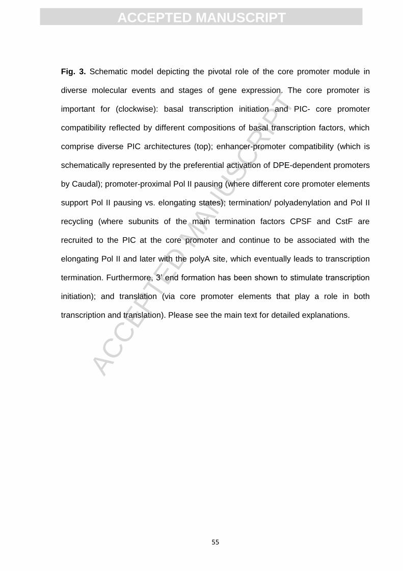
ACC
EPTE
D M
ANU
SCR
IPT
ACCEPTED MANUSCRIPT
55
Fig. 3. Schematic model depicting the pivotal role of the core promoter module in
diverse molecular events and stages of gene expression. The core promoter is
important for (clockwise): basal transcription initiation and PIC- core promoter
compatibility reflected by different compositions of basal transcription factors, which
comprise diverse PIC architectures (top); enhancer-promoter compatibility (which is
schematically represented by the preferential activation of DPE-dependent promoters
by Caudal); promoter-proximal Pol II pausing (where different core promoter elements
support Pol II pausing vs. elongating states); termination/ polyadenylation and Pol II
recycling (where subunits of the main termination factors CPSF and CstF are
recruited to the PIC at the core promoter and continue to be associated with the
elongating Pol II and later with the polyA site, which eventually leads to transcription
termination. Furthermore, 3‟ end formation has been shown to stimulate transcription
initiation); and translation (via core promoter elements that play a role in both
transcription and translation). Please see the main text for detailed explanations.

ACC
EPTE
D M
ANU
SCR
IPT
ACCEPTED MANUSCRIPT
56
Figure 1

ACC
EPTE
D M
ANU
SCR
IPT
ACCEPTED MANUSCRIPT
57
Figure 2
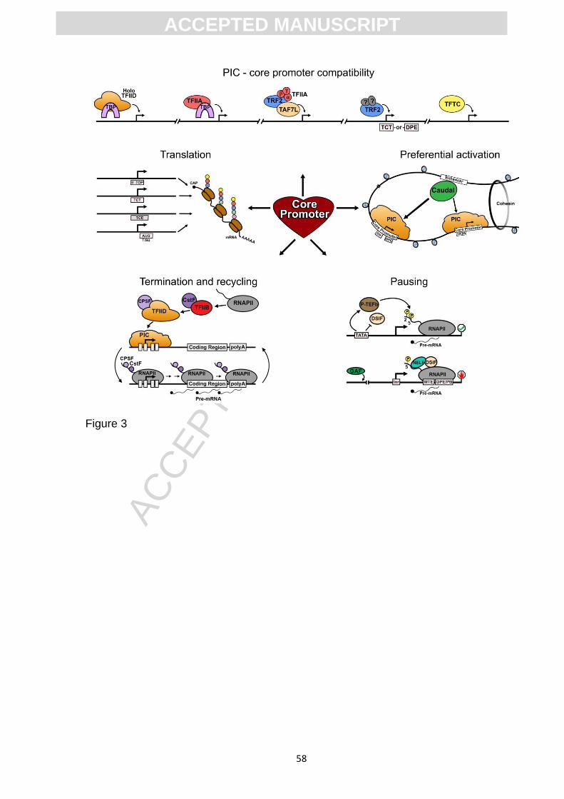
ACC
EPTE
D M
ANU
SCR
IPT
ACCEPTED MANUSCRIPT
58
Figure 3

ACC
EPTE
D M
ANU
SCR
IPT
ACCEPTED MANUSCRIPT
59
Graphical abstract

ACC
EPTE
D M
ANU
SCR
IPT
ACCEPTED MANUSCRIPT
60
Name Consensus Positions
(relative to the +1) Bound by
BREu
SSRCGCC
Immediately
upstream of the
TATA box
TFIIB
TATA box TATAWAAR -30/-31 to -23/-24 TBP
BREd
RTDKKKK
Immediately
downstream of
the TATA box
TFIIB
Mammalian Inr
/sINR
YYA+1NWYY
/CCA+1TYTT -2 to +5
TAF1 & TAF2
/TAF1
Drosophila Inr TCA+1KTY -2 to +4 TAF1 & TAF2
Human TCT YC+1TYTYY -1 to +6
Drosophila TCT YYC+1TTTYY -2 to +6
XCPE1 DSGYGGRAS+1M -8 to +2
XCPE2 VCYCRTTRCM+1Y -9 to +2
MTE CSARCSSAACGS +18 to +29 TAF6 & TAF9
DPE DSWYVY
(functional range set) +28 to +33 TAF6 & TAF9
Bridge Part I: CGANC
Part II: WYGT
Part I: +18 to +22
Part II: +30 to +33 TAF6 & TAF9 (?)
DCE
Necessary motifs:
Part I: CTTC
Pat II: CTGT
Part III: AGC
Part I: +6 to +11
Part II:+16 to +21
Part III:+30 to +34
TAF1
Table 1. The precisely positioned core promoter elements found in focused promoters
and the proteins that bind them.

ACC
EPTE
D M
ANU
SCR
IPT
ACCEPTED MANUSCRIPT
61
the heart of gene expressionat he core promoter: T - Highlights
There are three major types of transcription initiation patterns in metazoans.
Diverse PIC compositions are compatible with different core promoter architectures.
Diverse gene regulatory programs are regulated via enhancer-promoter specificity.
Core promoter elements affect Pol II pausing, as well as transcription termination.
Distinct core promoter elements link transcription and translation.