Embed Size (px)
Citation preview
ORIGINAL RESEARCHADULT BRAIN
Basilar Artery Changes in Fabry DiseaseX R. Manara, X R.Y. Carlier, X S. Righetto, X V. Citton, X G. Locatelli, X F. Colas, X M. Ermani, X D.P. Germain, and X A. Burlina
ABSTRACT
BACKGROUND AND PURPOSE: Dolichoectasia of the basilar artery is a characteristic finding of Fabry disease. However, its prevalence,severity, and course have been poorly studied. This study quantitatively evaluated, by MRA, a panel of basilar artery parameters in a largecohort of patients with Fabry disease.
MATERIALS AND METHODS: Basilar artery mean diameter, curved length, “origin-to-end” linear distance (linear length), and tortuosityindex ([curved length � linear length] � 1) were retrospectively measured on 1.5T MRA studies of 110 patients with Fabry disease (mean age,39.4 � 18.6 years; 40 males) and 108 control patients (mean age, 42.0 � 18.2 years; 40 males).
RESULTS: Patients with Fabry disease had increased basilar artery mean diameter (P � .001) and basilar artery linear length (P � .02)compared with control patients. Basilar artery curved length and tortuosity index correlated with age in both groups (P � .001), whereasbasilar artery linear length correlated with age only in patients with Fabry disease (P � .002). Patients with Fabry disease showed a basilarartery curved length mean increase of 4.2% (9.7% in male patients with Fabry disease versus male control patients), whereas the basilarartery mean diameter had a mean increase of 12.4% (14.3% in male patients with Fabry disease versus male control patients). Male patientswith Fabry disease had increased basilar artery mean diameter, curved length, and tortuosity index compared with female patients withFabry disease (P � .04, P � .02, and P � .001, respectively) and male control patients (P � .001, P � .01, and P � .006, respectively). Femalepatients with Fabry disease demonstrated an age-dependent increase of basilar artery mean diameter that became significant (P � .001)compared with female control patients above the age of 45 years.
CONCLUSIONS: The basilar artery of patients with FD is subjected to major remodeling that differs according to age and sex, thusproviding interesting clues about the pathophysiology of cerebral vessels in Fabry disease.
ABBREVIATION: FD � Fabry disease
Fabry disease (OMIM 301500; FD) is a rare X-linked (Xq22.1)
disease caused by mutations in the GLA gene causing defi-
ciency of the hydrolase �-galactosidase A (�-GalA, E.C.
3.2.1.22).1 The enzyme deficiency results in impaired sphingo-
lipid catabolism with lysosomal accumulation of upstream me-
tabolites (mainly globotriaosylceramide [Gb3] and its deacylated
compound globotriaosylsphingosine [lysoGb3]). All organs are
involved, with major damage to the kidneys, heart, and nervous
system. The brain might present white matter vascular-like ab-
normalities, TIAs, and stroke at a young age,2 suggesting that
micro- and macroangiopathy might have a pivotal role in the
pathogenesis of brain lesions. Indeed, both small and large blood
vessels have been consistently shown to present functional and
morphologic changes in patients with FD.3-5 Increased vessel tor-
tuosity has been found in the retina6 and in the skin,7,8 and intra-
cranial artery dolichoectatic changes have been repeatedly observed
in patients with FD during both pathologic and neuroimaging
evaluations.9-17
Hitherto, most of the latter studies either referred to small
samples or evaluated single specific aspects of artery dolichoecta-
sia (such as the vessel lumen diameter). In addition, these studies
Received August 20, 2016; accepted October 6.
From the Sezione di Neuroscienze (R.M.), University of Salerno, Salerno, Italy; Radi-ology Division (R.Y.C., F.C.) and Division of Medical Genetics (F.C., D.P.G.), Univer-sity of Versailles, Versailles, France; Department of Neurosciences (S.R., V.C., M.E.),University of Padova, Padova, Italy; S. Giovanni e Ruggi d’Aragona Hospital (G.L.),Salerno, Italy; and Neurological Unit (A.B.), St. Bassiano Hospital, Bassano delGrappa, Italy.
D.P.G. and A.B. should be considered co–senior authors.
Please address correspondence to Alessandro Burlina, MD, PhD, Neurological Unit,St. Bassiano Hospital, Via dei Lotti 40, 36061 Bassano del Grappa, Italy; e-mail:[email protected]
Indicates open access to non-subscribers at www.ajnr.org
Indicates article with supplemental on-line appendix and tables.
http://dx.doi.org/10.3174/ajnr.A5069
AJNR Am J Neuroradiol 38:531–36 Mar 2017 www.ajnr.org 531
applied different quantification methods or semiquantitative
scores for the severity of the intracranial FD-related vessel
changes, leading to conflicting results about the role of sex, age,
and treatments.
Our retrospective, transversal, case-control MRA study quan-
titatively investigated the morphologic basilar artery lumen
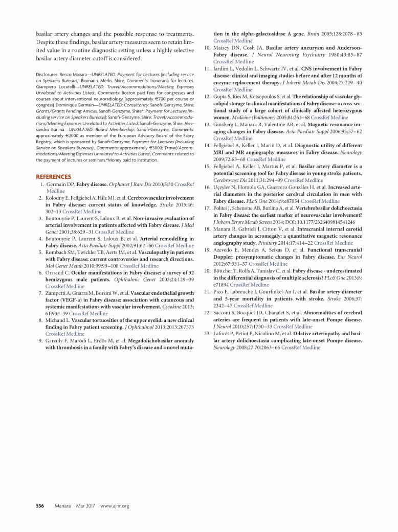
changes (Fig 1) by applying a comprehensive panel of measure-
ments encompassing all aspects of vessel dolichoectasia (diame-
ter, length, and tortuosity) on a large cohort of patients with FD,
aiming to provide a detailed picture of FD-related basilar artery
changes according to age and sex.
MATERIALS AND METHODSPatientsOne hundred thirteen patients with FD underwent MR imaging
and MRA and were retrospectively analyzed. Three patients were
excluded for anatomic reasons (see below): therefore, 110 patients
with FD were considered (mean age, 39.4 � 18.6 years; range,
6 –75 years; 40 males). The diagnosis of FD was confirmed by
�-galactosidase A enzyme activity and GLA mutation identifica-
tion. All patients gave written informed consent.
One hundred ten patients with no history of head trauma,
neurosurgery, or other neurologic or psychiatric disease who un-
derwent MRI and MRA for headache or TIA were analyzed. Two
patients were excluded for anatomic reasons (see below): there-
fore, 108 patients (mean age, 42.0 � 18.2 years; range: 4 –76 years;
40 males) represented our control group.
MR TechniqueAll MR examinations of patients and control patients were per-
formed between 2007–2014 with 1.5T scanners (Achieva or In-
tera; Philips Healthcare, Best, the Netherlands) with a standard
quadrature head coil.
The MR study protocol included conventional T1-weighted
and FLAIR axial images covering the whole brain and a TOF-
MRA sequence for the evaluation of the main intracranial arteries.
The latter sequence was acquired with the following parame-
ters: TR, 23 ms; TE, 2.274 ms; section thickness, 1.4 mm; ac-
quisition matrix, 512 � 512; and FOV, 16 cm on the Intera
scanner and TR, 25 ms; TE, 6.906 ms; section thickness, 1 mm;
acquisition matrix, 512 � 512; and FOV, 16 cm on the Achieva
scanner.
Basilar Artery EvaluationAll MRIs were evaluated by 2 neuroradi-ologists (R.M., V.C.) to exclude concomi-tant posterior cranial fossa lesions thatcould potentially interfere with the cister-nal course of the basilar artery (eg, neo-plasms, abscesses, and parenchymal orvascular malformations). No patient withFD or healthy patient was excluded for thisreason, and in no case was there disagree-ment between neuroradiologists.
The mean basilar artery diameter wasmeasured by a neuroradiologist (R.M.)on the acquisition MRA axial sections witha commercially available image viewer
(Medstation version 4.9; Exprivia, Molfetta, Italy) at 3 points, namelythe starting, middle, and ending points of the basilar artery after ad-justing the image window (On-line Appendix).
The basilar artery anatomic length (curved length of the wholecisternal segment) and the linear distance between the proximaland distal extremities of the basilar artery (linear length) weremeasured by a neuroradiologist (S.R.) with commercially avail-able software (InSpace, syngo MR Workplace; Siemens, Erlangen,Germany) dedicated to the vessel analysis (On-line Appendix). Be-cause the basilar artery landmarks were 1) the conjunction of the 2vertebral arteries (starting point) and 2) the basilar artery bifurcationinto the 2 posterior cerebral arteries (ending point), patients with FDand control patients with agenesia of 1 vertebral artery or of the pre-communicating segment of 1 or both posterior cerebral arteries wereexcluded from the study (2 control patients and 3 patients with FD,including a woman harboring a persistent trigeminal artery).
In addition, the tortuosity index was calculated as follows, ac-cording to previous studies18:
Tortuosity Index � (curved length � linear length) � 1
This index is equal to 0 when the vessel is perfectly straight, and
the index increases with the relative increase of the curved length
with respect to the linear length (Fig 1C).
In 15 patients, all of the aforementioned variables were remea-
sured after 3 months by 2 of the coauthors (R.M., S.R.) to assess
intrarater and interrater reproducibility.
Statistical AnalysisThe Student t test was performed to compare quantitative nor-
mally distributed variables between patients with FD and control
patients and between subgroups. Analysis of covariance was used
to test the dependence of groups, omitting the effect of age. The
linear relationships between age and diameter, length, and tortu-
osity measures were evaluated by using the Pearson coefficient.
Reproducibility of the measurements was also tested by Pearson
coefficient. Significance level was set at P � .05.
RESULTSMain findings are presented in On-line Table 1. FD and control
groups did not differ for age (P � .30) or sex (P � .91). All mea-
surements (mean diameter, curved length, linear length, and tor-
tuosity index) showed very high interrater and intrarater agree-
ment (r � 0.9).
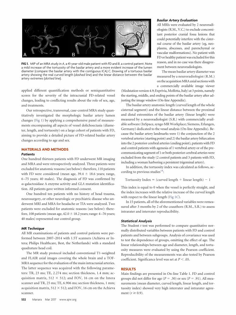
FIG 1. MIP of an MRA study in A, a 41-year-old male patient with FD and B, a control patient. Notea mild increase of the tortuosity of the basilar artery and a more evident increase of the lumendiameter (compare the basilar artery with the contiguous ICA) C, Drawing of a tortuous basilarartery showing the real curved length (dashed line) and the linear distance between the basilarartery extremes (dotted line).
532 Manara Mar 2017 www.ajnr.org
Diameter AnalysisCompared with control patients, patients with FD had increased
mean basilar artery diameter (4.16 � 0.62 mm versus 3.70 � 0.36
mm; P � .001); the mean basilar artery diameter showed mild
correlation with age among patients with FD (r � 0.3; P � .001)
and no correlation among control patients.
When subgrouping according to sex, the basilar artery diam-
eter did not correlate with age among male patients with FD and
male and female control patients. In contrast, female patients with
FD showed a good correlation between age and basilar artery di-
ameter (r � 0.54) (Fig 2).
Male patients with FD had increased mean basilar artery di-
ameter compared with female patients with FD (4.32 � 0.72 mm
versus 4.07 � 0.55 mm; P � .04) and male control patients
(4.32 � 0.72 mm versus 3.78 � 0.36 mm; P � .001). Female
patients with FD had increased mean basilar artery diameter com-
pared with female control patients (4.07 � 0.55 mm versus 3.66 �
0.36 mm; P � .001). No difference was found between male and
female control patients (3.70 � 0.36 mm versus 3.66 � 0.36 mm).
We divided patients and control patients according to sex and
age (younger or older than 45 years); male patients with FD
showed increased mean basilar artery diameter compared with
control patients at any age, whereas female patients with FD dif-
fered from control patients only in the older subgroup.
A diagnostic criterion based on a mean basilar artery diameter
�4.42 mm had a specificity of 97.2% and a sensitivity of 28.2%.
Length and Tortuosity AnalysisCompared with control patients, patients with FD had increased
linear length (28.34 � 4.03 mm versus 27.18 � 3.32 mm; P � .02)
and showed a trend toward increased curved length (31.13 � 5.38
mm versus 29.88 � 4.79 mm; P � .07). As a consequence, the
tortuosity index did not differ between patients with FD and con-
trol patients (0.097 � 0.083 versus 0.099 � 0.113; P � .86).
Curved length and tortuosity index showed a positive correlation
with age in both the patient and control groups (P � .001). In
contrast, the linear length correlated
with age only in the FD group (P �
.002).
Male patients with FD had increased
curved length and tortuosity index com-
pared with female patients with FD
(32.72 � 5.81 mm versus 30.21 � 4.92
mm; P � .02 and 0.132 � 0.094 versus
0.077 � 0.070; P � .001, respectively)
and male control patients (32.72 � 5.81
mm versus 29.83 � 4.5 mm; P � .01 and
0.132 � 0.094 versus 0.075 � 0.085; P �
.006). No significant difference was ob-
served between male and female control
patients. No significant difference was
found between female patients with FD
and female control patients after adjust-
ing for age.
Analysis of Diameter Versus Lengthand TortuosityThe mean basilar artery diameter showed
no correlation with curved length and tortuosity index in control
patients and showed a mild correlation (r � 0.35 and r � 0.43,
respectively) in patients with FD.
The curved length showed a mean increase of 4.2% in patients
with FD compared with control patients (9.7% in male patients
with FD compared with male control patients), whereas the mean
lumen diameter (ie, circumference) showed a mean increase of
12.4% in patients with FD compared with control patients (14.3%
in male patients with FD compared with male control patients).
DISCUSSIONThis MRA study quantitatively investigated the morphologic
basilar artery lumen changes in FD, showing the progressive and
profound vessel remodeling that ultimately leads to basilar artery
dolichoectasia. In particular, patients with FD presented in-
creased basilar artery diameter, length, and tortuosity that dif-
fered significantly according to patient sex and age.
Basilar Artery Diameter ChangesEctasia of intracranial arteries has been qualitatively observed in
patients with FD at both pathologic and neuroimaging evalua-
tion.9-13 A few studies applied a semiquantitative or quantitative
approach with MRA 3D reconstructions (2 studies with 1.5T and
1 with 3T scanner),14-16,19 T2 axial images (van der Tol L, per-
sonal communication), either T2 axial images or MRA,17 or axial
partitions of MRA (present study). Despite different methods,
sequences, and scanners used for the measurement and the differ-
ent ways to present study findings (different patient subgrouping,
mean or median value, etc), all studies consistently showed the
increase of the basilar artery diameter in patients with FD com-
pared with control patients (On-line Table 2).
As expected for an X-linked disease, the basilar artery diameter
in FD was shown to be significantly greater among male patients
compared with female patients in all previous studies except
one.19 The latter discordant result might have depended on the
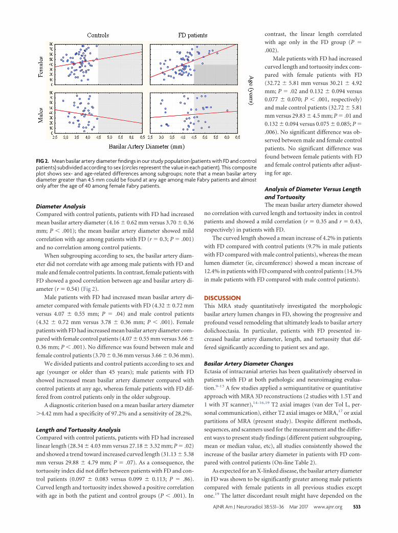
FIG 2. Mean basilar artery diameter findings in our study population (patients with FD and controlpatients) subdivided according to sex (circles represent the value in each patient). This compositeplot shows sex- and age-related differences among subgroups; note that a mean basilar arterydiameter greater than 4.5 mm could be found at any age among male Fabry patients and almostonly after the age of 40 among female Fabry patients.
AJNR Am J Neuroradiol 38:531–36 Mar 2017 www.ajnr.org 533
small sample size (5 males versus 7 females) or on the age of the
sex subgroups considered in the study (mean age of female study
participants, �40 years). In fact, according to our findings, the
basilar artery diameter appears to already be enlarged in male
patients with FD at a young age, whereas women show significant
basilar artery changes only above the age of 45 years. The different
behavior of basilar artery diameter according to sex is interesting
for several reasons. For example, it limits the efficacy of basilar
artery diameter as a criterion for differentiating female patients
with FD from patients with multiple sclerosis, as recently sug-
gested.20 In fact, the diagnostic dilemma usually involves young
women aged less than 45 years with neurologic symptoms and
nonspecific white matter signal abnormalities.20 In addition, be-
cause WM changes also seem to appear early in female patients
with FD, but basilar artery changes occur later in women, mac-
roangiopathy seems to have no role or a minimal role in the ap-
pearance of white matter signal abnormalities. Therefore, mi-
cro- and macroangiopathy might present different time courses
in FD.
Length and Tortuosity of the Basilar ArteryThe approach applied in this study provides quantitative infor-
mation on the effect of FD on the elongation of the basilar artery.
Indirect data have been provided by previous case reports, case
series, and small studies based on qualitative evaluations (On-line
Table 3).9,10,13 Recently, Politei et al17 applied a semiquantitative
approach with the Smoker criteria. According to the Smoker cri-
teria, in addition to a basilar diameter greater than 4.5 mm, the
position of the top of the basilar artery (indicative of the length of
the artery) and the basilar artery position with respect to the mid-
line (indicative of the tortuosity of the artery) also concur in the
identification of dolichoectasic basilar arteries. Politei et al17
found an increased rate of dolichoectasia in patients with FD,
especially among males (55.5% of male patients with FD versus
34.8% of female patients with FD; P � .09). Unfortunately, this
study did not include a control population and did not provide
the weight of each item (diameter, elongation, and tortuosity) in
determining dolichoectasia in the FD population, hampering
their precise analysis.
In the present study, FD-related vessel elongation was di-
rectly demonstrated by the increased curved length (effective
length of the basilar artery in its 3D cisternal course) in male
patients with FD compared with female patients with FD and
control patients. In addition, patients with FD showed in-
creased linear distance between the proximal and distal ex-
tremities of the basilar artery. The concomitant cranial dis-
placement of the top of the basilar artery likely determines a
higher score at the second item of the Smoker criteria, thus
contributing to the high rate of dolichoectasia observed in pa-
tients with FD.17 The measurement of both curved and linear
basilar artery length allowed us to precisely quantify the basilar
artery tortuosity by a tortuosity index. Although the concom-
itant increase of the linear length partly hampered the efficacy
in detecting basilar artery tortuosity changes, this measure fur-
ther highlighted the sex-related artery vulnerability in FD,
showing a significantly increased basilar artery tortuosity in
male patients with FD compared with both female patients
with FD and male control patients. Although the Smoker score
is easily applicable to conventional T2 axial images and is cer-
tainly less time-consuming, the quantitative evaluation of lin-
ear length, curved length, and tortuosity index on 3D-TOF
MRA proved to be highly reproducible in our study, providing
a powerful and reliable tool for the follow-up of the natural
history of artery changes in FD over the years as well as the
monitoring of any therapeutic intervention.
Evolution of Basilar Artery ChangesThe evolution of basilar artery changes is an obvious phenome-
non that can be inferred by the fact that megadolicho-arteries do
not appear, or at least have never been reported, in pediatric pa-
tients with FD. Nonetheless, the characterization of the evolution
of basilar artery changes in FD remains rather elusive. Azevedo et
al19 found increased basilar artery diameter values in older pa-
tients with FD, whereas Fellgiebel et al14,15 and Uceyler et al16
found no association with age. The present study showed that the
basilar artery diameter increases significantly with age in the fe-
male subgroup, whereas male patients with FD and both male and
female control patients do not change significantly over time. Ac-
cording to these findings, male patients with FD already present
increased mean basilar artery diameter at a young age, with scarce
changes over time, whereas female patients with FD progress from
values that do not differ from control patients to values that do
not differ from male patients with FD. Altogether, these contra-
dictory findings underlie 1) the heterogeneity of basilar artery
involvement in FD and 2) the difficulty in extrapolating complex
phenomena in rare diseases without well-designed, long-lasting
longitudinal studies in large patient cohorts with sex- and age-
matched control patients. For example, the lack of evolution of
basilar artery diameter in men might be related to a higher early
mortality in patients with more severe intracranial artery involve-
ment. Because stroke is associated with an increased basilar ar-
tery diameter in the healthy population21 and stroke occurs at
a younger age in male patients with FD,2 a biased selection
might have altered basilar artery diameter age-dependent find-
ings in a transverse study design.
Hitherto, the evolution of basilar artery elongation has never
been addressed, and all the information can be driven only from
the present study, which necessarily requires further validation.
According to our findings, basilar artery elongation seems to be an
age-dependent phenomenon recognizable in both patients with
FD and control patients. Nonetheless, patients with FD also
present an age-dependent significant increase of the linear dis-
tance between the proximal and distal extremities of the basilar
artery, which likely results in cranial ectopia of its distal
extremity.
Altogether, these artery changes involving diameter, length,
and tortuosity might explain the high rate of dolichoectasia pre-
viously detected semiquantitatively in patients with FD.17
Ectasia Versus ElongationDolichoectatic changes might represent different faces of the same
phenomenon (ie, the structural disruption of the artery vessel
wall) or underlie different pathogenic mechanisms. Actually,
534 Manara Mar 2017 www.ajnr.org
diameter, curved length, and tortuosity index showed no correla-
tion among control patients and displayed a relatively weak rela-
tionship within the FD cohort. In addition, the severity of the
curved length and diameter changes in the FD cohort compared
with control patients demonstrates that the artery wall is nearly 3
times more vulnerable circumferentially than longitudinally. This
observation seems to be consistent with functional and pathologic
findings in FD that report the primary involvement of the smooth
muscle cells in the accumulation of globotriaosylceramide, re-
duced sympathetic innervation of proximal cerebral arteries, and
enhanced release of nitric oxide from endothelial cells.5 Because
the smooth muscle cell tone is pivotal in controlling the vessel
radius according to brain parenchyma metabolic need, media
layer dysfunction might manifest prevalently as ectasia and, only
to a lesser extent, elongation. The presence of basilar artery ectasia
in other lysosomal storage disorders, like late-onset Pompe dis-
ease, might support the role of metabolite accumulation within
the smooth muscle cells.22,23
Diagnostic Role of Basilar Artery Changes in FDLimits inherent to the measurement of the basilar artery diameter
and to scanners, sequences, and methods applied have been
discussed above. For example, the evaluation of the basilar
artery diameter on MRA most likely leads to an underestima-
tion compared with T2 axial findings because MRA represents
only the vessel lumen, whereas T2 imaging measurement in-
cludes the vessel wall. In addition, sequences with long TR and
TE (T2 imaging) are prone to CSF pulsation artifacts around
the basilar artery that might alter the measurement of the vessel
diameter. Sequences on different scanners might differ for res-
olution, with a considerable impact on the measurement of the
basilar artery diameter. Nonetheless, at least 2 further ana-
tomic and methodologic aspects need to be discussed. In con-
trast to the internal carotid artery, the basilar artery shows high
diameter variability even among patients who do not have FD
(2.8 – 4.4 mm in the present study, but also 1.2–5.5 mm).16 The
caliper variability of the vertebral arteries and the rather com-
mon variants of the circle of Willis involving the precommu-
nicating P1 segment of the posterior cerebral arteries might
significantly influence the basilar artery diameter, hampering
its diagnostic value. In fact, nearly 20% of P1 segments are
hypoplastic, and the blood flow of the postcommunicating P2
segment might be partly or completely sustained by the ipsi-
lateral internal carotid artery via the posterior communicating
artery. A significant basilar artery diameter increment in a pa-
tient with FD with unilateral or bilateral P1 segment hypopla-
sia would probably not lead to “increased” values compared
with the normal mean value. This anatomic variability might
result in a decreased sensitivity of basilar artery diameter cutoff
values when looking for a high specificity (a cutoff of 4.42 mm
in the present study had a specificity of 97.2%, but a sensitivity
of 28.1%), especially among young women, in whom the dif-
ferences between patients and control patients are less evident,
if there are any.
The methodologic aspect regards the variability of the diame-
ter according to image windowing. Previous studies did not spec-
ify the window parameters used for the evaluation of the basilar
artery diameter because they depend on the investigator’s choice
and on image features that might vary from patient to patient,
even when using the same sequence parameters. Different image
windowing might lead to the inclusion or exclusion of a pixel at
the border of the vessel. Because the pixel size is usually 0.3– 0.4
mm and the mean basilar artery diameter in control patients is
approximately 3.7 mm, different image windowing might lead to
a 10%–20% variability in the measure obtained. This fact might
partly explain the difficulty of replicating cutoff values in different
studies. A previous study investigated 25 patients with FD and
showed that a basilar artery diameter cutoff of 2.98 mm could
differentiate patients with FD from young patients without FD
who have had a stroke with a specificity of 88.5% and a sensitivity
of 84%.15 Another study on 87 patients with FD demonstrated
that a basilar artery diameter cutoff of 3.2 mm had a sensitivity of
87% and a specificity of 86% in discriminating male patients with
FD from male control patients (no difference was found between
male patients with FD and male patients without FD who have
had a stroke or among female subgroups).16
Therefore, to minimize the measurement variability, we ap-
plied an easily replicable standardized method to optimize and
standardize the image windowing throughout the study (On-line
Appendix). Indeed, we obtained outstanding interrater and intra-
rater reproducibility of the basilar artery diameter measurements.
Nonetheless, because differences in image windowing, sequences,
sequence parameters, and scanners might significantly impact
the final measure of the vessel diameter, center-specific nor-
mative values are likely required, and literature cutoff values
can hardly be used in a diagnostic setting of FD. Taking into
account the anatomic variability of the circle of Willis, the use
of the basilar artery diameter as a unique marker of FD appears
rather inefficient.
Similarly, basilar artery linear and curved length and the rela-
tive tortuosity index proved to retain very poor diagnostic utility
because of the largely overlapping findings between patients with
FD and control patients. These limits are minimized in a longitu-
dinal study design because the anatomy of the circle of Willis does
not change significantly over time, thus allowing precise monitor-
ing of the basilar artery dolichoectatic changes.
To note, the inclusion of patients with headache and TIA in
our control group might represent a possible drawback of the
study. Even though no study has shown basilar artery changes in
patients with headache and TIA, these patients may potentially
present initial dolichoectatic abnormalities that might have
hampered the recognition of FD basilar artery changes. Ac-
cording to this hypothesis, basilar artery changes in FD may be
even more evident when using healthy patients as control pa-
tients. However, a positive control group has the advantage of
being closer to a clinical setting, where some of the above-
mentioned vascular changes have been proposed for diagnostic
purposes.
CONCLUSIONSThe panel of measurements (artery diameter, length, and tortu-
osity) applied in this study provides some relevant hints about the
pathophysiology of vessel remodeling in FD. Moreover, our study
provides an interesting tool for monitoring the natural course of
AJNR Am J Neuroradiol 38:531–36 Mar 2017 www.ajnr.org 535
basilar artery changes and the possible response to treatments.
Despite these findings, basilar artery measures seem to retain lim-
ited value in a routine diagnostic setting unless a highly selective
basilar artery diameter cutoff is considered.
Disclosures: Renzo Manara—UNRELATED: Payment for Lectures (including serviceon Speakers Bureaus): Biomarin, Merks, Shire, Comments: honoraria for lectures.Giampiero Locatelli—UNRELATED: Travel/Accommodations/Meeting ExpensesUnrelated to Activities Listed:, Comments: Boston paid fees for congresses andcourses about interventional neuroradiology (approximately €700 per course orcongress). Dominique Germain—UNRELATED: Consultancy: Sanofi-Genzyme, Shire;Grants/Grants Pending: Amicus, Sanofi-Genzyme, Shire*; Payment for Lectures (in-cluding service on Speakers Bureaus): Sanofi-Genzyme, Shire; Travel/Accommoda-tions/Meeting Expenses Unrelated to Activities Listed: Sanofi-Genzyme, Shire. Ales-sandro Burlina—UNRELATED: Board Membership: Sanofi-Genzyme, Comments:approximately €2000 as member of the European Advisory Board of the FabryRegistry, which is sponsored by Sanofi-Genzyme; Payment for Lectures (IncludingService on Speakers Bureaus):, Comments: approximately €3000; Travel/Accom-modations/Meeting Expenses Unrelated to Activities Listed:, Comments: related tothe payment of lectures or seminars.*Money paid to institution.
REFERENCES1. Germain DP. Fabry disease. Orphanet J Rare Dis 2010;5:30 CrossRef
Medline2. Kolodny E, Fellgiebel A, Hilz MJ, et al. Cerebrovascular involvement
in Fabry disease: current status of knowledge. Stroke 2015;46:302–13 CrossRef Medline
3. Boutouyrie P, Laurent S, Laloux B, et al. Non-invasive evaluation ofarterial involvement in patients affected with Fabry disease. J MedGenet 2001;38:629 –31 CrossRef Medline
4. Boutouyrie P, Laurent S, Laloux B, et al. Arterial remodelling inFabry disease. Acta Paediatr Suppl 2002;91:62– 66 CrossRef Medline
5. Rombach SM, Twickler TB, Aerts JM, et al. Vasculopathy in patientswith Fabry disease: current controversies and research directions.Mol Genet Metab 2010;99:99 –108 CrossRef Medline
6. Orssaud C. Ocular manifestations in Fabry disease: a survey of 32hemizygous male patients. Ophthalmic Genet 2003;24:129 –39CrossRef Medline
7. Zampetti A, Gnarra M, Borsini W, et al. Vascular endothelial growthfactor (VEGF-a) in Fabry disease: association with cutaneous andsystemic manifestations with vascular involvement. Cytokine 2013;61:933–39 CrossRef Medline
8. Michaud L. Vascular tortuosities of the upper eyelid: a new clinicalfinding in Fabry patient screening. J Ophthalmol 2013;2013:207573CrossRef Medline
9. Garzuly F, Marodi L, Erdos M, et al. Megadolichobasilar anomalywith thrombosis in a family with Fabry’s disease and a novel muta-
tion in the alpha-galactosidase A gene. Brain 2005;128:2078 – 83CrossRef Medline
10. Maisey DN, Cosh JA. Basilar artery aneurysm and Anderson-Fabry disease. J Neurol Neurosurg Psychiatry 1980;43:85– 87CrossRef Medline
11. Jardim L, Vedolin L, Schwartz IV, et al. CNS involvement in Fabrydisease: clinical and imaging studies before and after 12 months ofenzyme replacement therapy. J Inherit Metab Dis 2004;27:229 – 40CrossRef Medline
12. Gupta S, Ries M, Kotsopoulos S, et al. The relationship of vascular gly-colipid storage to clinical manifestations of Fabry disease: a cross-sec-tional study of a large cohort of clinically affected heterozygouswomen. Medicine (Baltimore) 2005;84:261–68 CrossRef Medline
13. Ginsberg L, Manara R, Valentine AR, et al. Magnetic resonance im-aging changes in Fabry disease. Acta Paediatr Suppl 2006;95:57– 62CrossRef Medline
14. Fellgiebel A, Keller I, Marin D, et al. Diagnostic utility of differentMRI and MR angiography measures in Fabry disease. Neurology2009;72:63– 68 CrossRef Medline
15. Fellgiebel A, Keller I, Martus P, et al. Basilar artery diameter is apotential screening tool for Fabry disease in young stroke patients.Cerebrovasc Dis 2011;31:294 –99 CrossRef Medline
16. Uceyler N, Homola GA, Guerrero Gonzalez H, et al. Increased arte-rial diameters in the posterior cerebral circulation in men withFabry disease. PLoS One 2014;9:e87054 CrossRef Medline
17. Politei J, Schenone AB, Burlina A, et al. Vertebrobasilar dolichoectasiain Fabry disease: the earliest marker of neurovascular involvement?J Inborn Errors Metab Screen 2014; DOI: 10.1177/2326409814541246
18. Manara R, Gabrieli J, Citton V, et al. Intracranial internal carotidartery changes in acromegaly: a quantitative magnetic resonanceangiography study. Pituitary 2014;17:414 –22 CrossRef Medline
19. Azevedo E, Mendes A, Seixas D, et al. Functional transcranialDoppler: presymptomatic changes in Fabry disease. Eur Neurol2012;67:331–37 CrossRef Medline
20. Bottcher T, Rolfs A, Tanislav C, et al. Fabry disease - underestimatedin the differential diagnosis of multiple sclerosis? PLoS One 2013;8:e71894 CrossRef Medline
21. Pico F, Labreuche J, Gourfinkel-An I, et al. Basilar artery diameterand 5-year mortality in patients with stroke. Stroke 2006;37:2342– 47 CrossRef Medline
22. Sacconi S, Bocquet JD, Chanalet S, et al. Abnormalities of cerebralarteries are frequent in patients with late-onset Pompe disease.J Neurol 2010;257:1730 –33 CrossRef Medline
23. Laforet P, Petiot P, Nicolino M, et al. Dilative arteriopathy and basi-lar artery dolichoectasia complicating late-onset Pompe disease.Neurology 2008;27:70:2063– 66 CrossRef Medline
536 Manara Mar 2017 www.ajnr.org
![CFM antonella shareWavelength [nm] Manara 201 7, SF Newsletter ACCRETION & STELLAR PROPERTIES DETERMINATION Manara et al. 2013b • • 0403.2019 Photospheric templates: Class Ill](https://img.pdfslide.us/doc/110x75/6067050c70d844432826bdea/cfm-antonella-share-wavelength-nm-manara-201-7-sf-newsletter-accretion-.jpg)