Embed Size (px)
DESCRIPTION
Please ask for permission before downloading. Notes taken from Dr Dasig's discussion for Neurophysiology
Citation preview

[02]: Neurophysiology 1Dr. Darwin Dasig20 Aug 2014
Physio 21
COLLEGE OF ALLIED MEDICAL PROFESSIONSUniversity of the Philippines Manila
Bachelor of Science in Physical Therapy Class of 2017
TopicsI. The Nervous systemII. Resting membrane potentialIII. Local potentialIV. Action potential or Nerve impulseV. Synapse and Synaptic transmissionVI. Somatosensory system
I. THE NERVOUS SYSTEM It is used for:
Control of the body. Integration
Integrates all the systems in order to maintain homeostasis
It is Contralateral. It senses danger from the external
environment and reacts accordingly in the other direction
The right brain controls the left side of the body and vice-versa
It interacts with the internal and external environment in order to maintain homeostasis.
It is composed of the Brain, Brain stem, Spinal cord, nerves and muscles.
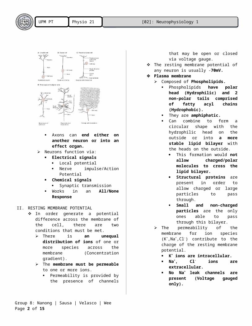
Brain Serves many different functions. It is comprised of Neurons.
These are the functional part of the brain.
It has a Cell Body (Soma). Contains the nucleus. Metabolic center of the cell. Controls all the
maintenance of the neuron. It has several processes
called Dendrites which extend outward and becomes a receptive area for an adjacent axon.
It also has a long process called an Axon that originates from the Axon Hillock.
Action potentials / Conducted impulses
generate in the initial segment of the axon.
This divides into presynaptic terminals, each of which ends in a number of synaptic knobs/terminal buttons or boutons.
Can be classified into unipolar, bipolar, or multipolar.
Axons can end either on another neuron or into an effect organ.
Neurons function via: Electrical signals
Local potential Nerve impulse/Action
Potential Chemical signals
Synaptic transmission Works in an All/None
Response
II. RESTING MEMBRANE POTENTIAL In order generate a potential difference
across the membrane of the cell, there are two conditions that must be met. There is an unequal distribution
of ions of one or more species across the membrane (Concentration gradient).
Group 8: Nanong | Sausa | Velasco | Wee Page 1 of 13
[02]: Neurophysiology 1Physio 21UPM PT 2017
The membrane must be permeable to one or more ions. Permeability is provided by the
presence of channels that may be open or closed via voltage gauge.
The resting membrane potential of any neuron is usually -70mV.
Plasma membrane Composed of Phospholipids.
Phospholipids have polar head (Hydrophilic) and 2 non-polar tails comprised of fatty acyl chains (Hydrophobic).
They are amphiphatic. Can combine to form a circular
shape with the hydrophilic head on the outside or into a more stable lipid bilayer with the heads on the outside. This formation would not
allow charged/polar molecules to cross the lipid bilayer.
Structural proteins are present in order to allow charged or large particles to pass through.
Small and non-charged particles are the only ones able to pass through this bilayer.
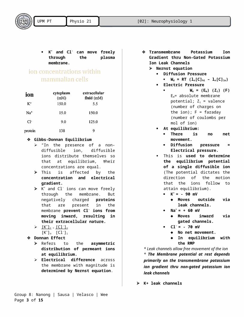
The permeability of the membrane for ion species (K+,Na+,Cl-) contribute to the charge of the resting membrane potential. K+ ions are intracellular. Na+, Cl- ions are
extracellular. No Na+ leak channels are
present (Voltage gauged only).
K+ and Cl- can move freely through the plasma membrane.
Gibbs-Donnan Equilibrium
“In the presence of a non-diffusible ion, diffusible ions distribute themselves so that at equilibrium, their concentrations are equal.”
This is affected by the concentration and electrical gradient.
K+ and Cl- ions can move freely through the membrane. But negatively charged proteins that are present in the membrane prevent Cl- ions from moving inward, resulting in their extracellular nature.
[K + ] i = [Cl - ] o
[K+]o [Cl-]i Donnan Effect
Refers to the asymmetric distribution of permeant ions at equilibrium.
Electrical difference across the membrane with magnitude is determined by Nernst equation.
Transmembrane Potassium Ion Gradient thru Non-Gated Potassium Ion Leak Channels Nernst equation
Diffusion Pressure Wd = RT (ln[C]hi – ln[C]lo)
Electric Pressure We = (Em) (Zi) (F)
Em= absolute membrane potential; Zi = valence (number of charges on the
Group 8: Nanong | Sausa | Velasco | Wee Page 2 of 13
[02]: Neurophysiology 1Physio 21UPM PT 2017
ion); F = faraday (number of coulombs per mol of ion)
At equilibrium: There is no net
movement. Diffusion pressure =
Electrical pressure. This is used to determine the
equilibrium potential of a single diffusible ion (The potential dictates the direction of the motion that the ions follow to attain equilibrium). K+ = - 90 mV
Moves outside via leak channels.
Na+ = + 60 mV Moves inward via
gated channels. Cl- = - 70 mV
No net movement. In equilibrium with the
RMP* Leak channels allow free movement of
the ion* The Membrane potential at rest depends primarily on the transmembrane potassium ion gradient thru non-gated potassium ion leak channels
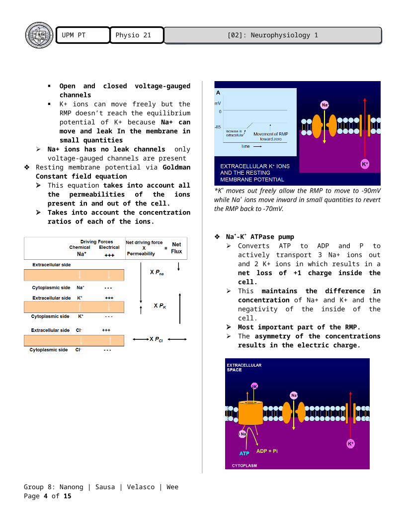
K+ leak channels Open and closed voltage-gauged
channels K+ ions can move freely but the RMP
doesn’t reach the equilibrium potential of K+ because Na+ can move and leak In the membrane in small quantities
Na+ ions has no leak channels only voltage-gauged channels are present
Resting membrane potential via Goldman Constant field equation This equation takes into account all the
permeabilities of the ions present in and out of the cell.
Takes into account the concentration ratios of each of the ions.
*K+ moves out freely allow the RMP to move to -90mV while Na+ ions move inward in small quantities to revert the RMP back to -70mV.
Na+-K+ ATPase pump
Converts ATP to ADP and P to actively transport 3 Na+ ions out and 2 K+ ions in which results in a net loss of +1 charge inside the cell.
This maintains the difference in concentration of Na+ and K+ and the negativity of the inside of the cell.
Most important part of the RMP. The asymmetry of the concentrations
results in the electric charge.
Group 8: Nanong | Sausa | Velasco | Wee Page 3 of 13
[02]: Neurophysiology 1Physio 21UPM PT 2017
The resting membrane potential is not in equilibrium. The RMP is only a steady state due to
the presence of the ATPase pump. Due to the usage of ATP (Equilibrium
doesn’t need energy in order to be maintained).
III. LOCAL POTENTIAL Local Potential
Transient shift of the membrane potential in a localized area of the cell (quick change).
Change in one portion of the membrane (any kind of change).
Lygan change – a neurotransmitter combines with receptor that opens (synaptic potential).
Graded response – amplitude is proportional to size of stimulus (more to more).
Hyperpolarization ↔ depolarization (hyper = increase negativity)
Rapid ↔ long
Local Potential Kickoff Local change in permeability of the
membrane to 1 or more ions. Synaptic potential
Lygan change Generator potential
chemical change Change in voltage/current applied from a
source. Electrotonic potential
Add voltage *increase Na and K conductance =
depolarization due to high driving force of Na (lesser degree when increased Na conductance).
Local Potentials Change in potential develops and subsides
over a few milliseconds. Remain localized in region where stimulus
is applied. Not sharply confined: falls off over a finite
distance on the membrane. Within milliseconds and within a single
area. Voltage change = current X resistance Some ions leak out across membrane.
• Lesser charges reach more distant sites.
• stable resistance : decreased current → decreased voltage
Local potential (Voltage change) – decreases with distance from the point of stimulation
Space constant
Group 8: Nanong | Sausa | Velasco | Wee Page 4 of 13
[02]: Neurophysiology 1Physio 21UPM PT 2017
Describes the distance of the voltage change along the membrane.
Distance at which the initial transmembrane voltage change has fallen to 37% of its peak value.
The larger the space constant, the farther along the membrane a voltage change is observed after a stimulus is applied.
Limit of the local potential (distance away the body).
λ=√Rm /RaRm = transmembrane resistance (ohm-cm) Ra = internal axoplasmic resistance
(ohm/cm)
↑ Diameter of axon or dendrite• ↓ Ra (internal axoplasmic
resistance)• ↑ space constant • ↑ current will flow farther along
cell↑ Rm (transmembrane resistance)
• ↑ space constant• less current leaks out
Directly related to the transmembrane resistance.
High transmembrane resistance – longer distance (how easy to leave; barrier will squish them out).
Low Internal axoplasmic resistance – high distance (how easy to travel; inside is very spacious).
Local Potentials CAN BE SUMMATED! Spatial summation
Add up beside each other (through space).
Remain localized in region where stimulus is applied.
Not sharply confined: falls off over a finite distance on the membrane.
Temporal summation Change in potential develops and
subsides over a few milliseconds. Same space (through time) Repeated stimulation
*Spatial summation (top) and temporal summation (bottom)*summated subthreshold potentials may reach threshold and produce an action potential*neurons = electrical and chemical communication
IV. Action potential or Nerve impulse ACTION POTENTIAL
Fleeting, self-renewing wave of membrane depolarization that propagates without decrement along the length of a nerve axon at high speed.
Is a rapid, all-or-none change in the membrane potential followed by a return to the resting membrane potential.
Voltage-dependent ion (Na+ and K+) channels in the plasma membrane are the basis for action potentials.
Group 8: Nanong | Sausa | Velasco | Wee Page 5 of 13
[02]: Neurophysiology 1Physio 21UPM PT 2017
An action potential is propagated with the same shape and size along the entire length of an axon.
Action potentials are usually initiated at the axon hillock of the axon.
The action potential is the basis of the signal- carrying ability of nerve cells.
The patterns of conducted action potentials encode the information conveyed by nerve cells.
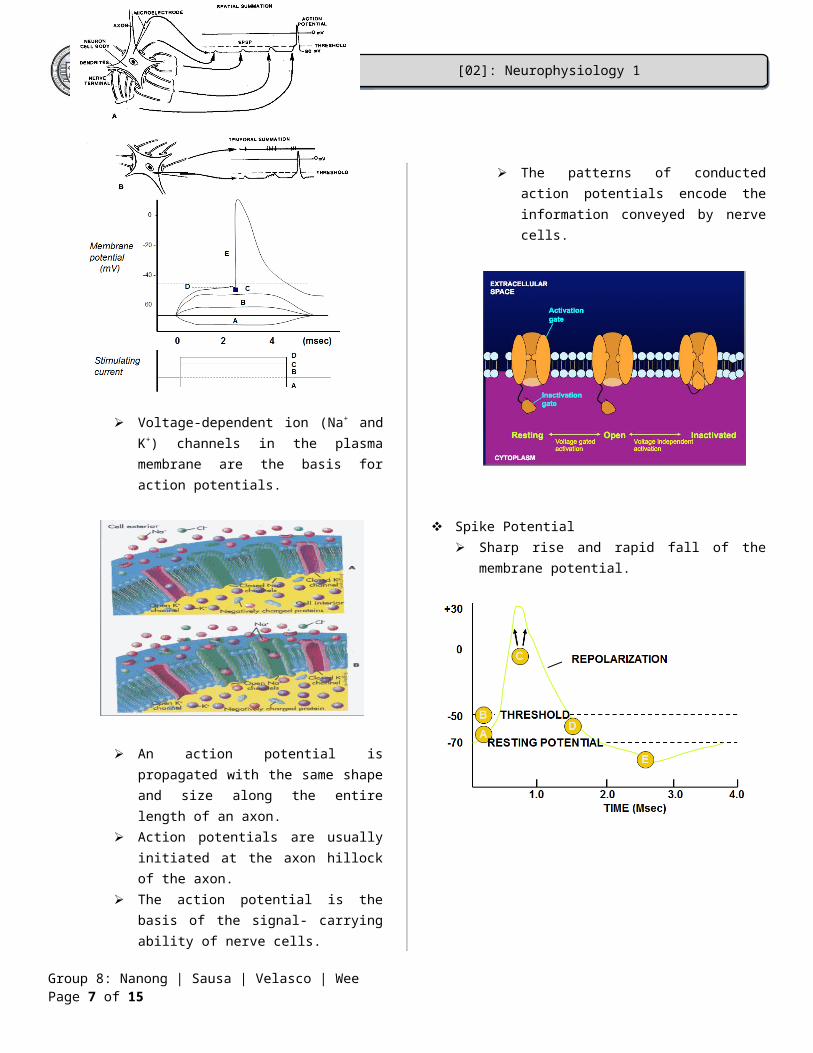
Spike Potential Sharp rise and rapid fall of the membrane
potential.
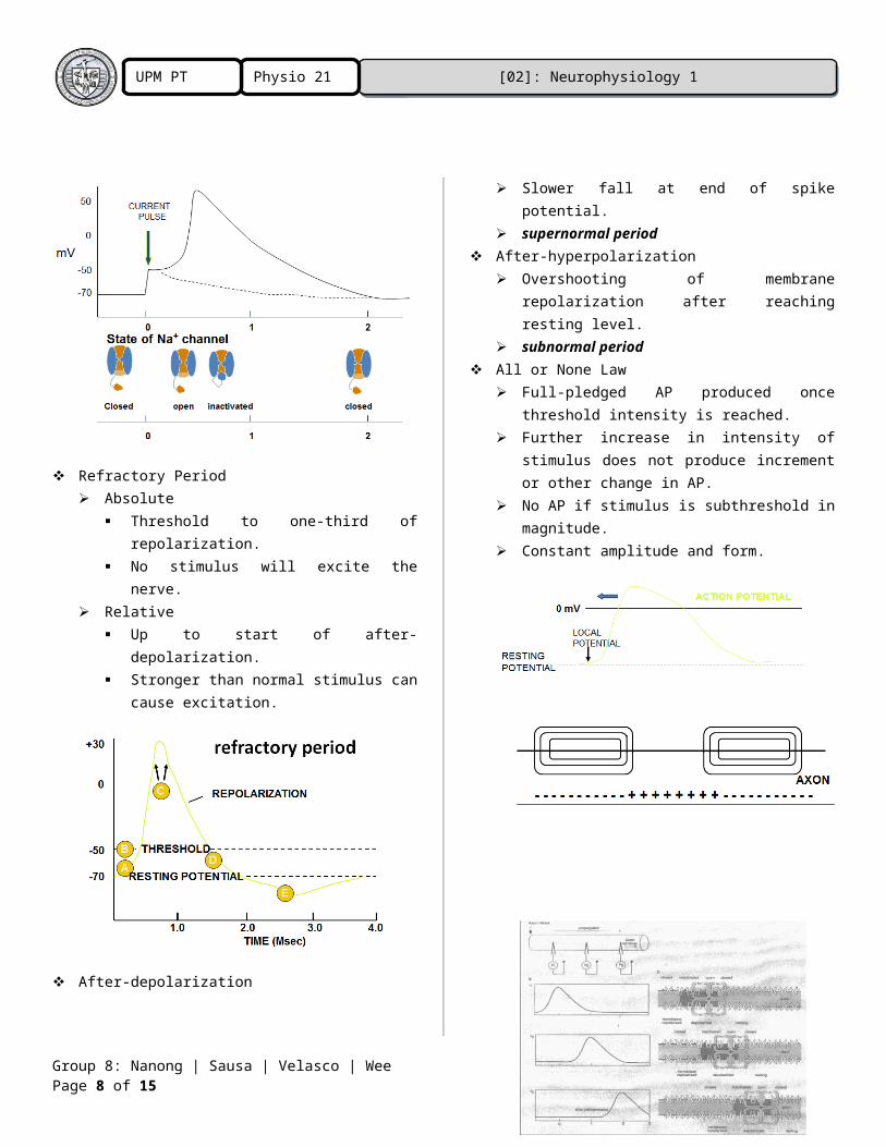
Refractory Period Absolute
Threshold to one-third of repolarization.
No stimulus will excite the nerve. Relative
Up to start of after-depolarization. Stronger than normal stimulus can
cause excitation.
Group 8: Nanong | Sausa | Velasco | Wee Page 6 of 13
[02]: Neurophysiology 1Physio 21UPM PT 2017
After-depolarization Slower fall at end of spike potential. supernormal period
After-hyperpolarization Overshooting of membrane repolarization
after reaching resting level. subnormal period
All or None Law Full-pledged AP produced once threshold
intensity is reached. Further increase in intensity of stimulus
does not produce increment or other change in AP.
No AP if stimulus is subthreshold in magnitude.
Constant amplitude and form.
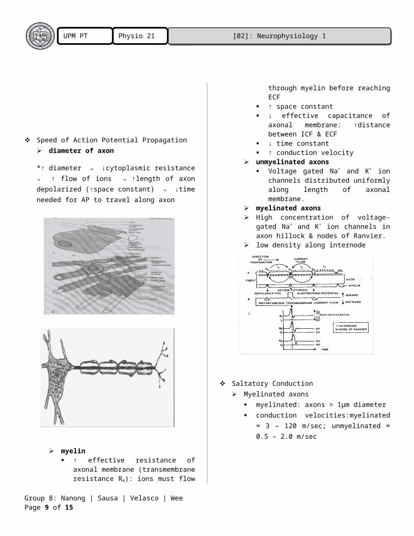
Speed of Action Potential Propagation diameter of axon
*↑ diameter → ↓cytoplasmic resistance → ↑ flow of ions → ↑length of axon depolarized (↑space constant) → ↓time needed for AP to travel along axon
myelin ↑ effective resistance of axonal
membrane (transmembrane
Group 8: Nanong | Sausa | Velasco | Wee Page 7 of 13
[02]: Neurophysiology 1Physio 21UPM PT 2017
resistance Rm): ions must flow through myelin before reaching ECF
↑ space constant ↓ effective capacitance of axonal
membrane: ↑distance between ICF & ECF
↓ time constant ↑ conduction velocity
unmyelinated axons Voltage gated Na+ and K+ ion
channels distributed uniformly along length of axonal membrane.
myelinated axons High concentration of voltage-gated
Na+ and K+ ion channels in axon hillock & nodes of Ranvier.
low density along internode
Saltatory Conduction Myelinated axons
myelinated: axons > 1μm diameter conduction velocities:myelinated = 3 –
120 m/sec; unmyelinated = 0.5 – 2.0 m/sec
V. SYNAPSE AND SYNAPTIC TRANSMISSION Synapse - A junction where the axon or
some other portion of one neuron (called the presynaptic neuron) terminates on the dendrites, soma, or axon of another neuron (called the postsynaptic neuron), muscle cell, or gland cell.
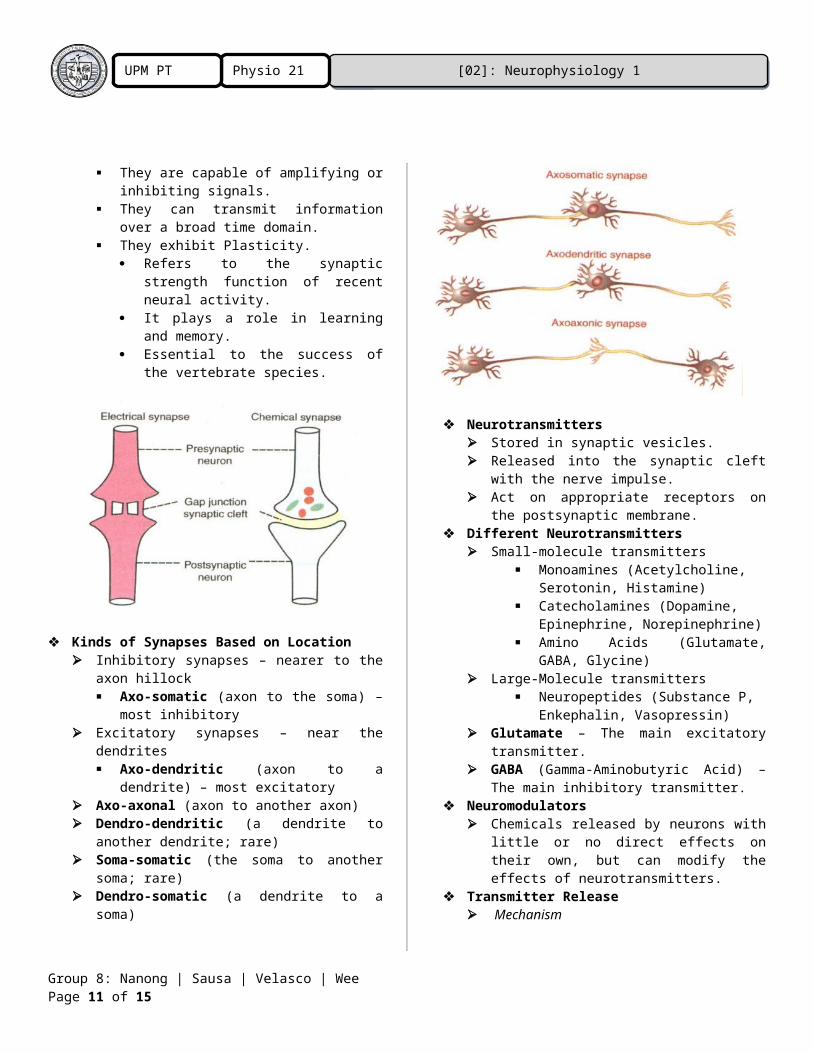
Types of Synapses Electrical
There is a gap junction formed between the presynaptic and postsynaptic neuron.
There is cytoplasmic continuity. Since the neurons are connected to
each other, they are inflexible (they cannot form new connections).
Each gap junction is formed by two hemichannels called Connexons, one contributed by each cell.
The gap junction forms low-resistance bridges through which ions pass with relative ease.
There is very little distance between the two neurons (3nm-5nm).
Ionic currents travel extremely fast through these bridges.
There is no synaptic delay. The structure allows for the
bidirectional travel of the ionic currents.
Uses very little metabolic energy and molecular machinery.
Highly reliable.
Group 8: Nanong | Sausa | Velasco | Wee Page 8 of 13
[02]: Neurophysiology 1Physio 21UPM PT 2017
Chemical Presynaptic and postsynaptic neurons
are separated by a space called the Synaptic Cleft. There is no cytoplasmic continuity. Since the neurons are separated
from each other, they are flexible (they can form new connections as required, such as when learning new skills or remembering something). This is termed “Plasticity.”
The presynaptic neuron uses chemical transmitters called Neurotransmitters that bind to the postsynaptic neuron to propagate potentials.
Neurotransmitters are able to travel much larger distances (30nm-50nm) between the presynaptic and postsynaptic neuron.
The neurotransmitter is unidirectional: from the presynaptic neuron to the postsynaptic neuron.
They are capable of amplifying or inhibiting signals.
They can transmit information over a broad time domain.
They exhibit Plasticity. Refers to the synaptic strength
function of recent neural activity. It plays a role in learning and
memory. Essential to the success of the
vertebrate species.
Kinds of Synapses Based on Location Inhibitory synapses – nearer to the axon
hillock Axo-somatic (axon to the soma) –
most inhibitory Excitatory synapses – near the dendrites
Axo-dendritic (axon to a dendrite) – most excitatory
Axo-axonal (axon to another axon) Dendro-dendritic (a dendrite to another
dendrite; rare) Soma-somatic (the soma to another
soma; rare) Dendro-somatic (a dendrite to a soma)
Neurotransmitters Stored in synaptic vesicles. Released into the synaptic cleft with the
nerve impulse. Act on appropriate receptors on the
postsynaptic membrane.
Group 8: Nanong | Sausa | Velasco | Wee Page 9 of 13
[02]: Neurophysiology 1Physio 21UPM PT 2017
Different Neurotransmitters Small-molecule transmitters
Monoamines (Acetylcholine, Serotonin, Histamine)
Catecholamines (Dopamine, Epinephrine, Norepinephrine)
Amino Acids (Glutamate, GABA, Glycine)
Large-Molecule transmitters Neuropeptides (Substance P,
Enkephalin, Vasopressin) Glutamate – The main excitatory
transmitter. GABA (Gamma-Aminobutyric Acid) – The
main inhibitory transmitter. Neuromodulators
Chemicals released by neurons with little or no direct effects on their own, but can modify the effects of neurotransmitters.
Transmitter Release Mechanism
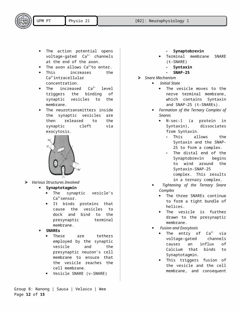
The action potential opens voltage-gated Ca2+ channels at the end of the axon.
The axon allows Ca2+to enter. This increases the
Ca2+intracellular concentration. The increased Ca2+ level triggers
the binding of synaptic vesicles to the membrane.
The neurotransmitters inside the synaptic vesicles are then released to the synaptic cleft via exocytosis.
Various Structures Involved Synaptotagmin
The synaptic vesicle’s Ca2+sensor.
It binds proteins that cause the vesicles to dock and bind to the presynaptic terminal membrane.
SNAREs These are tethers employed
by the synaptic vesicle and the presynaptic neuron’s cell membrane to ensure that the vesicle reaches the cell membrane.
Vesicle SNARE (v-SNARE) Synaptobrevin
Terminal membrane SNARE (t-SNARE) Syntaxin SNAP-25
Snare Mechanism Initial State
The vesicle moves to the nerve terminal membrane, which contains Syntaxin and SNAP-25 (t-SNAREs).
Formation of the Ternary Complex of Snares N-sec-1 (a protein in
Syntaxin), dissociates from Syntaxin. This allows the Syntaxin
and the SNAP-25 to form a complex.
The distal end of the Synaptobrevin begins to wind around the Syntaxin-SNAP-25 complex. This results in a ternary complex.
Tightening of the Ternary Snare Complex The three SNAREs continue to
form a tight bundle of helices. The vesicle is further drawn to
the presynaptic membrane. Fusion and Exocytosis
The entry of Ca2+ via voltage-gated channels causes an influx of Calcium that binds to Synaptotagmin.
This triggers fusion of the vesicle and the cell membrane, and consequent exocytosis of the neurotransmitter.
Disassembly of the Ternary Snare Complex
Group 8: Nanong | Sausa | Velasco | Wee Page 10 of 13
[02]: Neurophysiology 1Physio 21UPM PT 2017
α-SNAP and the ATPase NSF bind to the ternary SNARE complex.
They use the energy of ATP hydrolysis to disassemble the SNAREs.
Recycling of Snares The vesicle is endocytosed,
and the three SNAREs are again ready for use.
Additional Information Transmitter release is a
quantum. The number of
neurotransmitter molecules released by one vesicle is fixed.
Even if the neurotransmitters are quantized (Quantum Content), the number of quanta released when the synapse is activated depends on the level of Ca2+.
The greater the Ca2+, the more quantized neurotransmitters are released.
Burst of transmission terminates with rapid sequestration in organelles (such as the mitochondria) within the ending.
Ca2+is extruded via the Na+- Ca2+Antiport.
Neurotransmitter Receptors Ionotropic Receptors
The chemical receptor is also the channel.
Ligand-gated ion channel proteins
Rapid opening of ion channels Depolarization and
hyperpolarization Nicotinic Acetylcholine, GABA,
Glycine, Serotonin, Glutamate Metabotropic Receptors
The chemical receptor is not the channel.
The cell membrane uses proteins to facilitate the channeling of the neurotransmitter.
Seven-transmembrane-helix-receptors
G protein-linked
G proteins are nucleotide regulatory proteins that bind GTP.
Ion channel proteins or 2nd
messenger effector proteins Translate signals to biological
effects inside the cell. Glutamate, Muscarinic
Acetylcholine, α and β adrenergic Excitatory Postsynaptic Potential
(EPSP) Local potential of depolarization under
the active zone from a single stimulus. Increases the excitability of the neuron
to other stimuli. Ionic basis:
Na+ channels are opened. There is an increase in Na+
conductance through ligand-gated channels.
Closure of K+ channels. Inhibitory Postsynaptic Potentials
(IPSP) Local potential of hyperpolarization Postsynaptic direct inhibition. Stimulation to other stimuli is decreased. Ionic basis:
Opening of CI- channels. There is an increase in Cl-
conductance causing an outward flow of current.
Opening of K+ channels. Closure of Na+ or Ca2+ channels.
Excitation Spatial and Temporal summation.
Spatial summation refers to excitation over an area, where intersecting areas of excitation in the postsynaptic neuron are added-up.
Temporal summation refers to repeated excitation over time.
The axon hillock is the area of highest integration
Inhibition Presynaptic Inhibition
Provides a mechanism for controlling the efficacy of transmission at individual synapses.
Group 8: Nanong | Sausa | Velasco | Wee Page 11 of 13
[02]: Neurophysiology 1Physio 21UPM PT 2017
Mediated by Axo-axonal synapses.
Postsynaptic Inhibition Direct Inhibition
Inhibition with IPSP Not a consequence of
previous discharges of the postsynaptic neuron.
Indirect Inhibition Due to the effects of previous
postsynaptic neuron discharge (refractory period or during after-hyperpolarization).
Long-term Potentiation Characterized by the enhanced
transmission at synapses that follow high frequency stimulations.
First observed in the hippocampus, a part of the brain that plays an important role in memory.
Depends on the presence of NMDA (N-methyl-D-aspartate) receptors in the postsynaptic membrane.
Plays a role in memory and associative learning.
Convergence and Divergence One to one. Divergence:
One to many – The axon of the presynaptic neuron divides into many branches that end on many postsynaptic neurons.
Spatially focused – The branching is much less, usually confined to several effectors in close proximity.
Widely divergent – The branching goes over a wide area.
Convergence: Many to one – Many presynaptic
neurons converge on a single postsynaptic neuron.
REFERENCES:
Barret, K.E., Barman, S.M., Biotano, S., & company. (2012). Ganong’s Review of Medical Physiology, 24th ed. The McGraw-Hill Companies, Inc., United States of America.
Dasig, D. (2014). CAMP Physio 21 Neurophysiology 1 2014 Power point presentation.
Koeppen, B.M., Stanton, B.A. (2012). Berne and Levy Physiology, 6th ed. Molsby Inc., Elsevier Inc.
Waxman, S.G. (2013). Clinical Neuroanatomy, 27thed. The McGraw-Hill Companies, Inc., United States of America.
Authors’ Notes on the Subject Matter
Group 8: Nanong | Sausa | Velasco | Wee Page 12 of 13
[02]: Neurophysiology 1Physio 21UPM PT 2017
Group 8: Nanong | Sausa | Velasco | Wee Page 13 of 13