Embed Size (px)
Citation preview

Basic Skull Positioning- Parts 1 and 2
6/5/2014
Skull Positioning DMI 55

-form protective housing of brain (cranial vault)
14 Facial
2 types of Skull bones
8 Cranial
-provides structure, shape & support for face-protective housing for upper ends of respiratory & digestive tracts- with cranial-forms eye sockets
The Cranium
• The Brain• Of Jane• Stays mainly• In her
Cran(ium)


8Cranial Bones are:
• 1 Frontal
• 2 Parietal
• 1 Occipital
• 1 Ethmoid
• 1 Sphenoid
• 2 Temporal
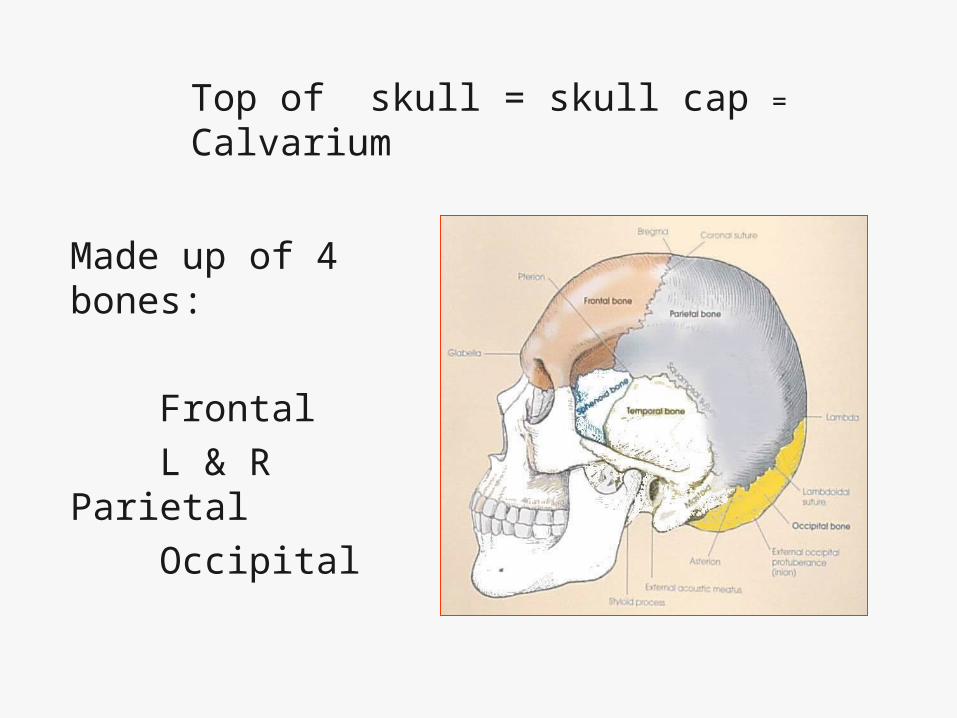
Top of skull = skull cap = Calvarium
Made up of 4 bones:
Frontal L & R Parietal Occipital
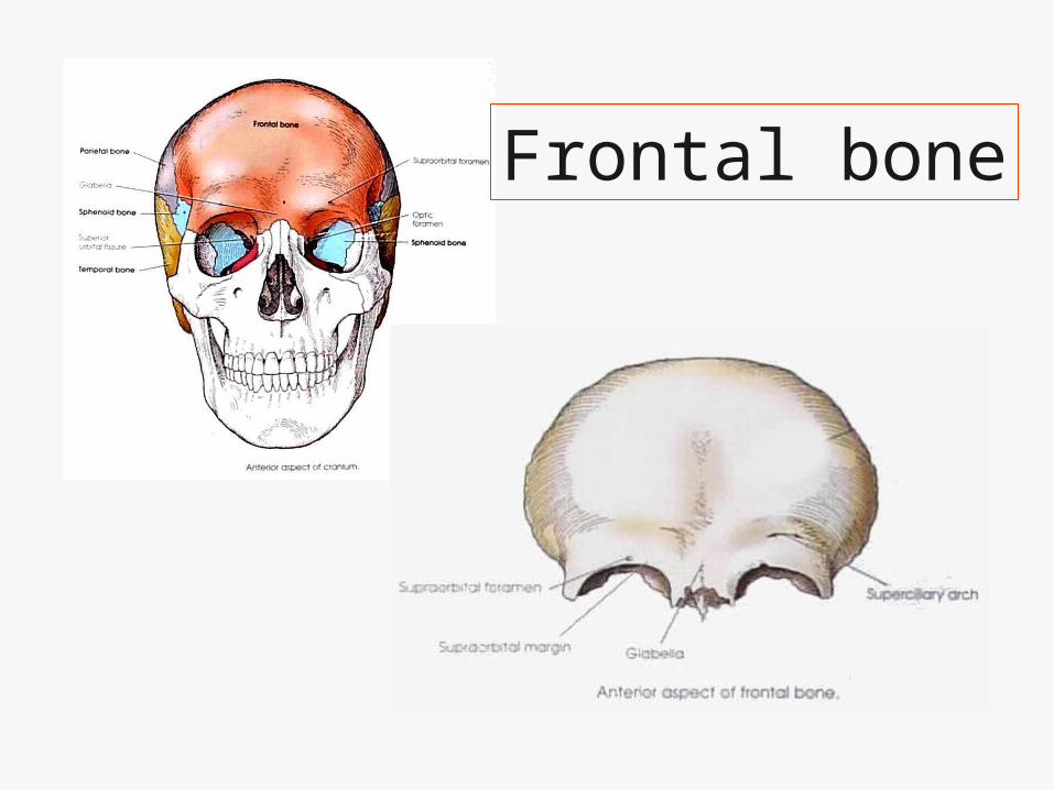
Frontal bone

2 parietal bones

Occipital
Floor of Cranium

Floor of Cranium is made of 4 bones
(The four on the floor!)
• Ethmoid• Sphenoid• Left & Right
Temporal bones

1 Ethmoid Bone

1 Sphenoid bone
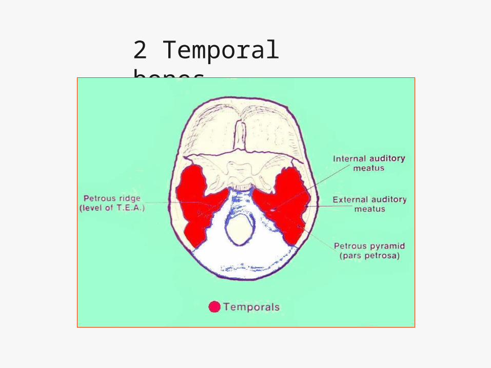
2 Temporal bones
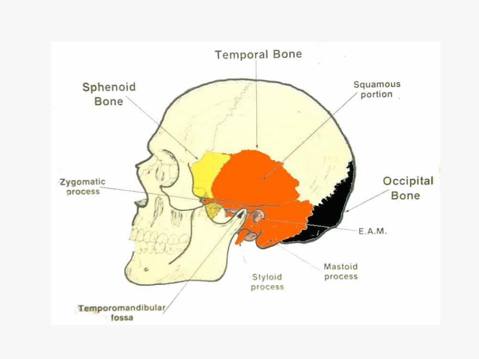

Temporal Bones
LATERAL
AP
PETROUS RIDGE

Temporal bones contain the organs of hearing and balance!

14 Facial Bones
• 2 maxillary bones• 2 nasal • 2 lacrimal• 2 Zygoma (malar)• 2 palatine• 2 inferior nasal conchae• 1 vomer• 1 mandible

2 Maxillary bones

2 nasal bones
2 lacrimal bones

2 Zygomas

2 Palatine bones

2 inferior nasal conchae
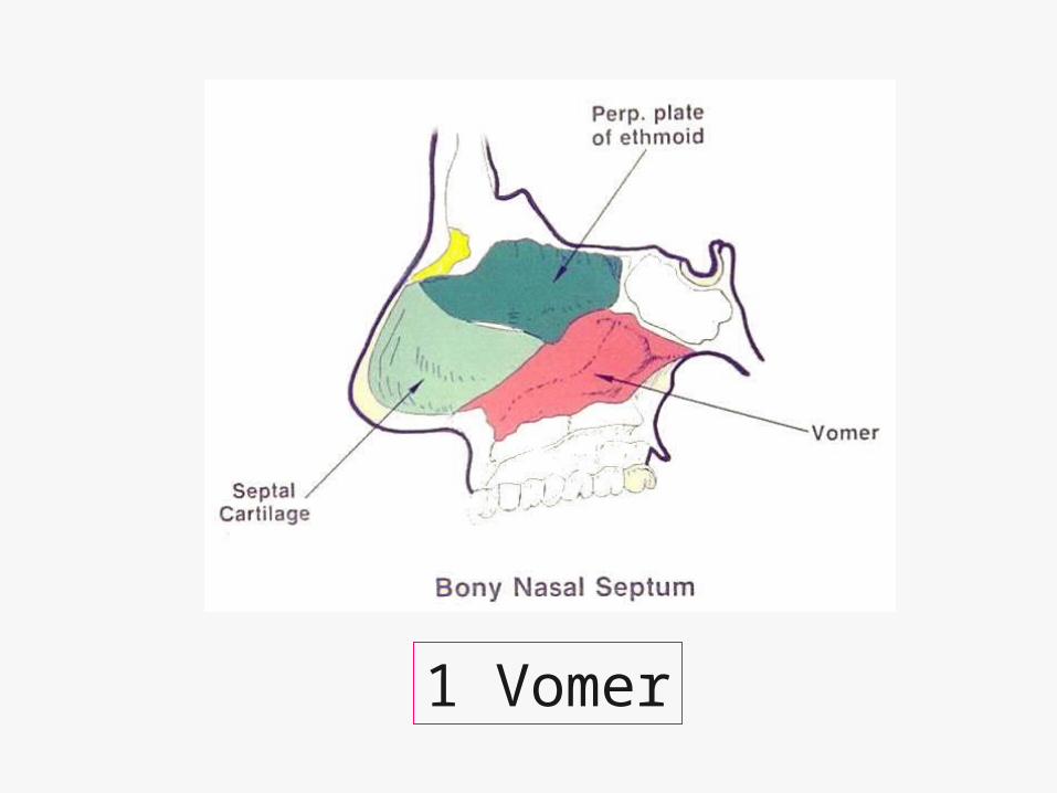
1 Vomer

1 Mandible
At approximately what age does the human eyeball reach maturity?
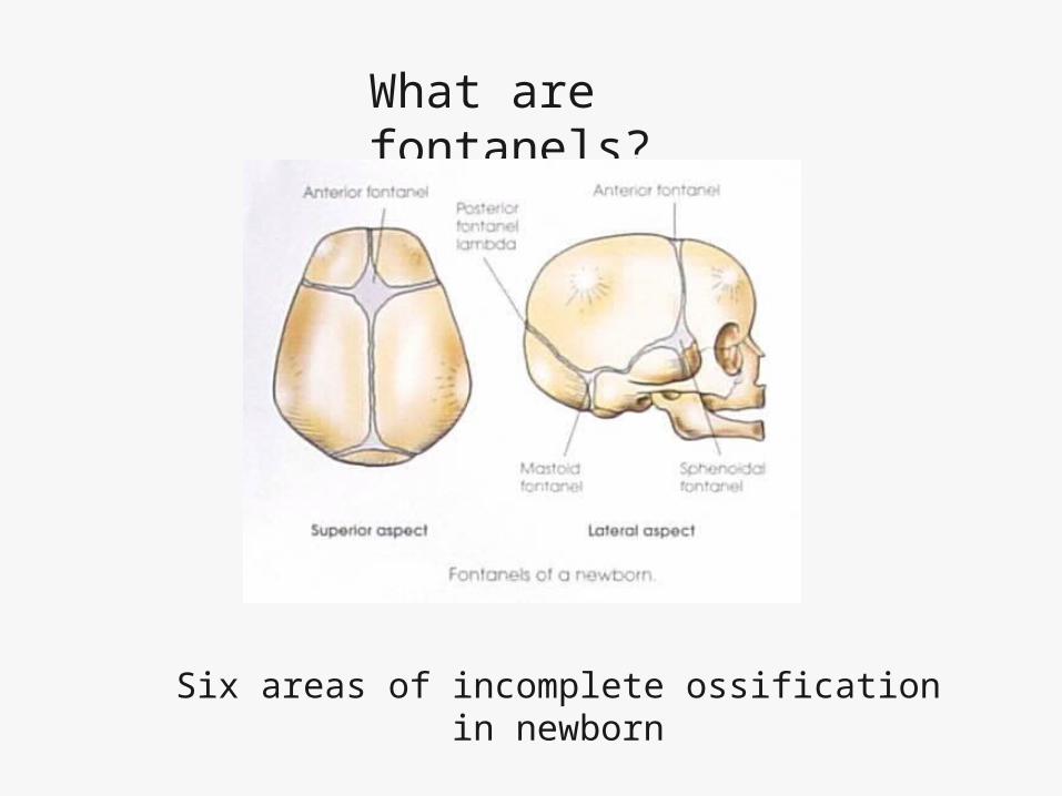
What are fontanels?
Six areas of incomplete ossification in newborn
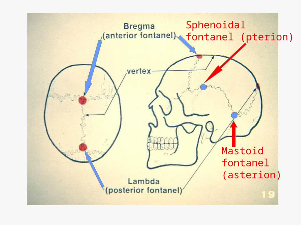
Mastoidfontanel(asterion)
Sphenoidalfontanel (pterion)

At what age do the fontanels close?
• Posterior and sphenoidal fontanels close during first 1-3 months after birth
• Anterior and mastoid fontanels close during 2nd year of life

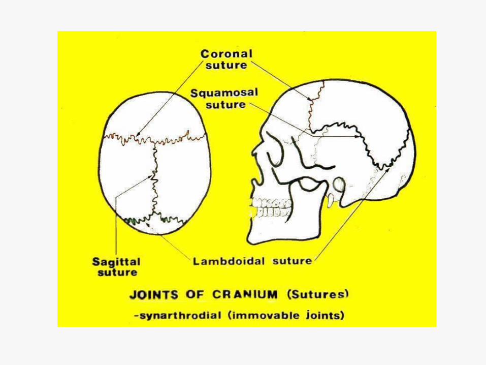
Radiographic Landmarks
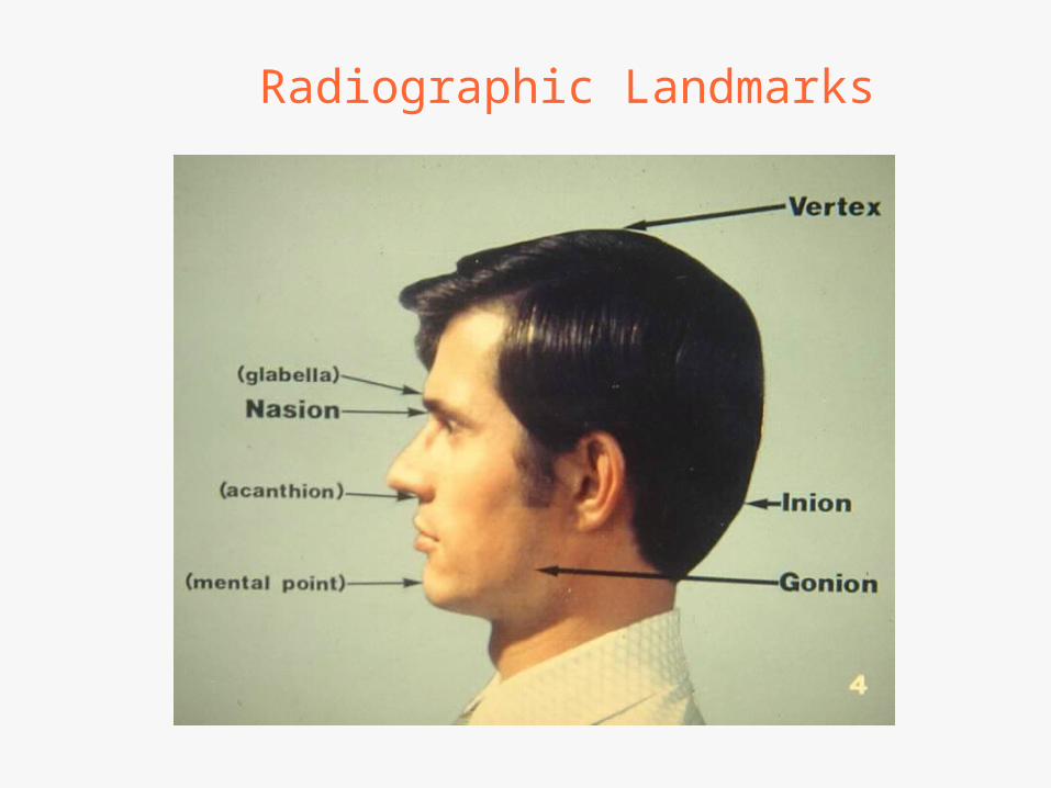
Radiographic Landmarks

Landmarks


Radiographic baselines

All skull positions are based on 3 factors
• Rotation
• Tilt
• Flexion-Extension

3 types of Skull Position change
• 1st type -
• Rotation -your head is rotating on an axis-your neck
• The “NO” position

2nd type of skull position change
• Flexion-extension
• Also called “Yes” position
ExtensionFlexion

3rd type of skull position change
• Tilt• Or “Maybe”
position

Skull Morphology
Mesocephalic Brachycephalic Dolichocephalic

Average Skull

Brachycephalic- (Broad)
Dolichocephalic-(thin)
Skull Morphology
Mesocephalic-(middle-average)


Review • A-Vomer
• B-Perp.plate
ethmoid
• C-Nasion
• D- inferior
nasal
conchae
• E- Anterior
nasal spine
A b
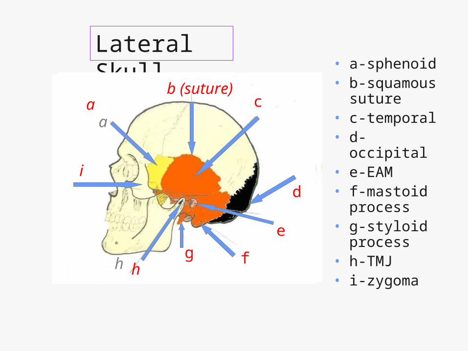
Lateral Skull • a-sphenoid
• b-squamous suture
• c-temporal• d-occipital• e-EAM• f-mastoid
process • g-styloid
process• h-TMJ• i-zygoma
c
d
e
fgh h
i
aa
b (suture)
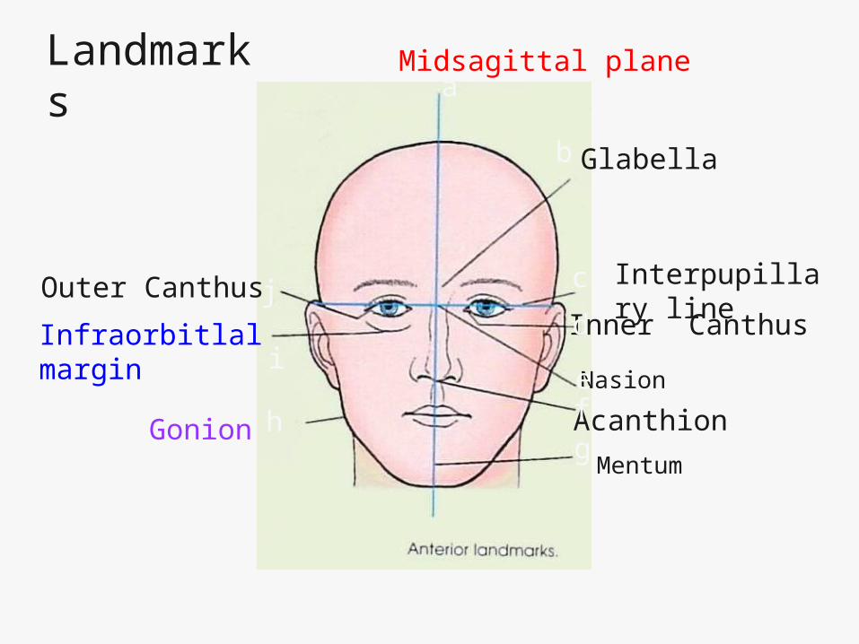
Landmarks
Midsagittal plane
Glabella
Interpupillary line
Inner Canthus
Nasion
Acanthion
Mentum
Outer Canthus
Gonion
Infraorbitlal margin
a
b
c
d
efg
h
i
j
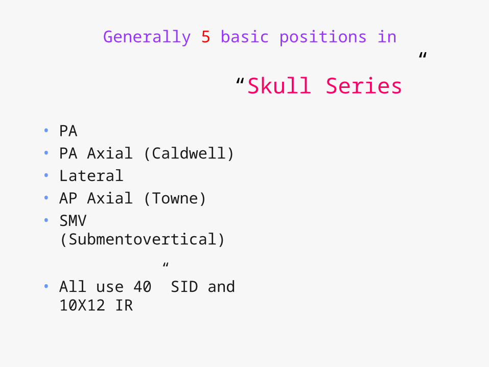
Generally 5 basic positions in
“Skull Series”
• PA• PA Axial (Caldwell)• Lateral• AP Axial (Towne)• SMV
(Submentovertical)
• All use 40” SID and 10X12 IR

PA projection

PA projection
• Forehead and nose touch IR
• CR perpendicular to IR (0 deg. Angle)
• Exit at nasion
• Cassette 10x12 lengthwise
O degrees
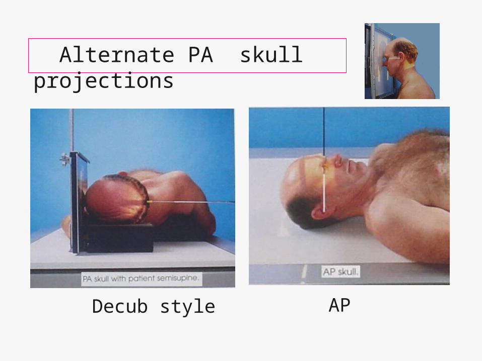
Alternate PA skull projections
Decub style AP

PA Skull- Evaluation Criteria
• Entire Cranium included
• Equal distance from lateral border of skull to lateral border of orbit on both sides
• Symmetric petrous pyramids filling orbits!
R

At approximately what age does human skull reach full size?
___ years old?
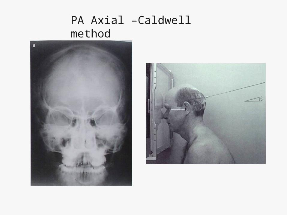
PA Axial –Caldwell method

PA Axial-Caldwell
• Exactly same as PA, except CR angled 15 degrees down!

PA Axial- Caldwell
Evaluation Criteria
• Same as PA except petrous ridges fill the lower 1/3 of orbits!

Which PA Axial projection is best?
A B

Compare the difference!
PA PA Axial
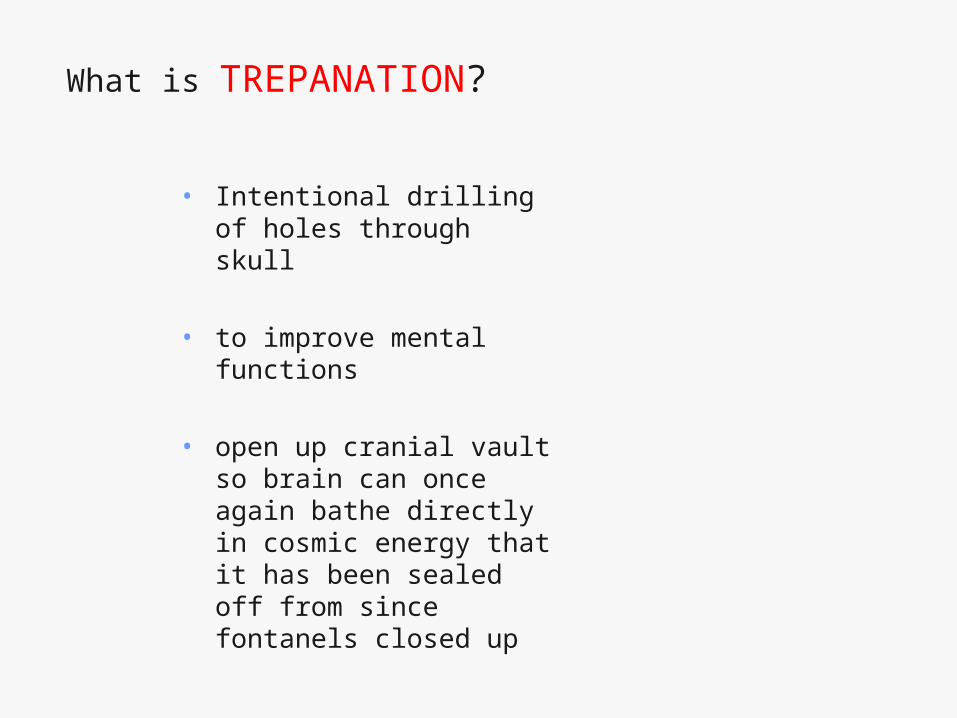
What is TREPANATION?
• Intentional drilling of holes through skull
• to improve mental functions
• open up cranial vault so brain can once again bathe directly in cosmic energy that it has been sealed off from since fontanels closed up
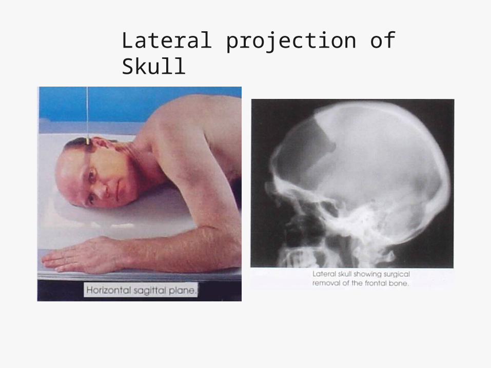
Lateral projection of Skull

Lateral projection of Skull
• 10x12 CW
• CR 2” superior to EAM
• Midsagittal plane parallel to IR
• Interpupillary line perpendicular to IR
• (IOML parallel to long axis of IR)

Lateral Skull- Evaluation Criteria
• Entire cranium without tilt or rotation
• Superimposed orbital roofs, and EAMs, TMJs
• Sella Turcica in profile
• No overlap of C-spine by mandible

What is wrong with this lateral?
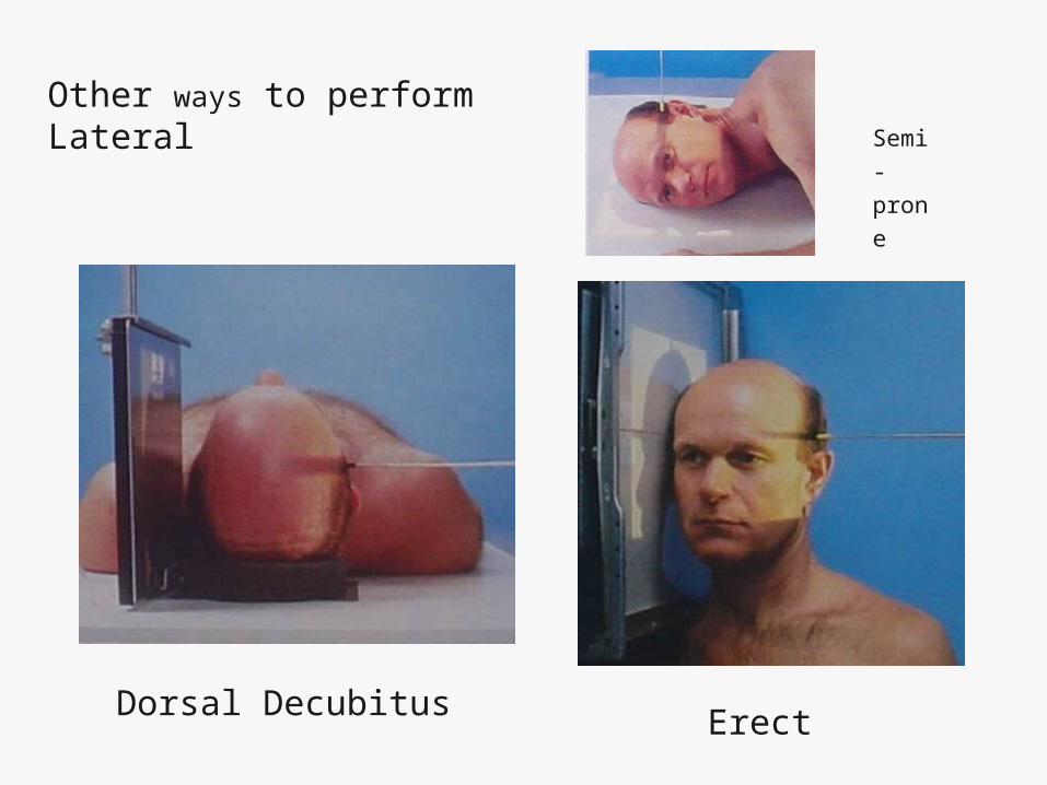
Other ways to perform Lateral
Dorsal Decubitus Erect
Semi-
prone

What projection and what is wrong?
R
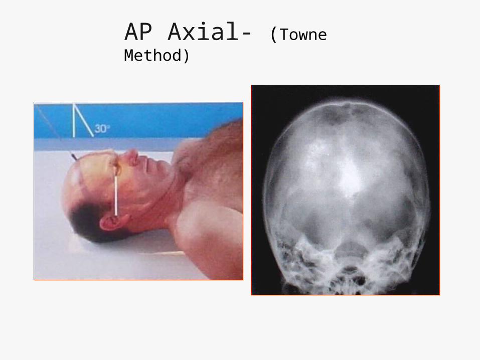
AP Axial- (Towne Method)
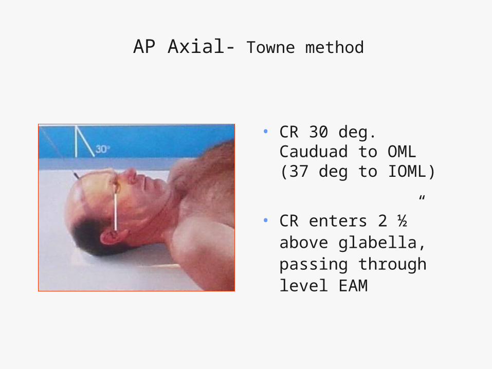
AP Axial- Towne method
• CR 30 deg. Cauduad to OML (37 deg to IOML)
• CR enters 2 ½” above glabella, passing through level EAM

AP Axial (Towne Method) - Evaluation Criteria
• No rotation (equal
distance from lateral
border of skull to lateral
margin of foramen
magnum)
• Symmetric petrous
ridges
• Dorsum sellae and
posterior clinoids
visible in foramen

Alternate ways to perform Towne
ErectLateral Decubitus

Submentovertical projection (SMV)

SMV
• CR-through Sella turcica (3/4” anterior
to EAM) Perpendicular to IOML
• IOML parallel to IR
• 10x12 cassette lengthwise
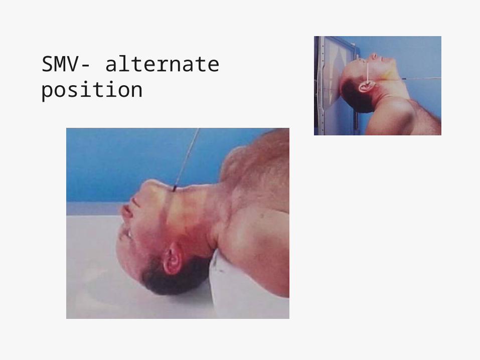
SMV- alternate position

SMV- Evaluation Criteria
• Equal distance from lateral border of skull to mandibular condyles on both sides (no tilt)
• Superimposition of mental protuberance over frontal bone
• Mandibular condyles anterior to petrous pyramids
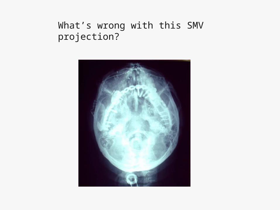
What’s wrong with this SMV projection?
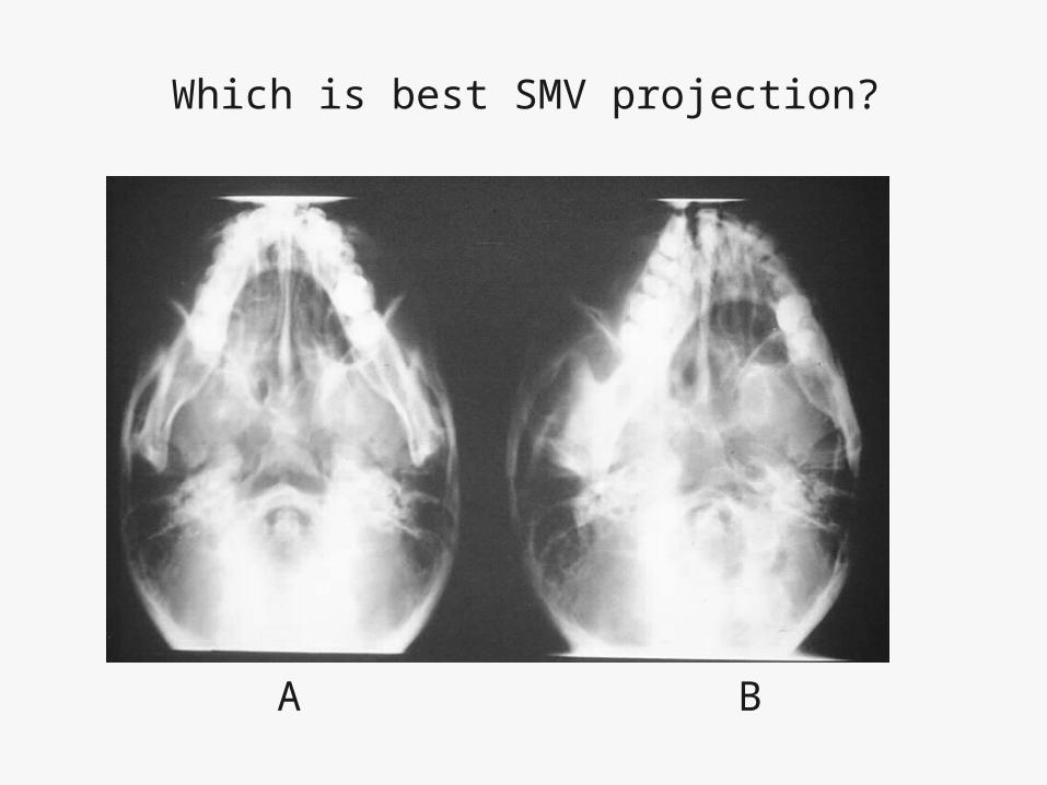
Which is best SMV projection?
A B

Which SMV projection is the best?
A B

? ?