Embed Size (px)
Citation preview

1463
Int J Ophthalmol, Vol. 14, No. 10, Oct.18, 2021 www.ijo.cnTel: 8629-82245172 8629-82210956 Email: [email protected]
·Basic Research·
Anti-viral activity of Staphylococcus aureus lysates against herpes simplex virus type-I infection: an in vitro and in vivo study
Tian-Lan Lin, Chao Cheng, Wei-Ting Zeng, Fang Duan, Yin-Hui Pei, Xiu-Ping Liu, Fu Shang, Kai-Li Wu
Zhongshan Ophthalmic Center, State Key Laboratory of Ophthalmology, Sun Yat-sen University, Guangzhou 510060, Guangdong Province, ChinaCorrespondence to: Kai-Li Wu. Zhongshan Ophthalmic Center, State Key Laboratory of Ophthalmology, Sun Yat-sen University, Guangzhou 510060, Guangdong Province, China. [email protected]: 2020-12-28 Accepted: 2021-02-05
Abstract● AIM: To investigate the effect of Staphylococcus aureus (S. aures) lysates (SALs) on herpes simplex virus type-I (HSV1) infection in human corneal epithelial (HCE) cells and in a mouse model of HSV1 keratitis.● METHODS: HCE, Vero, HeLa, and BV2 cells were infected with HSV1 [HSV1 f strain, HSV1f; HSV-1-H129 with green fluorescent protein (GFP) knock-in, HSV1g]. Pre- or post-infection, SAL at various concentrations was added to the culture medium for 24h. GFP fluorescence in HSV1g or plaque formation by HSV1f were examined. The effects of heat-treated SAL, precooled acetone-precipitated SAL, and SAL subjected to ultrafiltration (100 kDa) were evaluated. The effects of other bacterial components and lysates on HSV1 infection were also tested, including lipoteichoic acid (LTA), peptidoglycan (PGN), staphylococcal protein A (SPA), and α-hemolysin from S. aureus (α-toxin) as well as lysates from a wild-type S. aureus strain, S. epidermidis, and Escherichia coli (W-SAL, SEL, and ECL, respectively). In addition, SAL eye drops were applied topically to BALB/c mice with HSV1 keratitis, followed by in vivo observations. ● RESULTS: The cytopathic effect, plaque formation (HSV1f), and GFP expression (HSV1g) in infected cells were inhibited by SAL in a dose-dependent manner. The active component of SAL (≥100 kDa) was heat-sensitive and retained activity after acetone precipitation. In HSV1g-infected cells, treatment with LTA-sa, α-toxin, PGN-sa, or SPA did not inhibit GFP expression. SAL, W-SAL, and SEL
(but not ECL) decreased GFP expression. In mice with HSV1 keratitis, SAL reduced corneal lesions by 71%.● CONCLUSION: The results of this study demonstrate that SAL can be used to inhibit HSV1 infection, particularly keratitis. Further studies are needed to determine the active components and mechanism underlying the effects of SAL.● KEYWORDS: herpes simplex virus type-1; Staphylococcus aureus; infection; cornea epithelial cells; keratitisDOI:10.18240/ijo.2021.10.01
Citation: Lin TL, Cheng C, Zeng WT, Duan F, Pei YH, Liu XP, Shang F, Wu KL. Anti-viral activity of Staphylococcus aureus lysates against herpes simplex virus type-I infection: an in vitro and in vivo study. Int J Ophthalmol 2021;14(10):1463-1472
INTRODUCTION
H erpes simplex virus type 1 (HSV1) is a prevalent human pathogen that infects over 3.72 billion individuals
worldwide[1]. The virus causes a broad range of diseases, including labial herpes, ocular keratitis, genital disease, and encephalitis. Ocular infection with HSV1 and its associated sequelae account for the majority of corneal infections and can result in vision impairment or blindness[2]. HSV1 has a complex interaction with host responses. It establishes latent infection in neuronal cell bodies and becomes reactivated when triggered by internal or external factors[3].HSV1 is a linear double-stranded DNA virus and is a member of the α-herpes virus subfamily[4], characterized by a short replication cycle, high cytopathogenicity, and distinct neurotropism[5-8]. HSV1 replication is temporally regulated by immediate early (IE), early (E), and late (L) genes, usually occurring approximately 1-2h post-infection[9]. The main anti-HSV drugs are nucleoside analogs[10], such as acyclovir, which inhibits HSV DNA replication. A sufficiently safe and efficient vaccine is not currently available[11]. The development of new anti-HSV drugs is urgently needed because the extensive use of nucleoside analogs has led to drug resistance and side effects[10,12]. It has recently been

1464
noted that some natural products have anti-HSV activity, including compounds extracted from various plants, such as neem (Azadirachta indica L.) bark[13], Cornus canadensis[14], Houttuynia cordata[15-17], Phyllanthus urinaria[18], Prunella vulgaris[19], Euphorbia denticulata Lam[20], Baicalein [21], and Mallotus peltatus[22]. In addition, carrageenan-rich enzymatic extracted from Solieria filiformis[23], Brazilian propolis[24], and abalone hemocyanin[25] have anti-HSV1 effects. Importantly, some studies have reported that compounds derived from prokaryotes could block the replication of HSV. Tiwari et al[26] provide evidence that a novel 11-kDa sugar-binding antiviral protein, cyanovirin-N (CV-N), originally isolated from the cyanobacterium Nostoc ellipsosporum, acts as a potent inhibitor of HSV1 entry in natural target cells. Furthermore, human immunodeficiency virus (HIV) and HSV infections are inhibited by vaginal lactobacilli[27]. Antiviral effects of Lactobacillus crispatus against HSV-2 have been reported in mammalian cell lines[28]. Accordingly, prokaryotes, especially bacteria, are promising sources of potential antiviral drugs. Staphylococcus aureus (S. aureus), a gram-positive bacterium, is a human commensal or a potentially lethal opportunistic pathogen[29-30]. It is the leading cause of keratoconjunctivitis[31]. Lysates of S. aureus and other bacteria have been used empirically in Europe to prevent respiratory infections in children for the past 100y[32]; however, research has focused on their immunoregulatory properties and not on their impact on viruses. Purified components of S. aureus able to inhibit viral infection have been identified over the last few decades. For example, staphylococcal protein A (SPA) substantially increases anti-porcine circovirus type 2 (PCV2) monoclonal antibody bioactivity[33]. Lipoteichoic acid (LTA) is a potential innate immune stimulant that can be used against infectious laryngotracheitis virus (ILTV) in chickens[34]. Additionally, α-hemolysin from S. aureus (α-toxin) modulates the skin host response to HSV1 infection[35]. However, the effects of components of S. aureus on HSV1 infection are largely unknown. In this study, to address the gap in knowledge regarding the effects of S. aureus on HSV1 infection, the effects of S. aureus lysates (SALs) on HSV1 infection were evaluated in cultured cells and on the ocular surface of mice.SUBJECTS AND METHODSEthical Approval All procedures involving human participants were performed in accordance with the ethical standards of the institutional and/or national research committee and with the 1964 Helsinki declaration and its later amendments or comparable ethical standards. Informed consent was obtained from all individual participants included in the study. All procedures were conducted in accordance with the Association for Research in Vision and Ophthalmology (ARVO) Statement for the Use of Animals in Ophthalmic and
Vision Research and the guidelines of the Ophthalmic Animal Laboratory of the Zhongshan Ophthalmic Center. The research protocol was approved by the Animal Care Committee of the Zhongshan Ophthalmic Center at Sun Yat-sen University (Approval #: 2017-102).Culture of Cells, Viruses, and Bacteria Transformed human corneal epithelial (HCE) cells (RCB2280; RIKEN Cell Bank, Tokyo, Japan) were cultured in Dulbecco’s modified Eagle medium (DMEM)/F-12 (Gibco, Grand Island, NY, USA) containing 10% fetal bovine serum (FBS; Gibco), 10 ng/mL human epidermal growth factor (hEGF; Sigma-Aldrich, St. Louis, MO, USA), 5 μg/mL insulin (Sigma-Aldrich), and 1% penicillin/streptomycin (Gibco) at 37℃ in a humidified atmosphere containing 5% CO2. Both Vero cells (African green monkey kidney cells, CCL-81; American Type Culture Collection, ATCC, Manassas, VA, USA) and HeLa cells (Henrietta Lacks strain of cancer cells; ATCC CCL-2) were grown in DMEM (Invitrogen, Carlsbad, CA, USA) supplemented with 10% FBS and 1% penicillin/streptomycin in an incubator at 37℃ with 5% CO2 and saturated humidity. BV2 microglial cells (ATCC CRL-2467TM) were maintained in high-glucose DMEM (Invitrogen) with 5% FBS. The HSV1 F strain[36] (HSV1f) and green fluorescent protein (GFP)-expressing HSV1 strain (HSV-1 strain H129 with knocked-in GFP, HSV1g, with a cloned-in GFP gene, a gift from professor Minhua Luo, Wuhan Institute of Virology, Chinese Academy of Sciences) were propagated and titrated on Vero cells. A virus titer of 105 PFU/mL [multiplicity of infection (MOI)=1] was determined by a plaque assay, and viral particles with a MOI of 1.0 or 0.1 were used in all experiments[36].The bacterial culture was performed as described previously[37]. S. aureus (ATCC 25923) was obtained as a lyophilized powder and was stored at 4℃ until use. A wild-type S. aureus strain was isolated from a patient with blepharitis by individual skin swabs using cotton sticks. Pure cultures of E. coli (ECL, ATCC 25922) and Staphylococcus epidermidis (SEL, ATCC 29887) were grown overnight at 37℃ on LB media. All bacteria were initially subcultured on chocolate agar plates (bioMérieux, Craponne, France) for 24h at 37℃. Bacterial colonies were routinely identified in a clinical laboratory in the Zhongshan Ophthalmic Center. Preparation of Bacterial Lysates A bacterial colony was picked and suspended in sodium chloride broth, 7.5% (HKM, Ltd., Guangdong Province, China) for 24h to increase the bacterial yield. The cultures were centrifuged at 10 000× g for 10min and the supernatant was discarded. The pellet containing the bacteria was washed three times with pyrogen-free phosphate-buffered saline (PBS; Gibco). The pellet was resuspended in PBS, vortexed thoroughly, diluted to the proper
Anti-HSV-1 effect of S. aureus lysates

1465
Int J Ophthalmol, Vol. 14, No. 10, Oct.18, 2021 www.ijo.cnTel: 8629-82245172 8629-82210956 Email: [email protected]
concentration, and dispensed into EP tubes. TZP beads (0.4-0.6 mm; Hualian Hi-Tech Ltd., Suzhou, China) were added and the bacteria were lysed using a tissue grinder (Scientz, Ningbo, China) for 0.5min×65 Hz at 3-min intervals on ice. The bacterial lysates were centrifuged (12 000× g, 4℃) for 15min. The supernatants for SAL, SEL, or ECL were filtered with 0.22-μm filters (Merck Millipore, Tullagreen, Ireland) and stored at -80℃ for further use. The protein concentrations were measured using the BCA protein assay kit (Beyotime, Shanghai, China), as described previously, to quantify active substances in various lysates[38]. HSV1 Infection in Cultured Cells HCE, Vero, HeLa, or BV2 cells were infected with HSV1 (HSV1f or HSV1g) at different MOIs following a previously described method[39]. Pre- and post-virus infection, SAL was added at various concentrations to the culture medium, and the cells were cultured for 24h. GFP fluorescence of HSV1g in cells was examined under a fluorescence microscope and images were obtained. Plaque formation by HSV1f was examined as described previously[36]. Vero cells were grown in 12-well plates to 70%-80% confluence; cells were treated with SAL and infected with HSV1f as described above. After 1h, the cells were overlaid with 1 mL of a 1:1 mixture of low-melting-temperature agarose (NuSieve GTG Agarose; Lonzo, Morristown, NJ, USA) and 2×DMEM/high glucose to permit only cell-to-cell spread of the virus. At 48h, the agarose was removed carefully. The plates were stained with crystal violet for 20min and images were obtained. Finally, plaque sizes were measured.Cell Viability Assay Cell viability was assessed by a cell counting kit-8 (CCK-8) assay (Dojindo, Kumamoto, Japan) according to the manufacturer’s instructions. Briefly, HCE or Vero cells were seeded in 96-well culture plates at a density of 7000 cells per well and cultured overnight. Then, cells were incubated with various concentrations of SAL (0, 0.01, 0.05, 0.1, 0.3, and 0.5 mg/mL) for 24h. After treatment, cells were washed twice with PBS. Then 100 μL of cell viability detection solution (10 μL of CCK-8 reaction solution and 90 μL of serum-free medium) was added to each well, and the cells were incubated for 2h at 37℃ in the dark. Absorbance was measured at 450 nm using SynergyH1 (BioTek, Winooski, VT, USA). Treatment of HSV1-infected Cells SAL at the indicated concentrations was added to the medium of various cultured cells with or without HSV1 infection. Next, the effects of several purified components of S. aureus, i.e., LTA-sa[34] (0.02 mg/mL; InvivoGen, San Diego, CA, USA), peptidoglycan-sa (PGN-sa)[40] (0.04 mg/mL; InvivoGen), SPA[41] (0.04 mg/mL; Sigma-Aldrich), and α-toxin[35] (40 ng/mL; Sigma-Aldrich), on HSV1g infection in cultured cells were evaluated. Additionally, SAL was subjected to the following pretreatments: 1) heated
at 100℃ for 30min; 2) precooled, acetone-precipitated, and resuspended in PBS; and 3) ultrafiltration with a molecular weight cut-off of 100 kDa (Merck Millipore), separating SAL into a <100 kDa fraction and a fraction that did not permeate the ultrafiltration membrane. For validation, all pretreated SALs were separated by SDS-PAGE and subjected to Coomassie blue R-250 staining using a commercial kit following the manufacturer’s instructions (Beyotime, Shanghai, China). The effects of lysates of ATCC25923 (SAL), the wild-type strain of S. aureus (W-SAL), SEL, and ECL on HSV1g infection in target cells were compared.Effects of SAL on a Mouse Model of HSV1f Keratitis Twenty-four BALB/c female mice, aged 6-8wk were purchased from the Animal Supply Center of Sun Yat-sen University (Guangzhou, China). The mice were quarantined and acclimatized for a week before the experiments. The mice were free of clinically observable ocular surface disease before the study. The mice were randomly assigned to three groups (eight mice per group), and the right eye of each mouse was chosen for the experiment. Animal models of herpes simplex keratitis were established based on our previously described method[42]. SAL eye drops (1 mg/mL in 10 μL of PBS) or PBS drops only were applied topically four times the day before infection and continued for 3d post-infection. Corneal changes were examined daily using a slit-lamp microscope (Sun Kingdom Medical Instruments, Chongqing, China) and images were obtained using a digital camera. Alterations in epithelial lesions were evaluated as increased (ineffective), unchanged, or decreased (effective).Statistical Analysis All statistical analyses were conducted with ANOVA and post-hoc tests. Fluorescence was quantified using Image J (http://rsb.info.nih.gov/ij/). Values of P<0.05 were considered statistically significant.RESULTSAnti-HSV1 Infection Effects of SAL in Vitro To test the effects of SAL on HSV1 infection, we treated HCE cells with SAL pre- or post-HSV1g infection. As shown in Figure 1, the inhibitory effects of SAL on HSV1 infection were observed in HCE cells treated with SAL both pre- and post-HSV1g infection. When HCE cells were treated with SAL for 12h before infection, substantial reductions in the cytopathic effect (CPE) and GFP expression were observed. Similarly, the addition of SAL to the media after HSV1g infection resulted in the inhibition of viral infection. Thus, SAL treatment post-infection was used in subsequent experiments.Figure 2 summarizes the inhibitory effects of SAL on HSV1 infection in different cell types. When 0.04 mg/mL SAL was added to the culture media, the CPE and HSV1g GFP fluorescence decreased significantly in HCE, Vero, HeLa, and

1466
BV2 cell lines (Figure 2A, 2C). In HCE cells, as determined by a plaque assay, HSV1f induced the formation of a large number of plaques and treatment with SAL significantly decreased the plaque sizes and numbers (Figure 2B, 2D). Dose- and Time-dependent Antiviral Activity of SAL HSV1g infection was inhibited by SAL in a dose-dependent manner during the entire culture period (Figure 3). In contrast to the continuous increase in GFP expression in HSV1g-infected HCE cells, the addition of SAL decreased GFP fluorescence at 12 and 24h (Figure 3A, 3C) and at 36 and 48h (data not shown). Consistent with the results of the plaque assay for HSV1f, when HSV1g-infected HCE cells were cultured with various concentrations of SAL, GFP fluorescence decreased in a dose-dependent manner (Figure 3B, 3D). HSV1g replication was inhibited by greater than 90% by 0.06 mg/mL SAL.Effect of SAL on the Viability of HCE and Vero cells To determine the safe concentration range of SAL in cells, HCE or Vero cells were treated with different doses of SAL (0, 0.01, 0.05, 0.1, 0.3, and 0.5 mg/mL) for 24h, and cell viability
was evaluated by a CCK-8 assay. As shown in Figure 4, SAL concentrations ranging from 0.01 to 0.1 mg/mL did not reduce cell viability. However, when the concentrations of SAL were ≥0.3 mg/mL, the viability of HCE and Vero cells was significantly lower than that of untreated control cells.Antiviral Effects of Pretreated SAL HSV1g-affected HCE were treated by SAL that was prepared by various methods (Figure 5). When SAL was heat-treated, its inhibitory effect on viral infection was lost, indicating that the active component was heat-sensitive. When SAL was precipitated by cooled acetone and resuspended in PBS, it retained the ability to reduce GFP fluorescence in HSV1g-infected cells, indicating that the active components of SAL could be precipitated by cold acetone. After ultrafiltration, the SAL fraction that did not permeate the ultrafiltration membrane retained the inhibitory activity against GFP expression in HSV1g-infected cells. However, components of <100 kDa did not show obvious inhibitory effects on GFP expression. In addition, pretreated SALs were subjected to SDS-PAGE to examine changes
Figure 1 Comparison of the inhibitory effects of SAL on HSV1g infection when administered pre- or post-infection HCE cells were infected with HSV1g (HSV-1-H129 with GFP knock-in) at an MOI of 1.0. SAL (0.04 mg/mL) was added to the medium 12h before viral infection (upper panel) or 1h post-infection (lower panel); phase-contrast images and GFP fluorescence were obtained by microscopy under the same magnification (scale bar=50 μm). Both pre- and post-virus-infection, the addition of SAL to the culture medium resulted in substantial decreases in cell lesions and in HSV1g GFP expression.
Anti-HSV-1 effect of S. aureus lysates

1467
Int J Ophthalmol, Vol. 14, No. 10, Oct.18, 2021 www.ijo.cnTel: 8629-82245172 8629-82210956 Email: [email protected]
in protein components (Figure 5C). We found that heated-SAL and the low-molecular-weight fraction obtained by ultrafiltration (<100 kDa) displayed distinct band patterns compared with those for the other three protein samples.Effects of Purified Components of S. aureus on HSV1 Infection Figure 6 presents a comparison of the effects of SAL and different purified components of S. aureus. The addition of SAL obviously inhibited GFP expression in HSV1g-infected cells. Upon the addition of LTA, the fluorescence signal was weakened. However, target cells were sensitive to LTA,
with significant cell death observed by phase microscopy. In contrast to SAL, α-toxin resulted in strong GFP expression in cells. The addition of SPA or PGN did not significantly alter GFP fluorescence in HSV1g-infected cells; however, there were slightly increases in fluorescence intensities. Taken together, these results suggest that LTA, α-toxin, SPA, and PGN were not the main active components in SAL responsible for the inhibition of viral infection.Comparison Between SAL and Lysates of Different Bacteria Lysates of a gram-negative bacterium (ECL), gram-
Figure 2 Comparison of the inhibitory effects of SAL on different cells infected with HSV1, as determined by GFP fluorescence (A, C) and plaque assays (B, D) A: SAL at 0.04 mg/mL was added to HSV1g-infected HCE, Vero, HeLa, or BV2 cells for 24h. Phase-contrast images were obtained and GFP fluorescence was evaluated by microscopy under the same magnification (scale bar=50 µm). B: For a plaque test, HCE cells were infected with HSV1f (MOI 0.01) for 1h, followed by the addition of SAL (0, 0.02, 0.04, and 0.08 mg/mL) for 24h. The control group (Ctl) consisted of cells without viral infection and SAL treatment. C: Sum of fluorescence intensities in A. bP<0.01 vs without SAL. D: Average plaque areas calculated from B. bP<0.01 vs HSV1f-infected cells without SAL. Each experiment was repeated three or more times.

1468
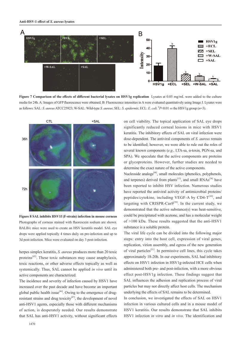
positive bacterium (SEL), and W-SAL were added at 0.05 mg/mL to the culture media to determine their effects on HSV1 infection (Figure 7). Compared with levels in untreated cells, SAL and W-SAL clearly inhibited GFP expression in cells infected with HSV1g. SEL displayed moderate inhibitory effects on GFP expression. In contrast, ECL increased GFP expression in target cells. These results suggested that S. aureus has unique anti-HSV1 substances.Anti-HSV1 Effects of SAL in Vivo We attempted to use SAL to treat HSV1f-infected corneas via eye drops. Disruptions of the corneal epithelium were observed in animals when
examined by slit-lamp microscopy and were stained with fluorescein sodium (FLS) post-infection (Figure 8). SAL eye drops or PBS drops were applied topically pre- (24h) and post-infection (up to 72h) every 4h to BALB/c mice with HSV1 keratitis. Corneal damage was obviously reduced by SAL compared with that in untreated animals on Day 3 post-infection (5 of 7 SAL-treated eyes vs 1 out of 8 control eyes; Table 1).DISCUSSION This study focused on the combined effects of bacterial lysates and HSV1 in infection models. Immunoregulatory and immunostimulatory effects of bacterial lysates in respiratory
Figure 3 SAL inhibits HSV1g infection in a dose-dependent manner and HCE cell lines were infected with HSV1g A: Comparison of the inhibitory effects of SAL at 12 and 24h. The control group (Ctl) consisted of cells without virus infection and with SAL treatment. B: HSV1g-infected HCE cells were incubated with 0, 0.02, 0.04, and 0.08 mg/mL SAL for 24h. Images of GFP fluorescence were obtained. C: Comparison of GFP fluorescence intensities in A. bP<0.01. D: Average fluorescence intensities calculated from B. bP<0.01 vs HSV1g infected cells without SAL. All photograms were obtained at the same magnification (scale bar=50 µm). Each experiment was repeated at least three times.
Figure 4 Effects of SAL on the viability of HCE and Vero cells HCE (A) and Vero cells (B) were treated with the indicated concentrations of SAL, and cell viability was determined by the CCK-8 assay (aP<0.05, bP<0.01 compared with the control group). Each experiment was repeated three times.
Anti-HSV-1 effect of S. aureus lysates

1469
Int J Ophthalmol, Vol. 14, No. 10, Oct.18, 2021 www.ijo.cnTel: 8629-82245172 8629-82210956 Email: [email protected]
infections and asthma have been reported[32]. For example, Broncho-Vaxom (OM85-BV), a mixture of extracts from eight strains of gram-positive (including S. aureus) and gram-negative bacteria, promotes anti-infection immune responses[43]. Antigens have been obtained from mass cultures
of appropriate bacterial strains by mechanical cell disruption (polyvalent mechanical bacterial lysate) or chemical proteolysis (polyvalent chemical bacterial lysate)[44]. Additionally, bacterial lysates have a long history of use for the prevention of respiratory infections in children[32]. In the present study, we discovered that SAL could inhibit HSV1 infection, providing a basis for the development of novel anti-HSV1 agents. To our knowledge, this is the first evaluation of the interaction between SAL and HSV infection. However, the precise active components remain to be identified. In addition, although the antiviral components are effective in our mouse models of
Figure 5 HSV1g infection was affected by SAL prepared under different conditions A: Similar to control cells (Ctl), GFP expression in HSV1g-infected HCE cells was retained in heated-SAL and the low-molecular-weight fraction obtained by ultrafiltration (<100 kDa). All cells were treated with 0.05 mg/mL protein, except for the control group. B: Fluorescence intensities in A were quantified using Image J (bP<0.01 vs Ctl, n=3). C: SDS-PAGE results for different SAL preparations used in A. UF_H_fraction included proteins that did not permeate the ultrafiltration membrane (cut-off 100 kDa), labeled ≥100 kDa in A and B, and UF_L_fraction contained proteins that penetrated the ultrafiltration membrane. For all lanes, 30 µg of protein was used, except UF_L_fraction, run with 8 μg of protein.
Figure 6 Comparison of the effects of purified components of S. aureus on cells infected with HSV1g A: SAL (0.04 mg/mL), LTA (0.02 mg/mL), PGN (0.04 mg/mL), SPA (0.04 mg/mL), and α-Toxin (40 ng/mL) from S. aureus were added to HeLa cells. Images of GFP fluorescence were obtained. B: Fluorescence intensities in A were calculated from triplicate experiments. aP<0.05, bP<0.01 vs the HSV1g group.
Table 1 SAL effects on corneal ulcers of mouse HSV1 keratitis
Group Decreased Unchanged Increased χ2 P
HSV1f+SAL 5 (71.43%) 1 (14.29%) 1 (14.29%)6.060 0.028
HSV1f+PBS 1 (12.50%) 1 (12.50%) 6 (75.00%)
SAL: Staphylococcus aureus lysates.
1470
herpes simplex keratitis, S. aureus produces more than 20 toxic proteins[45]. These toxic substances may cause anaphylaxis, toxic reactions, or other adverse effects topically as well as systemically. Thus, SAL cannot be applied in vivo until its active components are characterized.The incidence and severity of infection caused by HSV1 have increased over the past decade and have become an important global public health issue[46]. Owing to the emergence of drug-resistant strains and drug toxicity[47], the development of novel anti-HSV1 agents, especially those with different mechanisms of action, is desperately needed. Our results demonstrate that SAL has anti-HSV1 activity, without significant effects
on cell viability. The topical application of SAL eye drops significantly reduced corneal lesions in mice with HSV1 keratitis. The inhibitory effects of SAL on viral infection were dose-dependent. The antiviral components of S. aureus remain to be identified; however, we were able to rule out the roles of several known components (e.g., LTA-sa, α-toxin, PGN-sa, and SPA). We speculate that the active components are proteins or glycoproteins. However, further studies are needed to determine the exact nature of the active components. Nucleoside analogs[48], small molecules (phenolics, polyphenols, and terpenes) derived from plants[13], and small RNAs[36] have been reported to inhibit HSV infection. Numerous studies have reported the antiviral activity of antimicrobial proteins/peptides/cytokine, including VEGF-A by CD4-T[49], and targeting with CRISPR-Cas9[50]. In the current study, we demonstrated that the active substance(s) was heat-sensitive, could be precipitated with acetone, and has a molecular weight of >100 kDa. These results suggested that the anti-HSV1 substance is a soluble protein.The viral life cycle can be divided into the following major steps: entry into the host cell, expression of viral genes, replication, virion assembly, and egress of the new generation of viral particles[51]. In permissive cell lines, this cycle takes approximately 18-20h. In our experiments, SAL had inhibitory effects on HSV1 infection in HSV1g-infected HCE cells when administered both pre- and post-infection, with a more obvious effect post-HSV1g infection. These findings suggest that SAL influences the adhesion and replication process of viral particles but may not directly affect host cells. The mechanism underlying the effects of SAL remains to be determined.In conclusion, we investigated the effects of SAL on HSV1 infection in various cultured cells and in a mouse model of HSV1 keratitis. Our results demonstrate that SAL inhibits HSV1 infection in vitro and in vivo. The identification and
Figure 7 Comparison of the effects of different bacterial lysates on HSV1g replication Lysates at 0.05 mg/mL were added to the culture media for 24h. A: Images of GFP fluorescence were obtained; B: Fluorescence intensities in A were evaluated quantitatively using Image J. Lysates were as follows: SAL: S. aureus ATCC25923; W-SAL: Wild-type S. aureus; SEL: S. epidermis; ECL: E. coli. bP<0.01 vs the HSV1g group (n=3).
Figure 8 SAL inhibits HSV1f (F-strain) infection in mouse corneas Photographs of corneas stained with fluorescein sodium are shown. BALB/c mice were used to create an HSV keratitis model. SAL eye drops were applied topically 4 times daily on pre-infection and up to 3d post-infection. Mice were evaluated on day 3 post-infection.
Anti-HSV-1 effect of S. aureus lysates

1471
Int J Ophthalmol, Vol. 14, No. 10, Oct.18, 2021 www.ijo.cnTel: 8629-82245172 8629-82210956 Email: [email protected]
characterization of the active component of SAL merits further research and is expected to aid in the development of therapeutic agents against HSV infection. ACKNOWLEDGEMENTSWe thank Prof. Min-Hua Luo (Wuhan Institute of Virology, Chinese Academy, Chinese Academy of Sciences) for the gift of HSV1 strain-H129 knocked-in GFP gene, and Prof. Huangxuan Shen (Zhongshan Ophthalmic Center, Sun Yat-sen University) for the gifts of HeLa cells (ATCC CCL-2) and BV2 microglial cells (ATCC® CRL-2467TM). Foundations: Supported by the National Natural Science Foundation of China (No.81770896; No.81970848); the Guangzhou Science Technology and Innovation Commission (No.201607020011).Conflicts of Interest: Lin TL, None; Cheng C, None; Zeng WT, None; Duan F, None; Pei YH, None; Liu XP, None; Shang F, None; Wu KL, None.REFERENCES
1 Khan AA, Srivastava R, Chentoufi AA, Kritzer E, Chilukuri S, Garg S,
Yu DC, Vahed H, Huang L, Syed SA, Furness JN, Tran TT, Anthony
NB, McLaren CE, Sidney J, Sette A, Noelle RJ, BenMohamed L.
Bolstering the number and function of HSV-1-specific CD8+ effector
memory T cells and tissue-resident memory T cells in latently infected
trigeminal Ganglia reduces recurrent ocular herpes infection and
disease. J Immunol 2017;199(1):186-203.
2 Miner JJ, Platt DJ, Ghaznavi CM, Chandra P, Santeford A, Menos AM,
Dong ZY, Wang ER, Qian W, Karozichian ES, Philips JA, Apte RS.
HSV-1 and zika virus but not SARS-CoV-2 replicate in the human
cornea and are restricted by corneal type III interferon. Cell Rep
2020;33(5):108339.
3 Su AR, Wang HR, Li YL, Wang XH, Chen DY, Wu ZW. Opposite roles
of RNase and kinase activities of inositol-requiring enzyme 1 (IRE1)
on HSV-1 replication. Viruses 2017;9(9):E235.
4 Laine RF, Albecka A, van de Linde S, Rees EJ, Crump CM, Kaminski
CF. Structural analysis of herpes simplex virus by optical super-
resolution imaging. Nat Commun 2015;6:5980.
5 Poccardi N, Rousseau A, Haigh O, Takissian J, Naas T, Deback C,
Trouillaud L, Issa M, Roubille S, Juillard F, Efstathiou S, Lomonte P,
Labetoulle M. Herpes simplex virus 1 replication, ocular disease, and
reactivations from latency are restricted unilaterally after inoculation of
virus into the lip. J Virol 2019;93(24):e01586-e01519.
6 Szpara ML, Gatherer D, Ochoa A, Greenbaum B, Dolan A, Bowden
RJ, Enquist LW, Legendre M, Davison AJ. Evolution and diversity in
human herpes simplex virus genomes. J Virol 2014;88(2):1209-1227.
7 Musarrat F, Chouljenko V, Kousoulas KG. Cellular and viral
determinants of HSV-1 entry and intracellular transport towards nucleus
of infected cells. J Virol 2021:JVI.02434-JVI.02420.
8 Tognarelli EI, Palomino TF, Corrales N, Bueno SM, Kalergis AM,
González PA. Herpes simplex virus evasion of early host antiviral
responses. Front Cell Infect Microbiol 2019;9:127.
9 Suzich JB, Cliffe AR. Strength in diversity: understanding the pathways
to herpes simplex virus reactivation. Virology 2018;522:81-91.
10 Zinser E, Krawczyk A, Mühl-Zürbes P, Aufderhorst U, Draßner C,
Stich L, Zaja M, Strobl S, Steinkasserer A, Heilingloh CS. A new
promising candidate to overcome drug resistant herpes simplex virus
infections. Antiviral Res 2018;149:202-210.
11 Naidu SK, Nabi R, Cheemarla NR, Stanfield BA, Rider PJ,
Jambunathan N, Chouljenko VN, Carter R, Del Piero F, Langohr I,
Kousoulas KG. Intramuscular vaccination of mice with the human
herpes simplex virus type-1 (HSV-1) VC2 vaccine, but not its parental
strain HSV-1(F) confers full protection against lethal ocular HSV-1
(McKrae) pathogenesis. PLoS One 2020;15(2):e0228252.
12 Zeng ZY, Zhang RH, Hong W, Cheng YT, Wang HJ, Lang YG, Ji
ZL, Wu YL, Li WX, Xie YL, Cao ZJ. Histidine-rich modification of
a scorpion-derived peptide improves bioavailability and inhibitory
activity against HSV-1. Theranostics 2018;8(1):199-211.
13 Tiwari V, Darmani NA, Yue BY, Shukla D. In vitro antiviral activity
of neem (Azardirachta indica L.) bark extract against herpes simplex
virus type-1 infection. Phytother Res 2010;24(8):1132-1140.
14 Lavoie S, Côté I, Pichette A, Gauthier C, Ouellet M, Nagau-Lavoie
F, Mshvildadze V, Legault J. Chemical composition and anti-herpes
simplex virus type 1 (HSV-1) activity of extracts from Cornus
canadensis. BMC Complement Altern Med 2017;17(1):123.
15 Hung PY, Ho BC, Lee SY, Chang SY, Kao CL, Lee SS, Lee CN.
Houttuynia cordata targets the beginning stage of herpes simplex virus
infection. PLoS One 2015;10(2):e0115475.
16 Chen X, Wang Z, Yang Z, Wang J, Xu Y, Tan RX, Li E. Houttuynia
cordata blocks HSV infection through inhibition of NF-κB activation.
Antiviral Res 2011;92(2):341-345.
17 Li T, Liu LB, Wu HL, Chen SD, Zhu QC, Gao H, Yu XT, Wang Y,
Su WH, Yao XS, Peng T. Anti-herpes simplex virus type 1 activity of
Houttuynoid A, a flavonoid from Houttuynia cordata Thunb. Antiviral
Res 2017;144:273-280.
18 Yang CM, Cheng HY, Lin TC, Chiang LC, Lin CC. The in vitro
activity of geraniin and 1,3,4,6-tetra-O-galloyl-beta-D-glucose isolated
from Phyllanthus urinaria against herpes simplex virus type 1 and type
2 infection. J Ethnopharmacol 2007;110(3):555-558.
19 Zhang Y, But PP, Ooi VE, Xu HX, Delaney GD, Lee SH, Lee SF.
Chemical properties, mode of action, and in vivo anti-herpes activities
of a lignin-carbohydrate complex from Prunella vulgaris. Antiviral Res
2007;75(3):242-249.
20 Shamsabadipour S, Ghanadian M, Saeedi H, Rahimnejad MR,
Mohammadi-Kamalabadi M, Ayatollahi SM, Salimzadeh L.
Triterpenes and steroids from euphorbia denticulata lam. with anti-
herpes symplex virus activity. Iran J Pharm Res 2013;12(4):759-767.
21 Luo Z, Kuang XP, Zhou QQ, Yan CY, Li W, Gong HB, Kurihara H,
Li WX, Li YF, He RR. Inhibitory effects of baicalein against herpes
simplex virus type 1. Acta Pharm Sin B 2020;10(12):2323-2338.
22 Bag P, Chattopadhyay D, Mukherjee H, Ojha D, Mandal N, Sarkar
MC, Chatterjee T, Das G, Chakraborti S. Anti-herpes virus activities of

1472
bioactive fraction and isolated pure constituent of Mallotus peltatus: an
ethnomedicine from Andaman Islands. Virol J 2012;9:98.
23 Ana P, Nathalie B, Gilles B, Daniel R, Tomás MS, Yolanda FP. Anti-
Herpes simplex virus (HSV-1) activity and antioxidant capacity
of carrageenan-rich enzymatic extracts from Solieria filiformis
(Gigartinales, Rhodophyta). Int J Biol Macromol 2021;168:322-330.
24 Shimizu T, Takeshita Y, Takamori Y, Kai H, Sawamura R, Yoshida H,
Watanabe W, Tsutsumi A, Park YK, Yasukawa K, Matsuno K, Shiraki
K, Kurokawa M. Efficacy of Brazilian propolis against herpes simplex
virus type 1 infection in mice and their modes of antiherpetic efficacies.
Evid Based Complement Alternat Med 2011;2011:976196.
25 Talaei Zanjani N, Miranda-Saksena M, Valtchev P, Diefenbach RJ,
Hueston L, Diefenbach E, Sairi F, Gomes VG, Cunningham AL,
Dehghani F. Abalone hemocyanin blocks the entry of herpes simplex
virus 1 into cells: a potential new antiviral strategy. Antimicrob Agents
Chemother 2016;60(2):1003-1012.
26 Tiwari V, Shukla SY, Shukla D. A sugar binding protein cyanovirin-N
blocks herpes simplex virus type-1 entry and cell fusion. Antiviral Res
2009;84(1):67-75.
27 Zabihollahi R, Motevaseli E, Sadat SM, Azizi-Saraji AR, Asaadi-
Dalaie S, Modarressi MH. Inhibition of HIV and HSV infection by
vaginal lactobacilli in vitro and in vivo. Daru 2012;20(1):53.
28 Mousavi E, Makvandi M, Teimoori A, Ataei A, Ghafari S, Samarbaf-
Zadeh A. Antiviral effects of Lactobacillus crispatus against HSV-2 in
mammalian cell lines. J Chin Med Assoc 2018;81(3):262-267.
29 Horino T, Hori S. Metastatic infection during Staphylococcus aureus
bacteremia. J Infect Chemother 2020;26(2):162-169.
30 Balasubramanian D, Harper L, Shopsin B, et al. Staphylococcus aureus
pathogenesis in diverse host environments. Pathog Dis 2017;75(1): ftx005.
31 Orlans HO, Hornby SJ, Bowler IC. In vitro antibiotic susceptibility
patterns of bacterial keratitis isolates in Oxford, UK: a 10-year review.
Eye (Lond) 2011;25(4):489-493.
32 Kearney SC, Dziekiewicz M, Feleszko W. Immunoregulatory and
immunostimulatory responses of bacterial lysates in respiratory
infections and asthma. Ann Allergy Asthma Immunol 2015;114(5):364-369.
33 Luo BB, Wu SX, Zou WG, Zhang ZH, Zhao MF, Shi SH, Liu Y, Xi
XF, Zeng Z, Liang WW, Yan ZJ, Zhang L. Label-free immunoassay
for porcine circovirus type 2 based on excessively tilted fiber
grating modified with staphylococcal protein A. Biosens Bioelectron
2016;86:1054-1060.
34 Haddadi S, Thapa S, Kameka AM, Hui J, Czub M, Nagy E, Muench
G, Abdul-Careem MF. Toll-like receptor 2 ligand, lipoteichoic acid is
inhibitory against infectious laryngotracheitis virus infection in vitro
and in vivo. Dev Comp Immunol 2015;48(1):22-32.
35 Bin LH, Kim BE, Brauweiler A, Goleva E, Streib J, Ji YD, Schlievert
PM, Leung DY. Staphylococcus aureus α-toxin modulates skin host
response to viral infection. J Allergy Clin Immunol 2012;130(3):683-691.e2.
36 Duan F, Liao JY, Huang Q, Nie YH, Wu KL. HSV-1 miR-H6 inhibits
HSV-1 replication and IL-6 expression in human corneal epithelial
cells in vitro. Clin Dev Immunol 2012;2012:192791.
37 Song ZY, Liu XP, Zhu MY, Tan YW, Wu KL. Using MALDI-
TOF-MS to test Staphylococcus aureus-infected vitreous. Mol Vis
2017;23:407-415.
38 Yang Q, Zhang YF, Liu XP, Wang N, Song ZY, Wu KL. A comparison
of the effects of benzalkonium chloride on ocular surfaces between
C57BL/6 and BALB/c mice. Int J Mol Sci 2017;18(3):509.
39 Duan F, Ni SM, Nie YH, Huang Q, Wu KL. Small interfering RNA
targeting for infected-cell polypeptide 4 inhibits herpes simplex
virus type 1 replication in retinal pigment epithelial cells. Clin Exp
Ophthalmol 2012;40(2):195-204.
40 Cao FQ, Zhou W, Liu GH, Xia T, Liu MF, Mi BB, Liu Y. Staphylococcus
aureus peptidoglycan promotes osteoclastogenesis via TLR2-mediated
activation of the NF-κB/NFATc1 signaling pathway. Am J Transl Res
2017;9(11):5022-5030.
41 Wang Y, Liu X, Dou C, Cao Z, Liu C, Dong SW, Fei J. Staphylococcal
protein A promotes osteoclastogenesis through MAPK signaling during
bone infection. J Cell Physiol 2017;232(9):2396-2406.
42 Li Z, Duan F, Lin L, Huang Q, Wu K. A new approach of delivering
siRNA to the cornea and its application for inhibiting herpes simplex
keratitis. Curr Mol Med 2014;14(9):1215-1225.
43 Luan H, Zhang Q, Wang L, Wang CX, Zhang M, Xu XL, Zhou H, Li XG,
Xu Q, He F, Yuan J, Lv YM. OM85-BV induced the productions of IL-1β,
IL-6, and TNF-α via TLR4- and TLR2-mediated ERK1/2/NF-κB pathway
in RAW264.7 cells. J Interferon Cytokine Res 2014;34(7):526-536.
44 Jurkiewicz D, Zielnik-Jurkiewicz B. Bacterial lysates in the prevention
of respiratory tract infections. Pol Otolaryngol 2018;72(5):1-8.
45 Oliveira D, Borges A, Simões M. Staphylococcus aureus toxins and
their molecular activity in infectious diseases. Toxins 2018;10(6):252.
46 Ramchandani M, Kong M, Tronstein E, Selke S, Mikhaylova A,
Magaret A, Huang ML, Johnston C, Corey L, Wald A. Herpes simplex
virus type 1 shedding in tears and nasal and oral mucosa of healthy
adults. Sex Transm Dis 2016;43(12):756-760.
47 Mitterreiter JG, Titulaer MJ, van Nierop GP, van Kampen JJ, Aron GI,
Osterhaus AD, Verjans GM, Ouwendijk WJ. Prevalence of intrathecal
acyclovir resistant virus in herpes simplex encephalitis patients. PLoS
One 2016;11(5):e0155531.
48 Sundaram GS, Harpstrite SE, Kao JL, Collins SD, Sharma V. A
new nucleoside analogue with potent activity against mutant Sr39
herpes simplex virus-1 (HSV-1) thymidine kinase (TK). Org Lett
2012;14(14):3568-3571.
49 Yun HM, Yee MB, Lathrop KL, Kinchington PR, Hendricks RL, St
Leger AJ. Production of the cytokine VEGF-A by CD4+ T and myeloid
cells disrupts the corneal nerve landscape and promotes herpes stromal
keratitis. Immunity 2020;53(5):1050-1062.e5.
50 Yin D, Ling SK, Wang DW, Dai Y, Jiang H, Zhou XJ, Paludan SR,
Hong JX, Cai YJ. Targeting herpes simplex virus with CRISPR-
Cas9 cures herpetic stromal keratitis in mice. Nat Biotechnol
2021;39(5):567-577.
51 Tebaldi G, Pritchard SM, Nicola AV. Herpes simplex virus entry by a
non-conventional endocytic pathway. BioRxiv 2020.
Anti-HSV-1 effect of S. aureus lysates