Embed Size (px)
Citation preview

Basic Principles in the Assessment and Treatment of Fractures in Skeletally
Immature Patients
Joshua Klatt, MD
Original Author: Steven Frick, MD; March 2004 1st Revision: Steven Frick, MD; August 2006
2nd Revision: Joshua Klatt, MD; December 2010

Anatomy Unique to Skeletally Immature Bones
• Anatomy – Epiphysis – Physis – Metaphysis – Diaphysis
• Physis = growth plate

Anatomy Unique to Skeletally Immature Bones
• Periosteum – Thicker – More osteogenic – Attached firmly at
periphery of physes • Bone
– More porous – More ductile

Periosteum • Osteogenic • More readily elevated from
diaphysis and metaphysis than in adults
• Often intact on the concave (compression) side of the injury – Often helpful as a hinge for
reduction – Promotes rapid healing
• Periosteal new bone contributes to remodeling
From: The Closed Treatment of Fractures, John Charley

Physeal Anatomy
• Gross - secondary centers of ossification
• Histologic zones • Vascular anatomy

Centers of Ossification
• 1° ossification center – Diaphyseal
• 2° ossification centers – Epiphyseal – Occur at different
stages of development – Usually occurs earlier
in girls than boys
source: http://training.seer.cancer.gov

Physeal Anatomy
• Reserve zone – Matrix production
• Proliferative zone – Cellular proliferation – Longitudinal growth
• Hypertrophic zone – subdivided into
• Maturation • Degeneration • Provisional calcification
With permission from M. Ghert, MD McMaster University, Hamilton, Ontario
Epiphyseal side
Metaphyseal side

Examination of the Injured Child
• Assess location of deformity or tenderness
• Carefully assess and document specifically distal neurologic and circulatory function
• Radiographic evaluation

Radiographic Evaluation of the Injured Child
• At least 2 orthogonal views
• Include joint above and below fracture
• Understand normal ossification patterns
• Comparison radiographs rarely needed, but can be useful in some situations

Special Imaging • Evaluate intra-articular
involvement – Tomograms, CT scan, MRI,
arthrogram • Identify fracture through
nonossified area – Arthrogram, MRI
• Identify occult (or stress) fractures – Bone scan, MRI
• Assess vascularity (controversial) – Bone scan, MRI

Fractures common only in skeletally immature
• Physeal injuries – “weak link” = physis,
especially toward end of growth
• Buckle or Torus Fracture
• Plastic Deformation • Greenstick Fracture

Buckle or Torus Fracture
• Compression failure • Stable • Usually at
metaphyseal / diaphyseal junction
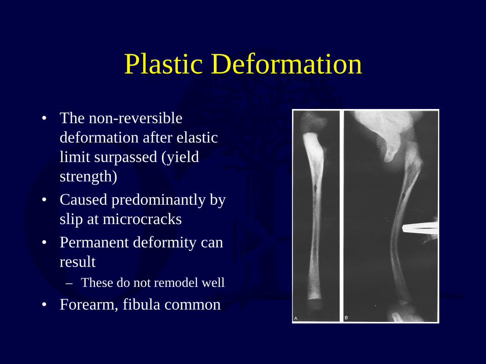
Plastic Deformation
• The non-reversible deformation after elastic limit surpassed (yield strength)
• Caused predominantly by slip at microcracks
• Permanent deformity can result – These do not remodel well
• Forearm, fibula common

Greenstick Fractures
• Bending mechanism • Failure on tension side • Incomplete fracture,
plastic deformation on compression side
• May need to complete fracture to realign

Salter - Harris Classification • Type I
– Through physis only • Type II
– Through physis & metaphysis • Type III
– Through physis & epiphysis • Type IV
– Through metaphysis, physis & epiphysis
• Type V – Crush injury to entire physis
• Others added later by subsequent authors
Described by Robert B. Salter and W. Robert Harris in 1963.

Salter Harris Classification General Treatment Principles
• Type I & Type II – Closed reduction &
immobilization – Exceptions
• Proximal femur • Distal femur

Salter Harris Classification General Treatment Principles
• Type III & IV – Intra-articular and
physeal step-off needs anatomic reduction
– ORIF, if necessary
Physeal Fractures
• Traditionally believed to occur primarily through zone of hypertrophy
• Recent studies show fractures often traverse more than one zone
• Growth disturbance/arrest potentially related to – Location of fracture within physeal zones – Disruption of vascularity
Jaramillo et al, Radiology, 2000. Johnson et al, Vet Surg, 2004. Kleinman & Marks, Am J Roentgenol, 1996.

Fracture Treatment in Children General Principles
• Children heal faster (factors) – Age – Mechanism of injury – Fracture location – Initial displacement – Open vs. closed injury
• Growing bones remodel more readily • Need less immobilization time • Stiffness of adjacent joints less likely

Treatment Principles
• When possible, restore: – Length, alignment & rotation
• Maintain residual angulation as small as possible using closed treatment methods – molded casts, cast changes, cast wedging, etc.
• Displaced intra-articular fractures will not remodel – anatomic reduction mandatory

Treatment Principles Closed Methods
• Achieve adequate pain control and relaxation – Anesthesia
• Local • Regional • General
– Conscious sedation (often combination of drugs) • Propofol • Ketamine • Benzodiazepines • Narcotics

Treatment Principles Closed Methods
• Vast majority of pediatric fractures treated by closed methods. – Exceptions - open fractures, intra-articular
fractures, multi-trauma • Attempt to restore alignment (do not always
rely on remodeling) • Gentle reduction of physeal injuries
(adequate relaxation, traction)

Treatment Principles Closed Methods
• Well molded casts/splints – Use 3-point fixation principle
• Consider immobilization method on day of injury that will last through entire course of treatment – Limit splint or cast changes
• Consider likelihood of post-reduction swelling – Cast splitting or splint
• If fracture is unstable, repeat radiographs at weekly intervals to document maintenance of acceptable position until early bone healing

Excellent reduction maintained with thin, well-molded cast/splint

Fiberglass cast applied with proper technique and split/spread is excellent way to safely immobilize limb, maintain reduction and
accommodate swelling

Treatment Principles Loss of Reduction
• Metaphyseal/diaphyseal fractures can be remanipulated with appropriate anesthesia/analgesia up to 3 weeks after injury
• In general, do not remanipulate physeal fractures after 5-7 days – increased risk of physeal damage
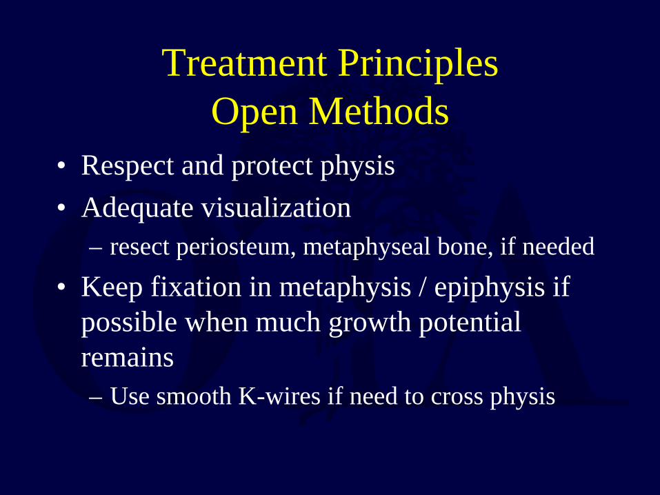
Treatment Principles Open Methods
• Respect and protect physis • Adequate visualization
– resect periosteum, metaphyseal bone, if needed • Keep fixation in metaphysis / epiphysis if
possible when much growth potential remains – Use smooth K-wires if need to cross physis

ORIF Salter IV Distal Tibia
* Note epiphyseal/metaphyseal wires to track postoperative growth

Complications of Fractures - Bone -
• Malunion • Limb length
discrepancy • Physeal arrest • Nonunion (rare) • Crossunion • Osteonecrosis

Complications of Fractures - Soft Tissue -
• Vascular Injury – Especially elbow/knee
• Neurologic Injury – Usually neuropraxia
• Compartment Syndrome – Especially leg/forearm
• Cast sores/pressure ulcers • Cast burns
– Use care with cast saw

Complications of Fractures - Cast Syndrome -
• Patient in spica/body cast
• Acute gastric distension, vomiting
• Possibly mechanical obstruction of duodenum by superior mesenteric artery

Location Specific Pediatric Fracture Complications
Complication Fracture
Cubitus varus Supracondylar humerus fracture
Volkmann’s ischemic contracture Supracondylar humerus fracture
Refracture Femur fracture Mid-diaphyseal radius/ulna fractures
Overgrowth Femur fracture (especially < 5 years)
Nonunion Lateral humeral condyle fracture
Osteonecrosis Femoral neck fracture Talus fracture
Progressive valgus Proximal tibia fractures

Remodeling of Children’s Fractures
• Occurs by physeal & periosteal growth changes
• Greater in younger children
• Greater if near a rapidly growing physis

Treatment Principles Immobilization Time
• In general, physeal injuries heal in half the time it takes for nonphyseal fracture in the same region
• Healing time dependent on fracture location, displacement
• Stiffness from immobilization rare, thus err towards more time in cast if in doubt

Remodeling of Children’s Fractures
• Not as reliable for: – Midshaft angulation – Older children – Large angulation (>20-30º)
• Will not remodel for: – Rotational deformity – Intraarticular deformity

Remodeling more likely if:
• 2 years or more growth remaining
• Fractures near end of bone
• Angulation in plane of movement of adjacent joint
10 weeks post-injury 1 week post-injury

Healing Salter I Distal Tibia Fracture

Growth Arrest Secondary to Physeal Injury
• Complete cessation of longitudinal growth – leads to limb length
discrepancy • Partial cessation of
longitudinal growth – angular deformity, if
peripheral – progressive shortening,
if central

Physes Susceptible to Growth Arrest
• Large cross sectional area
• Large growth potential • Complex geometric
anatomy • Distal femur > distal
tibia, proximal tibia > distal radius

Growth Arrest Lines • Transverse lines of Park-
Harris Lines • Occur after fracture/stress • Result from temporary
slowdown of normal longitudinal growth
• Thickened osseous plate in metaphysis
• Should parallel physis

Growth Arrest Lines
• Appear 6-12 weeks after fracture
• Look for them in follow-up radiographs after fracture
• If parallel physis - no growth disruption
• If angled or point to physis - suspect bar

Physeal Bar - Imaging -
• Scanogram / Orthoroentgenogram
• Tomograms/CT scans • MRI • Map bar to determine
location and extent
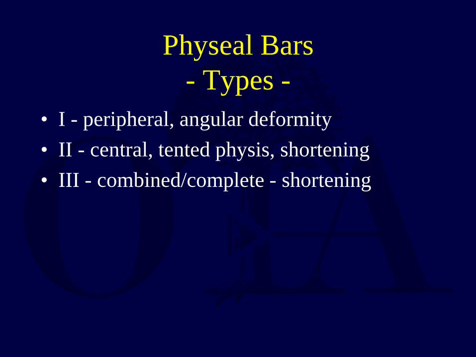
Physeal Bars - Types -
• I - peripheral, angular deformity • II - central, tented physis, shortening • III - combined/complete - shortening

Physeal Bar - Treatment -
• Address – Angular deformity – Limb length discrepancy
• Assess – Growth remaining – Amount of physis involved – Degree of angular
deformity – Projected LLD at maturity

Physeal Bar Resection - Indications -
• >2 years remaining growth • <50% physeal involvement (cross-sectional) • Concomitant osteotomy for >15-20º
deformity • Completion epiphyseodesis and
contralateral epiphyseodesis may be more reliable in older child

Physeal Bar Resection - Techniques
• Direct visualization • Burr/currettes • Interpositional
material (fat, cranioplast) to prevent reformation
• Wire markers to document future growth

Epiphysis or Apophysis?
• Epiphysis - forces are compressive on physeal plate
• Apophysis - forces are tensile
• Histologically distinct – Apophysis has less
proliferating cartilage and more fibrocollagen to help resist tensile forces

Apophyseal Injuries
• Tibial tubercle • Medial Epicondyle
– Often associated with dislocation
• May be preceded by chronic injury/reparative processes

Pathologic Fractures
• Diagnostic workup important – Local bone lesion – Generalized bone
weakness • Prognosis dependent
on biology of lesion • Often need surgery

Polyostotic Fibrous Dysplasia

Open Fractures Principles
• IV antibiotics, tetanus prophylaxis
• Emergent irrigation & debridement – Ideally within 6-8
hours of injury • Skeletal stabilization • Soft tissue coverage

Chronic Osteomyelitis following Open Femur Fracture
• Extremely rare in children • Serial debridement • Followed by simultaneous
bone graft and soft tissue coverage
Monsivais, J South Orthop Assoc, 1996.

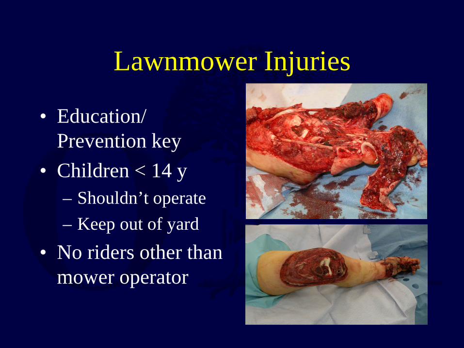
Lawnmower Injuries • Common cause of open
fractures & amputations in children
• Most are – A rider or bystander (70%) – Under 5 years old (78%)
• High complication rate – Infection – Growth arrest – Amputation
• > 50% poor results
Loder, JBJS-Am, 2004
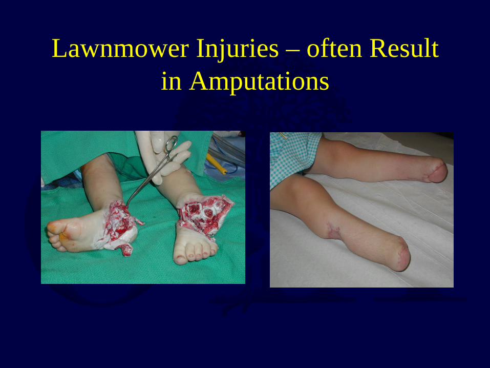
Lawnmower Injuries – often Result in Amputations
Lawnmower Injuries
• Education/ Prevention key
• Children < 14 y – Shouldn’t operate – Keep out of yard
• No riders other than mower operator

Overuse Injuries
• More common as children and adolescents participate in high level athletics
• Soccer, dance, baseball, gymnastics
• Ask about training regimens
• Mechanical pain
Femoral stress fracture Heyworth, Curr Opin Pediatr, 2008.

Overuse Injuries • Diagnosis
– History/Exam – Serial radiographs – Bone scan – CT/MRI
• Treatment – Abstinence from
sport/activity – Cast if child is overly active – Spica/Fixation for all
femoral neck stress fxs
Femoral stress fracture Heyworth, Curr Opin Pediatr, 2008.

Femoral Shaft Stress Fracture in 12 year old Male Runner

Metal Removal in Children
• Controversial • Historically recommended
if significant growth remaining
• Indications evolving • Intramedullary devices
and plates /screws around hip still removed by many in young patients
Kim, Injury 2005. Peterson, J Pediatr Orthop, 2005.

Summary
• Pediatric musculoskeletal injuries are relatively common
• General orthopaedic surgeons can treat majority of fractures
• Remember pediatric musculoskeletal differences
• Most fractures heal, regardless of treatment

Summary
• Most important factors: – Patient age – Mechanism of injury – Associated injuries
• Good results – possible with all types treatment • Trend for more invasive treatment • Must use good clinical judgment and good
technique to get good results

Bibliography • Salter R, Harris WR: Injuries Involving the Epiphyseal Plate. J Bone Joint Surg
Am. 1963;45:587-622. • Jaramillo D, Kammen B, Shapiro F: Cartilaginous path of physeal fracture-
separations: evaluation with MR imaging--an experimental study with histologic correlation in rabbits. Radiology 2000;215:504-11.
• Johnson J, Johnson A, Eurell J: Histological appearance of naturally occurring canine physeal fractures. Vet Surg 1994;23:81-6.
• Kleinman & Marks: A regional approach to the classic metaphyseal lesion in abused infants: the proximal humerus. Am J Roentgenol 1996;167:1399-403.
• Monsivais J: Effective management of osteomyelitis after grade III open fractures. J South Orthop Assoc 1996;5:30-6.

Bibliography • Loder R: Demographics of tramatic amputations in children. Implications for
prevention strategies. J Bone Joint Surg Am 2004;86:923-8. • Heyworth B & Green D: Lower extremity stress fractures in pediatric and
adolescent athletes. Curr Opin Pediatr 2008;20:58-61. • Kim W, et al: The removal of forearm plates in children. Injury 2005;36:1427-
30 • Peterson H: Metallic implant removal in children. J Pediatr Orthop
2005;25:107-15. • Wenger D, Pring M & Rand M: Rang’s Children’s Fractures, 3rd ed.
Philadelphia: Lippincott Williams & Wilkins, 2005. • Rockwood C & Wilkins K: Fractures in Children, 7th ed. Philadelphia:
Lippincott Williams & Wilkins, 2009.
Return to Pediatrics
Index
If you would like to volunteer as an author for the Resident Slide Project or recommend updates to any of the following slides, please send an e-mail to [email protected]