Embed Size (px)
Citation preview
1
Vital Signs
Chapter 12
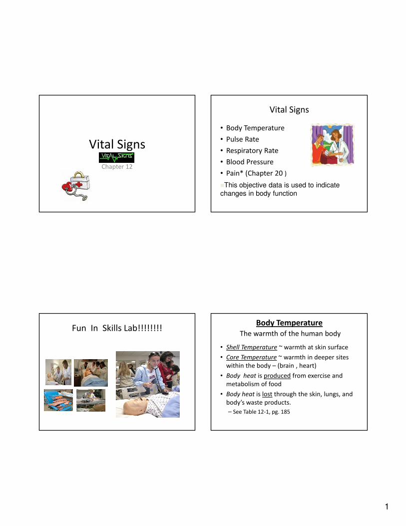
Vital Signs
• Body Temperature
• Pulse Rate
• Respiratory Rate
• Blood Pressure
• Pain* (Chapter 20 )
�This objective data is used to indicate
changes in body function
Fun In Skills Lab!!!!!!!!Body Temperature
The warmth of the human body
• Shell Temperature ~ warmth at skin surface
• Core Temperature ~ warmth in deeper sites
within the body – (brain , heart)
• Body heat is produced from exercise and
metabolism of food
• Body heat is lost through the skin, lungs, and
body’s waste products.
– See Table 12-1, pg. 185
2
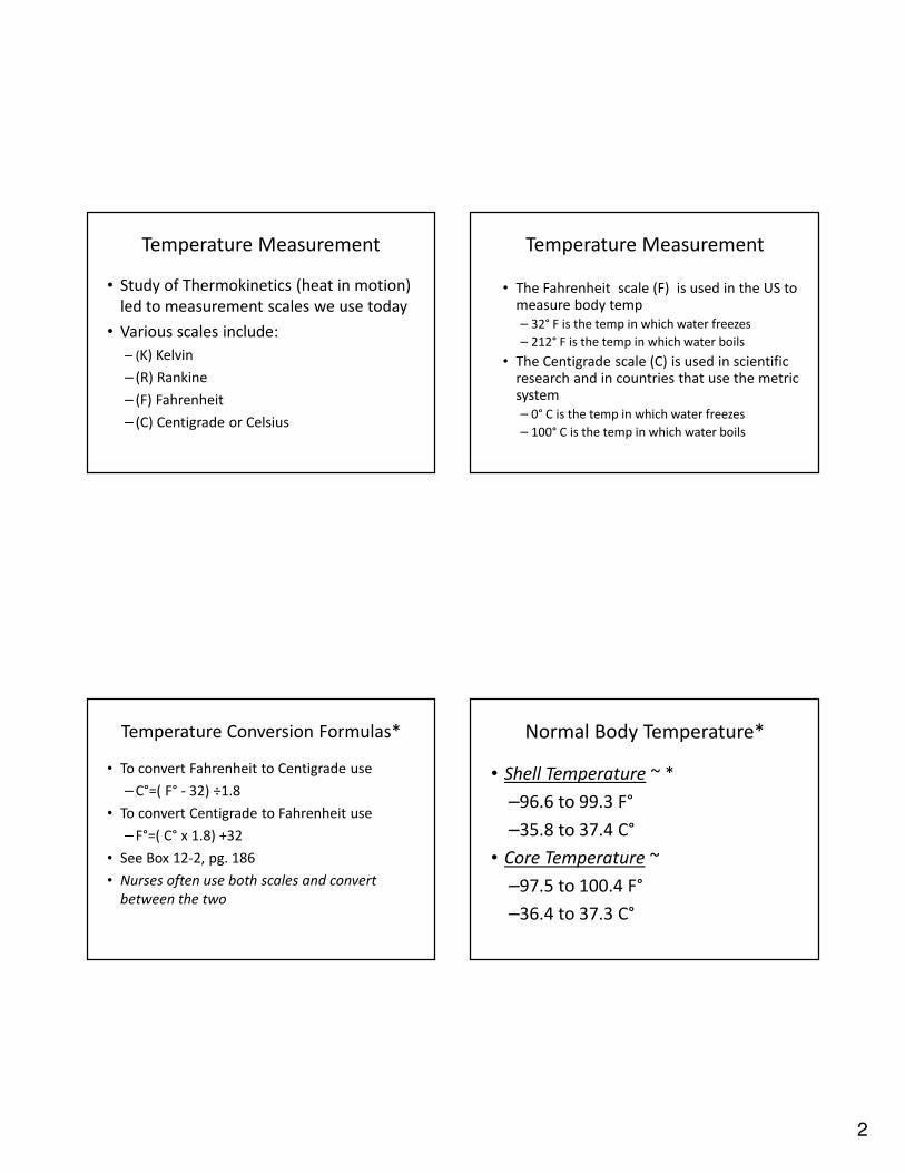
Temperature Measurement
• Study of Thermokinetics (heat in motion)
led to measurement scales we use today
• Various scales include:
– (K) Kelvin
– (R) Rankine
– (F) Fahrenheit
– (C) Centigrade or Celsius
Temperature Measurement
• The Fahrenheit scale (F) is used in the US to measure body temp
– 32° F is the temp in which water freezes
– 212° F is the temp in which water boils
• The Centigrade scale (C) is used in scientific research and in countries that use the metric system
– 0° C is the temp in which water freezes
– 100° C is the temp in which water boils
Temperature Conversion Formulas*
• To convert Fahrenheit to Centigrade use
– C°=( F° - 32) ÷1.8
• To convert Centigrade to Fahrenheit use
– F°=( C° x 1.8) +32
• See Box 12-2, pg. 186
• Nurses often use both scales and convert
between the two
Normal Body Temperature*
• Shell Temperature ~ *
–96.6 to 99.3 F°
–35.8 to 37.4 C°
• Core Temperature ~
–97.5 to 100.4 F°
–36.4 to 37.3 C°
3
Temperature Regulation*
• The Hypothalamus
– A structure within the brain that helps control various metabolic activities
– Acts as the center for temperature regulation
• The anterior Hypothalamus promotes heat loss through vasodilatation and sweating
• The posterior Hypothalamus promotes:
– Heat conservation
– Heat production
Temperature Regulation
Hypothalamus
• Heat Conservation
– Adjusting where blood circulates
– Causing piloerection (goose bumps or flesh)
– Promoting a shivering response
• Heat Production (thermogenesis)
– Increasing metabolism ~ thyroid hormone
– Releasing epinephrine and norepineprine
• Maintains the core temperature
Temperature Regulation
by the Hypothalamus continued:
• Maintains the core temperature (set point) to
within 1°C
• See Figure 12-1, pg 186
• Temp above 105.8°F and below 93.2°F
– Indicates impairment of the hypothalamus
• Temp above 110°F and below 84°F
– The chance for survival is diminished
Factors Affecting Body Temp
• Food Intake ~ Thermogenesis
• Age ~ Metabolic Rate
• Climate
• Gender
• Exercise and activity
• Circadian Rhythm
• Emotions
• Illness or Injury
• Medications
• Food Intake
4
Assessment Sites
• Core Body Temperature ~ Most Accurate
• Brain – no technology
• Heart, lower third of esophagus and urinary bladder–
use thermistor catheter.
• Most convenient and practical sites include the
mouth, rectum, axilla and ear
Practical and Convenient Assessment
Sites
• Oral
• Rectal
• Axillary
• Ear
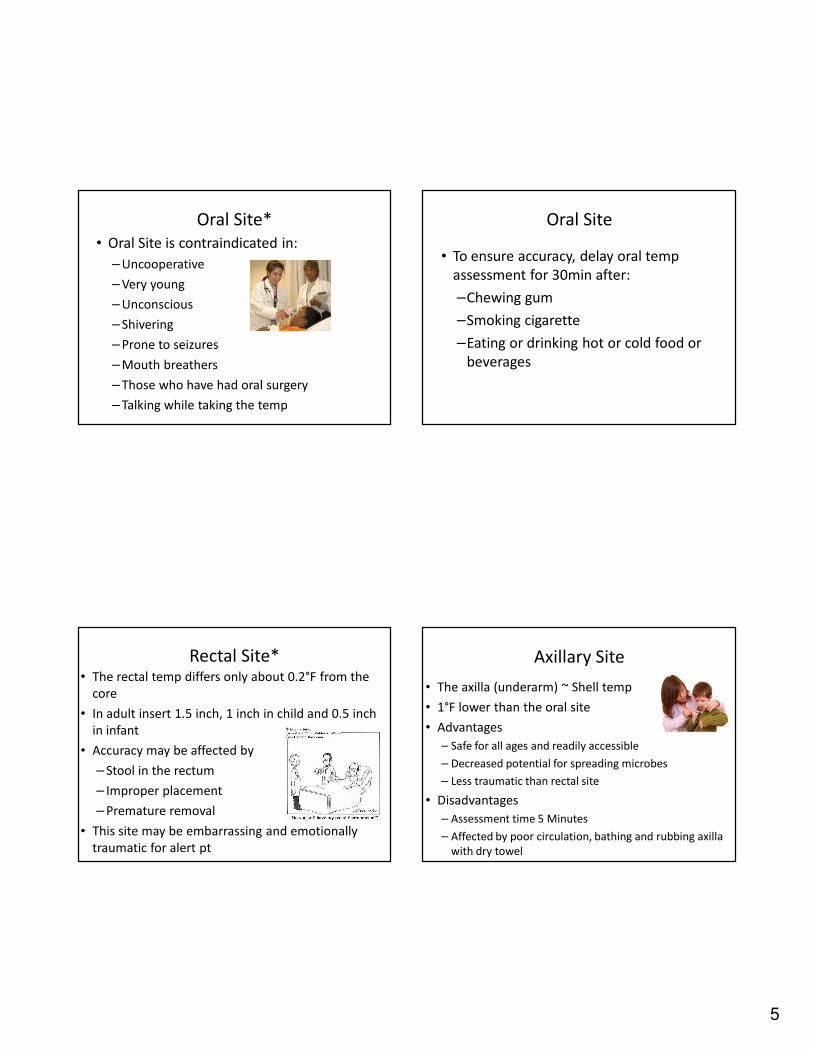
Oral Site
• Considered a shell temp
• Measures 0.8° to 1.0°F below the core temp
• Place the oral thermometer
– Under the tongue
– In direct proximity to the sublingual artery
– Sublingual pocket- See fig 12-3, page 188
– Poor placement or premature removal can lead to
an inaccurate reading
Oral temperature measurement
5
Oral Site*
• Oral Site is contraindicated in:
– Uncooperative
– Very young
– Unconscious
– Shivering
– Prone to seizures
– Mouth breathers
– Those who have had oral surgery
– Talking while taking the temp
Oral Site
• To ensure accuracy, delay oral temp
assessment for 30min after:
–Chewing gum
–Smoking cigarette
–Eating or drinking hot or cold food or
beverages
Rectal Site*• The rectal temp differs only about 0.2°F from the
core
• In adult insert 1.5 inch, 1 inch in child and 0.5 inch
in infant
• Accuracy may be affected by
– Stool in the rectum
– Improper placement
– Premature removal
• This site may be embarrassing and emotionally
traumatic for alert pt
Axillary Site
• The axilla (underarm) ~ Shell temp
• 1°F lower than the oral site
• Advantages
– Safe for all ages and readily accessible
– Decreased potential for spreading microbes
– Less traumatic than rectal site
• Disadvantages
– Assessment time 5 Minutes
– Affected by poor circulation, bathing and rubbing axilla
with dry towel
6
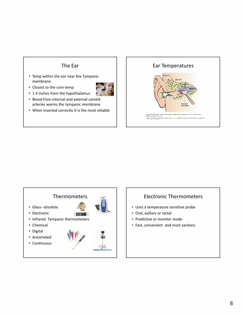
The Ear
• Temp within the ear near the Tympanic
membrane
• Closest to the core temp
• 1.4 inches from the hypothalamus
• Blood from internal and external carotid
arteries warms the tympanic membrane
• When inserted correctly it is the most reliable
Ear Temperatures
Thermometers
• Glass- obsolete
• Electronic
• Infrared Tympanic thermometers
• Chemical
• Digital
• Automated
• Continuous
Electronic Thermometers
• Uses a temperature sensitive probe
• Oral, axillary or rectal
• Predictive or monitor mode
• Fast, convenient and most sanitary
7
Infrared (Tympanic) Thermometers
• Probe contains infrared sensor
• Detects warmth from tympanic membrane
• Convert heat to a temp in 2-5 seconds
• Will produce inaccurate measurements if
– Ear canal is not straightened
– The probe is too large for ear canal (6-8mm)
– Directed at the ear canal not the tympanic membrane
– Impacted with cerumen ( ear wax)
– There is fluid behind the tympanic membrane
– The drawdown effect (cooling on contact)
Other Thermometers
• Chemical Thermometers - heat sensitive tape or chemical dot thermometers (Isolation)
• Digital Thermometers – includes a sensing tip at the end, on/off button and display area
• Automated Monitoring Devices – save time and money
• Continuous Monitoring Devices - internal thermistor probes used in ICU and are used only in acute patients.
Elevated Body Temperature*
• Fever ~ Body temp over 99.3
• Pyrexia ~ (Greek work for fire) warmer than normal set
point
• Febrile ~ condition in which temp is elevated
• Afebrile ~ condition in which there is no fever*
• Hyperthermia ~ excessively high core temp over 105.8 F
– Increased metabolic demands
– Will lead to Brain damage or death
Signs and Symptoms associated with
Fever*
• Pinkish, red (flushed) skin
• Restlessness or sleepiness
• Irritability, headache
• Poor appetite
• Glassy eyes and sensitivity to light
• Increased pulse, respiration or perspiration
• Disorientation and confusion
• Convulsions ( in small children)
• Fever Blister in clients with HSV
8
Phases of a Fever• Prodromal phase
– Nonspecific symptoms just
before fever
• Onset or invasion phase
– Mechanism for increased
temp ~ shiver, pale, or feel
cold
• Stationary phase
– Fever is sustained
• Resolution or
Defervescence phase
– Fever returns to normal
Nursing Management
• Fever is an important defense mechanism against microorganisms
• Fever below 102°F with no chronic medical condition – Provide fluids and rest
• Fever 102 F to 104 F– Antipyretics ~ Drugs that reduce fever
• Tylenol or Aspirin
• Fever between104-105.8°F – Cooling blanket and other physical cooling
measures
Subnormal Body Temperature
• Hypothermia ~ core body temp, less than 95°F
– Mild - 95-93.2°F
– Moderate - 93-86°F
– Severe - below 86°F (Fatal)
– Cold body temps are best be measured with
tympanic thermometers
– See guidelines 12-2, pg 194
Subnormal Body Temperature
• Signs and Symptoms– Shivering
– Pale, cool and puffy skin
– Impaired muscle coordination
– Listlessness
– Decreased pulse, respirations
– Irregular Heart rate
– Impaired ability to think and use good judgment
– Impaired ability to feel pain
9
Pulse * • A wavelike sensation that can be palpated in a
peripheral artery
– The pulse rate – the number of peripheral pulsations in one minute
• Normal 60-100 BPM at rest
– Tachycardia ~ a fast heart rate
• 100-150 at rest
– Palpitation ~ awareness of your own HR
– Bradycardia ~ Slow heart rate (less than 60 BPM)
Factors Affecting Pulse and HR
• Age
• Circadian rhythm
• Gender
• Body build
• Exercise and activity
• Stress and emotion
• Body temperature
• Blood volume
• Drugs
Pulse Rhythm & Volume
• Pulse Rhythm ~ Pattern of the pulsations and the pauses between
them. It is normally regular.
– Arrhythmia or dysrhythmia ~ irregular pattern
• Pulse volume ~ quality of the pulse related to the force of the
heart’s contraction
– Thready ~ not easily felt, slight pressure (disappears)
– Weak ~ stronger than thready, light pressure (disappears)
– Normal ~ easily felt, moderate pressure (disappears)
– Bounding ~ strong will not disappear with pressure
– See Table 12-6, pg 196-Identifying Pulse Volume
Assessment Site
• The arteries used for pulse assessment lie close
to the skin ~ peripheral pulses
• Most named for the bone it’s located near
• The radial artery (inner aspects of wrist – thumb
side) is used most for pulse assessment
• Other techniques include
– Apical heart rate
– Apical radial rate
– Doppler Ultrasound
10
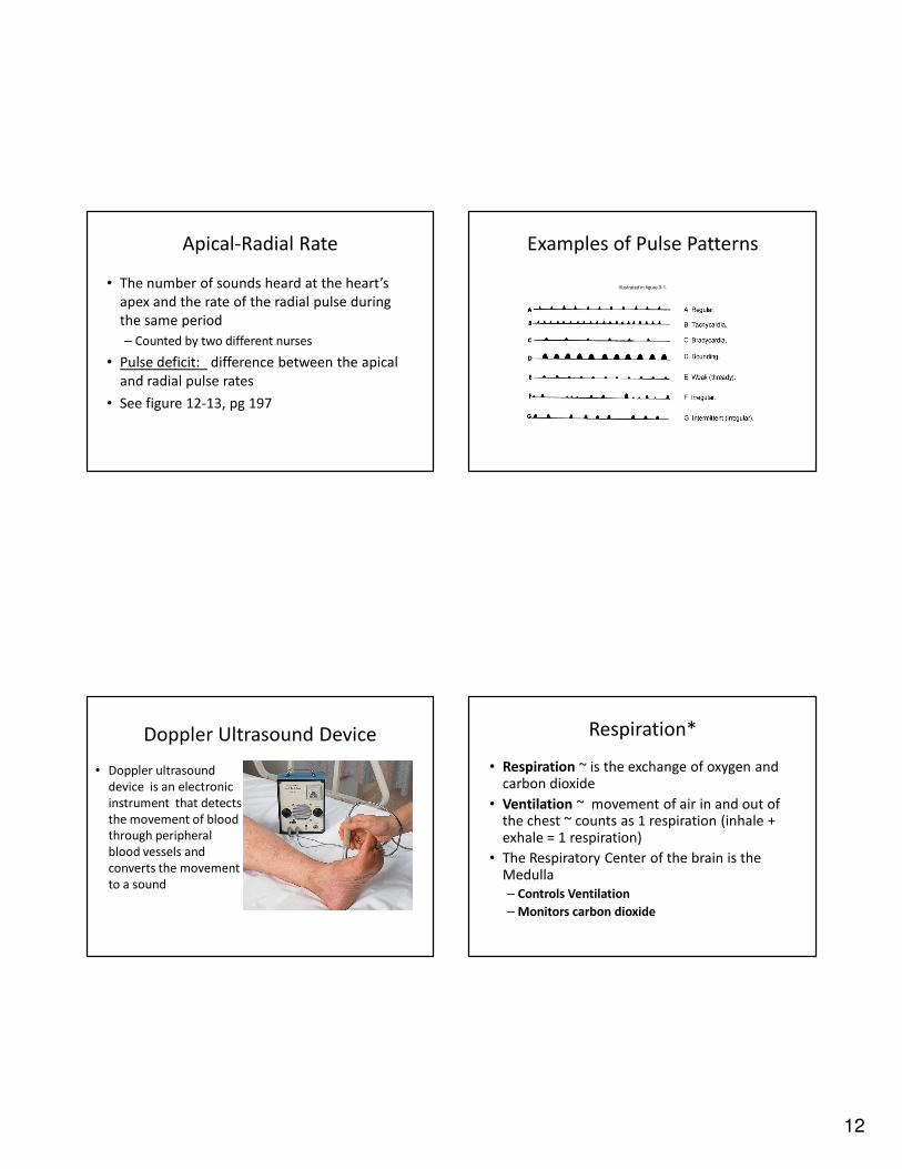
Carotid pulse site Radial Pulse site
Brachial Pulse site Apical Pulse Site
• ***Apical pulse – Learning Activity
Show Examples!!
11
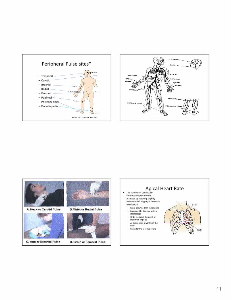
Peripheral Pulse sites*
– Temporal
– Carotid
– Brachial
– Radial
– Femoral
– Popliteal
– Posterior tibial
– Dorsalis pedis
Pulse Apical Heart Rate• The number of ventricular
contractions per minute ~
assessed by listening slightly
below the left nipple, in line with
left clavicle
– More accurate than radial pulse
– Is counted by listening with a
stethoscope
– Or by feeling at the point of
maximum impulse
– At the apex or lower tip of the
heart
– Listen for the lub/dub sound
12
Apical-Radial Rate
• The number of sounds heard at the heart’s
apex and the rate of the radial pulse during
the same period
– Counted by two different nurses
• Pulse deficit: difference between the apical
and radial pulse rates
• See figure 12-13, pg 197
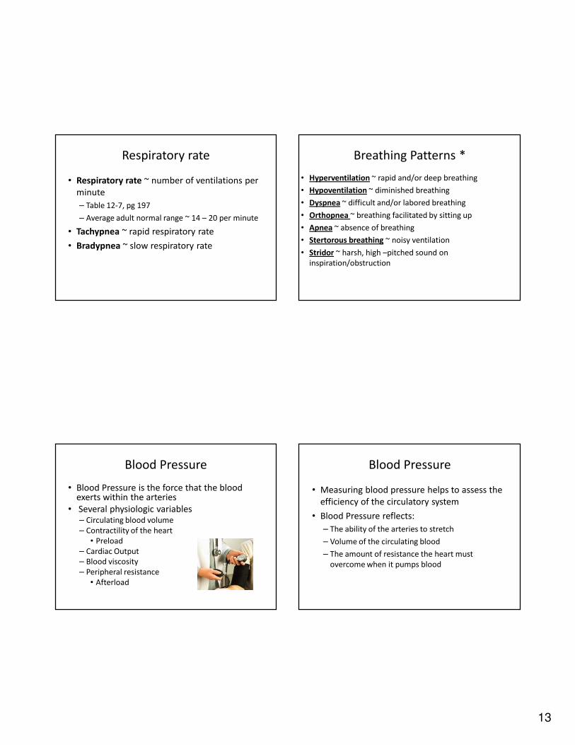
Examples of Pulse Patterns
illustrated in figure 3-1.
Doppler Ultrasound Device
• Doppler ultrasound
device is an electronic
instrument that detects
the movement of blood
through peripheral
blood vessels and
converts the movement
to a sound
Respiration*
• Respiration ~ is the exchange of oxygen and carbon dioxide
• Ventilation ~ movement of air in and out of the chest ~ counts as 1 respiration (inhale + exhale = 1 respiration)
• The Respiratory Center of the brain is the Medulla
– Controls Ventilation
– Monitors carbon dioxide
13
Respiratory rate
• Respiratory rate ~ number of ventilations per
minute
– Table 12-7, pg 197
– Average adult normal range ~ 14 – 20 per minute
• Tachypnea ~ rapid respiratory rate
• Bradypnea ~ slow respiratory rate
Breathing Patterns *
• Hyperventilation ~ rapid and/or deep breathing
• Hypoventilation ~ diminished breathing
• Dyspnea ~ difficult and/or labored breathing
• Orthopnea ~ breathing facilitated by sitting up
• Apnea ~ absence of breathing
• Stertorous breathing ~ noisy ventilation
• Stridor ~ harsh, high –pitched sound on
inspiration/obstruction
Blood Pressure
• Blood Pressure is the force that the blood exerts within the arteries
• Several physiologic variables– Circulating blood volume
– Contractility of the heart
• Preload
– Cardiac Output
– Blood viscosity
– Peripheral resistance
• Afterload
Blood Pressure
• Measuring blood pressure helps to assess the
efficiency of the circulatory system
• Blood Pressure reflects:
– The ability of the arteries to stretch
– Volume of the circulating blood
– The amount of resistance the heart must
overcome when it pumps blood
14
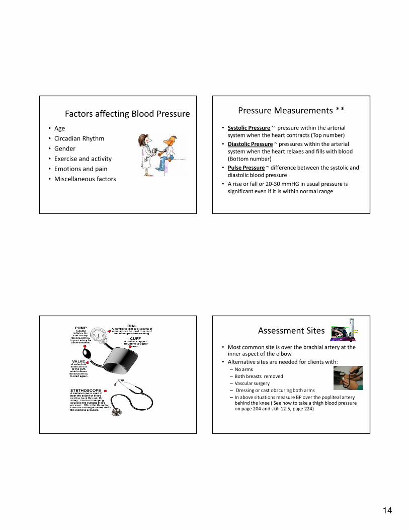
Factors affecting Blood Pressure
• Age
• Circadian Rhythm
• Gender
• Exercise and activity
• Emotions and pain
• Miscellaneous factors
Pressure Measurements **
• Systolic Pressure ~ pressure within the arterial
system when the heart contracts (Top number)
• Diastolic Pressure ~ pressures within the arterial
system when the heart relaxes and fills with blood
(Bottom number)
• Pulse Pressure ~ difference between the systolic and
diastolic blood pressure
• A rise or fall or 20-30 mmHG in usual pressure is
significant even if it is within normal range
Assessment Sites
• Most common site is over the brachial artery at the inner aspect of the elbow
• Alternative sites are needed for clients with:
– No arms
– Both breasts removed
– Vascular surgery
– Dressing or cast obscuring both arms
– In above situations measure BP over the popliteal artery behind the knee ( See how to take a thigh blood pressure on page 204 and skill 12-5, page 224)
15
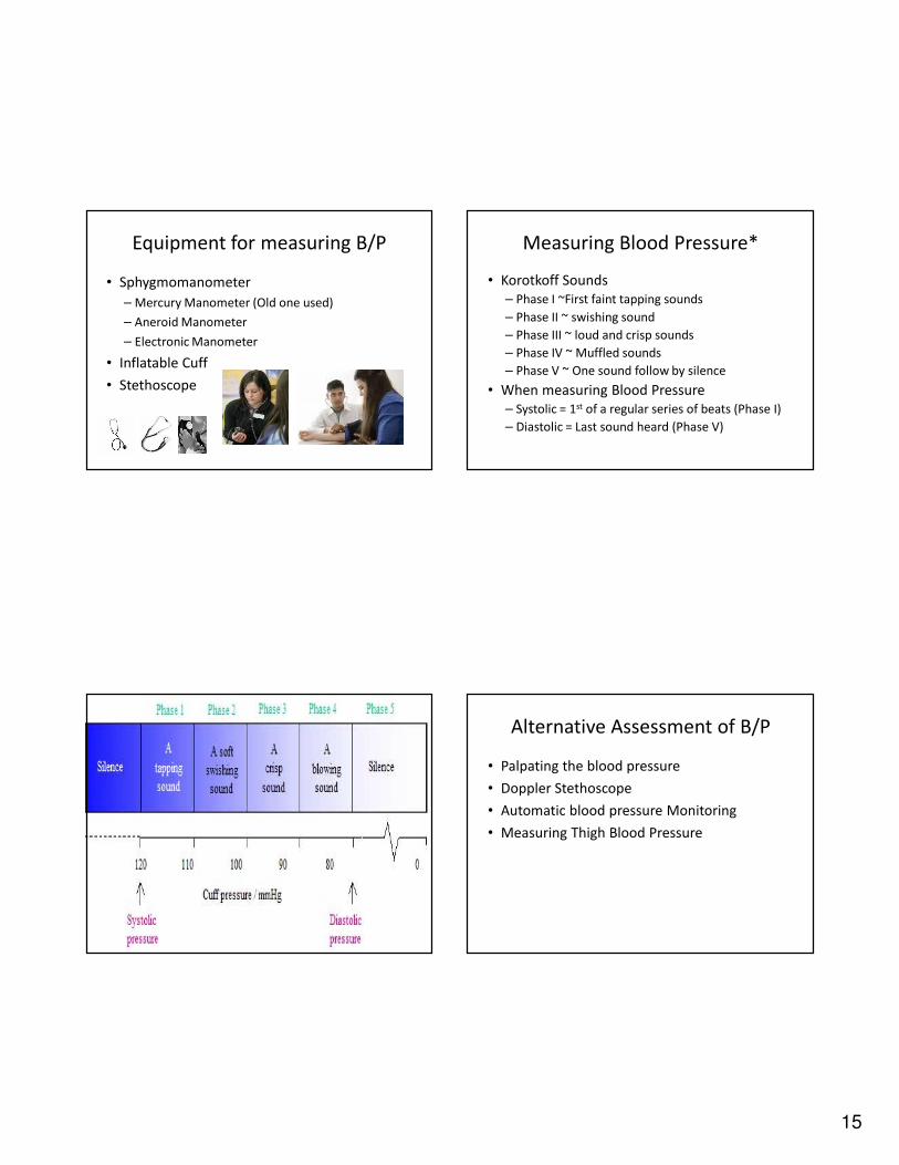
Equipment for measuring B/P
• Sphygmomanometer
– Mercury Manometer (Old one used)
– Aneroid Manometer
– Electronic Manometer
• Inflatable Cuff
• Stethoscope
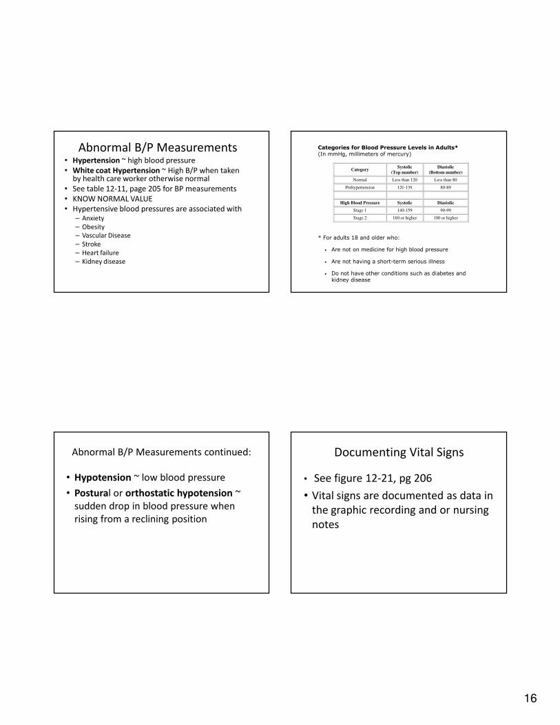
Measuring Blood Pressure*
• Korotkoff Sounds
– Phase I ~First faint tapping sounds
– Phase II ~ swishing sound
– Phase III ~ loud and crisp sounds
– Phase IV ~ Muffled sounds
– Phase V ~ One sound follow by silence
• When measuring Blood Pressure
– Systolic = 1st of a regular series of beats (Phase I)
– Diastolic = Last sound heard (Phase V)
Alternative Assessment of B/P
• Palpating the blood pressure
• Doppler Stethoscope
• Automatic blood pressure Monitoring
• Measuring Thigh Blood Pressure
16
Abnormal B/P Measurements• Hypertension ~ high blood pressure
• White coat Hypertension ~ High B/P when taken by health care worker otherwise normal
• See table 12-11, page 205 for BP measurements
• KNOW NORMAL VALUE
• Hypertensive blood pressures are associated with– Anxiety
– Obesity
– Vascular Disease
– Stroke
– Heart failure
– Kidney disease
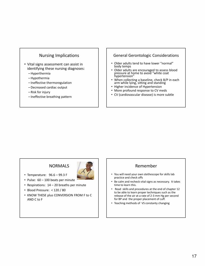
Categories for Blood Pressure Levels in Adults* (In mmHg, millimeters of mercury)
Category Systolic
(Top number)
Diastolic
(Bottom number)
Normal Less than 120 Less than 80
Prehypertension 120-139 80-89
High Blood Pressure Systolic Diastolic
Stage 1 140-159 90-99
Stage 2 160 or higher 100 or higher
* For adults 18 and older who:
• Are not on medicine for high blood pressure
• Are not having a short-term serious illness
• Do not have other conditions such as diabetes and kidney disease
Abnormal B/P Measurements continued:
• Hypotension ~ low blood pressure
• Postural or orthostatic hypotension ~
sudden drop in blood pressure when
rising from a reclining position
Documenting Vital Signs
• See figure 12-21, pg 206
• Vital signs are documented as data in
the graphic recording and or nursing
notes
17
Nursing Implications
• Vital signs assessment can assist in identifying these nursing diagnoses:
– Hyperthermia
– Hypothermia
– Ineffective thermoregulation
– Decreased cardiac output
– Risk for injury
– Ineffective breathing pattern
General Gerontologic Considerations
• Older adults tend to have lower “normal” body temps
• Older adults are encouraged to assess blood pressure at home to avoid “white coat hypertension”
• When collecting a baseline, check B/P in each arm while lying, sitting and standing
• Higher incidence of Hypertension• More profound response to CV meds
• CV (cardiovascular disease) is more subtle
NORMALS
• Temperature: 96.6 – 99.3 F
• Pulse: 60 – 100 beats per minute
• Respirations: 14 – 20 breaths per minute
• Blood Pressure: < 120 / 80
• KNOW THESE plus CONVERSION FROM F to C
AND C to F
Remember
• You will need your own stethescope for skills lab practice and check offs
• Be calm and recheck vital signs as necessary. It takes time to learn this.
• Read skills and procedures at the end of chapter 12 to be able to learn proper techniques such as the release of the air at a rate of 2-3 mm Hg per second for BP and the proper placement of cuff.
• Teaching methods of VS constanty changing
18
2.5 point participation activity
• Put your name and ID# on the 2.5 point
participation activity. Turn in before leaving
class. Not accepted late and cannot make up if
absent.