Embed Size (px)
Citation preview
ACG CASE REPORTS JOURNAL
acgcasereports.gi.org ACG Case Reports Journal | Volume 2 | Issue 3 | April 2015161
CASE REPORT | COLON
Basaloid Squamous Cell Carcinoma of the Sigmoid Colon Salih Samo, MD1, Muhammed Sherid, MD2, Kevin Liu, MD3, Leila Kia, MD4, and Glynn J. Elliott, MD, FACP5
1Department of Medicine, Division of Hospital Medicine, Northwestern Memorial Hospital, Northwestern University Feinberg School of Medicine, Chicago, IL2Department of Medicine, Division of Gastroenterology, Georgia Regents University, Augusta, GA 3Department of Medicine, Northwestern Memorial Hospital, Northwestern University Feinberg School of Medicine, Chicago, IL4Department of Medicine, Division of Gastroenterology and Hepatology, Northwestern Memorial Hospital, Northwestern University Feinberg School of Medicine, Chicago, IL5Department of Medicine, Presence Saint Francis Hospital, Evanston, IL
AbstractBasaloid squamous cell carcinoma (BSCC) of the colon is rarely found proximal to the anal canal. We report a case of an 81-year-old woman who was diagnosed with squamous cell carcinoma (SCC) of the lung without metastasis and BSCC of the sigmoid with differing histologic findings suggesting that these tumors were separate primary neoplasms. SCC of the colon has a dismal prognosis. Surgery is the primary method of treatment when feasible, in addition to chemotherapeutic agents.
IntroductionAdenocarcinomas represent the vast majority of colorectal cancers (CRCs). Other histologic types, including squa-mous cell carcinoma (SCC) are relatively uncommon. SCC is of epithelial origin and is commonly seen in glandular tissues such as the lung, but is rarely seen as primary colorectal tumors outside the anal canal.1 In colorectal adenocarcinoma, scattered areas of Paneth and neuroendocrine cells and small foci of squamous cell differentia-tion can be seen, but a pure colorectal SCC is an extremely rare tumor entity.1 Basaloid squamous cell carcinoma (BSCC) is a unique subtype of SCC that typically affects the upper digestive tract, respiratory tract, and anal canal. On histologic examination, BSCC of the anal canal has been characterized by hyperchromatic basaloid cells and tumor nests with eosinophilic infiltration.2
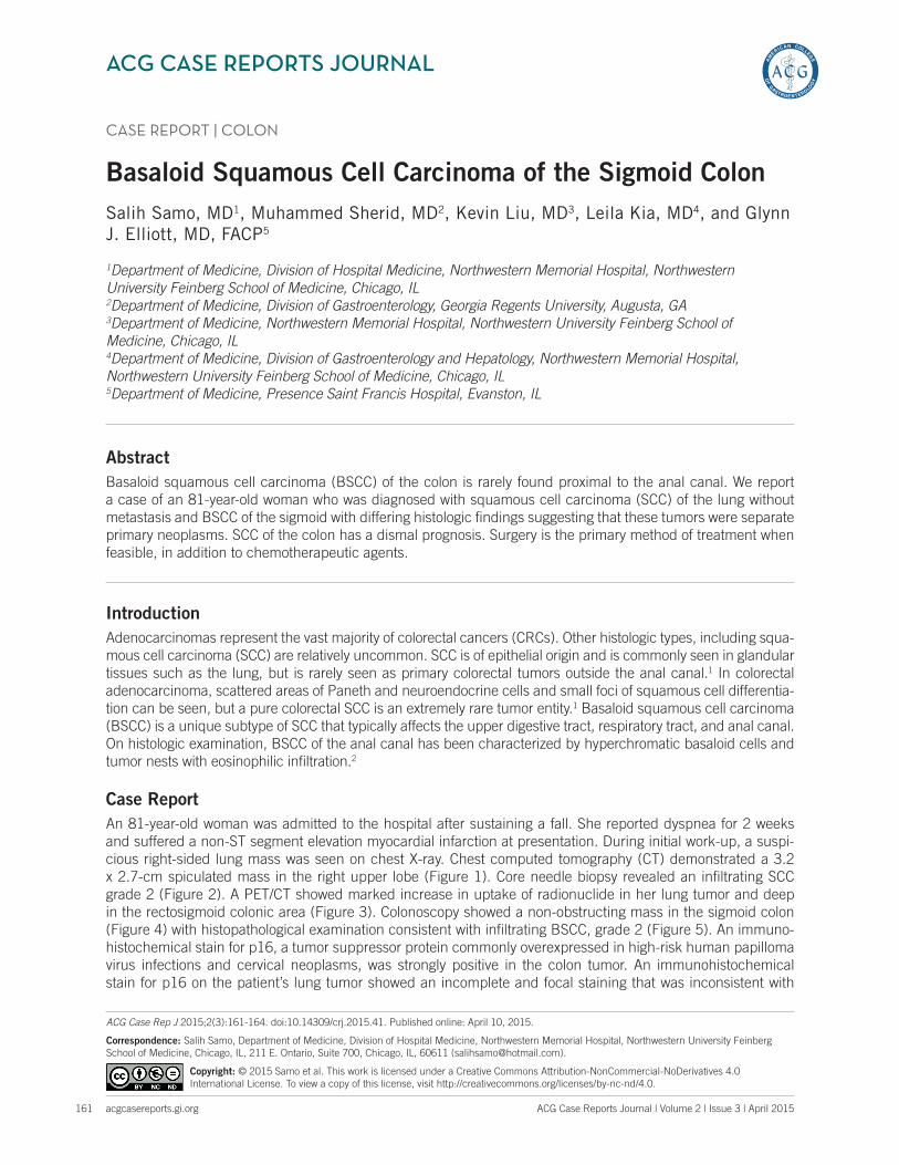
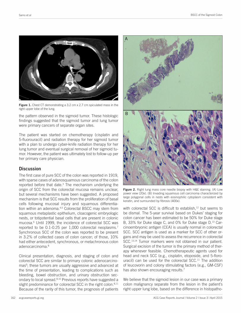
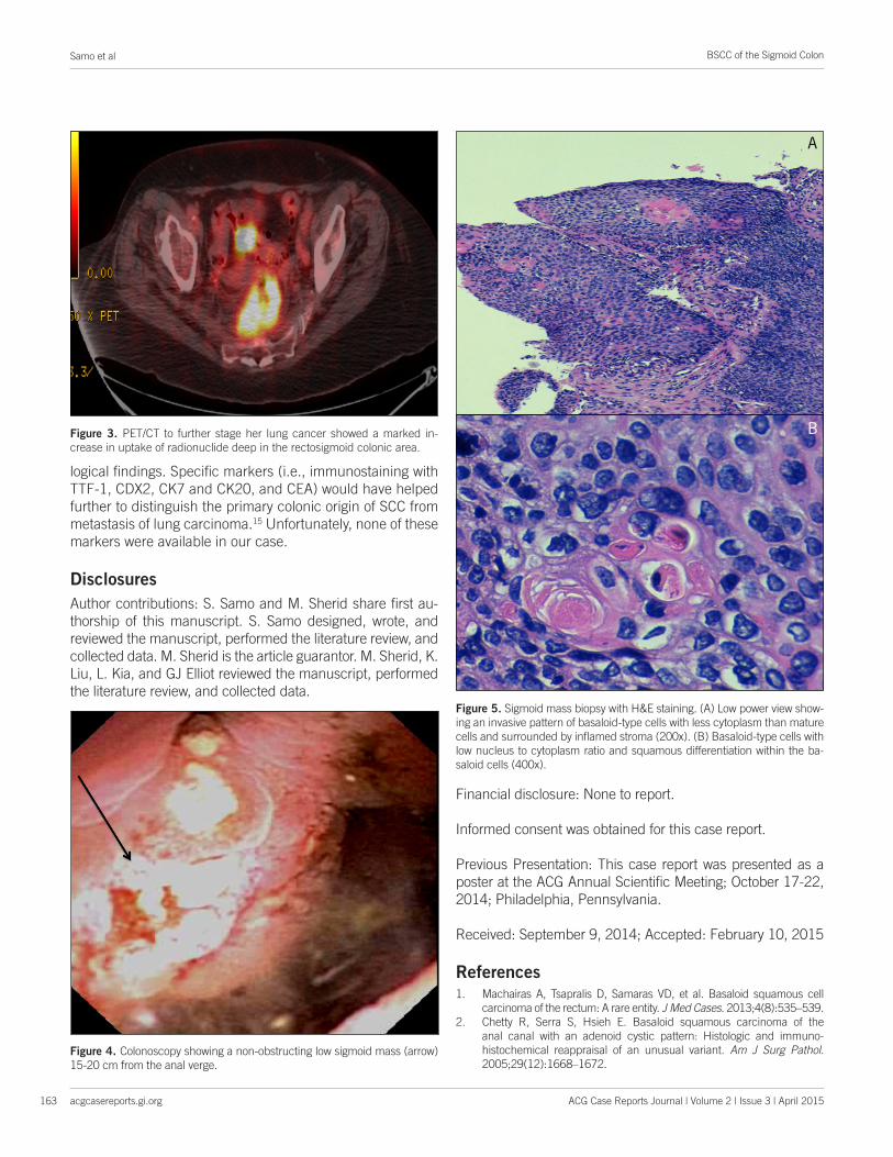
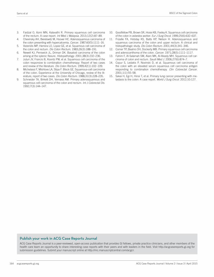
Case ReportAn 81-year-old woman was admitted to the hospital after sustaining a fall. She reported dyspnea for 2 weeks and suffered a non-ST segment elevation myocardial infarction at presentation. During initial work-up, a suspi-cious right-sided lung mass was seen on chest X-ray. Chest computed tomography (CT) demonstrated a 3.2 x 2.7-cm spiculated mass in the right upper lobe (Figure 1). Core needle biopsy revealed an infiltrating SCC grade 2 (Figure 2). A PET/CT showed marked increase in uptake of radionuclide in her lung tumor and deep in the rectosigmoid colonic area (Figure 3). Colonoscopy showed a non-obstructing mass in the sigmoid colon (Figure 4) with histopathological examination consistent with infiltrating BSCC, grade 2 (Figure 5). An immuno-histochemical stain for p16, a tumor suppressor protein commonly overexpressed in high-risk human papilloma virus infections and cervical neoplasms, was strongly positive in the colon tumor. An immunohistochemical stain for p16 on the patient’s lung tumor showed an incomplete and focal staining that was inconsistent with
ACG Case Rep J 2015;2(3):161-164. doi:10.14309/crj.2015.41. Published online: April 10, 2015.
Correspondence: Salih Samo, Department of Medicine, Division of Hospital Medicine, Northwestern Memorial Hospital, Northwestern University Feinberg School of Medicine, Chicago, IL, 211 E. Ontario, Suite 700, Chicago, IL, 60611 ([email protected]).
Copyright: © 2015 Samo et al. This work is licensed under a Creative Commons Attribution-NonCommercial-NoDerivatives 4.0 International License. To view a copy of this license, visit http://creativecommons.org/licenses/by-nc-nd/4.0.
BSCC of the Sigmoid Colon
acgcasereports.gi.org ACG Case Reports Journal | Volume 2 | Issue 3 | April 2015
Samo et al
162
the pattern observed in the sigmoid tumor. These histologic findings suggested that the sigmoid tumor and lung tumor were primary cancers of separate organ sites.
The patient was started on chemotherapy (cisplatin and 5-fluorouracil) and radiation therapy for her sigmoid tumor with a plan to undergo cyber-knife radiation therapy for her lung tumor and eventual surgical removal of her sigmoid tu-mor. However, the patient was ultimately lost to follow-up per her primary care physician.
DiscussionThe first case of pure SCC of the colon was reported in 1919, with sparse cases of adenosquamous carcinoma of the colon reported before that date.3 The mechanism underlying the origin of SCC from the colorectal mucosa remains unclear, but several mechanisms have been suggested. A proposed mechanism is that SCC results from the proliferation of basal cells following mucosal injury and squamous differentia-tion within an adenoma.4,5 Colorectal BSCC may stem from squamous metaplastic epithelium, cloacogenic embryologic nests, or totipotential basal cells that are present in colonic mucosa.6 Until 1999, the incidence of colorectal SCC was reported to be 0.1-0.25 per 1,000 colorectal neoplasms.7 Synchronous SCC of the colon was reported to be present in 3.2% of collected cases of colon cancer; of those, 10% had either antecedent, synchronous, or metachronous colon adenocarcinoma.8
Clinical presentation, diagnosis, and staging of colon and colorectal SCC are similar to primary colonic adenocarcino-mas9; these tumors are usually aggressive and advanced at the time of presentation, leading to complications such as bleeding, bowel obstruction, and urinary obstruction sec-ondary to local spread.8,10 Previous reports have suggested a slight predominance for colorectal SCC in the right colon.8,11 Because of the rarity of this tumor, the prognosis of patients
with colorectal SCC is difficult to establish,12 but seems to be dismal. The 5-year survival based on Dukes’ staging for colon cancer has been estimated to be 50% for Duke stage B, 33% for Duke stage C, and 0% for Duke stage D.13 Car-cinoembryonic antigen (CEA) is usually normal in colorectal SCC. SCC antigen is used as a marker for SCC of other or-gans and may be used to assess the recurrence in colorectal SCC.13,14 Tumor markers were not obtained in our patient. Surgical excision of the tumor is the primary method of ther-apy whenever feasible. Chemotherapeutic agents used for head and neck SCC (e.g., cisplatin, etoposide, and 5-floro-uracil) can be used for the colorectal SCC.11 The addition of leucovorin and colony stimulating factors (e.g., GM-CSF) has also shown encouraging results.7
We believe that the sigmoid lesion in our case was a primary colon malignancy separate from the lesion in the patient’s right upper lung lobe, based on the difference in histopatho-
Figure 1. Chest CT demonstrating a 3.2 cm x 2.7 cm spiculated mass in the right upper lobe of the lung.
A B
Figure 2. Right lung mass core needle biopsy with H&E staining. (A) Low power view (20x). (B) Invading squamous cell carcinoma characterized by large polygonal cells in nests with eosinophilic cytoplasm consistent with keratin, and surrounded by fibrosis (400x).
A
B
BSCC of the Sigmoid Colon
acgcasereports.gi.org ACG Case Reports Journal | Volume 2 | Issue 3 | April 2015
Samo et al
163
logical findings. Specific markers (i.e., immunostaining with TTF-1, CDX2, CK7 and CK20, and CEA) would have helped further to distinguish the primary colonic origin of SCC from metastasis of lung carcinoma.15 Unfortunately, none of these markers were available in our case.
Disclosures
Author contributions: S. Samo and M. Sherid share first au-thorship of this manuscript. S. Samo designed, wrote, and reviewed the manuscript, performed the literature review, and collected data. M. Sherid is the article guarantor. M. Sherid, K. Liu, L. Kia, and GJ Elliot reviewed the manuscript, performed the literature review, and collected data.
Financial disclosure: None to report.
Informed consent was obtained for this case report.
Previous Presentation: This case report was presented as a poster at the ACG Annual Scientific Meeting; October 17-22, 2014; Philadelphia, Pennsylvania.
Received: September 9, 2014; Accepted: February 10, 2015 References1. Machairas A, Tsapralis D, Samaras VD, et al. Basaloid squamous cell
carcinoma of the rectum: A rare entity. J Med Cases. 2013;4(8):535–539. 2. Chetty R, Serra S, Hsieh E. Basaloid squamous carcinoma of the
anal canal with an adenoid cystic pattern: Histologic and immuno-histochemical reappraisal of an unusual variant. Am J Surg Pathol. 2005;29(12):1668–1672.
Figure 5. Sigmoid mass biopsy with H&E staining. (A) Low power view show-ing an invasive pattern of basaloid-type cells with less cytoplasm than mature cells and surrounded by inflamed stroma (200x). (B) Basaloid-type cells with low nucleus to cytoplasm ratio and squamous differentiation within the ba-saloid cells (400x).
Figure 4. Colonoscopy showing a non-obstructing low sigmoid mass (arrow) 15-20 cm from the anal verge.
Figure 3. PET/CT to further stage her lung cancer showed a marked in-crease in uptake of radionuclide deep in the rectosigmoid colonic area.
A
B
Publish your work in ACG Case Reports JournalACG Case Reports Journal is a peer-reviewed, open-access publication that provides GI fellows, private practice clinicians, and other members of the health care team an opportunity to share interesting case reports with their peers and with leaders in the field. Visit http://acgcasereports.gi.org for submission guidelines. Submit your manuscript online at http://mc.manuscriptcentral.com/acgcr.
Samo et al
acgcasereports.gi.org
BSCC of the Sigmoid Colon
164 ACG Case Reports Journal | Volume 2 | Issue 3 | April 2015
3. Faidzal O, Azmi MN, Kalavathi R. Primary squamous cell carcinoma of the rectum: A case report. Int Med J Malaysia. 2013;12(2):87–89.
4. Chevinsky AH, Berelowitz M, Hoover HC. Adenosquamous carcinoma of the colon presenting with hypercalcemia. Cancer. 1987;60(5):1111–16.
5. Vezeridis MP, Herrera LO, Lopez GE, et al. Squamous cell carcinoma of the colon and rectum. Dis Colon Rectum. 1983;26(3):188–191.
6. Newell KJ, Penswick JL, Driman DK. Basaloid carcinoma of the colon arising at the splenic flexure. Histopathology. 2001;38(3):232–236.
7. Juturi JV, Francis B, Koontz PW, et al. Squamous cell carcinoma of the colon responsive to combination chemotherapy: Report of two cases and review of the literature. Dis Colon Rectum. 1999;42(1):102–109.
8. Michelassi F, Mishlove LA, Stipa F. Block GE. Squamous-cell carcinoma of the colon. Experience at the University of Chicago, review of the lit-erature, report of two cases. Dis Colon Rectum. 1988;31(3):228–235.
9. Schneider TA, Birkett DH, Vernava AM. Primary adenosquamous and squamous cell carcinoma of the colon and rectum. Int J Colorectal Dis. 1992;7(3):144–147.
10. Goodfellow PB, Brown SR, Hosie KB, Feeley K. Squamous cell carcinoma of the colon in asbestos worker. Eur J Surg Oncol. 1999;25(6):632–637.
11. Frizelle FA, Hobday KS, Batts KP, Nelson H. Adenosquamous and squamous carcinoma of the colon and upper rectum: A clinical and histopathologic study. Dis Colon Rectum. 2001;44(3):341–346.
12. Comer TP, Beahrs OH, Dockerty MB. Primary squamous cell carcinoma and adenocanthoma of the colon. Cancer. 1971;28(5):1111–1117.
13. Fahim F, Al-Salamah SM, Alam MK, Al-Akeely MH. Squamous cell car-cinoma of colon and rectum. Saudi Med J. 2006;27(6):874–7.
14. Copur S, Ledakis P, Novinski D, et al. Squamous cell carcinoma of the colon with an elevated serum squamous cell carcinoma antigen responding to combination chemotherapy. Clin Colorectal Cancer. 2001;1(1):55–58.
15. Sakai H, Egi H, Hinoi T, et al. Primary lung cancer presenting with me-tastasis to the colon: A case report. World J Surg Oncol. 2012;10:127.