Embed Size (px)
Citation preview

Research Article
Barrett’s Esophagus with Epithelial Changes Indefinite for Dysplasia: What we Have Learnt from Recent Studies - Prashanthi N Thota1, Ashwini K.Esnakula2, David Hernandez Gonzalo2, Xiuli Liu3*
1Department of Gastroenterology, Digestive Disease Institute, Cleveland Clinic, Cleveland, OH9500 Euclid Avenue, Cleveland, Ohio 44195, United States of America2Department of Pathology, Immunology and Laboratory Medicine,University of Florida College of Medicine, P.O. Box 100275,1600 SW Archer Road, Gainesville, FL, 32610-02753Department of Anatomic Pathology, Cleveland Clinic, 9500 Euclid Avenue, Cleveland, Ohio 44195, United States of America
*Address for Correspondence: : Xiuli Liu, Department of Anatomic Pathology Cleveland Clinic, 9500 Euclid Avenue/L25 Cleveland, Ohio 44195, Fax: 216-445-6967 ; Tel: 216-445-8745 ; E-mail: [email protected]
Submitted: 09 December 2015 Approved: 28 December 2015 Published: 31 December 2015
Citation this article: Thota PN, Esnakula AK, Gonzalo DH, Liu X. Barrett’s Esophagus with Epithelial Changes Indefinite for Dysplasia: What we Have Learnt from Recent Studies. Int J Hepatol Gastroenterol. 2015;1(1): 009-013.
Copyright: © 2015 Liu X et al. This is an open access article distributed under the Creative Commons Attribution License, which permits unrestricted use, distribution, and reproduction in any medium, provided the original work is properly cited.
International Journal ofHepatology & Gastroenterology
SCIRES Literature - Volume 1 Issue 1 - www.scireslit.com Page - 010
International Journal of Hepatology & Gastroenterology
INTRODUCTIONBarrett`s esophagus (BE) is a complication of chronic
gastroesophageal reflux disease (GERD); it is defined as the extension of salmon-colored mucosa into the tubular esophagus ≥1 cm proximal to the gastroesophageal junction (GEJ) with biopsy confirmation of intestinal metaplasia as defined by the presence of goblet cells histologically [1]. Patients with BE are at increased risk of esophageal adenocarcinoma (EAC), and as such, undergo endoscopic surveillance and biopsy with the goal of detecting dysplasia or early adenocarcinoma. Histologic criteria for dysplasia in BE were well described in 1988 by Reid et al. [2]. Routinely, the biopsies are classified as negative for dysplasia,IND or positive for dysplasia, the latter can be further divided into low-grade (LGD) and high-grade (HGD).
The management of LGDand HGDin BE has been reviewed extensively and discussed in many guidelines. Experienced gastrointestinal pathologists can diagnose HGD and intra-mucosal adenocarcinoma (IMAC) with a high degree of agreement [2]. Many studies have focused on the high end of neoplasia in BE, HGD and IMAC, leading to a much improved and less invasive management [3,4,5]. However, the category of BE with epithelial changes indefinite for dysplasia (BE IND) represents the diagnosis with the greatest interobserver variability, the most uncertain clinical significance, and
the least known natural history.
This review examines the available evidence for the histologic criteria for BE IND and the clinical significance of BE IND as revealed in several recent studies, particularly regarding the prevalence and progression to advanced neoplasia. It also summarizes the results of possible clinicopathologic and biomarkers predictors of these risks.
METHODS
PubMed was searched using key word “Barrett’s esophagus indefinite for dysplasia” as of November 1, 2015. Histologic criteria used for defining BE IND were reviewed and studies with synchronous or prior HGD/EAC were excluded.One study shared part of the same database and was excluded [6]. Studies were reviewed for prevalence and incidence rates of HGD/EAC (advanced neoplasia) in BE-IND as well as biomarkers or predictors for progression in IND.
RESULTSDefinition of BE with IND: It has been agreed upon that the
diagnosis “indefinite for dysplasia” is used by pathologists when they are genuinely concerned for but not absolutely sure about the presence of dysplasia. In routine pathology practice, many of such cases were related to the presence of inflammation and/or ulceration interfering with the interpretation. This diagnostic category was also
ABSTRACTBarrett’s esophagus (BE), a complication of chronic gastroesophageal reflux disease (GERD), is defined as the extension of salmon-
colored mucosa into the tubular esophagus ≥1 cm proximal to the gastroesophageal junction (GEJ) with biopsy confirmation of intestinal metaplasia, defined by the presence of goblet cells histologically. Patients with BE are at increased risk of esophageal adenocarcinoma (EAC), and as such, need to undergo endoscopic surveillance with biopsy to detect dysplasia or earlyEAC. Histologic criteria for dysplasia in BE were well described in 1988 by Reid et al. and classified as BE with low grade dysplasia (LGD), BE with high grade dysplasia (HGD) and BE with changes indefinite for dysplasia (IND). Biopsies are classified as IND when the epithelial abnormalities are not sufficient to diagnose dysplasia or the nature of the epithelial abnormalities is uncertain due to inflammation or technical issues. Specific diagnostic criteria for indefinite for dysplasia (IND) are not well established and its clinical significance has not been well studied. Previous studies have focused on the higher end of neoplasia in BE and led to revolutionary changes and improvement in the management of BE with HGD and early EAC. Only recently, the lower end of dysplasia in BE attracted researchers’ interest. This reviewsummarizes the findings in most recent studies on the neoplastic risk and thus the management of BE IND.
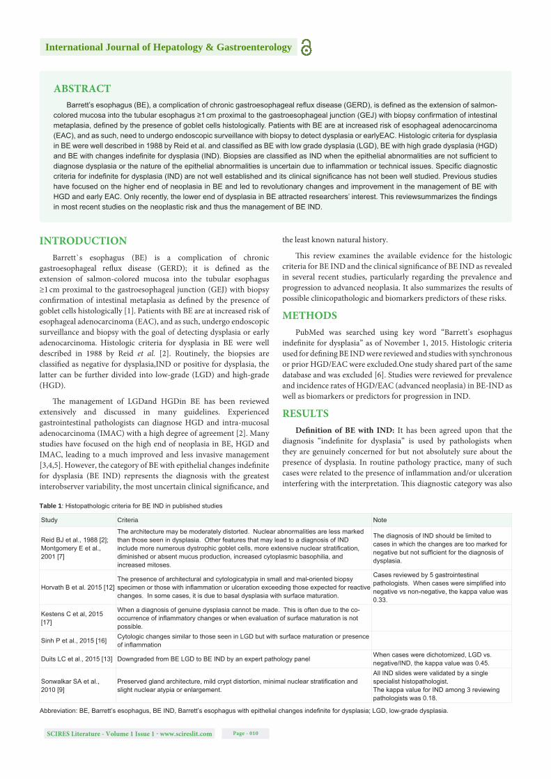
Study Criteria Note
Reid BJ et al., 1988 [2]; Montgomery E et al., 2001 [7]
The architecture may be moderately distorted. Nuclear abnormalities are less marked than those seen in dysplasia. Other features that may lead to a diagnosis of IND include more numerous dystrophic goblet cells, more extensive nuclear stratification, diminished or absent mucus production, increased cytoplasmic basophilia, and increased mitoses.
The diagnosis of IND should be limited to cases in which the changes are too marked for negative but not sufficient for the diagnosis of dysplasia.
Horvath B et al. 2015 [12]The presence of architectural and cytologicatypia in small and mal-oriented biopsy specimen or those with inflammation or ulceration exceeding those expected for reactive changes. In some cases, it is due to basal dysplasia with surface maturation.
Cases reviewed by 5 gastrointestinal pathologists. When cases were simplified into negative vs non-negative, the kappa value was 0.33.
Kestens C et al, 2015 [17]
When a diagnosis of genuine dysplasia cannot be made. This is often due to the co-occurrence of inflammatory changes or when evaluation of surface maturation is not possible.
Sinh P et al., 2015 [16] Cytologic changes similar to those seen in LGD but with surface maturation or presence of inflammation
Duits LC et al., 2015 [13] Downgraded from BE LGD to BE IND by an expert pathology panel When cases were dichotomized, LGD vs. negative/IND, the kappa value was 0.45.
Sonwalkar SA et al., 2010 [9]
Preserved gland architecture, mild crypt distortion, minimal nuclear stratification and slight nuclear atypia or enlargement.
All IND slides were validated by a single specialist histopathologist. The kappa value for IND among 3 reviewing pathologists was 0.18.
Table 1: Histopathologic criteria for BE IND in published studies
Abbreviation: BE, Barrett’s esophagus, BE IND, Barrett’s esophagus with epithelial changes indefinite for dysplasia; LGD, low-grade dysplasia.
SCIRES Literature - Volume 1 Issue 1 - www.scireslit.com Page - 011
International Journal of Hepatology & Gastroenterology
used when technicalissues such as biopsy crushing artifact, thick tissue sectioning, marked thermal artifact and tangential embedding and sectioning precluded a reliablediagnostic interpretation of dysplasia. Occasional caseswere secondary to the use of certain types of fixatives. For example, tissue fixation in Hollande’s and Bouin fixatives resulted in vesicular nucleus and prominent nucleolusleading to overinterpretation of IND by pathologists not familiar with this phenomenon [7]. In rare cases, the diagnosis of IND may be due to the so called “basal crypt dysplasia-like atypia”, where the dysplasia-like atypia is limited to the bases of the crypts, without involvement of the surface epithelium in BE [8].
Despite the attempted description and illustration of BE IND in initial publication [2], BE IND is diagnostically challenging and it is clear that its diagnostic reproducibility is poor [7,9,10]. Histologic criteria used to diagnose BE IND varied in different studies (Table 1) and even more so by pathologists in routine practice. For instance, the criteria for IND described by Reid BJ et alincluded moderate architectural distortion, nuclear abnormalities less marked than those seen in dysplasia, frequent dystrophic goblet cells, more extensive nuclear stratification, diminished or absent mucus production, increased cytoplasmic basophilia, and increased mitoses. The diagnosis of IND should be limited to cases in which the changes are worrisome but not sufficient for the diagnosis of dysplasia [2]. Using similar criteria, other groups performed intraobserver and interobserver reproducibility studies and found that BE IND has significant interobserver variability [7,11]. In daily pathology practice, the BE IND category appears to expand, one such example being basal crypt dysplasia-like atypia. The concept of basal crypt dysplasia-like atypia remains controversial and is interpreted by some groups as IND while others believe that it truly represents dysplasia without surface involvement.
Clinical significance of BE IND: Regardless of the definition, illustration, and intraobserver interobserver variability, BE IND category is not uncommonly used in daily pathology practice. Several studies recently investigated the clinical significance of BE IND and the results are reviewed and summarized in Tables2 and 3.
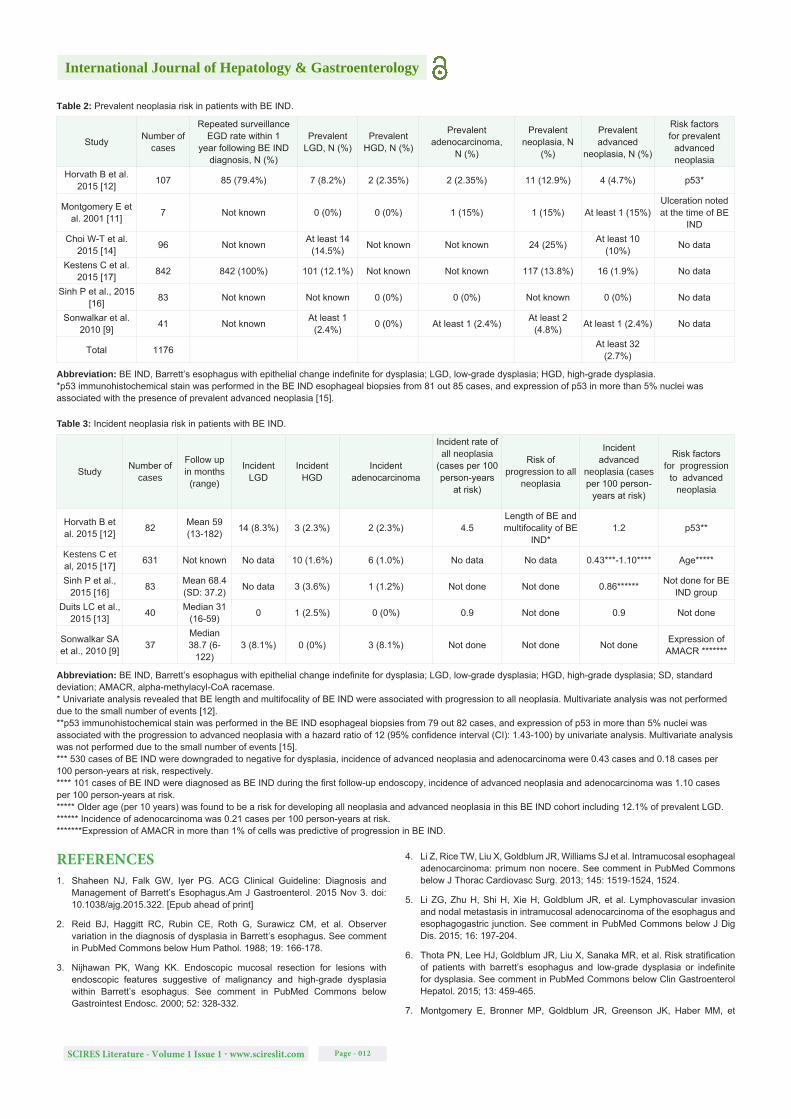
1. Prevalent neoplasia risk in patients with BE IND: The results are summarized in Table 2. Three studies addressed the prevalent neoplasia (defined as LGD, HGD or EAC detected within 1 year of the diagnosis of BE IND), and concluded that it ranged from 12.9% to 25%. Four studies addressed the prevalence of advanced neoplasia as defined byHGD or EAC detected within 1 year of the diagnosis of BE INDandit varied between 1.9% and 15%. When a6-month interval was used as a cut-off, the prevalence of LGDand advanced neoplasia in BE IND was at least 2.8% [9]. The presence of mucosal ulcerationwas associated with EAC in one study [11].
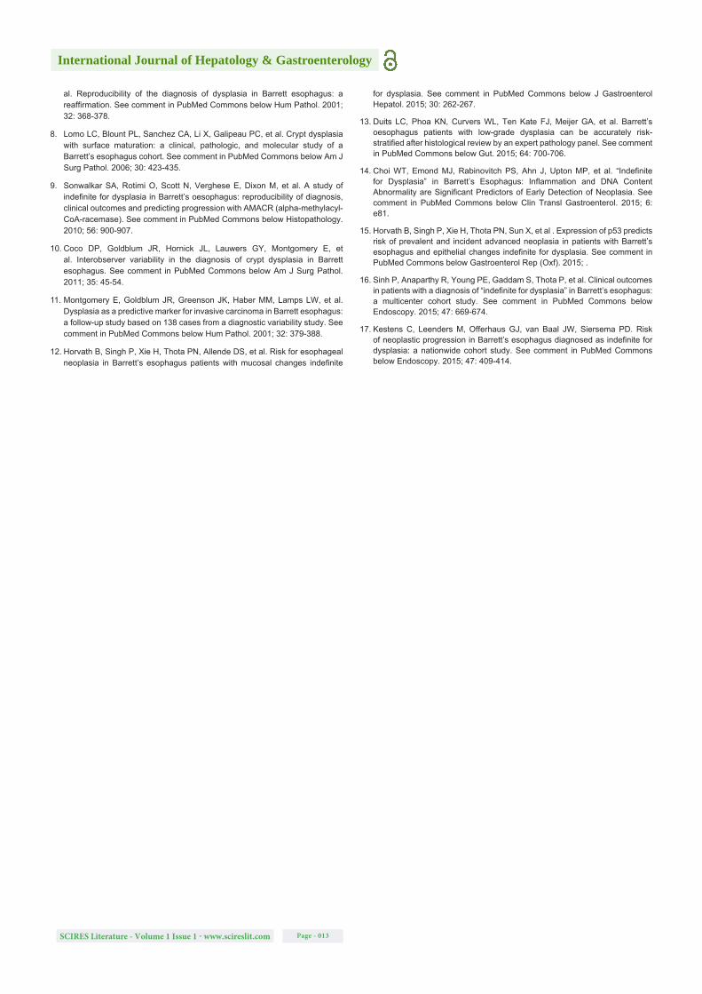
2. Incident neoplasia risk in patients with BE IND: The results are summarized in Table 3. The incidence of all neoplasia in BE-IND is reported to be 4.5 cases per 100 person-years at risk. The length of BE segment and multifocality of BE IND were associated with progression [12]. The progression to advanced neoplasia was 0.43 to 1.2 cases per 100 person-years at risk. The progression to EACwas 0.18 to 1.10 cases per 100 person-years at risk. One study examined the progression to advanced neoplasia in a cohort of BE IND (n=36) which was downgraded from an original diagnosis of BE LGD and reported an advanced neoplasia incidence of 0.9 cases per 100 person-years at risk, similar to a rate of 0.6 cases per 100 person-years at risk in patients with BE negative for dysplasia (n=153) [13]. In contrast,
BE LGD (n=75) agreed upon by a panel of expert pathologists had an advanced neoplasia incidence of 9.1 cases per 100 person-years at risk [13]. Using 6-monthfollow-up as a cutoff, Sonwalkar SA et al. (2010) reported that 8.1% of BE IND patients progressed to LGD and 8.1% BE IND progressed to EACduring a medium followup of 38.7 months (range: 6-122) [9]. Interestingly, none of the 6 patients with BE IND progression had a consensus diagnosis of IND by all three reviewing pathologists.
Somestudies addressed the neoplasia risk of BE IND, but did not distinguish between prevalent and incident cases of progression. For example, in the study by Montgomery E et al., adenocarcinoma was detected in 4 of 22 (18%) patients with the diagnosis of INDwith a median progression-free survival of 62 months and a median progression-free follow-up of 36 months [11]. In another study, Choi W-T et al reported that, in a group of BE IND patients without synchronous or previous neoplasia, the 1-, 2-, and 3-year detection rates of HGD or EAC were 10%, 13% and 20%, respectively [14].
3. Biomarkers for risk stratification of BE IND patients: Choi W-T et al identified active inflammation and DNA flow cytometric results as significant risk factors of neoplasia in patients with BE IND and reported that the hazard ratio for combined markers (active inflammation and abnormal DNA flow cytometric results, either DNA aneuploidy and/or 4N fractions greater than 6% of the nuclei) was 18.8[14].Sonwalkar SA et alreported thatthe expression of alpha-methylacyl-CoA racemase (AMACR) in more than 1% of cells predicted progression in BE IND [9]. However, the role of AMACR expression in risk stratifying BE IND was not substantiated in a study by Horvath B et al, and they instead showed that high expression of p53 (defined as intense staining in>5% nuclei) was associated with prevalent advanced neoplasia andprogression to advanced neoplasia in BE IND [15].
CONCLUSIONSIn summary, the diagnosis of BE IND is challenging. Recent data
revealsthat BE IND carries a significant risk of prevalent advanced neoplasia (at least 2.8%, 31 out of 1135 patients, ranging from 0% to 15%) (Table 2). In addition, the diagnosis of BE IND is associated with risk of progression to advanced neoplasia (0.43 to 1.2 cases person-years at risk) (Table 3), similar to the calculated progression risk of LGD without histology review [16], but much lower than the progression risk in consensus diagnosis of LGD[13]. Also, 73% of cases with a diagnosis of BE LGD originally rendered by practicing pathologists were down-graded to BE IND or BE negative for dysplasia by an expert pathology panel [13]. These results strongly suggest that cases with initial impression of BE IND or LGD should be reviewed by additional GI pathologists to confirm the diagnosis.The current knowledge regarding the clinical significance of BE IND as revealed by recent studies supports a close followup (short intervals between surveillance within 1 year) afterintensiveacid suppressive therapy and extensive biopsy sampling to detect prevalent neoplasia. BE IND patients with follow-up biopsies which are negative for dysplasia have low risk of neoplasia progression and may be reverted to routine surveillance as suggested by Kestens C et al., 2015 [17]. Although the length of BE, multifocality of BE IND, older age (>60 years old), abnormal p53 expression, active inflammation, and abnormal DNA content as detected by flow cytometry may provide useful information to risk-stratify this patient population, additional large prospectivestudies are needed to address their role in clinical management of patients with BE IND.
SCIRES Literature - Volume 1 Issue 1 - www.scireslit.com Page - 012
International Journal of Hepatology & Gastroenterology
Study Number of cases
Repeated surveillance EGD rate within 1
year following BE IND diagnosis, N (%)
Prevalent LGD, N (%)
Prevalent HGD, N (%)
Prevalent adenocarcinoma,
N (%)
Prevalent neoplasia, N
(%)
Prevalent advanced
neoplasia, N (%)
Risk factors for prevalent
advanced neoplasia
Horvath B et al. 2015 [12] 107 85 (79.4%) 7 (8.2%) 2 (2.35%) 2 (2.35%) 11 (12.9%) 4 (4.7%) p53*
Montgomery E et al. 2001 [11] 7 Not known 0 (0%) 0 (0%) 1 (15%) 1 (15%) At least 1 (15%)
Ulceration noted at the time of BE
IND Choi W-T et al.
2015 [14] 96 Not known At least 14 (14.5%) Not known Not known 24 (25%) At least 10
(10%) No data
Kestens C et al. 2015 [17] 842 842 (100%) 101 (12.1%) Not known Not known 117 (13.8%) 16 (1.9%) No data
Sinh P et al., 2015 [16] 83 Not known Not known 0 (0%) 0 (0%) Not known 0 (0%) No data
Sonwalkar et al. 2010 [9] 41 Not known At least 1
(2.4%) 0 (0%) At least 1 (2.4%) At least 2 (4.8%) At least 1 (2.4%) No data
Total 1176 At least 32 (2.7%)
Table 2: Prevalent neoplasia risk in patients with BE IND.
Abbreviation: BE IND, Barrett’s esophagus with epithelial change indefinite for dysplasia; LGD, low-grade dysplasia; HGD, high-grade dysplasia.*p53 immunohistochemical stain was performed in the BE IND esophageal biopsies from 81 out 85 cases, and expression of p53 in more than 5% nuclei was associated with the presence of prevalent advanced neoplasia [15].
Study Number of cases
Follow up in months (range)
Incident LGD
Incident HGD
Incident adenocarcinoma
Incident rate of all neoplasia
(cases per 100 person-years
at risk)
Risk of progression to all
neoplasia
Incident advanced
neoplasia (cases per 100 person-
years at risk)
Risk factors for progression
to advanced neoplasia
Horvath B et al. 2015 [12] 82 Mean 59
(13-182) 14 (8.3%) 3 (2.3%) 2 (2.3%) 4.5 Length of BE and multifocality of BE
IND*1.2 p53**
Kestens C et al, 2015 [17] 631 Not known No data 10 (1.6%) 6 (1.0%) No data No data 0.43***-1.10**** Age*****
Sinh P et al., 2015 [16] 83 Mean 68.4
(SD: 37.2) No data 3 (3.6%) 1 (1.2%) Not done Not done 0.86****** Not done for BE IND group
Duits LC et al., 2015 [13] 40 Median 31
(16-59) 0 1 (2.5%) 0 (0%) 0.9 Not done 0.9 Not done
Sonwalkar SA et al., 2010 [9] 37
Median 38.7 (6-
122)3 (8.1%) 0 (0%) 3 (8.1%) Not done Not done Not done Expression of
AMACR *******
Table 3: Incident neoplasia risk in patients with BE IND.
Abbreviation: BE IND, Barrett’s esophagus with epithelial change indefinite for dysplasia; LGD, low-grade dysplasia; HGD, high-grade dysplasia; SD, standard deviation; AMACR, alpha-methylacyl-CoA racemase.* Univariate analysis revealed that BE length and multifocality of BE IND were associated with progression to all neoplasia. Multivariate analysis was not performed due to the small number of events [12].**p53 immunohistochemical stain was performed in the BE IND esophageal biopsies from 79 out 82 cases, and expression of p53 in more than 5% nuclei was associated with the progression to advanced neoplasia with a hazard ratio of 12 (95% confidence interval (CI): 1.43-100) by univariate analysis. Multivariate analysis was not performed due to the small number of events [15]. *** 530 cases of BE IND were downgraded to negative for dysplasia, incidence of advanced neoplasia and adenocarcinoma were 0.43 cases and 0.18 cases per 100 person-years at risk, respectively.**** 101 cases of BE IND were diagnosed as BE IND during the first follow-up endoscopy, incidence of advanced neoplasia and adenocarcinoma was 1.10 cases per 100 person-years at risk.***** Older age (per 10 years) was found to be a risk for developing all neoplasia and advanced neoplasia in this BE IND cohort including 12.1% of prevalent LGD. ****** Incidence of adenocarcinoma was 0.21 cases per 100 person-years at risk.*******Expression of AMACR in more than 1% of cells was predictive of progression in BE IND.
REFERENCES1. Shaheen NJ, Falk GW, Iyer PG. ACG Clinical Guideline: Diagnosis and
Management of Barrett’s Esophagus.Am J Gastroenterol. 2015 Nov 3. doi: 10.1038/ajg.2015.322. [Epub ahead of print]
2. Reid BJ, Haggitt RC, Rubin CE, Roth G, Surawicz CM, et al. Observer variation in the diagnosis of dysplasia in Barrett’s esophagus. See comment in PubMed Commons below Hum Pathol. 1988; 19: 166-178.
3. Nijhawan PK, Wang KK. Endoscopic mucosal resection for lesions with endoscopic features suggestive of malignancy and high-grade dysplasia within Barrett’s esophagus. See comment in PubMed Commons below Gastrointest Endosc. 2000; 52: 328-332.
4. Li Z, Rice TW, Liu X, Goldblum JR, Williams SJ et al. Intramucosal esophageal adenocarcinoma: primum non nocere. See comment in PubMed Commons below J Thorac Cardiovasc Surg. 2013; 145: 1519-1524, 1524.
5. Li ZG, Zhu H, Shi H, Xie H, Goldblum JR, et al. Lymphovascular invasion and nodal metastasis in intramucosal adenocarcinoma of the esophagus and esophagogastric junction. See comment in PubMed Commons below J Dig Dis. 2015; 16: 197-204.
6. Thota PN, Lee HJ, Goldblum JR, Liu X, Sanaka MR, et al. Risk stratification of patients with barrett’s esophagus and low-grade dysplasia or indefinite for dysplasia. See comment in PubMed Commons below Clin Gastroenterol Hepatol. 2015; 13: 459-465.
7. Montgomery E, Bronner MP, Goldblum JR, Greenson JK, Haber MM, et
SCIRES Literature - Volume 1 Issue 1 - www.scireslit.com Page - 013
International Journal of Hepatology & Gastroenterology
al. Reproducibility of the diagnosis of dysplasia in Barrett esophagus: a reaffirmation. See comment in PubMed Commons below Hum Pathol. 2001; 32: 368-378.
8. Lomo LC, Blount PL, Sanchez CA, Li X, Galipeau PC, et al. Crypt dysplasia with surface maturation: a clinical, pathologic, and molecular study of a Barrett’s esophagus cohort. See comment in PubMed Commons below Am J Surg Pathol. 2006; 30: 423-435.
9. Sonwalkar SA, Rotimi O, Scott N, Verghese E, Dixon M, et al. A study of indefinite for dysplasia in Barrett’s oesophagus: reproducibility of diagnosis, clinical outcomes and predicting progression with AMACR (alpha-methylacyl-CoA-racemase). See comment in PubMed Commons below Histopathology. 2010; 56: 900-907.
10. Coco DP, Goldblum JR, Hornick JL, Lauwers GY, Montgomery E, et al. Interobserver variability in the diagnosis of crypt dysplasia in Barrett esophagus. See comment in PubMed Commons below Am J Surg Pathol. 2011; 35: 45-54.
11. Montgomery E, Goldblum JR, Greenson JK, Haber MM, Lamps LW, et al. Dysplasia as a predictive marker for invasive carcinoma in Barrett esophagus: a follow-up study based on 138 cases from a diagnostic variability study. See comment in PubMed Commons below Hum Pathol. 2001; 32: 379-388.
12. Horvath B, Singh P, Xie H, Thota PN, Allende DS, et al. Risk for esophageal neoplasia in Barrett’s esophagus patients with mucosal changes indefinite
for dysplasia. See comment in PubMed Commons below J Gastroenterol Hepatol. 2015; 30: 262-267.
13. Duits LC, Phoa KN, Curvers WL, Ten Kate FJ, Meijer GA, et al. Barrett’s oesophagus patients with low-grade dysplasia can be accurately risk-stratified after histological review by an expert pathology panel. See comment in PubMed Commons below Gut. 2015; 64: 700-706.
14. Choi WT, Emond MJ, Rabinovitch PS, Ahn J, Upton MP, et al. “Indefinite for Dysplasia” in Barrett’s Esophagus: Inflammation and DNA Content Abnormality are Significant Predictors of Early Detection of Neoplasia. See comment in PubMed Commons below Clin Transl Gastroenterol. 2015; 6: e81.
15. Horvath B, Singh P, Xie H, Thota PN, Sun X, et al . Expression of p53 predicts risk of prevalent and incident advanced neoplasia in patients with Barrett’s esophagus and epithelial changes indefinite for dysplasia. See comment in PubMed Commons below Gastroenterol Rep (Oxf). 2015; .
16. Sinh P, Anaparthy R, Young PE, Gaddam S, Thota P, et al. Clinical outcomes in patients with a diagnosis of “indefinite for dysplasia” in Barrett’s esophagus: a multicenter cohort study. See comment in PubMed Commons below Endoscopy. 2015; 47: 669-674.
17. Kestens C, Leenders M, Offerhaus GJ, van Baal JW, Siersema PD. Risk of neoplastic progression in Barrett’s esophagus diagnosed as indefinite for dysplasia: a nationwide cohort study. See comment in PubMed Commons below Endoscopy. 2015; 47: 409-414.