Embed Size (px)
DESCRIPTION
Genetics
Citation preview

SEX CHROMATIN BODY
Prof. M. Kamal
April 2008

Females are XX, males are XY
What is the consequence for females of having two X chromosomes, while males have only one?
Shouldn’t XX females produce twice the amount of X-linked gene products (proteins) as XY males?

Dosage Compensation
because XX females “compensate” by inactivating one of their X chromosomes to make a single “dosage” of X-linked genes.
• Measure the expression of X-linked genes revealed: • The level of mRNA or protein for various
X-linked genes (like autosomal genes) are similar between males and females
• Example – Factor VIII
No!
X Inactivation - one X chromosome in each female cell is inactivated- inactivation is a random process
Some cells - turn off paternal XSome cells - turn off maternal X
Bottom line: X-inactivation balances (compensates) dosage of X-linked genes between male and female cells
How is the dosage for X-linked genes adjusted to be equivalent in males and females?

In 1961 Mary Francis Lyon - British geneticist
- studied color coat in mice - knew that coat color was X-linked
Lyon concluded :
Male mice - hemizygous - have to be uniform color because they only
have one allele
Female mice - both alleles had to be active, but apparently not in the same cell
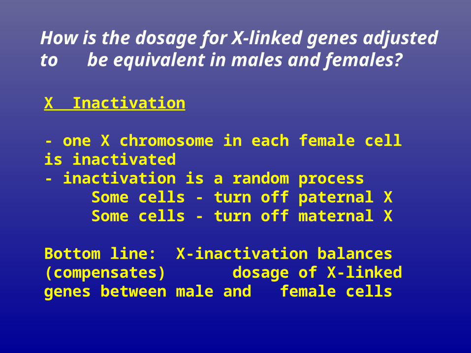
Barr Body
Inactivated X chromosome can be seen in females cells as the Barr body -
Murray Barr (1949)

X-inactivation reveals alleles in cats heterozygous for the fur color gene
Genotype is Xyellow/Xblack
Yellow patches: black allele is inactive Black patches: yellow allele is inactive
Xyellow/Xblack
Xyellow/Xblack
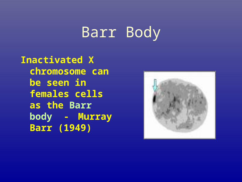
Barr Bodies are Inactivated X Chromosomes in Females
0 1
2 3
Normal male,Turner female
Normal female,Klinefelter male
# Barr bodies=N-1 rule
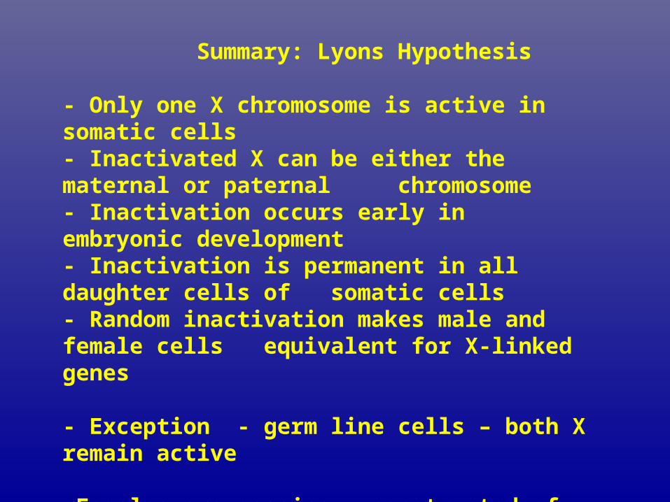
Summary: Lyons Hypothesis - Only one X chromosome is active in somatic cells - Inactivated X can be either the maternal or paternal
chromosome- Inactivation occurs early in embryonic development- Inactivation is permanent in all daughter cells of
somatic cells- Random inactivation makes male and female cells
equivalent for X-linked genes - Exception - germ line cells – both X remain active -Females are mosaics – constructed of two different cell
types


Anhidrotic ectodermal dysplasia
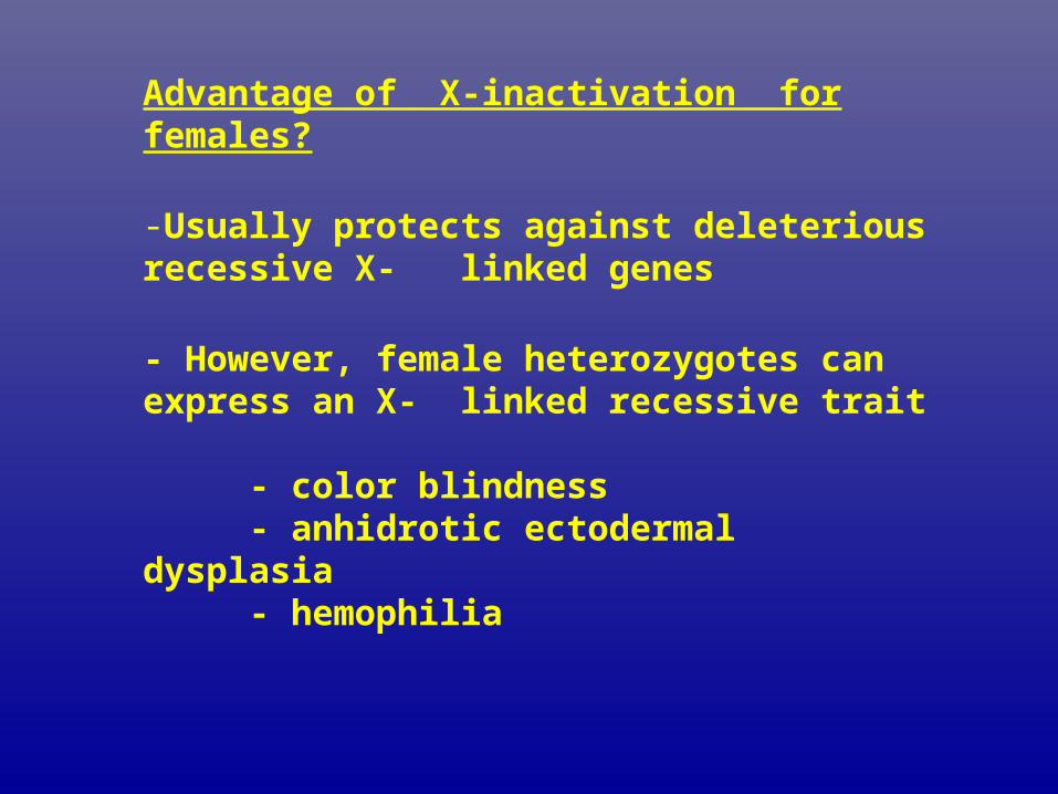
Advantage of X-inactivation for females?
-Usually protects against deleterious recessive X-linked genes
- However, female heterozygotes can express an X-linked recessive trait
- color blindness - anhidrotic ectodermal dysplasia
- hemophilia

Manifesting heterozygote• A carrier of an X-linked trait who expresses the
phenotype of the trait.• A higher proportion of normal X chromosomes if
inactivated in a given individual, may result in appearance of symptoms of disease in various degrees.
• Ornithine transcarbamylase deficiency (an enzyme deficiency resulting in high blood levels of ammonia and impaired urea formation),...
• Severe disease in males who inherit the mutant X chromosome).
• However, can also affect females who are “manifesting heterozygotes” presenting with severe disease during infancy or later in life during times of metabolic stress—for instance, during viral...
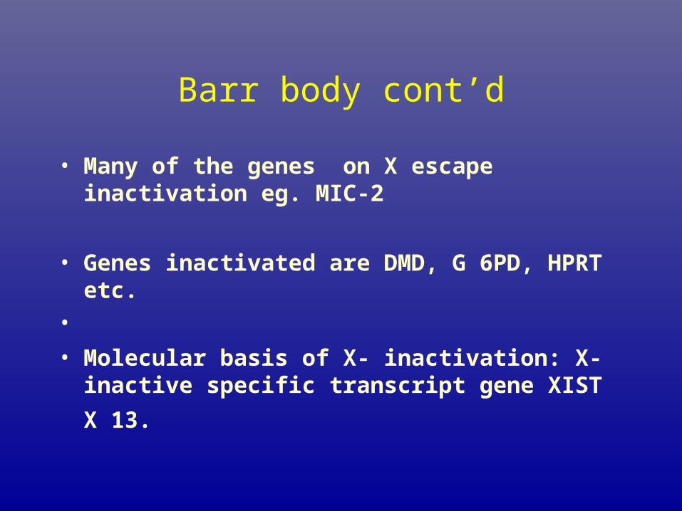
Barr body cont’d
• Many of the genes on X escape inactivation eg. MIC-2
• Genes inactivated are DMD, G 6PD, HPRT etc.
•
• Molecular basis of X- inactivation: X- inactive specific
transcript gene XIST X 13.
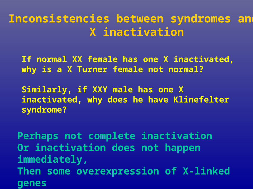
If normal XX female has one X inactivated, why is a X Turner female not normal?
Similarly, if XXY male has one X inactivated, why does he have Klinefelter syndrome?
Inconsistencies between syndromes and X inactivation
Perhaps not complete inactivationOr inactivation does not happen immediately,Then some overexpression of X-linked genes

HERMAPHRODITISM

What determines maleness and femaleness?
Two kinds of sex determination.
1. Environmental sex determination2. Genotypic sex determination
XX normal female XY normal male

Sex chromosomes determine gender.
Human males are the heterogametic sex with two different sex chromosomes, (XY).
•Human females are the homogametic sex (XX).

Genetic sex in humans
XX - normal femaleX - female phenotype –infertile (Turner’s)XXX - normal female (triplo-X)
- the X chromosome relates to the female phenotype - minimum of XX for normal female
XY - normal maleXXY - “normal male” – usually fertile (Klinefelters) XXXXY - severe Klinefelters syndrome
- male phenotypeY - monosomy Y - embryonic lethal
Y - chromosome directs male phenotypeminimum XY genotype necessary for a male
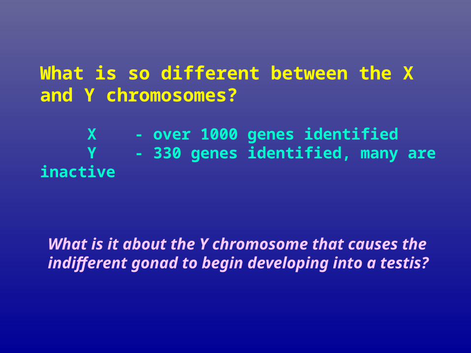
What is so different between the X and Y chromosomes?
X - over 1000 genes identifiedY - 330 genes identified, many are inactive
What is it about the Y chromosome that causes the indifferent gonad to begin developing into a testis?

Genes on the Y chromosome
There are three classes of genes on the Y.
Genes shared with X chromosome define the pseudoautosomal regions (PAR)
Genes similar to X chromosome genes are X-Y homologs
Genes unique to the Y including
SRY gene

What is SRY?
SRY (Sex-determining Region Y) is a sex-determining gene on the Y chromosome in humans
SRY – starts male development by - turning on testis-determining genes- turning off ovary-determining genes
Phenotypic males that were XX - sterilePhenotypic females who were XY - - turner’s
syndrome
TDF – testis determining factor


Summary of TDF 1. Initiates the process that directs the indifferent gonads
toward testis development 2. Activates Sertoli cells to produce Mullerian inhibiting
hormone, causing Mullerian duct degeneration 3. Stimulates Leydig cells to secrete testosterone, which then directs development of the Wolffian ducts towards epidiymides, vas deferens and seminal vesicles
- Testosterone conversion to dihydrotestosterone (DHT) - directs development of the urethra,
prostate gland and penis
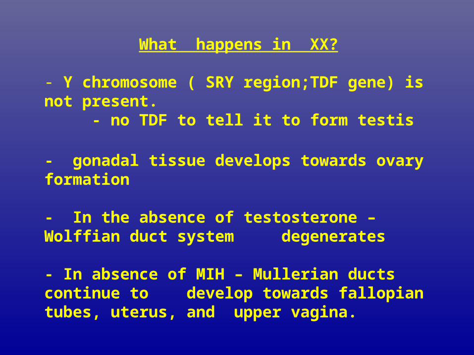
What happens in XX? - Y chromosome ( SRY region;TDF gene) is not present.
- no TDF to tell it to form testis
- gonadal tissue develops towards ovary formation
- In the absence of testosterone – Wolffian duct system degenerates
- In absence of MIH – Mullerian ducts continue to develop towards fallopian tubes, uterus, and upper vagina.

Summary of sex determination
At fertilization - have genotypic sex determination At 6-7 weeks - have gonadal sex determination
- if TDF present - male gonad development- if TDF absence - female gonad development
TDF - Referred to a the “master switch” How do we go from gonadal sex to phenotypic sex?
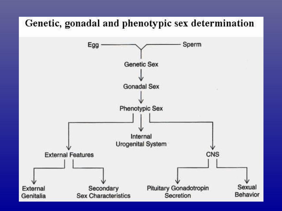

Levels of sexual identity
Development to childhood
Strong feelings of being
male or female develop
Gender identity
TimingEventsLevel
FertilizationXY=male
XX=female
Chromosomal
/genetic sex
9-16 weeks post fertilization
Undifferentiated structures becomes testis or ovary
Gonadal sex
8 weeks post fertilization to puberty
Internal and external reproductive structures
Phenotypic sex

Effect of castration and testosterone on adult male and female behavior

What is an abnormal sexual phenotypes?
There is an inconsistency between the observed genetic sex, gonadal sex and sexual differentiation

Abnormal sexual phenotypes result from mutations in genes involved in sexual development.
SRY gene Normal female development
Anti-Mullerian hormone Mullerian ducts persist in male
•gene
Testosterone gene Early development as female
Masculinization at puberty
DHT converting enzyme External structures lack signal
and develop as female,
internal structures are male.

Abnormal Development
Hermaphroditism • True hermaphrodism:
– possessing both male and female sexual anatomy
– example: one ovary, one testis, vaginal opening and penis
• Pseudohermaphrodism:– ovaries or testes, but not both
– if ovaries, then male external sexual anatomy
– if testes, then female external sexual anatomy

TRUE HERMAFHRODITISM
• Very rare
• Have both TESTICULAR and OVARIAN tissue.
• Internal & External sex organs variable
• Sex hormones also variable
• Majority XX, some XY some XX/ XY

PSEUDO HERMAPHRODITISM
• Have gonad of one sex i.e. testis OR ovary
• Ambiguous genetalia
• Various cause (cytogenetic, mendelian, Teratoganic)

MALE PSEUDO HERMAPHRODITISM
• Hetergenous group. genetically as well as clinically• • TESTICULAR FEMINZATION • X Linked disorder• genetic males (XY) with a female phenotype• - gonadal sex correct - gonads differentiate to testis • - produce MIH – females duct system has degenerated- produce testosterone and DHT

TESTICULAR FEMINZATION
• No uterus, Fallopian tube or ovary•TESTIS intrabdominal or in inguinal canal•Breast develop at puberty, sparse pubic / axillary hair •child appears to be a girl
- raised as girls- at puberty, genetically driven male phenotype emerges from an apparent female
phenotype
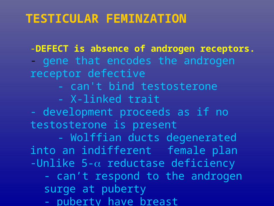
TESTICULAR FEMINZATION
-DEFECT is absence of androgen receptors.- gene that encodes the androgen receptor defective
- can't bind testosterone- X-linked trait
- development proceeds as if no testosterone is present
- Wolffian ducts degenerated into an indifferent female plan-Unlike 5- reductase deficiency
- can’t respond to the androgen surge at puberty- puberty have breast development, but no menstruation
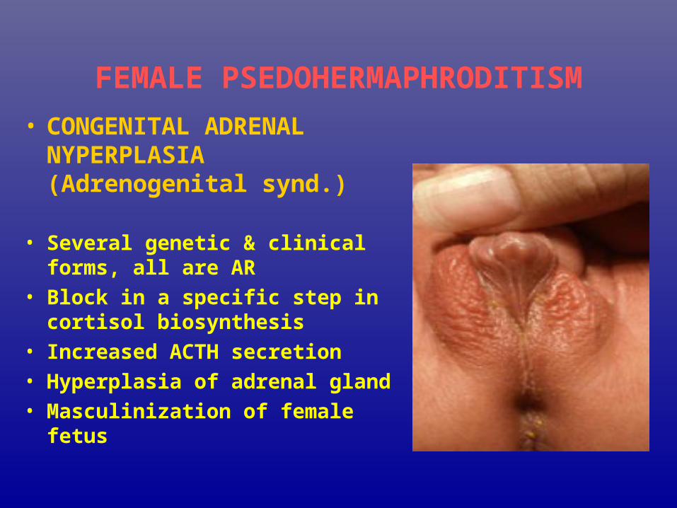
FEMALE PSEDOHERMAPHRODITISM
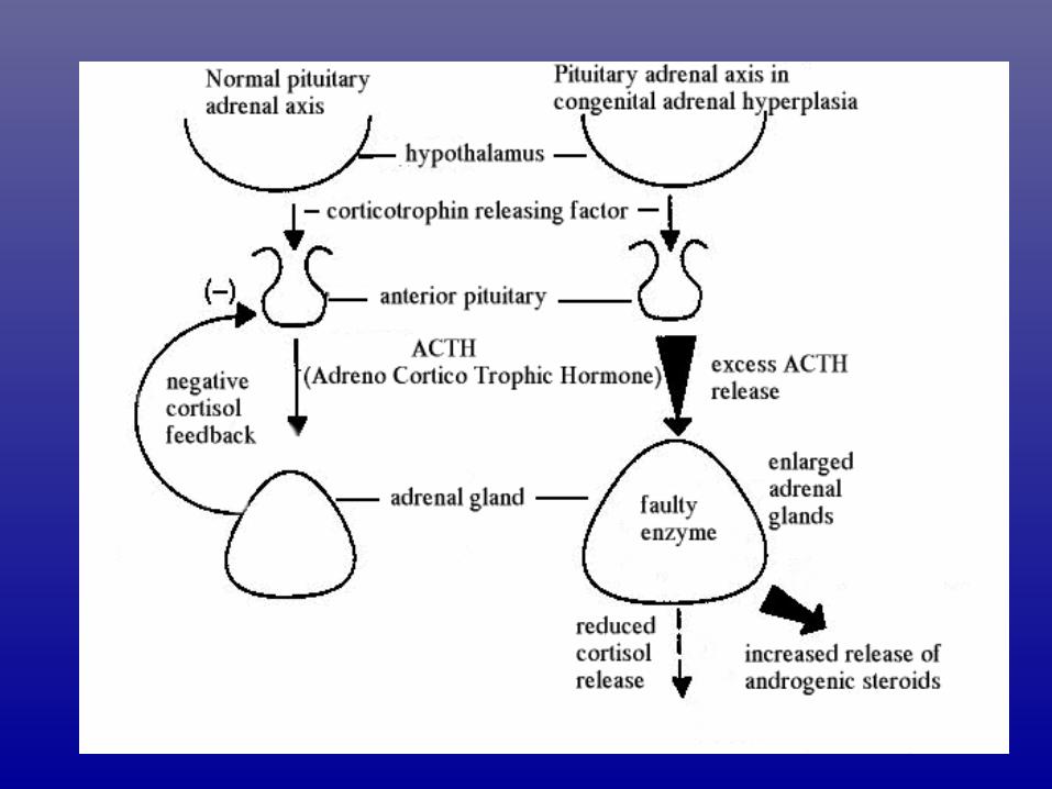
• CONGENITAL ADRENAL NYPERPLASIA (Adrenogenital synd.)
• Several genetic & clinical forms, all are AR
• Block in a specific step in cortisol biosynthesis
• Increased ACTH secretion
• Hyperplasia of adrenal gland
• Masculinization of female fetus

CONGENITAL ADRENAL
NYPERPLASIA • Most common form is 21 – hydroxylase deficiency
• Results in 3 different clinical presentations:
- Salt losing
- Simple virilizing
- Late onset virilization
• Diagnostic dues – Absence of testis in scrotum
- Presence of a uterus
- Elevated 17- ketosteroid.