Embed Size (px)
Citation preview

G-CSF Has a Therapeutic Effect on Cardıomyopathy in Diabetic Rats
Oytun Erbas, et al., BAOJ Diabet 2017 3: 13: 017
BAOJ Diabet, an open access journal Volume 3; Issue 1; 017
Oytun Erbas1, Hande Yapislar 2*, Fatih Oltulu3, Altug Yavasoglu3, Huseyin Aktug3 and Dilek Taskiran4
1İstanbul Bilim University MedicalFaculty,Department of Physiology2Acibadem University Medical Faculty, Department of Physiology
3Ege University MedicalFaculty, Department of HistologyandEmbriology4Ege University Medical Faculty, Department of Physiology
BAOJ Diabetes
*Corresponding author: Hande Yapislar, Acibadem Unv.Medical Fa-culty Physiology Department. Kerem Aydınlar Kampusu.Kayisdagi Cd. No: 32, Kucukbakkalkoy-Atasehir/Istanbul, Tel: 009 0532 472 74 97; E-mail: [email protected]
Sub Date: January 6, 2017, Acc Date: January 13, 2017, Pub Date: January 16, 2017.
Citation: Oytun Erbas, Hande Yapislar, Fatih Oltulu, Altug Yavaso-glu, Huseyin Aktug, et al. (2017) G-CSF Has a Therapeutic Effect on Cardıomyopathy in Diabetic Rats. BAOJ Diabet 3: 017.
Copyright: © 2017 Oytun Erbas, et al. This is an open-access article distributed under the terms of the Creative Commons Attribution Li-cense, which permits unrestricted use, distribution, and reproduction in any medium, provided the original author and source are credited.
Research
IntroductionThe major cause of morbidity and mortality in diabetes mellitus is cardiovascular disease. Coronary atherosclerosis and cardiomyopathy occur as a result of metabolic abnormalities and hyperglycemia associated with diabetes [1]. An abnormal increase in blood glucose was found to be related with cardiomyocyte to death by apoptosis. Apoptosis, result of myocardial abnormalities might be an important cause of cardiomyopathy [2]. Consequences of such abnormalities occur in diabetes and the contractile function of the heart is ultimately altered. The electro-cardiogram of many diabetes patients show several alterations from normal patterns. Increase of QT interval duration is one of them. Increase of QT interval reflects the abnormalities of ventricular myocardial re polarization. Prolongation of QT interval has been associated with sudden death in diabetes patients [3-4].
It was found that impaired fasting glucose and acute hyperglycemia was associated with QT prolongation. Also QT prolongation was found to be associated with hypertension and left ventricular hypertrophy, these conditions are related with cardiovascular diseases in diabetes mellitus [5].
Granulocyte colony stimulating factor (G-CSF) is a glycoprotein and a member of hematopoietic growth factor family which is capable of mobilizing bone marrow derived hematopoietic stem cells into the blood stream [6]. Recent years it’s been involved in investigating different treatment techniques for varied of diseases as a novel regenerative strategy. It’s been used in treatment of various diseases due to its role on multiplication of hemapoetic stem cells. In our previous study, we demonstrated that G-CSF has nephro-protective effect in diabetic rats [7]. This evidence is important in nephropathy which is one of the most frequent complications seen in advanced diabetes. Cardiomyopathy is another most common complication of diabetes. Increased hematopoietic stem cells induced by G-CSF could contribute to the tissue healing process. Our aim in this study is, inspired by our previous study, investigate the possible effects of G-CSF on QT-prolongation in diabetic rats and shed light on whether G-CSF could have a protective effect from cardiomyopathy.
Material and MethodsAnimals21 male Sprague Dawley albino mature rats were used in this study. Rats were 8 weeks old and weighing 200-220 g. All
animals were fed ad libitum and housed in pairs in steel cages having a temperature-controlled environment (22 ± 2°C) with 12-h light/dark cycles. The protocol of this study was approved by the Committee for Animal Research of Ege University. All animal studies are strictly conformed to the animal experiment guidelines of the Committee for Human Care.
Experimental Protocol
Diabetes was induced by intraperitoneal (i.p.) injection of streptozocin (STZ, Sigma-Aldrich, Inc.; Saint Louis, MO, USA) (60 mg/kg in 0.9% NaCl, adjusted to a pH 4.0 with 0.2M sodium citrate) for 14 rats. Control group didn’t receive any drugs(n=7) ,(control group). Diabetes was verified after 24 hours by evaluating blood glucose levels with the help of glucose oxidase reagent strips (Boehringer- Mannheim, Indianapolis). The rats with blood glucose levels of 250 mg/dl and higher were included in the study. 14 diabetic rats were randomly divided into 2 groups; diabetes group was treated with 1 mL/kg saline (Diabetes) (n=7), and diabetes+ G-CSF group was treated with 100 µg/kg/day G-CSF (Neupogen®, 48 MIU/0,5 ML) (Diabetes +G-CSF) (n=7) by i.p. injection for four weeks.
At the end of this four week, the animals were euthanized and electrocardiography( ECG) was taken as derivation I, blood samples were collected by cardiac puncture for blood glucose and removal of the heart were performed for histopathological examination.
Histopathological Examination of Heart Tissue
For histological and immunohistochemical studies, all animals were anesthetized by an i.p. injection of 40 mg/kg ketamin

BAOJ Diabet, an open access journal Volume 3; Issue 1; 017
Page 2 of 4Citation: Oytun Erbas, Hande Yapislar, Fatih Oltulu, Altug Yavasoglu, Huseyin Aktug, et al. (2017) G-CSF Has a Therapeutic Effect on Cardıomyopathy in Diabetic Rats. BAOJ Diabet 3: 017.
(Alfamine®, Ege Vet, Alfasan International B.V., Holland)/4 mg/kg xylazine (Alfazyne®, Ege Vet, Alfasan International B.V., Holland) and perfused with 200 ml of 4% formaldehyde in 0.1 M phosphate- buffer saline (PBS). Formalin-fixed heart sections (5μm) were stained with hematoxylin and eosin(H&E). All sections were photographed with Olympus C-5050 digital camera mounted on Olympus BX51 microscope.
Morphological analysis was assessed by computerized image analysis system. The degree of heart muscle cell hypertrophy were examined by light microscopy. Thickness of muscle cells was calculated from the cross-sectional image. The cross section yielding the maximum diameter of the muscle fiber was photographed and converted into a digital image by an examiner blinded to the source of the tissue. Muscle fibers were measured with the image analysis software Image-Pro Express 1.4.5, Media Cybernetics, Inc. USA. Fifty cardiac muscle cell from each animal were examined, and the average was used for analysis.
TGF-Β1, CD-34, Akt Immuno Expression
For immunohistochemistry, sections were incubated with H2O2 (10%) for 30 min to eliminate endogenous peroxidase activity and blocked with 10% normal goat serum (Invitrogen) for 1 hour at room temperature. Subsequently, sections were incubated in primary antibodies (TGF-β1, CD-34, Akt, Bioss, Inc.; 1/100) for 24 h at 4°C. Antibody detection was performed with the Histostain-Plus Bulk kit (Bioss, Inc) against rabbit IgG, and 3,3’ diaminobenzidine (DAB) was used to visualise the final product. All sections were washed in PBS and photographed with an Olympus C-5050 digital camera mounted on Olympus BX51 microscope. Brown cytoplasmic staining was scored positive for immunoexpression. The number of immunoexpression positive cell was assessed by systematically scoring at least fifty cardiac muscle cell per field in 10 fields of tissue sections at a magnification of 100x
Assessment of Cardiac Autonomic Neuropathy
QT interval and T wave duration was recorded electrocardio-graphically (Derivation I) . While the length of the QT interval is rate dependent, the QT analysis was realized on the transformed parameter QTc according to Bazett’s formula:
Statistical Analysis
All quantitative data were analyzed by using non-parametric (Mann-Whitney U) test. Student’s-t test was used to evaluate the differences between the groups. Data are presented as mean values ± standard error of the mean (SEM). p values of 0.05 or less were regarded as statistically significant.
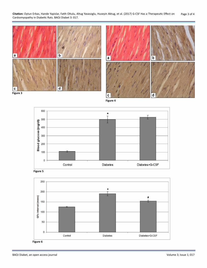
ResultsIn diabetic rats, cardiac muscle cell thickness (hypertrophy), TGF-β1 expression, QT interval ,T wave duration and blood glucose levels were increased significantly when compared to control group (p<0.05), (Figure 1-8) Administration of G-CSF
in diabetic rats causes a significant reduction both in cardiac muscle cell thickness, TGF-β1 expression and QT interval and T wave duration in these rats(p<0.05), (Figure 1, 4,5, 6,7,8) In terms of CD 34+ and Akt expression, there was no difference between control and diabetic rats (p>0.05), (Figure 2,3,8) Akt expression increased significantly in G-CSF administrated diabetic groups when compared to diabetic rats (p<0.05), (Figure 4,8). CD 34+ expression there was no difference between diabetic rats and G-CSF administered diabetic rats (p>0.05), (Figure 3,4,8).
Briefly, G-CSF administrated diabetic rats there was a decrease in cardiac muscle cell thickness and TGF-β1 expressions; Akt expressions were increased significantly when compared to diabetic rats (rats (p<0.05). CD 34+ expression showed no difference between G-CSF administrated groups and diabetic rats (p>0.05), (Figure 1-6)
Figure 1
Figure 2
BAOJ Diabet, an open access journal Volume 3; Issue 1; 017
Page 3 of 4Citation: Oytun Erbas, Hande Yapislar, Fatih Oltulu, Altug Yavasoglu, Huseyin Aktug, et al. (2017) G-CSF Has a Therapeutic Effect on Cardıomyopathy in Diabetic Rats. BAOJ Diabet 3: 017.
Figure 3
Figure 4
Figure 5
Figure 6

BAOJ Diabet, an open access journal Volume 3; Issue 1; 017
Page 4 of 6Citation: Oytun Erbas, Hande Yapislar, Fatih Oltulu, Altug Yavasoglu, Huseyin Aktug, et al. (2017) G-CSF Has a Therapeutic Effect on Cardıomyopathy in Diabetic Rats. BAOJ Diabet 3: 017.
DiscussionIn this study we showed the beneficial effects of G-CSF on both cardiac muscle and ECG on diabetic rats. Diabetes mellitus is a common disease affects many organs in the body adversely and the heart is one of them. Cardiomyopathy is one of the consequences of advanced diabetes, Especially “death in bed” syndrome in diabetes patients has been suggested to be caused by hypoglycemia. It was hypothesized that fatal cardiac arrhythmia has been triggered by hypoglycemia [8], researchers have found that hypoglycemia is associated with prolongation of QT interval and this is related to increased risk of sudden cardiac death [9]. QT interval prolongation reflects the abnormality in ventricular myocardial re polarization [10]. In summary, difficulty in re polarization which is shown in
ECG as QT interval prolongation and very common in diabetic patients cause arrhythmia and this situation triggers sudden death in diabetes mellitus.
There is plenty of data in literature demonstrating the connection between QT interval prolongation an diabetes. Jankyova et al searched out that diabetic rats have prolonged QT interval [11] . In two different studies Veglio et al found the relationship between QT prolongation and increased risk of death in diabetes mellitus [12-13]. We hypothesized that if we could fix QT interval prolongation, sudden deaths could be decrease among diabetes patients.
We administered G-CSF to the diabetic rats to carry out the effects of G-CSF on QT interval and other important factors as TGF-β1,
Figure 7
Figure 8
BAOJ Diabet, an open access journal Volume 3; Issue 1; 017
Page 5 of 6Citation: Oytun Erbas, Hande Yapislar, Fatih Oltulu, Altug Yavasoglu, Huseyin Aktug, et al. (2017) G-CSF Has a Therapeutic Effect on Cardıomyopathy in Diabetic Rats. BAOJ Diabet 3: 017.
References
1. Grundy SM, Benjamin IJ, Burke GL, Chait A, Eckel RH, et al. (1999) Diabetes and cardiovascular disease: a statement for healthcare pro-fessionals from the American Heart Association. Circulation 100(10): 1134-1146.
2. Tomei LD, Umansky SR (2001) Apoptosis and the heart: a brief review. Ann N Y Acad Sci 946:160-8.
3. Yap YG, Camm AJ (2003) Drug induced QT prolongation and torsades de pointes. Heart 89(11): 1363-72.
4. Straus SM, Kors JA, De Bruin ML, van der Hooft CS, Hofman A, Heer-inga J, et al. (2006) Prolonged QTc interval and risk of sudden cardiac death in a population of older adults. J Am Coll Cardiol 47(2): 362-367.
5. Marfella R, Rossi F, Giugliano D (2001) Hyperglycemia and QT interval: time for re-evaluation. Diabetes Nutr Metab 14(2): 63-5.
6. Kojima H, Otani A, Oishi A, Makiyama Y, Nakagawa S, et al. (2011) Granulocyte colony-stimulating factor attenuates oxidative stress-in-duced apoptosis in vascular endothelial cells and exhibits functional and morphologic protective effect in oxygen-induced retinopathy. Blood 117(3): 1091-100.
7. Yapislar H, Taskin E (2014) L-carnosine alters some hemorheologic and lipid peroxidation parameters in nephrectomized rats. Med Sci Monit 20: 399-405.
8. Weston PJ, Gill GV (1999) Is undetected autonomic dysfunction re-sponsible for sudden death in Type 1 diabetes mellitus? The ‘dead in bed’ syndrome revisited. Diabet Med 16(8): 626-631.
9. van Noord C, Sturkenboom MC, Straus SM, Hofman A, Kors JA, et al. (2010) Serum glucose and insulin are associated with QTc and RR in-tervals in nondiabetic elderly. Eur J Endocrinol 162(2): 241-248.
10. Rossing P, Breum L, Major-Pedersen A, Sato A, Winding H, et al. (2001) Prolonged QTc interval predicts mortality in patients with Type 1 dia-betes mellitus. Diabet Med 18(3): 199-205.
11. Jankyova S, Kmecova J, Cernecka H, Mesarosova L, Musil P, et al. (2012) Glucose and blood pressure lowering effects of Pycnogenol(R) are inefficient to prevent prolongation of QT interval in experimental diabetic cardiomyopathy. Pathol Res Pract 208(8): 452-457.
Akt and CD 34+ cells. We chose G-CSF due to our previous studies. In our latest study we demonstrated the protective effect of G-CSF on kidneys in diabetic rats. G-CSF is a glycoprotein and a member of growth factor family. G-CSF mobilizes hematopoietic stem cells from bone marrow into the blood stream [14]. In our study we found that G-CSF causes a significant reduction both in cardiac muscle cell thickness, QT interval and T wave duration in diabetic rats. Among diabetic patients besides QT interval duration increase in T wave duration and cardiac muscle cell thickness are other risk factors for cardiomyopathy and sudden death. Since T wave in ECG reflects the re polarization in diastole, increase in T wave duration demonstrates the difficulty in re polarization and resting phase of heart in diabetics. Hypertrophy is one of the risk factors for coronary failure [15-16]. We found that diabetic rats cardiac muscle cell thickness (hypertrophy) was increased when compared to control group. Administration of G-CSF decreased the cardiac muscle cell thickness in these rats.
G-CSF has been involved in diabetic and cardiovascular studies recently. Protective effects of G-CSF was demonstrated on acute myocardial infarction (G-CSF- MI). In another study, Shin et al studied on OLETF rats and they demonstrated that G-CSF might have a cardio-protective effect in diabetic cardiomyopathy. Our data is parallel to other findings in literature. We found that G-CSF administered diabetic rats QT interval was decreased than regular diabetic rats.
To find out the mechanism of G-CSF’s therapeutic cardiac effects on diabetic rats, we studied, CD34+, TGF-β1 and Akt expression levels in these rats. CD34+ cells are bone marrow derived immature cells in blood stream. It’s been reported that circulating CD 34+ cell levels are associated with cerebral infarction [17]. Makino et al found that increased CD 34+ levels in circulation might have anti-atherosclerotic effects [18]. Akt (Protein kinase B) is a molecule activated downstream of the PI3K(Phosphoinositide-3-OH kinase) signaling pathway [19]. Akt includes in regulation of cellular processes as cell survival, growth and proliferation and metabolism as well [20]. Akt also regulates the glucose uptake into tissues as a response to insulin. Akt deregulation has been reported in diabetes [21]. Studies on animal models demonstrated that Akt is an important molecule in regulating cardiovascular function and and plays a role in pathologic cardiac hypertrophy [22].
In our study there were no differences between control and dia-betic rats in terms of CD 34+ and Akt expressions. After G-CSF ad-ministration Akt expression increased significantly in diabetic rats.
Previous data showed that transforming growth factor beta-1 (TGF-β1) is a fibrogenic and an inflammatory cytokine that plays role in nephropathy in diabetes [22]. Also it has been demonstrated that Because TGF-β1 is a pivotal mediator of matrix accumulation it plays a key role in fibrosis in the heart [23]. According to the study of Yu et al down regulation of TGF-β1 in myocardial tissue might be protective against cardiomyopathy [24]. We found over-expression of TGF-β1 in diabetic rats. TGF-β1 expression significantly decreased in diabetic rats after G-CSF administration.
Our results show that G-CSF prevents diabetic rats from cardiomyopathy via increasing Akt and TGF-β1 expression but not via increment of CD 34+ cells.
In diabetic rats, cardiac muscle cell thickness (hypertrophy), TGF-β1 expression, QT interval and T wave duration were increased significantly when compared to control group. Administration of G-CSF in diabetic rats causes a significant reduction both in cardiac muscle cell thickness, TGF-β1 expression and QT interval and T wave duration in these rats. In terms of CD 34+ and Akt expression, there was no difference between control and diabetic rats. Akt expression increased significantly in G-CSF administrated diabetic groups when compared to diabetic rats. CD 34+expression there was no difference between control and diabetic rats.
AcknowledgementThis study was supported by Ege University Research Foundation. The authors declare that they have no conflict of interest
BAOJ Diabet, an open access journal Volume 3; Issue 1; 017
Page 6 of 6Citation: Oytun Erbas, Hande Yapislar, Fatih Oltulu, Altug Yavasoglu, Huseyin Aktug, et al. (2017) G-CSF Has a Therapeutic Effect on Cardıomyopathy in Diabetic Rats. BAOJ Diabet 3: 017.
12. Veglio M, Chinaglia A, Cavallo-Perin P (2004) QT interval, cardiovas-cular risk factors and risk of death in diabetes. J Endocrinol Invest 27(2): 175-81.
13. Veglio M, Bruno G, Borra M, Macchia G, Bargero G, et al. (2002) Prevalence of increased QT interval duration and dispersion in type 2 diabetic patients and its relationship with coronary heart disease: a population-based cohort. J Intern Med 251(4): 317-24.
14. Zohlnhofer D, Dibra A, Koppara T, de Waha A, Ripa RS, et al. (2008) Stem cell mobilization by granulocyte colony-stimulating factor for myocardial recovery after acute myocardial infarction: a meta-anal-ysis. J Am Coll Cardiol 51(15): 1429-1437.
15. Guo WT, Dong DL (2014) Bone morphogenetic protein-4: a novel therapeutic target for pathological cardiac hypertrophy/heart failure. Heart Fail Rev.
16. Ochi Y, Kubo T, Kitaoka H (2014) Repeated heart failure in a 74-year-old man with left ventricular hypertrophy. Heart 100(9): 710-discus-sion 742.
17. Taguchi A, Matsuyama T, Moriwaki H, Hayashi T, Hayashida K, et al. (2004) Circulating CD34-positive cells provide an index of cerebrovas-cular function. Circulation 109(24): 2972-2975.
18. Makino H, Okada S, Nagumo A, Sugisawa T, Miyamoto Y, et al. (2008) Pioglitazone treatment stimulates circulating CD34-positive cells in type 2 diabetes patients. Diabetes Res Clin Pract 81(3): 327-330.
19. Vanhaesebroeck B, Guillermet-Guibert J, Graupera M, Bilanges B (2010)The emerging mechanisms of isoform-specific PI3K signalling. Nat Rev Mol Cell Biol 11(5): 329-341.
20. Alessi DR, Caudwell FB, Andjelkovic M, Hemmings BA, Cohen P (1996) Molecular basis for the substrate specificity of protein kinase B; com-parison with MAPKAP kinase-1 and p70 S6 kinase. FEBS Lett 399(3): 333-338.
21. Cho H, Thorvaldsen JL, Chu Q, Feng F, Birnbaum MJ (2001) Akt1/PKBalpha is required for normal growth but dispensable for mainte-nance of glucose homeostasis in mice. J Biol Chem 276(42): 38349-38352.
22. Shiojima I, Sato K, Izumiya Y, Schiekofer S, Ito M, et al. (2005) Disrup-tion of coordinated cardiac hypertrophy and angiogenesis contrib-utes to the transition to heart failure. J Clin Invest 115(8): 2108-2118.
23. Ares-Carrasco S, Picatoste B, Benito-Martin A, Zubiri I, Sanz AB, et al. (2009) Myocardial fibrosis and apoptosis, but not inflammation, are present in long-term experimental diabetes. Am J Physiol Heart Circ Physiol 297(6): H2109-H2119.
24. Yu J, Fei J, Azad J, Gong M, Lan Y, et al. (2012) Myocardial protection by Salvia miltiorrhiza Injection in streptozotocin-induced diabetic rats through attenuation of expression of thrombospondin-1 and trans-forming growth factor-beta1. J Int Med Res 40(3): 1016-1024.