Embed Size (px)
Citation preview
CORYNEBACTERIA
&
MORAXELLA
Istar Dolapci, MD, PhDAnkara University School of Medicine
Department of Medical Microbiology

Learning Objectives
Identify the distinguishing characteristics of the species
within the genera Corynebacteria and Moraxella
Explain the media used for culture for this group of
organisms, including the chemical principle and
composition
Correlate patient signs and symptoms with laboratory
data, and identify the most likely etiologic agent

CONTENTS (Description Headings)
Corynebacterium spp.
Physiology and Structure
Pathogenesis and Immunity
Epidemiology
Clinical Diseases
Laboratory diagnosis
Treatment, prevention & control
Moraxella spp.

Corynebacterium spp.
A large, heterogeneous collection of more than
100 species and subspecies…
They have a cell wall with short-chain mycolic
acid (in most species)
But they are not acid-fast!
They are stained with Gram stain

Corynebacterium spp.

Corynebacterium spp.
Aerobic or facultative anaerobic
Gram positive rods
Nonmotile
Catalase positive
Ferment carbohydrates, producing lactic acid by
product (most species but not all)
Grow well on common laboratory media (many
species)
Some species require supplementation of media with
lipids for good growth

Corynebacterium spp.
They normally colonize the skin, upper
respiratory tract, gastrointestinal tract, and
urogenital tract in humans
Can function as opportunistic pathogens
Few are associated with human disease
e.g. Corynebacterium diphtheriae

Corynebacterium diphtheriae
Physiology and Structure
C.diphtheriae is an irregularly staining, pleomorphic rod
0.3-0.8 x 1.0-8.0 mm
Grow on blood agar
Large, 1-3 mm colonies
Four biotypes: belfanti, gravis, intermedius, and mitis
Most diseases are caused by biotype mitis

Corynebacterium
diphtheriae
Pathogenesis and
Immunity
Major virulence factor:
Diphtheria toxin
This 58,300-Da protein is an
example of the classic A-B
exotoxin
The exotoxin is coded by tox
gene
The gene is carried by a
lysogenic bacteriophage, b-
phage
Microbial Pathogenesis, W. W. Norton & Company.

Diphteria Toxin
Toxins 2010, 2, 2519-2583; doi: 10.3390/toxins2112519

Toxins 2013, 5(8), 1362-1380

Diphteria Toxin
www.peertechz.com/articles/GJIDCR-3-114.php

Corynebacterium diphtheriae
Epidemiology
Worldwide distribution maintained in asymptomatic
carriers and infected patients
Humans are the only known reservoir for this
organism
Spread person to person by exposure to respiratory
droplets or skin contact

Diphtheria – United States1980-2011
Diphtheria – Age Distribution of Reported Cases, United States
https://www.cdc.gov/vaccines/pubs/pinkbook/dip.html

Corynebacterium diphtheriae
Clinical Diseases
The clinical presentation of diphtheria is determined by
the;
1. Site of infection,
2. Immune status of the patient, and
3. Virulence of the organism
Toxigenic strains
Nontoxigenic strains

The organism
does not need to
enter the blood to
produce disease
&
does not typically
invade deep
tissues

Corynebacterium diphtheriae
Clinical Diseases
1. Respiratory Diphtheria
2. Cutaneous Diphtheria

Respiratory Diphtheria
Incubation period: 2- to 4-days
Organisms multiply locally on epithelial cells in
the pharynx
Initially cause localized damage as a result of
exotoxin activity

Respiratory Diphtheria
The onset is sudden,
with;
malaise,
sore throat,
exudative
pharyngitis, and
a low-grade fever

Respiratory Diphtheria
A thick pseudo membrane
composed of bacteria,
lymphocytes, plasma cells,
fibrin, and dead cells
can cover the tonsils, uvula, and
palate
firmly adheres to the underlying
tissue
is difficult to dislodge without
making the tissue bleed
unique to diphtheria
https://en.wikipedia.org/wiki/Diphtheria

Respiratory Diphtheria
Complications of diphtheria are attributable to effects of
the toxin
The toxin, when absorbed, affects organs and tissues distant
from the site of invasion
The most frequent complications of diphtheria are
myocarditis and neuritis
If myocarditis occurs early, it is often fatal
Neuritis most often affects motor nerves and usually resolves
completely
Paralysis of the soft palate, eye muscles, limbs, and diaphragm etc.

Cutaneous Diphtheria
is acquired through skin contact with other infected
persons
papule develops first and then evolves into a chronic,
nonhealing ulcer
sometimes covered with a grayish membrane
N Engl J Med 2018; 378:e17

Laboratory Diagnosis
Microscopy
Culture
Identification
Toxigenicity testing
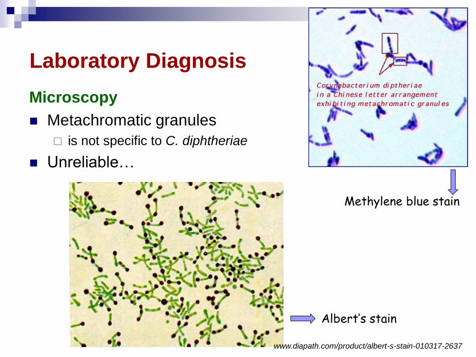
Laboratory Diagnosis
Microscopy
Metachromatic granules
is not specific to C. diphtheriae
Unreliable…
Albert’s stain
Methylene blue stain
www.diapath.com/product/albert-s-stain-010317-2637

Laboratory Diagnosis
Culture
Specimen: from both the nasopharynx and
throat
Culture media:
A nonselective, enriched blood agar plate
A selective medium Cysteine-tellurite blood agar [CTBA],
Tinsdale medium,
Colistin-nalidixic agar [CNA]

Laboratory Diagnosis
Culture
Cysteine-tellurite blood agar [CTBA]
A long shelf life
Inhibits some strains of C. diphtheria
Tinsdale medium The best medium for recovering C. diphtheriae in clinical specimens
A short shelf life and requires addition of horse serum
Colistin-nalidixic agar [CNA] Commonly used for the selective recovery of gram-positive bacteria
A practical alternative medium

Laboratory Diagnosis
Corynebacterium diphtheriae on tellurite agar
http://www.medical-labs.net/corynebacterium-diphtheriae-on-tellurite-agar-2180/

Laboratory Diagnosis
Corynebacterium diphtheriae in Tinsdale Agar medium
Look for black colonies with brown halo
https://microbeonline.com/tinsdale-agar-composition-preparation/

Laboratory Diagnosis
Identification
Presence of
cystinase
Absence of
pyrazinamidase

Laboratory Diagnosis
Toxigenicity testing
Elek test
PCR
ELISA
https://microbeonline.com/elek-test-principle-procedure-results/
http://www.rahulgladwin.com/noteblog/bacteriology/what-is-an-eleks-test.php

Elek Test
A filter paper disk
containing antitoxin (10
IU/disk) is placed on an
agar plate
The cultures to be tested for toxigenicity are spot
inoculated 7–9 mm away from the disk
After 48 hours of incubation, the antitoxin diffusing from
the paper disk has precipitated the toxin diffusing from
toxigenic cultures and has resulted in precipitin bands
between the disk and the bacterial growth

PCR An alternative method is the detection of the exotoxin gene
by a polymerase chain reaction (PCR)–based nucleic acid
amplification method
This test can detect the tox gene in clinical isolates and
directly in clinical specimens
Result of PCR analysis of the C. diphtheriae tox gene, using DNA templates;JOURNAL OF CLINICAL MICROBIOLOGY, 2000, 38: 2400–02


Treatment
Diphtheria antitoxin
early administration is important
Penicillin or erythromycin
can eliminate C. diphtheriae and terminate toxin
production
Immunization with toxoid
to enhance the production of protective antibodies

Prevention & Control
DPT vaccine
At ages 2, 4, 6, 15 to 18 months, and 4 to 6 years
Booster vaccinations with diphtheria toxoid, every 10 years
People in close contact with patients who have
documented diphtheria are at risk for acquiring the
disease
Nasopharyngeal specimens for culture should be collected from
all close contacts and antimicrobial prophylaxis with
erythromycin or penicillin should be started immediately

Other Corynebacteria Species
Nonlipophilic Corynebacteria spp.
Corynebacterium ulcerans
Corynebacterium pseudotuberculosis
Corynebacterium minutissimum
Corynebacterium amycolatum
Lipophilic Corynebacteria spp.
Corynebacterium jeikeium
Corynebacterium urealyticum

Organism Clinical Features Epidemiologic
Features
Treatment
C. ulserans Respiratory diphtheria Normal microbiota:
Humans and cattle
Mode of transmission:
Uncertain
Zoonosis
No definitive
guidelines.
All are
susceptible to
vancomycin and
teicoplanin
C. pseudotuberculosis Lymphadenitis,
ulcerative
lymphangitis, abscess
formation, respiratory
diphtheria
Normal microbiota:
Animals such as sheep,
goats, and horses
Zoonosis: Close animal
contact, but infections in
humans are rare
No definitive
guidelines.
All are
susceptible to
vancomycin and
teicoplanin
Corynebacterium ulserans &
Corynebacterium pseudotuberculosis

Corynebacterium jeikeium &
Corynebacterium urealyticum
Organism Clinical Features Epidemiologic
Features
Treatment
C. jeikeium Opportunistic
infections;
bacteremia
Immunocompromised
patients at increased risk
Vancomycin
C. urealyticum Urinary tract
infections, including
pyelonephritis;
bacteremia
Risk factors include
immunosuppression,
underlying genitourinary
disorders, antecedent
urologic procedures, prior
antibiotic therapy
Vancomycin

Pseudomonas and related bacteria
Nonfermentative rods
Opportunistic pathogens of plants, animals, and humans
Most clinically significant isolates are members of five
genera:
1. Pseudomonas,
2. Burkholderia,
3. Stenotrophomonas,
4. Acinetobacter, and
5. Moraxella

Moraxella spp. The most important
species is M. catarrhalis
strictly aerobic,
oxidase-positive,
gram negative diplococci
considered to be a part of
the normal oropharyngeal
flora
can become pathogenic
The cellular morphology of
this species is more similar
to that of Neisseria spp.
than that of the other
Moraxella spp.
http://hit-micrscopewb.hc.msu.edu/Microbiology/Lab/S1-
Resp_Image_11.html

Moraxella spp.
When a breakdown of the patient’s mucosal or
epidermal defensive barriers occurs;
Bronchitis
Bronchopneumonia
in elderly patients with chronic pulmonary disease
Sinusitis
Otitis

Moraxella spp.
rarely cause infection…
low virulence…
contaminants… ?

Moraxella spp./ Cultivation
5% Sheep blood
Chocolate agar
MacConkey agar
Commercial blood culture systems
Nutrient broths
Such as thioglycollate and brain-heart infusion
35°C in carbon dioxide or ambient air for a minimum of 48
hours
Clinically important isolates should be sent to a reference
laboratory for definitive identification

Moraxella spp.
Organism Therapeutic Options Potential Resistance
to Therapeutic
Options
Moraxella spp. No definitive guidelines;
generally susceptible to
penicillins and
cephalosporins
Beta-lactamase–mediated
resistance to penicillins
common

Moraxella spp.
Organism Habitat (Reservoir) Mode of
Transmission
Spectrum
of Disease
and
Infections
Moraxella
nonliquefaciens,
Moraxella lacunata,
Moraxella osloensis,
Moraxella lincolnii,
Moraxella canis,
Moraxella atlantae
Normal human
microbiota that inhabit
mucous membranes
covering the nose,
throat, other parts of the
upper respiratory tract,
conjunctiva, and, for
some species (i.e., M.
osloensis), the urogenital
tract; may also colonize
the skin
Infections are
rare; when they
occur, they are
probably caused
by the patient’s
endogenous
strains; person-to-
person
transmission may
be possible, but
this has not been
documented
Eye
infections,
bacteremia,
endocarditis,
septic
arthritis,
and,
possibly,
respiratory
infections

References
Medical Microbiology; Murray, Rosenthal, Pfaller; 7th Ed;
Elsevier Saunders; 2013
Jawetz, Melnick & Adelberg’s Medical Microbiology;
Brooks G, Carroll KC, Butel J, Morse S (Eds); 27th Ed;
McGraw Hill Lange; 2016
Sherris Medical Microbiology; 6th Ed; Ryan KJ, Ray CG;
McGraw Hill Education; 2014

THE END
THANKS FOR LISTENING