Embed Size (px)
Citation preview
1121
http://journals.tubitak.gov.tr/biology/
Turkish Journal of Biology Turk J Biol(2016) 40: 1121-1128© TÜBİTAKdoi:10.3906/biy-1601-76
Baculoviral vector loaded mesenchymal stem cells as efficient gene therapy tools for cancer treatment
Sedef Hande AKTAŞ1, Hakan AKBULUT1,*, Ayşe Eser ELÇİN2, Mahmut PARMAKSIZ2,Arzu Aktan KESKİN3, Arzu ÇÖLERİ CİHAN4, Yaşar Murat ELÇİN2, Fikri İÇLİ1
1Department of Medical Oncology, Ankara University School of Medicine, Ankara, Turkey2Tissue Engineering, Biomaterials and Nanobiotechnology Laboratory, Ankara University Faculty of Science, and Ankara University
Stem Cell Institute, Ankara, Turkey3Department of Physiology, Gazi University, Ankara, Turkey
4Department of Biology, Ankara University Faculty of Science, Ankara, Turkey
* Correspondence: [email protected]
1. IntroductionMesenchymal stem cells (MSCs) have important advantages as vehicles of gene therapy vectors. Recently, MSCs transduced with lentiviruses, retroviruses, or plasmids have been successfully tested in cancer treatment studies (Sasportas et al., 2009; Huang et al., 2010; Matuskova et al., 2010). Along with bone marrow-derived MSCs (BM-MSC), adipose tissue-derived MSCs (AT-MSC) can also home to tumor sites when administered intravenously (Elçin, 2003; Kucerova et al., 2007). AT-MSCs transduced with a retroviral vector carrying the cytosine deaminase (CD) suicide gene caused significant tumor regression in a colon cancer model (Ho et al., 2005). Although lentiviral and retroviral vectors have the capability for long-term gene expression in transduced cells, the high rate of insertional mutagenesis limits their use in cancer treatment.
Recently, baculoviral vectors derived from an insect virus of Autographa californica multiple nucleopolyhedrovirus have been shown to be used in gene transfer studies. Ho et al. have shown that the BV can enter
mammalian cells without causing any significant toxicity (Ho et al., 2005). The BV genome is approximately 130 kb in size, which allows the transfer of large-sized genes. BVs have been shown to transduce mammalian cells, including mesenchymal stem cells (Chuang et al., 2007, 2009; Chen et al., 2011). The potential of transduction and lack of innate immunity against baculoviruses in mammals make these vectors attractive tools for gene targeting and cancer gene therapy.
In the current study, we constructed baculoviral vectors carrying CD (BV-CD) or green fluorescence protein (GFP) genes (BV-GFP) and tested the transduction efficiency of the vectors in tumor and mesenchymal stem cells. We also tested the antitumor efficacy of the BM-MSCs transduced with the BV-CD vector in a colon cancer model.
2. Materials and methods2.1. Plasmids, cells, and animalsIn order to construct the recombinant BV, we used the following plasmids: pfastBac1 (Invitrogen, Carlsbad, CA,
Abstract: Insufficient targeting of the therapeutic genes to tumor cells is one of the major reasons for failure in cancer gene therapy. Mesenchymal stem cells (MSCs) seem to be a good candidate as a carrier for gene therapy because of its selective tumor tissue-homing properties. In the current study, we constructed baculoviral vectors (BVs) carrying cytosine deaminase (CD) (BV-CD) or green fluorescence protein (GFP) genes (BV-GFP) and tested the transduction efficiency of the vectors in tumor and mesenchymal stem cells. We also tested the in vivo efficacy of the BV-CD vector in a colon cancer model. Our results showed that the recombinant baculoviral vectors can efficiently transduce mammalian cells and express genes of interest. The BV-CD vector treatment caused significant in vitro cytotoxicity when used with 5-fluorocytosine. MSCs loaded with the BV-CD vector caused a significant delay in tumor growth and increased survival when compared to control and MSC alone treated groups bearing colon cancer. Our results show that the recombinant BV-CD vector could be used either alone or loaded into MSCs in the treatment of established tumors.
Key words: Gene therapy, mesenchymal stem cells, baculoviral vector, cancer
Received: 26.01.2016 Accepted/Published Online: 08.06.2016 Final Version: 08.11.2016
Research Article
AKTAŞ et al. / Turk J Biol
1122
USA), pORF-codAupp (InvivoGen, San Diego, CA, USA), pCITE (2+) (Novagen, Germany), pShuttle-CMVGFP (Addgene, Teddington, UK), pCR-blunt (Invitrogen), and pcDNA.1 (Invitrogen). The plasmids were grown in chemical competent Top 10 E. coli or DH10Bac E. coli (Invitrogen). Sf-9 insect cells (Invitrogen) were used to produce baculoviral vectors. HEK293 human renal epithelial cells (ATCC, Teddington, UK), a mouse colon cancer cell line (CRL 2638) (ATCC), and primary mouse mesenchymal stem cells (MSCs) obtained from the bone marrow of BALB/c mice were used in in vitro transduction experiments or as vehicles of the recombinant therapeutic vector in a tumor model. Six- to eight-week-old BALB/c mice (Refik Saydam Health Institute, Ankara, Turkey) were used either to obtain BM-MSCs or to establish a colon cancer model for in vivo experiments. The experimental protocol was approved by the Animal Experiments Local Ethics Committee of Ankara University.2.2. Construction of baculoviral vectorsIn the current study, we constructed two different BVs carrying either the GFP reporter gene or CD therapeutic gene. The BVs were constructed according to the manufacturer’s protocol (BAC-to-BAC Baculoviral Expression System, Invitrogen). Briefly, we first amplified the CD gene from pORF-codAupp plasmid by using the forward 5’-ACCATGAGCAA TAACGCTTTACA-3’ and reverse 5’-GTAACCCAG TCGTTCAACGTTT-3’ primers, GFP gene using forward 5’-ACGCGCATGCGTAGA CACACCATGGT GAGCAAGG-3’ and reverse 5’-CGGAGGTACCTCGGTCATGGTGGCCT TGT ACAG-3’ primers, and CMV promoter using forward 5’-ACCACTAGTAA CCGAGTAAGATTTGGC-3’ and reverse 5’-TTTATGTTTCAGGTTCAG-3’ primers from pShuttle-CMVGFP plasmid. Then those genes were first cloned to either pCR blunt or pcDNA.1 shuttle plasmids and then the genes were cut and cloned to pFastBa/HBM-TOPO donor plasmid vector.
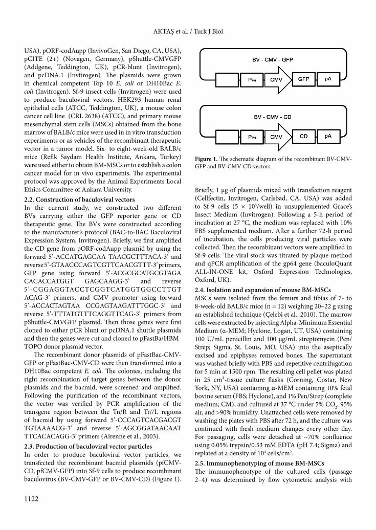
The recombinant donor plasmids of pFastBac-CMV-GFP or pFastBac-CMV-CD were then transformed into a DH10Bac competent E. coli. The colonies, including the right recombination of target genes between the donor plasmids and the bacmid, were screened and amplified. Following the purification of the recombinant vectors, the vector was verified by PCR amplification of the transgene region between the Tn/R and Tn7L regions of bacmid by using forward 5’-CCCAGTCACGACGT TGTAAAACG-3’ and reverse 5’-AGCGGATAACAAT TTCACACAGG-3’ primers (Airenne et al., 2003). 2.3. Production of baculoviral vector particlesIn order to produce baculoviral vector particles, we transfected the recombinant bacmid plasmids (pfCMV-CD, pfCMV-GFP) into Sf-9 cells to produce recombinant baculovirus (BV-CMV-GFP or BV-CMV-CD) (Figure 1).
Briefly, 1 µg of plasmids mixed with transfection reagent (Cellfectin, Invitrogen, Carlsbad, CA, USA) was added to Sf-9 cells (5 × 10⁵/well) in unsupplemented Grace’s Insect Medium (Invitrogen). Following a 5-h period of incubation at 27 °C, the medium was replaced with 10% FBS supplemented medium. After a further 72-h period of incubation, the cells producing viral particles were collected. Then the recombinant vectors were amplified in Sf-9 cells. The viral stock was titrated by plaque method and qPCR amplification of the gp64 gene (baculoQuant ALL-IN-ONE kit, Oxford Expression Technologies, Oxford, UK). 2.4. Isolation and expansion of mouse BM-MSCsMSCs were isolated from the femurs and tibias of 7- to 8-week-old BALB/c mice (n = 12) weighing 20–22 g using an established technique (Çelebi et al., 2010). The marrow cells were extracted by injecting Alpha-Minimum Essential Medium (α-MEM; Hyclone, Logan, UT, USA) containing 100 U/mL penicillin and 100 µg/mL streptomycin (Pen/Strep; Sigma, St. Louis, MO, USA) into the aseptically excised and epiphyses removed bones. The supernatant was washed briefly with PBS and repetitive centrifugation for 5 min at 1500 rpm. The resulting cell pellet was plated in 25 cm²-tissue culture flasks (Corning, Costar, New York, NY, USA) containing α-MEM containing 10% fetal bovine serum (FBS; Hyclone), and 1% Pen/Strep (complete medium; CM), and cultured at 37 °C under 5% CO2, 95% air, and >90% humidity. Unattached cells were removed by washing the plates with PBS after 72 h, and the culture was continued with fresh medium changes every other day. For passaging, cells were detached at ~70% confluence using 0.05% trypsin/0.53 mM EDTA (pH 7.4; Sigma) and replated at a density of 104 cells/cm2. 2.5. Immunophenotyping of mouse BM-MSCsThe immunophenotype of the cultured cells (passage 2–4) was determined by flow cytometric analysis with
Figure 1. The schematic diagram of the recombinant BV-CMV-GFP and BV-CMV-CD vectors.
AKTAŞ et al. / Turk J Biol
1123
FACSCanto II (BD Biosciences, San Jose, CA, USA) using a CD antigen panel consisting of TER114-PE, Sca-1-PE, CD90.2-PE, CD106-PE, CD31-PE, CD45-FITC, CD11b-FITC, CD44-FITC, and CD29-FITC (all from BD Biosciences) as described previously (Odabaş et al., 2014). Briefly, the cells were incubated with conjugated antibodies, washed twice with FACSCanto II buffer containing 2% FCS, then fixed in 1% formaldehyde flow buffer containing 0.1% sodium azide and 0.5% BSA, and scanned by using the FACSDiva (BD Biosciences) analysis program.2.6. Trilineage differentiation of mouse BM-MSCsThe trilineage differentiation potential of mouse BM-MSCs was evaluated by differentiating passage-3 cells into the osteogenic, adipogenic and chondrogenic lineages as described previously (Çelebi and Elçin, 2009; Odabaş et al., 2014). While the osteogenic and adipogenic cultures were performed in 6-well plates, chondrogenic differentiation was carried out as micropellet culture.
Osteogenic differentiation was induced by switching to the osteogenic medium consisting of Dulbecco’s Modified Eagle’s Medium (DMEM; low glucose), 10% FBS, 50 µM ascorbic acid-2-phosphate (AA), 10 mM β-glycerophosphate, 0.1 µM dexamethasone (Dex), 1% L-glutamine, and 1% Pen/Strep (all from Sigma) (Baykan et al., 2014). Osteogenic culture was continued for 21 days with fresh medium changes every 3 days. The cells were fixed after 21 days in cold methanol and osteogenic differentiation was evaluated by immunohistochemistry using anti-Osteonectin staining (Alexis, San Diego, CA, USA).
Adipogenic differentiation was carried out for 21 days in adipogenic medium consisting of high glucose DMEM supplemented with 10% FBS, 1 μM Dex, 5 μM 3-isobutyl-1-methyl-xanthine (IBMX), 5 μM indomethacine, 5 µg/mL insulin, 1% L-glutamine, and 1% Pen/Strep (all from Sigma), with fresh medium changes every 3 days. After 21 days, the cells were fixed with 4% paraformaldehyde (PF; in PBS), washed in PBS and stained for 20 min with Oil Red O solution (0.16%; Sigma) to evaluate adipogenic differentiation.
Chondrogenic differentiation was performed on centrifuged cell pellets (1 × 10⁶ each) inside centrifuge tubes using chondrogenic medium consisting of high glucose DMEM supplemented with 10% FBS, 0.1 µM Dex, 50 µM AA, 1 mM sodium pyruvate, 10 ng/mL TGF-β3, 1% insulin-transferrin-selenous acid (ITS) mixture, 1% L-glutamine, and 1% Pen/Strep (all from Sigma) for 21 days with fresh medium changes every 3 days (Emin et al., 2008). After 21 days, the cells were fixed in 4% PF, washed with PBS, and stained with Alcian blue (Sigma) to evaluate chondrogenic differentiation.
2.7. Transduction of recombinant BVs to mammalian cellsIn order to test the transduction efficiency of the recombinant BVs we used mouse colon cancer cells (CRL2638) and BM-MSCs. Following the culture of the cells on 6-well plates for 12 h, the medium was replaced with fresh medium without serum. Then the cells were transduced overnight with the BV-GFP vector at a multiplicity of infection (MOI) of 100. Following the 12-h incubation with the vector, the medium was replaced with fresh medium including serum. At the end of 96-h incubation, the cells were trypsinized and examined under a fluorescent microscope. 2.8. The protein expression of the CD gene and functional assay The CD protein expression of transfected cells was assayed by western blotting. Briefly, the mouse colon cancer cells were transduced with the BV-CD vector at the dose of 100 MOI. Following a 72-h period of incubation, the cells were harvested and analyzed by western blotting. The CD protein band was determined by using an anti-CD primer Ab (rat anti-CD IgG1, eBioscience, San Diego, CA, USA) and a secondary antibody bound to HRP.
To test the function of the CD gene, an XTT cell proliferation assay was performed (Cell Biolabs Inc, San Diego, CA, USA). The CRL2638 cells were seeded at a density of 50,000 cells/well in 96-well plates. The cells were then exposed to the BV-CD vector at 7 different MOIs between 0 and 1600 for 12 h. The cells were incubated for a further 72 h at 500 µM dose of 5-fluorocytosine (5-FC). Then 50 μL of XTT (2,3-bis-(2-methoxy-4-nitro-5-sulfophenyl)-2H-tetrazolium-5-carboxanilid) reaction solution prepared according to the protocol of the manufacturer was added to the wells and incubated at 37 °C for 2 h. The color change was measured at 450 nm with a 630 nm reference wavelength on a colorimetric plate reader. 2.9. In vivo efficacy of the vectorIn order to test the in vivo efficacy of the new construct of chemotherapy sensitizing vector we used a syngeneic BALB/c colon cancer model. First, 5 × 10⁵ CRL-2638 cells were suspended in PBS and injected subcutaneously (sc) into 18 BALB/c mice at 6–8 weeks of age. On the seventh day of tumor injection, when the tumor nodules in all mice reached a size of approximately 50 mm3, the mice were randomly divided into 3 groups with 6 mice in each and given the assigned treatments. While the first group (control group) was given intravenous (iv) PBS, the second group was administered 1 × 10⁵ MSCs and the third group received 1 × 10⁵ BV-CD vector loaded MSCs (transduced with the vector at the dose of 100 MOI for 12 h prior to injection) via the tail vein. On the same day as the iv injections, we started a 10-day course of intraperitoneal (ip) therapy (5-FC at 500 mg/kg per day to the assigned
AKTAŞ et al. / Turk J Biol
1124
groups). Tumor sizes were measured every other day and the tumor volume calculated as volume = length × width/2 (Akbulut et al., 2006).2.10. Statistical analysisResults of the in vitro cytotoxicity tests were evaluated by the Student’s t test using SPSS 10.0 (SPSS, Chicago, IL, USA). One-way ANOVA (with LSD post-hoc comparisons) and Mann–Whitney tests were used for the comparison of tumor volumes. The survival curves were evaluated by Kaplan–Meier method compared by log-rank test.
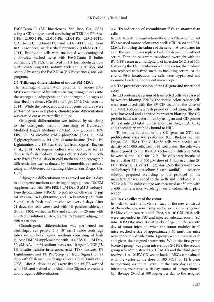
3. Results3.1. Isolation, expansion, and characterization of BM-MSCsIn contrast to human BM-MSCs, mouse BM-MSCs cannot be easily harvested by attachment to a plastic surface, since they typically contain significant levels of contaminating hematopoietic cells. We used a protocol that resulted in cultures largely composed of pure mouse BM-MSCs; thus we did not apply an extra immunomagnetic technique or sorting. The immunophenotypical findings of mouse BM-MSCs are presented in Figure 2. It was found that the cells were highly positive to MSC-related antigens, i.e. CD106-PE (89%), CD44-FITC (100%), CD90.2-PE (99%), Sca-1-PE (98%), and CD29-pure+FITC (99%). On
the other hand, BM-MSCs were highly negative to CD45-FITC (100%), CD11b-FITC (99%), CD31-PE (99%), and TER119-PE (99%) (Figure 2).
Mouse BM-MSC cultures basically demonstrated typical bipolar fibroblastoid morphology starting from the second passage, while we used MSCs from the third and fourth passages in the transduction studies (Figure 3a).
To fulfill the third standard (described by ISCT) for defining MSC requirements, we evaluated the in vitro trilineage mesoderm (namely, osteogenic, chondrogenic, and adipogenic) differentiation capacity of the cells. Our findings showed that BM-MSCs could be differentiated into the osteogenic (Figure 3b), chondrogenic (Figure 3c), and adipogenic (Figure 3d) lineages in vitro, confirmed by the anti-Osteonectin, Alcian blue, and Oil red O stainings, respectively (Figure 3b–3d).3.2. Recombinant baculoviral vector efficiently transduced the mammalian cells We tested the transduction efficacy of the recombinant vector carrying the GFP gene in Sf-9, CRL-2638, and MSCs. Following 12-h incubation of the recombinant vector at the dose of 100 MOI and cells in serum-free medium, the medium was replaced with fresh medium supplemented with serum. After 96 h, the GFP positive cells were determined by fluorescence microscopy. The recombinant vector transduced almost all of the Sf-9
Figure 2. Immunophenotypical characterization of mouse bone marrow mesenchymal stem cells at the third passage. Cells were highly positive to the MSC-related antigens, CD106-PE (89%), CD44-FITC (100%), CD90.2-PE (99%), Sca-1-PE (98%), and CD29-pure+FITC (99%). Cells were highly negative to the following antigens: CD45-FITC (100%), CD11b-FITC (99%), CD31-PE (99%), and TER119-PE (99%).

AKTAŞ et al. / Turk J Biol
1125
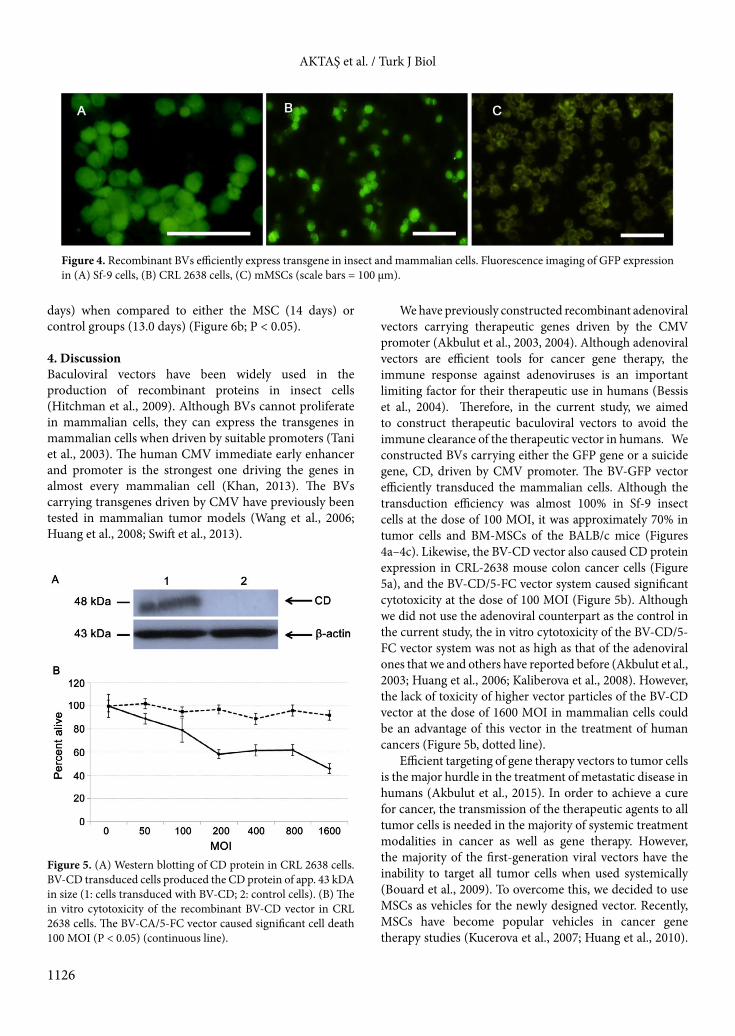
cells (Figure 4a). Although the transduction efficiency in mammalian cells was lower than in the insect cells (Figures 4b and 4c), approximately 70% of the CRL-2638 cells and mouse BM-MSCs were GFP positive (Figures 4b and 4c). 3.3. The recombinant BV-CD vector expressed the enzyme CD in colon cancer cells Following a 7-h infection of mouse colon cancer cells with the recombinant BV-CD vector, we detected the CD protein expression by western blotting (Figure 5a). To test the functional ability of the CD gene that converts the prodrug 5-FC into a cytotoxic drug 5-FU, we performed an in vitro cytotoxicity test by using the BV-CD vector. The vector itself did not cause significant cell death when used alone. However, the addition of 5-FC caused significant cytotoxicity at the dose of 100 MOI of the BV-CD vector (Figure 5b).
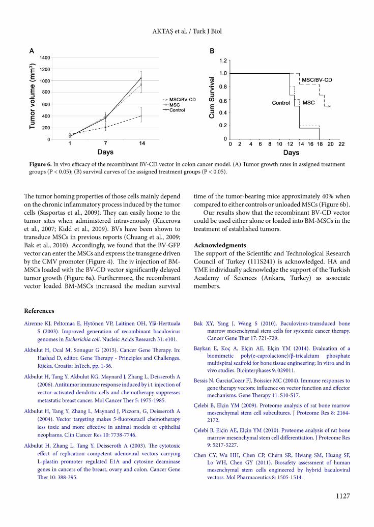
3.4. In vivo efficacy of the BV-CD vector loaded MSCs In order to test the in vivo efficacy of the BV-CD vector, we used a syngeneic BALB/c colon cancer model. First, 5 × 10⁵ CRL-2638 cells were injected sc into BALB/c mice. Three groups of mice with 6 mice in each having approximately 50 mm3 of tumor nodules were established. The control group was given iv PBS, the second group iv injection of 1 × 10⁵ BM-MSCs, and the third group iv injection of 1 × 10⁵ BM-MSCs loaded with 100 MOI of the BV-CD vector. All the mice were given ip 500 mg/kg 5-FC in 0.5 mL volume for 10 days. The tumor growth rates of both the control and MSC treated groups were significantly higher than those of the BV-CD vector loaded BM-MSCs group (Figure 6a; P < 0.05).
The median overall survival time was significantly longer in the BV-CD vector loaded BM-MSCs group (19
Figure 3. Photomicrographs of mouse bone marrow mesenchymal stem cells in culture and their trilineage differentiation. (A) Confluent culture at the third passage demonstrating typical bipolar fibroblastoid morphology. (B) Formation of a dense mineralized ECM typical to osteoblastic cells can be seen by anti-Osteonectin staining after 21 days of osteogenic differentiation. (C) Accumulation of glycosaminoglycans and some mucopolysaccharides typical to chondrocytes was visualized by Alcian blue staining after 21 days of chondrogenic differentiation. (D) Adipogenic induction was apparent by the accumulation of lipid-rich vacuoles within cells, visualized by staining with Oil red O after 21 days (scale bars = 150 µm).
AKTAŞ et al. / Turk J Biol
1126
days) when compared to either the MSC (14 days) or control groups (13.0 days) (Figure 6b; P < 0.05).
4. DiscussionBaculoviral vectors have been widely used in the production of recombinant proteins in insect cells (Hitchman et al., 2009). Although BVs cannot proliferate in mammalian cells, they can express the transgenes in mammalian cells when driven by suitable promoters (Tani et al., 2003). The human CMV immediate early enhancer and promoter is the strongest one driving the genes in almost every mammalian cell (Khan, 2013). The BVs carrying transgenes driven by CMV have previously been tested in mammalian tumor models (Wang et al., 2006; Huang et al., 2008; Swift et al., 2013).
We have previously constructed recombinant adenoviral vectors carrying therapeutic genes driven by the CMV promoter (Akbulut et al., 2003, 2004). Although adenoviral vectors are efficient tools for cancer gene therapy, the immune response against adenoviruses is an important limiting factor for their therapeutic use in humans (Bessis et al., 2004). Therefore, in the current study, we aimed to construct therapeutic baculoviral vectors to avoid the immune clearance of the therapeutic vector in humans. We constructed BVs carrying either the GFP gene or a suicide gene, CD, driven by CMV promoter. The BV-GFP vector efficiently transduced the mammalian cells. Although the transduction efficiency was almost 100% in Sf-9 insect cells at the dose of 100 MOI, it was approximately 70% in tumor cells and BM-MSCs of the BALB/c mice (Figures 4a–4c). Likewise, the BV-CD vector also caused CD protein expression in CRL-2638 mouse colon cancer cells (Figure 5a), and the BV-CD/5-FC vector system caused significant cytotoxicity at the dose of 100 MOI (Figure 5b). Although we did not use the adenoviral counterpart as the control in the current study, the in vitro cytotoxicity of the BV-CD/5-FC vector system was not as high as that of the adenoviral ones that we and others have reported before (Akbulut et al., 2003; Huang et al., 2006; Kaliberova et al., 2008). However, the lack of toxicity of higher vector particles of the BV-CD vector at the dose of 1600 MOI in mammalian cells could be an advantage of this vector in the treatment of human cancers (Figure 5b, dotted line).
Efficient targeting of gene therapy vectors to tumor cells is the major hurdle in the treatment of metastatic disease in humans (Akbulut et al., 2015). In order to achieve a cure for cancer, the transmission of the therapeutic agents to all tumor cells is needed in the majority of systemic treatment modalities in cancer as well as gene therapy. However, the majority of the first-generation viral vectors have the inability to target all tumor cells when used systemically (Bouard et al., 2009). To overcome this, we decided to use MSCs as vehicles for the newly designed vector. Recently, MSCs have become popular vehicles in cancer gene therapy studies (Kucerova et al., 2007; Huang et al., 2010).
Figure 4. Recombinant BVs efficiently express transgene in insect and mammalian cells. Fluorescence imaging of GFP expression in (A) Sf-9 cells, (B) CRL 2638 cells, (C) mMSCs (scale bars = 100 µm).
Figure 5. (A) Western blotting of CD protein in CRL 2638 cells. BV-CD transduced cells produced the CD protein of app. 43 kDA in size (1: cells transduced with BV-CD; 2: control cells). (B) The in vitro cytotoxicity of the recombinant BV-CD vector in CRL 2638 cells. The BV-CA/5-FC vector caused significant cell death 100 MOI (P < 0.05) (continuous line).
AKTAŞ et al. / Turk J Biol
1127
The tumor homing properties of those cells mainly depend on the chronic inflammatory process induced by the tumor cells (Sasportas et al., 2009). They can easily home to the tumor sites when administered intravenously (Kucerova et al., 2007; Kidd et al., 2009). BVs have been shown to transduce MSCs in previous reports (Chuang et al., 2009; Bak et al., 2010). Accordingly, we found that the BV-GFP vector can enter the MSCs and express the transgene driven by the CMV promoter (Figure 4). The iv injection of BM-MSCs loaded with the BV-CD vector significantly delayed tumor growth (Figure 6a). Furthermore, the recombinant vector loaded BM-MSCs increased the median survival
time of the tumor-bearing mice approximately 40% when compared to either controls or unloaded MSCs (Figure 6b).
Our results show that the recombinant BV-CD vector could be used either alone or loaded into BM-MSCs in the treatment of established tumors.
AcknowledgmentsThe support of the Scientific and Technological Research Council of Turkey (111S241) is acknowledged. HA and YME individually acknowledge the support of the Turkish Academy of Sciences (Ankara, Turkey) as associate members.
Figure 6. In vivo efficacy of the recombinant BV-CD vector in colon cancer model. (A) Tumor growth rates in assigned treatment groups (P < 0.05); (B) survival curves of the assigned treatment groups (P < 0.05).
References
Airenne KJ, Peltomaa E, Hytönen VP, Laitinen OH, Ylä-Herttuala S (2003). Improved generation of recombinant baculovirus genomes in Escherichia coli. Nucleic Acids Research 31: e101.
Akbulut H, Ocal M, Sonugur G (2015). Cancer Gene Therapy. In: Hashad D, editor. Gene Therapy - Principles and Challenges. Rijeka, Croatia: InTech, pp. 1-36.
Akbulut H, Tang Y, Akbulut KG, Maynard J, Zhang L, Deisseroth A (2006). Antitumor immune response induced by i.t. injection of vector-activated dendritic cells and chemotherapy suppresses metastatic breast cancer. Mol Cancer Ther 5: 1975-1985.
Akbulut H, Tang Y, Zhang L, Maynard J, Pizzorn, G, Deisseroth A (2004). Vector targeting makes 5-fluorouracil chemotherapy less toxic and more effective in animal models of epithelial neoplasms. Clin Cancer Res 10: 7738-7746.
Akbulut H, Zhang L, Tang Y, Deisseroth A (2003). The cytotoxic effect of replication competent adenoviral vectors carrying L-plastin promoter regulated E1A and cytosine deaminase genes in cancers of the breast, ovary and colon. Cancer Gene Ther 10: 388-395.
Bak XY, Yang J, Wang S (2010). Baculovirus-transduced bone marrow mesenchymal stem cells for systemic cancer therapy. Cancer Gene Ther 17: 721-729.
Baykan E, Koç A, Elçin AE, Elçin YM (2014). Evaluation of a biomimetic poly(e-caprolactone)/β-tricalcium phosphate multispiral scaffold for bone tissue engineering: In vitro and in vivo studies. Biointerphases 9: 029011.
Bessis N, GarciaCozar FJ, Boissier MC (2004). Immune responses to gene therapy vectors: influence on vector function and effector mechanisms. Gene Therapy 11: S10-S17.
Çelebi B, Elçin YM (2009). Proteome analysis of rat bone marrow mesenchymal stem cell subcultures. J Proteome Res 8: 2164-2172.
Çelebi B, Elçin AE, Elçin YM (2010). Proteome analysis of rat bone marrow mesenchymal stem cell differentiation. J Proteome Res 9: 5217-5227.
Chen CY, Wu HH, Chen CP, Chern SR, Hwang SM, Huang SF, Lo WH, Chen GY (2011). Biosafety assessment of human mesenchymal stem cells engineered by hybrid baculoviral vectors. Mol Pharmaceutics 8: 1505-1514.
AKTAŞ et al. / Turk J Biol
1128
Chuang CK, Sung LY, Hwang SM, Lo WH, Chen HC, Hu YC (2007). Baculovirus as a new gene delivery vector for stem cell engineering and bone tissue engineering. Gene Ther 14: 1417-1424.
Chuang CK, Wong TH, Hwang SM, Chang YH, Chen GY, Chiu YC (2009). Baculovirus transduction of mesenchymal stem cells: in vitro responses and in vivo immune responses after cell transplantation. Mol Ther 17: 889-896.
Bouard D, Alazard-Dany N, Cosset FL (2009). Viral vectors: from virology to transgene expression. Br J Pharmacol 157: 153-165.
Elçin YM (2003). Tissue Engineering, Stem Cells and Gene Therapies, In: Elçin M, editor, Advances in Experimental Medicine and Biology Series: New York, NY, USA: Kluwer Academic-Plenum Press.
Emin N, Koç A, Durkut S, Elçin AE, Elçin YM (2008). Engineering of rat articular cartilage on porous sponges: effects of tgf-beta 1 and microgravity bioreactor culture. Artif Cells Blood Substit Immobil Biotechnol 36: 123-137.
Hitchman RB, Possee RD, King LA (2009). Baculovirus expression systems for recombinant protein production in insect cells. Recent Pat Biotechnol 3: 46-54.
Ho YC, Chung YC, Hwang SM, Wang KC, Hu YC (2005). Transgene expression and differentiation of baculovirustransduced human mesenchymal stem cells. J Gene Med 7: 860-868.
Huang Q, Chen D, Fu X, Zu Y (2006). Inhibitory effect of pulmonary carcinoma by adenovirus-mediated CD/UPRT gene. J Huazhong Univ Sci Technolog Med Sci 26: 591-593.
Huang Q, Liu XZ, Kang CS, Wang GX, Zhong Y, Pu PY (2010). The anti-glioma effect of suicide gene therapy using BMSC expressing HSV/TK combined with overexpression of Cx43 in glioma cells. Cancer Gene Ther 17: 192-202.
Huang W, Tian XL, Wu YL, Zhong J, Yu LF, Hu SP, Li B (2008). Suppression of gastric cancer growth by baculovirus vector-mediated transfer of normal epithelial cell specific-1 gene. World J Gastroenterol 14: 5810-5815.
Kaliberova LN, Della Manna DL, Krendelchtchikova V, Black ME, Buchsbaum DJ, Kaliberov SA (2008). Molecular chemotherapy of pancreatic cancer using novel mutant bacterial cytosine deaminase gene. Mol Cancer Ther 7: 2845-2854.
Khan KH (2013). Gene expression in mammalian cells and its applications. Adv Pharm Bull 3: 257–263.
Kidd S, Spaeth E, Dembinski JL, Dietrich M, Watson K, Klopp A, Battula VL, Weil M, Andreeff M, Marini FC (2009). Direct evidence of mesenchymal stem cell tropism for tumor and wounding microenvironments using in vivo bioluminescent imaging. Stem Cells 27: 2614-2623.
Kucerova L, Altanerova V, Matuskova M, Tyciakova S, Altaner C (2007). Adipose tissue-derived human mesenchymal stem cells mediated prodrug cancer gene therapy. Cancer Res 67: 6304-6313.
Matuskova M, Hlubinova K, Pastorakova A, Hunakova L, Altanerova V, Altaner C, Kucerova L (2010). aHSV-tk expressing mesenchymal stem cells exert bystander effect on human glioblastoma cells. Cancer Letters 290: 58-67.
Odabaş S, Elçin AE, Elçin YM (2014). Isolation and characterization of mesenchymal stem cells. Methods Mol Biol 1109: 47-63.
Sasportas LS, Kasmieh R, Wakimoto H, Hingten S, van de Water JA, Mohapatra G, Fiuiredo JL, Martuza RL, Weissleder R, Shah K (2009). Assessment of therapeutic efficacy and fate of engineered human mesenchymal stem cells for cancer therapy. Proc Natl Acad Sci USA 106: 4822-4827.
Swift SL, Rivera GC, Dussupt V, Leadley RM, Hudson LC, de Ridder CMA, Kraaij R, Burns JE, Maitland NJ, Georgopoulos LJ (2013). Evaluating baculovirus as a vector for human prostate cancer gene therapy. PLoS ONE 8(6): e65557.
Tani H, Limn CK, Yap CC, Onishi M, Nozaki M, Nishimune Y, Okahashi N, Kitagawa Y, Watanabe R, Mochizuki R et al. (2003). In vitro and in vivo gene delivery by recombinant baculoviruses. J Virol 77: 9799-9808.
Wang CY, Li F, Yang Y, Guo HY, Wu CX, Wang S (2006). Recombinant baculovirus containing the diphtheria toxin a gene for malignant glioma therapy. Cancer Res 66: 57985806.