Embed Size (px)
Citation preview

fmicb-07-01391 September 6, 2016 Time: 20:9 # 1
ORIGINAL RESEARCHpublished: 08 September 2016
doi: 10.3389/fmicb.2016.01391
Edited by:Peter Mullany,
University College London, UK
Reviewed by:Marcin Łos,
University of Gdansk, PolandDeborah M. Hinton,
National Institutes of Health, USAChristopher King Mathews,
Oregon State University, USA
*Correspondence:Daniel Bryan
[email protected] M. Kutter
†These authors have contributedequally to this work.
Specialty section:This article was submitted to
Antimicrobials, Resistanceand Chemotherapy,
a section of the journalFrontiers in Microbiology
Received: 23 May 2016Accepted: 23 August 2016
Published: 08 September 2016
Citation:Bryan D, El -Shibiny A, Hobbs Z,
Porter J and Kutter EM (2016)Bacteriophage T4 Infection
of Stationary Phase E. coli: Life afterLog from a Phage Perspective.
Front. Microbiol. 7:1391.doi: 10.3389/fmicb.2016.01391
Bacteriophage T4 Infection ofStationary Phase E. coli: Life afterLog from a Phage PerspectiveDaniel Bryan1*†, Ayman El-Shibiny1,2,3, Zack Hobbs1, Jillian Porter1 andElizabeth M. Kutter1*†
1 Kutter Bacteriophage Lab, The Evergreen State College, Olympia, WA, USA, 2 Biomedical Sciences, University of Scienceand Technology, Zewail City of Science and Technology, Giza, Egypt, 3 Faculty of Environmental Agricultural Sciences, ArishUniversity, Arish, Egypt
Virtually all studies of phage infections investigate bacteria growing exponentially inrich media. In nature, however, phages largely encounter non-growing cells. Bacteriaentering stationary phase often activate well-studied stress defense mechanisms thatdrastically alter the cell, facilitating its long-term survival. An understanding of phage-host interactions in such conditions is of major importance from both an ecologicaland therapeutic standpoint. Here, we show that bacteriophage T4 can efficiently bindto, infect and kill E. coli in stationary phase, both in the presence and absence ofa functional stationary-phase sigma factor, and explore the response of T4-infectedstationary phase cells to the addition of fresh nutrients 5 or 24 h after that infection.An unexpected new mode of response has been identified. “Hibernation” mode isa persistent but reversible dormant state in which the infected cells make at leastsome phage enzymes, but halt phage development until appropriate nutrients becomeavailable before producing phage particles. Our evidence indicates that the block inhibernation mode occurs after the middle-mode stage of phage development; hostDNA breakdown and the incorporation of the released nucleotides into phage DNAindicate that the enzymes of the nucleotide synthesizing complex, under middle-modecontrol, have been made and assembled into a functional state. Once fresh glucoseand amino acids become available, the standard lytic infection process rapidly resumesand concentrations of up to 1011 progeny phage (an average of about 40 phage perinitially present cell) are produced. All evidence is consistent with the hibernation-modecontrol point lying between middle mode and late mode T4 gene expression. We havealso observed a “scavenger” response, where the infecting phage takes advantage ofwhatever few nutrients are available to produce small quantities of progeny within 2 to5 h after infection. The scavenger response seems able to produce no more than anaverage of one phage per originally available cell, and few if any further progeny areproduced by cells in this mode even if fresh nutrients are made available later.
Keywords: bacteriophage T4, E. coli, hibernation mode, multiplicity of infection (MOI), scavenger response,stationary phase, sigma S, T4 nucleotide synthesizing complex
Frontiers in Microbiology | www.frontiersin.org 1 September 2016 | Volume 7 | Article 1391

fmicb-07-01391 September 6, 2016 Time: 20:9 # 2
Bryan et al. T4 Infection of Stationary E. coli
INTRODUCTION
Bacteriophage infection has traditionally been studied usingbacterial hosts growing exponentially, with active aeration, inone of a few well-studied media. The resurgence of interest intherapeutic, prophylactic, and agricultural phage applications,as well as growing awareness of the substantial environmentalimpact of phages, make it increasingly important to study thedetails of phage-host interactions under conditions that aremore similar to those encountered in natural environments.Such exploration is especially important since the emergenceof multi-drug resistant pathogenic bacteria has become a majorpublic health concern (World Health Organization [WHO],2014). Understanding how cellular changes affect phage infectionunder natural conditions is essential for the success of the manyproposed antimicrobial phage applications. We here explore theability of coliphage T4 to infect hosts that are in stationaryphase. Such studies are particularly relevant as close relatives ofthis carefully studied model organism are ubiquitous in natureand have been very widely used in cocktails for treating entericinfections (Brüssow, 2005; Sarker et al., 2012; Kutter et al., 2014;Sarker et al., 2016).
T4 infection of E. coli has been one of the most thoroughlystudied model systems in molecular biology and microbiology forover 60 years (Mathews et al., 1983; Karam et al., 1994; Milleret al., 2003). T4 infection of exponentially growing E. coli quicklydisrupts host genome structure and expression, largely by makinguse of T4’s complete substitution of HMdC for dC in its DNA(Kutter et al., 1994c). Transcription of all cytosine-containingDNA is blocked, translation of residual host RNA is universallycut off, host DNA is gradually degraded, and the infected cellcannot respond to a change in available carbon sources. However,it was widely believed that T4 does not multiply in stationaryphase cells, since T4 plaques don’t continue to grow in size afterthe lawn is fully formed (Benzer, 1952; Adams, 1959). Schraderet al. (1997a,b) reported that T4 was incapable of multiplyingin E. coli AB1157 that had been starved for 24 h in LysogenyBroth (LB), even though 94% of the T4 phage particles boundto the host. They found, in contrast, that both coliphage T7and P. aeruginosa phage UT1 infected their hosts successfullyin identical conditions, though with extended latent periods andreduced burst sizes; plaques of T7 on a plate will also continueto grow in size indefinitely (Yin, 1991). Other indications havebeen found that T4 may multiply less efficiently than T7 in thegut environment. For example, in axenic mice monocolonizedwith E. coli, the amount of T4 was seen to increase only 300-foldduring transit of the gut as compared to passive transit in controlaxenic mice, while T7 showed a 106 fold increase (Weiss et al.,2009).
Optimal infection strategies might well be quite differentwhen T4 tries to infect stationary-phase cells – as mustfrequently happen in the mammalian colon, E. coli’s primaryhabitat, where the microbial density is extremely high andcompetition for nutrients is intense. Whereas σ70 is the mainregulator for “housekeeping” gene transcription in exponential-phase enteric bacteria, σS (regulated by gene RpoS) plays acentral role for cell adaptation during stationary phase. There,
E. coli maintains a basal metabolic level that lets it scavengefor nutrients from dead cells and respond quickly to a widerange of new nutrients (Hengge-Aronis, 1993; Zambrano andKolter, 1996). Many new pathways are activated as the overallmetabolic rate decreases during the transition to stationary phase,largely under the control of σS. E. coli in stationary phasehave denser cell membranes, with altered phospholipids andexpanded periplasmic spacing. They pack their chromosomemore tightly and produce a range of protective enzymes such ascatalase (Huisman et al., 1996; Págan and Mackey, 2000). Thecells maintain a low level of synthesis of some proteins suchas various transporters throughout stationary phase, enablingthem to respond to new nutrients and to quickly resumevegetative growth (Huisman et al., 1996). The number ofribosomes is reduced (Cangelosi and Brabant, 1997), and manyof those remaining are dimerized to a storage format, to berapidly released when nutrients are supplied (Wada et al.,2000).
Here, we describe in substantial detail the patterns of theinteraction when T4 infects E. coli 48 h after inoculation intofresh minimal medium, identifying a new “hibernation” modeof long-term interaction there, and explore the ability of T4 toproduce infective centers in E. coli that are up to several weeksinto stationary phase.
MATERIALS AND METHODS
MediaStationary phase studies were conducted in M9 minimal medium[25 mL 20X M9 salts (120 g Na2HPO4·H2O, 60 g KH2PO4(anhydrous), 10 g NaCl, and 20 g NH4Cl per 790 mL DIH2O),0.5 mL 0.1 M FeCl3, 5 mL, 0.1 M MgSO4, 0.5 mL 0.1 M CaCl2,per 461 mL DI H2O, sterile filtered and supplemented with 5 mLcasamino acids (CAA; 10% w/v) and 3 mL glucose (20% w/v)].Tryptic soy broth (30 g tryptic soy broth powder per 1000 mLof DI H2O) was used for standard cultivation of bacteria andpropagation of phage. Phage buffer (10 mL 1 M Tris pH 7.6, 0.1 ggelatin, 4 g NaCl, 990 mL DI H2O, boiled and pH balanced to 7.6at 25◦C, autoclaved) was used for phage stocks and as a diluentwhen titering phage.
Bacterial StrainsStudies were conducted in E. coli W3110 derivatives ZK126(1lacZ rpoS+), and ZK1000 (1lacZ rpoS-) (the kind gift ofDr. Steve Finkel) and phage were routinely plated on ZK126.Amber suppressor strain CR63 from the Evergreen collectionwas also used. Stocks were maintained in 20% (v/v) glycerolat −80◦C. All cultures were grown and infections carried outin Erlenmeyer flasks in a gyratory water bath at 37◦C and120 RPM.
Phage Strains and Their PropagationPhage T4D was from the Evergreen collection, as was T4 gene 43(DNA polymerase) amber mutant am4332. T4 was propagated inZK126 while am4332 was propagated in amber suppressor E. colistrain CR63. Briefly, a 500 mL culture at ∼0.4 OD600nm (OD, all
Frontiers in Microbiology | www.frontiersin.org 2 September 2016 | Volume 7 | Article 1391

fmicb-07-01391 September 6, 2016 Time: 20:9 # 3
Bryan et al. T4 Infection of Stationary E. coli
optical density readings used throughout are done at 600 nm) inTSB was infected at a multiplicity of infection (MOI) of ∼0.1,then incubated at 120 RPM and 37◦C for ∼4 h before lysingwith 1–2 mL of CHCl3. The lysates were incubated statically atroom temperature overnight to allow endogenous nucleases todegrade host DNA and then centrifuged for 10 min at 4642× g topellet bacterial debris. The supernatant was decanted away fromthe pellet, spun at low speed a second time, and then centrifugedfor 2 h at 23,975 × g to obtain a concentrated phage pellet. Thiswas resuspended without agitation in a few mL of phage bufferovernight at 4◦C.
Infection of E. coli in Stationary PhaseOvernight cultures used for stationary phase infections or forphage plating were grown either from 3 to 5 colonies takenfrom plate cultures no more than 3 days old or directly froma scraping of a glycerol freezer stock stored at −80◦C. Thislatter, less common method is used routinely in the Finkel andKolter labs for stationary phase work, and usually gives lessvariable bacterial behavior in stationary phase cultures than doespicking individual colonies. We adopted this technique in thefall of 2014 and the experiments presented in Figures 1 and 2were inoculated by this method. Stationary phase infectionswere performed with cultures of E. coli ZK126 or ZK1000in M9 at a range of multiplicities of infection (MOI), asindicated. Either 5 or 24 h post-infection, nutrients wereadded to bring the concentration in the infection flask upto 0.12% w/v glucose and 0.1% w/v casamino acids. Theinfected culture was tracked for an additional 3–5 h afternutrient addition (representative figures showing the nutrientresponse of uninfected cells are provided as SupplementalMaterial).
Phage were tracked as plaque forming units (PFU),enumerated by taking a 30 µL sample and adding it to amicrocentrifuge Eppendorf tube containing 270 µL of phagebuffer and 30 µL of chloroform. The samples were shaken welland allowed to settle for at least 20 min before 30 µL of thesample was serially diluted through 96 well plates containing270 µL of phage buffer. Then 100 µL of each appropriate dilutionwas added to a tube containing 3 mL of molten top agar held at45◦C and 100 µL of ZK126 at an OD of about 0.5, gently mixedwell and poured onto a TSA plate. Bacterial survivors (BS) weredetermined by taking 30 µL samples, serial diluting through 96well plates containing 270 µL M9 and spread plating 100 µLaseptically on round TSA plates. Optical density was determinedafter diluting samples either two or fourfold in M9 as needed tobring the OD below 1.0.
Duplicate samples collected at the same times had ∼100 µLof chloroform added to the dilution medium. These sampleswere shaken and left at least 20 min to allow the chloroform tosettle; then the OD of a 600 µL aliquot was read. (Chloroformis used to indicate successful production of lysozyme, indicatingsome phage early gene expression. When exposed to chloroform,phage-infected cells that have already produced some lysozymewill lyse, while uninfected cells will die but generally remainstructurally intact Adams, 1959).
Host DNA Degradation ExperimentsTo explore how stationary-phase phage infection affects the hostDNA, cultures of exponential-phase ZK126 were labeled withtritiated deoxythymidine (3H-dT). At OD = 0.4 on the first day,5 mL were transferred to flasks containing 0.25 mL dA (2 mg/mLin H2O) and 0.25 mL dT (100 µg/mL dT with 10 µCi/mL methyl3H-dT) and incubated at 37◦C in a shaking water bath whilethe remainder was grown in parallel without label to enumerateCFUs and PFUs and check the response to nutrient addition.At 48 h, both radio-labeled and unlabeled cultures were infectedwith either T4D+ or T4 am4332 at an MOI of about 10 phage percell. The standard nutrient mixture was re-added to the unlabeledflasks at 5 h after infection (0.12% w/v glucose and 0.1% w/vcasamino acids) to follow the ability of these infected cells torespond to nutrients by producing phage.
Labeled DNA samples were collected by spotting 50 µLsamples onto filter paper disks and letting them dry for one min,placing them into a bath of 10% cold acetic acid for at least10 min, washing them twice with cold 5% acetic acid for 10 min,then with 90% ethanol for 10 min and drying them. Sampleswere counted in a Packard 2200CA Tri-Carb Liquid ScintillationCounter using Perkin-Elmer Cybergold as the fluor.
[Note that E. coli can incorporate external deoxythymidine(dT) into its DNA only in the presence of 100 mg mL−1
deoxyadenosine (dA), to compete for a resident glycosidase(Boyce and Setlow, 1962)]. At least in exponential phase, T4reuses released dT so efficiently for its own DNA synthesis thatthe breakdown can be observed only by using a phage mutantunable to make DNA, even when orders of magnitude moreunlabeled dT is added to the culture (Kutter and Wiberg, 1968).Therefore, T4 DNA polymerase mutant am4332 was used forthese studies in parallel with wild-type T4 to follow host DNAdegradation in the absence of potential reincorporation of labeledhost DNA into progeny phage.
RESULTS
Patterns of T4 Infection of E. coli inStationary Phase:To explore T4 infection of starved E. coli, a 48 h old cultureof ZK126 grown in M9 supplemented with glucose and CAAwas infected at a range of different MOIs. By 5–10 min post-infection, more than 90% phage binding and MOI-dependentbacterial killing was routinely observed in all conditions. Thepattern of phage production is consistently different betweenwhen there are only a few infecting phage per cell and whenthe MOI is much higher (cf. Figure 1A). At lower MOIs, nophage are produced until fresh nutrients are provided, but theinfected cells respond and produce phage very rapidly whenglucose and CAA are re-added. This phage production is seenso quickly as to suggest that early stages of phage infectionhad already been initiated, rapidly yielding a substantial burstof progeny phage. We refer to this behavior as “hibernation”mode.
In contrast, with high MOIs, we also observe what we calla “scavenger” response, where some phage production begins
Frontiers in Microbiology | www.frontiersin.org 3 September 2016 | Volume 7 | Article 1391

fmicb-07-01391 September 6, 2016 Time: 20:9 # 4
Bryan et al. T4 Infection of Stationary E. coli
FIGURE 1 | Representative figures of (A) phage production and (B) bacterial survivors (BS) when the infection was carried out at three differentmultiplicity of infection (MOI) in parallel flasks split from the same 48 h old culture of E. coli ZK126, with 0.12% w/v glucose and 0.1% w/v CAAre-added 5 h after infection (As described in Section “Materials and Methods,” chloroform is always added to the phage samples, so these valuesinclude all complete phage, both intracellular and free). (C) Bacterial killing and phage production when ZK126 was infected after 48 h with T4 at an MOI of16.5 and no further nutrients were added until 25 h after infection. Both a long hibernation mode and a small scavenger response are observed. SupplementalMaterial with further representative figures showing similar behavior are provided for Figure 1, Figure 2, and Figure 3A as well as for the response of uninfectedcells to nutrient addition.
within a couple of hours after the phage are added to the cellswithout any addition of fresh nutrients, seldom extending beyond4 h after infection and showing little if any ability to produce morephage when nutrients are re-added either 5 or 24 h after phageinfection, presumably due to the fact that the phage infectionprocess has been completed.
The behavior of T4 during the infection process appears tobe determined on a cell by cell basis, and at least two differentsorts of responses can be observed in a single infected culture,as seen in the MOI 9 infection in Figures 1A,B and the MOI16.5 infection in Figure 1C. There, we observed an increase inphage titer before nutrient addition, but each infected culture
still rapidly produced large quantities of progeny phage afternutrient addition. Those infected stationary phase cells that enterhibernation mode are still able to respond with a similarly largeburst of phage (about 200 phage per initial cell over 4 h) if thenutrients are not added until 25 h after infection (Figure 1C).The drop in bacterial titer starting by 2 h post nutrient additionindicates that some of the phage-producing cells have been lysing,releasing phage that can now kill any still-uninfected phage-sensitive cells.
While patterns of phage production remained quite consistentfrom experiment to experiment, bacterial parameters such as thetiter at 48 h, extent of killing after infection at a particular MOI,
Frontiers in Microbiology | www.frontiersin.org 4 September 2016 | Volume 7 | Article 1391

fmicb-07-01391 September 6, 2016 Time: 20:9 # 5
Bryan et al. T4 Infection of Stationary E. coli
FIGURE 2 | Representative figures of phage production, bacterial killing, and optical density without (OD) and with (ODC) the addition of chloroform.0.12% w/v glucose and 0.1% w/v CAA added 5 hr after infection. (A) ZK126, MOI: 3.3, (B) ZK1000, MOI: 3.4, (C) ZK126, MOI: 28, and (D) ZK1000, MOI: 28.
and grow-back of BS after infection varied between experiments.However, bacterial parameters in infections started in parallelfrom the same overnight culture remained very consistent. Wehypothesize that the variation is due to differences in the initialbacterial cells in the cultures, some of which may have thendeveloped into faster growing cells such as Growth Advantagein Stationary Phase (GASP) mutants or something that couldmore easily be killed etc. during the periods of stationary-phaseincubation between the inoculation of the overnight culture froma freezer stock and infection of the experimental culture 72 h later(Zambrano and Kolter, 1996).
Further Characterization of Scavengerversus Hibernation ModesFollowing the optical density of each culture with and withoutadding chloroform to the sample (ODC and OD) providesa basis for better understanding what is happening in eachof the two observed modes of infection of stationary phase
cells. A drop in the OD very shortly after phage additionis probably indicative of “lysis from without” due to damagecaused by the infection process (Abedon, 2011). A drop in theODC indicates that at least some lysozyme has already beenproduced in the cell. The T4 lysozyme gene is transcribedat low levels from an early promoter 2.9 kb away (69.9 kbon the genetic map) with an immediately adjacent stronglate promoter directing much higher levels later at 67 kbon the genetic map (Kutter et al., 1994b), but the lysozymecan’t reach the peptidoglycan layer as long as the inner cellmembrane is intact. Exposing infected cells to chloroforminduces lysis if any lysozyme has already been produced, whilemost uninfected cells remain unlysed even though they arekilled (Adams, 1959). Thus, by following the susceptibility ofthe culture to lysis by chloroform, one can quickly determinewhether early phage proteins are indeed being made. Toexplore the role of the stationary-phase sigma factor indetermining response to phage, we here also include data
Frontiers in Microbiology | www.frontiersin.org 5 September 2016 | Volume 7 | Article 1391
fmicb-07-01391 September 6, 2016 Time: 20:9 # 6
Bryan et al. T4 Infection of Stationary E. coli
on infection of ZK1000, isogenic except that RpoS has beendeleted.
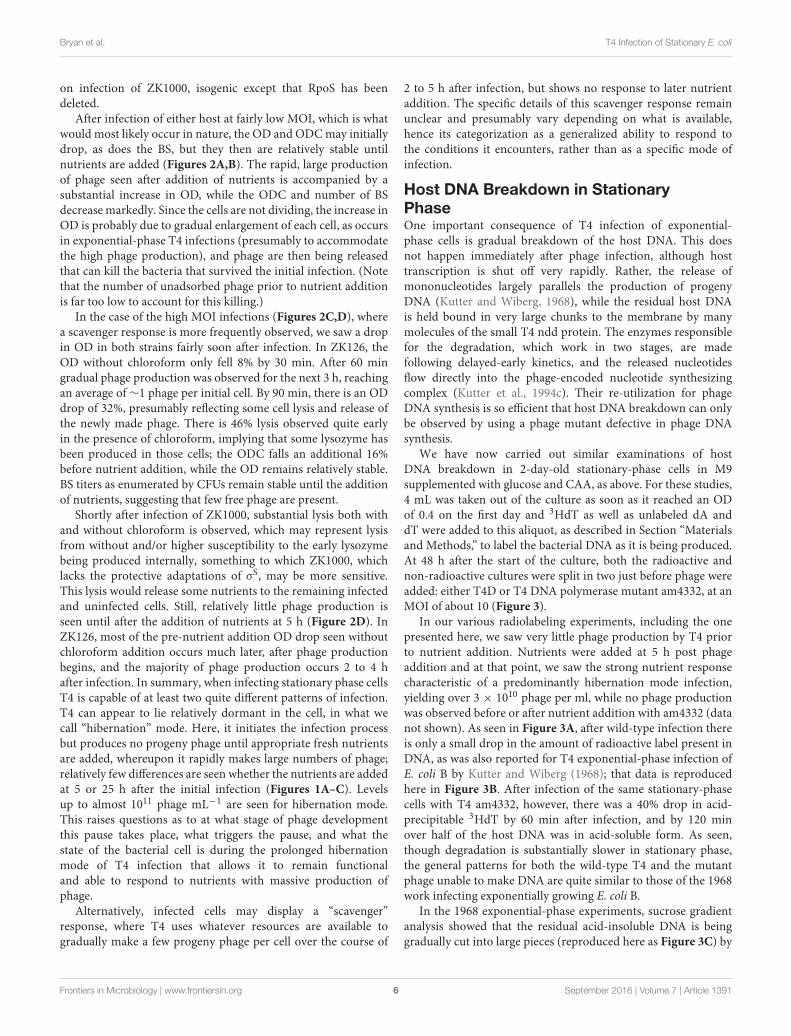
After infection of either host at fairly low MOI, which is whatwould most likely occur in nature, the OD and ODC may initiallydrop, as does the BS, but they then are relatively stable untilnutrients are added (Figures 2A,B). The rapid, large productionof phage seen after addition of nutrients is accompanied by asubstantial increase in OD, while the ODC and number of BSdecrease markedly. Since the cells are not dividing, the increase inOD is probably due to gradual enlargement of each cell, as occursin exponential-phase T4 infections (presumably to accommodatethe high phage production), and phage are then being releasedthat can kill the bacteria that survived the initial infection. (Notethat the number of unadsorbed phage prior to nutrient additionis far too low to account for this killing.)
In the case of the high MOI infections (Figures 2C,D), wherea scavenger response is more frequently observed, we saw a dropin OD in both strains fairly soon after infection. In ZK126, theOD without chloroform only fell 8% by 30 min. After 60 mingradual phage production was observed for the next 3 h, reachingan average of∼1 phage per initial cell. By 90 min, there is an ODdrop of 32%, presumably reflecting some cell lysis and release ofthe newly made phage. There is 46% lysis observed quite earlyin the presence of chloroform, implying that some lysozyme hasbeen produced in those cells; the ODC falls an additional 16%before nutrient addition, while the OD remains relatively stable.BS titers as enumerated by CFUs remain stable until the additionof nutrients, suggesting that few free phage are present.
Shortly after infection of ZK1000, substantial lysis both withand without chloroform is observed, which may represent lysisfrom without and/or higher susceptibility to the early lysozymebeing produced internally, something to which ZK1000, whichlacks the protective adaptations of σS, may be more sensitive.This lysis would release some nutrients to the remaining infectedand uninfected cells. Still, relatively little phage production isseen until after the addition of nutrients at 5 h (Figure 2D). InZK126, most of the pre-nutrient addition OD drop seen withoutchloroform addition occurs much later, after phage productionbegins, and the majority of phage production occurs 2 to 4 hafter infection. In summary, when infecting stationary phase cellsT4 is capable of at least two quite different patterns of infection.T4 can appear to lie relatively dormant in the cell, in what wecall “hibernation” mode. Here, it initiates the infection processbut produces no progeny phage until appropriate fresh nutrientsare added, whereupon it rapidly makes large numbers of phage;relatively few differences are seen whether the nutrients are addedat 5 or 25 h after the initial infection (Figures 1A–C). Levelsup to almost 1011 phage mL−1 are seen for hibernation mode.This raises questions as to at what stage of phage developmentthis pause takes place, what triggers the pause, and what thestate of the bacterial cell is during the prolonged hibernationmode of T4 infection that allows it to remain functionaland able to respond to nutrients with massive production ofphage.
Alternatively, infected cells may display a “scavenger”response, where T4 uses whatever resources are available togradually make a few progeny phage per cell over the course of
2 to 5 h after infection, but shows no response to later nutrientaddition. The specific details of this scavenger response remainunclear and presumably vary depending on what is available,hence its categorization as a generalized ability to respond tothe conditions it encounters, rather than as a specific mode ofinfection.
Host DNA Breakdown in StationaryPhaseOne important consequence of T4 infection of exponential-phase cells is gradual breakdown of the host DNA. This doesnot happen immediately after phage infection, although hosttranscription is shut off very rapidly. Rather, the release ofmononucleotides largely parallels the production of progenyDNA (Kutter and Wiberg, 1968), while the residual host DNAis held bound in very large chunks to the membrane by manymolecules of the small T4 ndd protein. The enzymes responsiblefor the degradation, which work in two stages, are madefollowing delayed-early kinetics, and the released nucleotidesflow directly into the phage-encoded nucleotide synthesizingcomplex (Kutter et al., 1994c). Their re-utilization for phageDNA synthesis is so efficient that host DNA breakdown can onlybe observed by using a phage mutant defective in phage DNAsynthesis.
We have now carried out similar examinations of hostDNA breakdown in 2-day-old stationary-phase cells in M9supplemented with glucose and CAA, as above. For these studies,4 mL was taken out of the culture as soon as it reached an ODof 0.4 on the first day and 3HdT as well as unlabeled dA anddT were added to this aliquot, as described in Section “Materialsand Methods,” to label the bacterial DNA as it is being produced.At 48 h after the start of the culture, both the radioactive andnon-radioactive cultures were split in two just before phage wereadded: either T4D or T4 DNA polymerase mutant am4332, at anMOI of about 10 (Figure 3).
In our various radiolabeling experiments, including the onepresented here, we saw very little phage production by T4 priorto nutrient addition. Nutrients were added at 5 h post phageaddition and at that point, we saw the strong nutrient responsecharacteristic of a predominantly hibernation mode infection,yielding over 3 × 10¹⁰ phage per ml, while no phage productionwas observed before or after nutrient addition with am4332 (datanot shown). As seen in Figure 3A, after wild-type infection thereis only a small drop in the amount of radioactive label present inDNA, as was also reported for T4 exponential-phase infection ofE. coli B by Kutter and Wiberg (1968); that data is reproducedhere in Figure 3B. After infection of the same stationary-phasecells with T4 am4332, however, there was a 40% drop in acid-precipitable 3HdT by 60 min after infection, and by 120 minover half of the host DNA was in acid-soluble form. As seen,though degradation is substantially slower in stationary phase,the general patterns for both the wild-type T4 and the mutantphage unable to make DNA are quite similar to those of the 1968work infecting exponentially growing E. coli B.
In the 1968 exponential-phase experiments, sucrose gradientanalysis showed that the residual acid-insoluble DNA is beinggradually cut into large pieces (reproduced here as Figure 3C) by
Frontiers in Microbiology | www.frontiersin.org 6 September 2016 | Volume 7 | Article 1391

fmicb-07-01391 September 6, 2016 Time: 20:9 # 7
Bryan et al. T4 Infection of Stationary E. coli
FIGURE 3 | (A) Host DNA degradation analysis of ZK126 that was labeled with tritiated thymidine during exponential growth and then infected at 48 h in stationaryphase with either T4D or T4 am4332 (DNA polymerase−), in parallel with infection of cultures being tracked in terms of phage production and BS. (B) Status of DNAlabeled with tritiated thymidine over the course of similar infections of E. coli B with T4D and T4 amN55x5 (dCMP HMase−) in exponential phase, as reported byKutter and Wiberg (1968). Both T4 am4332 and T4 amN55x5 produce DNA− phenotype phage when grown on a non-amber suppressor strain such as E. coli B orZK126. (C) Sucrose gradient analysis determining the size of the acid-insoluble fraction of the host DNA at various times after this exponential phase infection ofE. coli B by T4D. “T4” and “T7” refer to phage DNAs used as sedimentation markers.
a combination of cytosine-specific phage-encoded endonucleasesII and IV, while the degradation from there to mononucleotidesfor reutilization was shown to require T4’s gene 46- and 47-encoded exonuclease, which is also involved in T4 recombinationand thus not specific for cytosine DNA. In the absence ofexpression of genes 46 and 47, the sizes of the remainingfragments at various times after infection were the same as thoseseen here for T4, but all of the DNA was in the respective peaksrather than only the fraction indicated in Figure 3B as not yethaving been degraded to mononucleotides.
Since the host DNA is so substantially degraded to an acid-soluble form after infection by the DNA polymerase mutant instationary phase, we infer that it is similarly degraded in the
parallel wild-type T4 infection, as was shown in the exponentialphase sucrose gradient experiments, with the released hostnucleotides being efficiently re-incorporated into phage DNA.The endoII and endoIV that initiate host DNA degradation areunder middle-mode regulation, as are genes 46 and 47 and themany enzymes involved in nucleotide biosynthesis and DNAreplication (Cowan et al., 1994).
Additionally, since the host DNA is being degraded, it seemshighly probable that the observed rapid response to addednutrients in hibernation mode is dependent on stable hostproteins and/or stable host RNAs. It appears unlikely to be theresult of a transcriptional response, particularly assuming thatthe host DNA in infected cells is broken down in two steps
Frontiers in Microbiology | www.frontiersin.org 7 September 2016 | Volume 7 | Article 1391

fmicb-07-01391 September 6, 2016 Time: 20:9 # 8
Bryan et al. T4 Infection of Stationary E. coli
in stationary phase as it is in exponential phase (Figure 3C),making it likely that the residual acid-insoluble DNA in cellsin hibernation mode is in relatively small fragments, as it isin exponential phase. Such fragments of DNA might be largeenough to be transcribed, except for the fact that one of the first-made T4 proteins, gpAlc, blocks all transcription of cytosine-containing DNA (Kutter et al., 1981). The efficiency with whichthe 3HdTMP from host DNA degradation is reused suggests thatthe elaborate T4 nucleotide synthesizing complex (diagrammedin Figure 4) has already been produced by the time of thedegradation to acid-soluble form, that it is capable of efficientlychanneling the nucleotides from host DNA breakdown into theDNA replication fork as it does in exponential phase, and thatenough DNA is already being synthesized to largely use thosenucleotides. It thus appears most likely that the lack of phageproduction in hibernation mode is due to some sort of blockageof late-gene transcription and/or translation, which is quicklyreversed once the glucose and CAA again become available.
Ability of E. coli Infected with T4 well intoStationary Phase to form InfectiveCentersAll of the above stationary-phase experiments were conducted at48 h after inoculation of the bacteria into fresh medium, leavingthe question of how late into stationary phase T4 can still infectE. coli. A simple assessment of that question was carried outby exploring the ability of T4 to form infective centers (visibleas plaques) when added at very low MOI to a culture at up to19 days after inoculation into the same medium used for theabove experiments.
T4 infection of E. coli in exponential phase is highly efficient;when phage are added to exponential phase E. coli at an MOI<<1,diluted as necessary and plated, the number of infective centersobserved is equal to the number of phage that were added to thatsample, as determined by other methods. This is, in fact, the basisfor the way phages like T4 are normally counted. To determinehow well the ability to form infective centers is preserved as thebacteria progress further into stationary phase, 107 phage permL (MOI ∼0.01) were added to stationary phase E. coli ZK126and ZK1000 at various times up to 19 days after inoculation.At each time, samples were taken at 15 min, 4 and 24–27 hafter infection, diluted on ice through unsupplemented M9, andimmediately plated using TSA plates, where ample nutrients areavailable (Figure 5). To determine the level of unadsorbed phage,a sample was also taken at 10 min after infection into phage bufferwith several drops added chloroform, vortexed well, and allowedto sit for at least 20 min before further dilution in phage bufferand plating on the same host. This lyses the infected cells beforethey would have completed making any progeny phage, so onlythe unadsorbed phage can form plaques, allowing determinationof the actual numbers of infected cells that are producing infectivecenters.
At all times tested, at least 99.9% of the phage were able toadsorb. Interestingly, even better adsorption was observed after11 days of growth for both host strains. For at least the first 3 days,over half of the infected cells were able to make infective centers,
which were stable for at least 24 h, regardless of whether or not thehost had a functioning stationary-phase sigma factor. On ZK126,10–20% of the added phages formed infective centers even after8–11 days, and a large fraction of those were stable for over 24 h.By 19 days, only about 1% still had found hosts where they couldform infective centers; those were also still stable for 24 h.
For ZK1000, where the stationary-phase sigma factor genehas been deleted, the level of binding was similar to that seenwith ZK126, but only about 5% of the cells infected at 8 daysafter inoculation could make infective centers, and this droppedto 1% for cells infected at 11 days and beyond. These results,coupled with the greater susceptibility to early lysis at higherMOIs (Figures 2C,D), indicate that while σS status may not bemuch involved in the regulation of T4’s infection patterns, it doesplay a role in providing a more stable host for infection underlonger term and more stressful conditions.
DISCUSSION
T4 infection of exponentially growing E. coli quickly disrupts hostgenome structure and expression, largely by making use of T4’scomplete substitution of HMdC for dC in its DNA (Kutter et al.,1994c). Elongation of transcription of all cytosine-containingDNA is terminated and host DNA replication is disrupted. Thehost DNA is gradually degraded, very efficiently funneling thereleased nucleotides into T4’s elaborate nucleotide synthesizingcomplex, as discussed above (Figure 4). In addition, translationof host mRNA is very rapidly and universally terminated (Wibergand Karam, 1983); the mechanism for this has still not beendetermined, but very little of the lac mRNA transcribed beforeor just after T4 infection is bound to ribosomes, whereasvirtually all lac mRNA in uninfected cells is ribosome associated(Kennell, 1970). At least eight new proteins, ranging fromless than 10 kilodaltons (KD) to about 48 KD, are foundbound to the ribosome following infection, but none of theseproteins have yet been identified genetically. T4 infection alsorapidly makes E. coli resistant to streptomycin because theribosomes can no longer bind the antibiotic (Wiberg and Karam,1983).
Little is known about the effects of these various mechanismswhen T4 encounters cells which are in stationary phase and whichtherefore have made drastic metabolic and structural adaptationsto the lack of cell growth. The data presented here indicatethat there are at least two possible outcomes when T4 infectsstationary phase cells.
Firstly, results presented here indicate that T4 has the optionof responding to the starvation state of the cell by entering what,we call a “hibernation” mode, a reversible persistent dormantstate in which T4 initiates phage protein synthesis but thenhalts phage development midstream. Once needed nutrientsbecome available, phage development resumes and the standardlytic infection process continues to completion, producing largenumbers of progeny phage. This sort of “hibernation” modewould protect the phage from environmental factors, giving thephage a safe haven for a prolonged period of time, and wouldalso give it a competitive advantage against unbound phage
Frontiers in Microbiology | www.frontiersin.org 8 September 2016 | Volume 7 | Article 1391

fmicb-07-01391 September 6, 2016 Time: 20:9 # 9
Bryan et al. T4 Infection of Stationary E. coli
FIGURE 4 | The tight T4 nucleotide synthesizing complex, funneling NDP nucleotides very efficiently into the dNTP nucleotides needed forsynthesizing T4 DNA, in precisely the 2:1 ratio of A and T to G and C required by T4. The dNMP breakdown products of host DNA are also funneled throughthis complex, which feeds directly into T4’s DNA synthesizing complex. This unpublished diagram was designed by Chris Matthews at Oregon State University, wholong led its study.
FIGURE 5 | Infective center production and stability after low MOI T4 infection at various lengths of time after E. coli inoculation into M9supplemented with CAA and glucose. At each time point, 1 × 10⁷ phage/mL were added: (A) in ZK126 and (B) in ZK1000. The number of infective centers andtheir stability was measured at 15 min, 4 and 24 h by serially diluting the cells and plating 0.1 ml of the appropriate dilutions on ZK126, as previously described.
when nutrient conditions abruptly change to allow bacterialgrowth. Interactions somewhat similar to this have sometimesbeen referred to as a “pseudolysogenic” state (Łos et al.,2003; Abedon, 2009; Łos and Wegrzyn, 2012). However, theterm “pseudolysogeny” is used to describe a number of verydifferent phenomena in the literature (Abedon, 2009; Hobbsand Abedon, 2016) so, we choose to avoid using the termhere. This state is not to be considered a potential path tolysogeny, since the bacterial DNA is substantially degraded,
making it is very clear that the host can never go on toreplicate.
Secondly, it appears that T4 can also engage in what we calla “scavenger” response that takes advantage of whatever fewnutrients are present, including nutrients released by any cellslysed from without or within, to produce small quantities ofprogeny. In this case, the cycle appears to run to completionwithin a few hours, with no ability to make further phage ifnutrients later become available. This may be similar to T7’s
Frontiers in Microbiology | www.frontiersin.org 9 September 2016 | Volume 7 | Article 1391

fmicb-07-01391 September 6, 2016 Time: 20:9 # 10
Bryan et al. T4 Infection of Stationary E. coli
behavior during infection of stationary phase cells, leading to itsreported ability to produce a six orders of magnitude increasein phage titer while transiting the axenic mouse gut, wherethe level of T4 increased only 300-fold – i.e., the size of oneburst in exponential phase (Weiss et al., 2009). T7’s infectionof stationary-phase cells on plates – allowing the plaques togrow ever larger on the plates as each infected cell releasesenough phage to continue plaque enlargement even once thelawn is well into stationary phase – is probably related to thatfinding; a similar phenomenon is observed for some small, taillesscoliphages such as phiX174.
What regulates the choice between hibernation mode and thescavenger response is unclear, but it appears to be determinedon a cell-by-cell basis and not to be specifically linked to hostσs expression. We suggest that hibernation mode is the phageproduction state that is actively regulated, with the scavengerresponse representing any possible phage production usingwhatever resources are available if hibernation mode is nottriggered. The detection of host DNA breakdown and nucleotidereincorporation into phage DNA within the first few hours afterphage infection is indicative that there is indeed middle-modeprotein synthesis even before nutrient addition. However, we seeno sign of an increase in completed phage particles in hibernationmode until after the addition of nutrients. All of our evidenceto date suggests that it is late-mode transcription (or possiblytranslation) which is delayed by an unknown mechanism untiladequate resources become available. We know that the infectiondoes not progress to the production of phage and completion ofthe lytic cycle until after nutrient addition, but we cannot yetidentify the precise point at which hibernation mode suspendsphage production.
In stationary phase, we know that at least enough DNA isproduced to incorporate most of the nucleotides from the hostDNA. We do not yet know if progress is blocked at the levelof late-gene transcription or translation or later. A very unusualsigma factor, gp55, is essential for T4 late-gene transcription(Williams et al., 1994). It recognizes a promoter that has no −35region; it only interacts with a −10 stretch of nucleotides inthe usual promoter region. However, gp55 also interacts directlythrough gp45 with the DNA synthesizing complex which isdepicted in Figure 4, coupling the level of late mRNA and thuslate protein synthesis to a substantial degree to the productionof DNA to be packaged into the procapsids (Geiduschek andKassavetis, 2010). During exponential phase infections, the T4DNA presents as a long multi-branched concatemer, containingapproximately 50 T4 equivalents of DNA. The DNA is packagedinto the heads from the ends of the branches by a head-fullmechanism, with any nicks being repaired and side branchesclipped off in the process by the packaging enzymes. Sincethe control of late transcription is specifically linked to DNAreplication at least in exponential phase, this suggests that at leastsome late-mode transcription may well have already occurred,and the possibility that hibernation mode control of the phageprogram happens at the translational level should be considered.For example, one factor which could conceivably play a role in therapidity of the response to new nutrients is the sudden increasein functional ribosome numbers as the ribosomal dimerization
characteristic of stationary-phase cells is virtually instantaneouslyreversed (Shimada et al., 2013).
Stationary-phase changes in the bacterial surface could alsointerfere with the infection process, making some phages unableeither to adsorb properly or to transfer their DNA into thepotential host’s cytoplasm, but we see no indication of bindingproblems for the interaction of T4 (or coliphage T5, data notshown) with the tested lab strains of E. coli in stationaryphase. We did, however, find that some other coliphages in ourcollection were far less capable of binding to stationary phaseZK126 and ZK1000 and showed no phage growth either beforeor after nutrient addition (data not shown).
Integration of these Observations withmore Ecological Studies of T4 andRelated PhagesThe ability of any given phage to successfully infect stationaryphase cells is likely to be influenced by its natural hosts andhabitat, population reservoirs and life cycle.
Various lines of evidence indicate that T4 is specificallyadapted to the colonic environment of mammals and hasdeveloped strategies to optimize survival in those conditions. Forexample, T4 requires specific monovalent cation concentrationsmost commonly found in colonic environments to efficientlybind to E. coli. It is incapable of binding in fresh water,and binding efficiency was greatly reduced in the high saltconcentrations commonly found in sea water (Abedon, 1990).Furthermore, T4B also needs tryptophan – a nutrient notcommonly found in extra-colonic environments, but, presentin fecal water – to successfully bind, though T4D has no suchknown requirements. Furthermore, T4’s optimum temperaturefor infection is about 37–42◦C, above which phage productiondrops off precipitously, while the intracellular osmolarity hassurprisingly little effect over a range from 58 to 630 mOsm,despite its effects on phage assembly in vitro (Kutter et al., 1994a).
In the ever-changing colonic environment, the ability to eitherproduce low levels of phage as quickly as possible or to soeffectively ‘reserve’ a host for greater future progeny productionwould give T4 a competitive advantage against other coliphages.A clinical study in Bangladesh showed that some of the T4-relatedphages orally administered to diarrheal patients experiencedelevated phage titers in stool samples, suggesting that intestinalphage amplification can occur (Bourdin et al., 2013), thoughlittle or no increase was observed in Nestle’s Bangladesh infantdiarrheal clinical trials, where lower than expected levels ofE. coli were found in the stools (Sarker et al., 2016). Scavengerresponse growth would maintain a small population of freeT4 in the colonic environment. Hibernation mode, in contrast,would give T4 a longer-term survival mechanism as well as atemporal advantage when more nutrients become available, sincehibernation mode infected cells start producing phage almostimmediately after re-addition of usable nutrients, as opposed tohaving to wait until their hosts have started growing to beginthe infection process. This would be consistent with the broadrange of adaptive strategies known for T4 and related phages.For example, T4 has evolved a mechanism that allows infected
Frontiers in Microbiology | www.frontiersin.org 10 September 2016 | Volume 7 | Article 1391

fmicb-07-01391 September 6, 2016 Time: 20:9 # 11
Bryan et al. T4 Infection of Stationary E. coli
cells to detect related extracellular phages attempting to super-infect the cell and delay the lysis of phage-pregnant cells torelease progeny for about 6 h, giving T4 an advantage wherethere are substantially more phage around than susceptible hosts(Paddison et al., 1998).
CONCLUSION
T4 is able to enter and take over stationary-phase cells, findinga longer-term safe haven in at least some of the cells it infectsin stationary phase. In this “hibernation” mode, the phageproduction process pauses at some point after DNA synthesis is inprogress – but before the completion of the T4 capsid – until newnutrients become available. It then produces substantial numbersof phage in short order – without having to wait through theusual lag phase before uninfected cells would start multiplying,and much faster than it would start releasing phage in exponentialphase. T4 is also capable, after exposure to stationary phasecells, of producing progeny phage over a period of 2–5 h, arate far slower than that seen in cells growing exponentially.This “scavenger” response infection renders the cell incapable ofresponding to fresh nutrients, and some of the cells appear to lysevery early without making progeny, especially in E. coli lackingthe stationary-phase sigma factor. This early lysis may provideadditional nutrients to any intact infected cells, allowing for asmall amount of phage production without additional nutrientaddition.
Further work is planned to explore what if any T4 genes areinvolved in hibernation mode, whether other T4-related phagesshare this extra capability, and how widely it has evolved withinthat subfamily and beyond – particularly whether it is found inthe RB49/MEV12 group of phages, which do not have HMdC intheir DNA and thus are more limited in their tools for taking overthe host.
AUTHOR CONTRIBUTIONS
Primary responsibility for conception and design of the work:EK. Substantial contributions to the design of the workand acquisition, analysis and interpretation of the data: DBand AE-S. Substantial contribution to the acquisition, analysisand interpretation of the data: EK, ZH, and JP. Drafting thework and revising it critically for important intellectual content:EK, DB, and AE-S. Revising the work critically for important
intellectual content: ZH and JP. Final approval of the version tobe published AND agreement to be accountable for all aspects ofthe work in ensuring questions related to the accuracy or integrityof any part are appropriately investigated and resolved: EK, DB,AE-S, ZH, and JP.
FUNDING
The work was initiated under NSF Collaborative Research atUndergraduate Institutions grant BIR-9510214, which mainlyfocused on analysis of a number of the non-essential T4“monkey-wrench” proteins but supported students in variousprojects. Pieces of this work, including some using other phagesor T4 deletion mutants, were continued sporadically, supportedby short NIH grants and donations to the Evergreen PhagebioticsFund, operating under the Evergreen State College Foundation.The major final piece was explicitly supported by NIHgrant 2R15GM063637-03A1 and the Evergreen PhageBioticsFoundation.
ACKNOWLEDGMENTS
Experiments related to this question have long been carried outby Evergreen undergraduates. Chelsea Thomas (now Altrum)carried out the low-MOI experiments of Figure 1 in 1998. Otherswho have been particularly involved include Elizabeth Thomas,Tor Nelson, Barbara Anderson, Reid Bennett, Stefan Wheat,Bob Blasdel, Erin Brewster, Guillermo Rangel, Joni Anderson,and Sofia Gulyas. Drs. John Wiberg, Eduard Kellenberger,Fred Neidhardt, and Roberto Kolter were major inspirationsin thinking about host physiological state and stationary-phase issues, while Steve Finkel has been our consultant andsupplier of strains throughout. Evergreen colleagues BurtonGuttmann, Jim Neitzel, and Andrew Brabban were all involvedin various ways and times in these experiments, which werealso supported by Peter Robinson and our excellent EvergreenScientific Instructional Technicians.
SUPPLEMENTARY MATERIAL
The Supplementary Material for this article can be foundonline at: http://journal.frontiersin.org/article/10.3389/fmicb.2016.01391
REFERENCESAbedon, S. T. (1990). The Ecology of Bacteriophage T4. Doctoral dissertation,
University of Arizona, Tuscon, AZ.Abedon, S. T. (2009). “Disambiguating bacteriophage pseudolysogeny: an
historical analysis of lysogeny, pseudolysogeny, and the phage carrier state,” inContemporary Trends in Bacteriophage Research, ed. H. T. Adams (New York,NY: Nova Science Publishers), 285–307.
Abedon, S. T. (2011). Lysis from without. Bacteriophage 1, 46–49. doi:10.4161/bact.1.1.13980
Adams, M. H. (1959). Bacteriophages. New York, NY: Interscience Publishing.
Benzer, S. (1952). Resistance to ultraviolet light as an index to the reproduction ofbacteriophage. J. Bacteriol. 63, 59–72.
Bourdin, G., Schmitt, B., Guy, L. M., Germond, J.-E., Zuber, S.,Michot, L., et al. (2013). Amplification and purification of T4-LikeEscherichia coli phages for phage therapy: from laboratory to pilotscale. Appl. Environ. Microbiol. 4, 1469–1476. doi: 10.1128/AEM.03357-13
Boyce, P., and Setlow, R. B. (1962). Simple method of increasing the incorporationof thymidine into the DNA of E.coli. Biochim. Biophys. Acta 61, 618–620.
Brüssow, H. (2005). Phage therapy: the Escherichia coli experience. Microbiology151, 2133–2140. doi: 10.1099/mic.0.27849-0
Frontiers in Microbiology | www.frontiersin.org 11 September 2016 | Volume 7 | Article 1391

fmicb-07-01391 September 6, 2016 Time: 20:9 # 12
Bryan et al. T4 Infection of Stationary E. coli
Cangelosi, G. A., and Brabant, W. H. (1997). Depletion of pre-16S rRNA in starvedEscherichia coli cells. J. Bacteriol. 14, 4457–4463.
Cowan, J., D’Acci, K., Guttman, B., and Kutter, E. (1994). “Gel analysis of T4prereplicative proteins,” in Molecular Biology of Bacteriophage T4, ed. J. Karam(Washington, DC: American Society of Microbiology), 520–527.
Geiduschek, E. P., and Kassavetis, G. A. (2010). Transcription of the T4 late genes.Virol. J. 7:288. doi: 10.1186/1743-422X-7-288
Hengge-Aronis, R. (1993). Survival of hunger and stress: the role of rpoS in earlystationary phase gene regulation in E. coli. Cell 72, 165–168. doi: 10.1016/0092-8674(93)90655-A
Hobbs, Z., and Abedon, S. T. (2016). Diversity of phage infection types andassociated terminology: the problem with “Lytic or Lysogenic”. FEMSMicrobiol.Lett. 363:fnw047. doi: 10.1093/femsle/fnw047
Huisman, G. W., Siegele, D. W., Zambrano, M. M., and Kolter, R. (1996).“Morphological and physiological changes during stationary phase,” inEscherichia coli and Salmonella: cellular and molecular biology, ed. J. F.Neidhardt (Washington, DC: ASM Press), 1672–1682.
Karam, J. D., Drake, J. W., Kreuzer, K. N., Mosig, G., Hall, D. H., Eiserling, F. A.et al. (eds). (1994). Molecular Biology of Bacteriophage T4. Washington, DC:American Society of Microbiology.
Kennell, D. (1970). Inhibition of host protein synthesis during infection of E. coliby bacteriophage T4. II. Induction of host messenger ribonucleic acid and itsexclusion from polysomes. J. Virol. 6, 208–217.
Kutter, E. M., Borysowski, J., Miedzybrodzki, R., Górski, A., Weber-Dabrowska, B.,Kutateladze, M., et al. (2014). “Clinical phage therapy,” in Phage TherapyCurrent Research and Clinical Applications, ed. J. Borysowski (Norfolk: CaisterAcademic Press), 257–288.
Kutter, E. M., Bradley, D., Schenck, R., Guttman, B., and Laiken, R. (1981).Bacteriophage T4 alc gene product: general inhibitor of transcription fromcytosine-containing DNA. J. Virol. 40, 822–829.
Kutter, E. M., Kellenberger, E., Carlson, K., Eddy, S., Neitzel, J., Messinger, L.,et al. (1994a). “Effects of bacterial growth conditions and physiology on T4infection,” in Molecular Biology of Bacteriophage T4, ed. J. Karam (Washington,DC: American Society of Microbiology), 406–418.
Kutter, E. M., Stidham, T., Guttman, B., Kutter, E., Batts, D., Peterson, S.,et al. (1994b). “Genomic map of bacteriophage T4,” in Molecular Biologyof Bacteriophage T4, ed. J. Karam (Washington DC: American Society ofMicrobiology), 491–519.
Kutter, E. M., White, T., and Kashlev, M. (1994c). “Effects on host genomestructure and expression,” inMolecular Biology of Bacteriophage T4, ed. J. Karam(Washington DC: American Society of Microbiology), 357–368.
Kutter, E. M., and Wiberg, J. S. (1968). Biological effects of substituting cytosinefor 5-hydroxymethylcytosine in the deoxyribonucleic acid of bacteriophage T4.J. Virol. 4, 439–453.
Łos, M., and Wegrzyn, G. (2012). Pseudolysogeny. Adv. Virus Res. 82, 339–349. doi:10.1016/B978-0-12-394621-8.00019-4
Łos, M., Wegrzyn, G., and Neubauer, P. (2003). A role for bacteriophage T4 rIgene function in the control of phage development during pseudolysogeny andin slowly growing host cells. Res. Microbiol. 154, 547–552. doi: 10.1016/S0923-2508(03)00151-7
Mathews, C. K., Kutter, E. M., Mosig, G., and Berget, P. B. (1983). BacteriophageT4. Washington, DC: American Society for Microbiology.
Miller, E., Kutter, E. M., Mosig, G., Arisaka, F., Kunisawa, T., and Rueger, W.(2003). Bacteriophage T4 genome. Microbiol. Mol. Biol. Rev. 67, 86–156. doi:10.1128/MMBR.67.1.86-156.2003
Paddison, P., Abedon, S. T., Dressman, H. K., Gailbreath, K., Tracy, J., Mosser, E.,et al. (1998). The roles of the bacteriophage T4 rapid-lysis genes in lysisinhibition and fine-structure genetics: a new perspective. Genetics 148, 1539–1550.
Págan, R., and Mackey, B. (2000). Relationship between membrane damageand cell death in pressure-treated Escherichia coli cells: differences betweenexponential- and stationary-phase cells and variation among strains. Appl.Environ. Microbiol. 66, 2829–2834. doi: 10.1128/AEM.66.7.2829-2834.2000
Sarker, S. A., McCallin, S., Barretto, C., Berger, B., Pittet, A. C., Sultana, S.,et al. (2012). Oral T4-like phage cocktail application to healthy adultvolunteers from Bangladesh. Virology 434, 222–232. doi: 10.1016/j.virol.2012.09.002
Sarker, S. A., Sultana, S., Reuteler, G., Moine, D., Descombes, P., Charton, F.,et al. (2016). Oral phage therapy of acute bacterial diarrhea with two coliphagepreparations: a randomized trial in children from Bangladesh. EBioMedicine 4,124–137. doi: 10.1016/j.ebiom.2015.12.023
Schrader, H. S., Schrader, J. O., Walker, J. J., Bruggeman, N. B., Vanderloop,J. M., Shaffer, J. J., et al. (1997a). “Effects of host starvation on bacteriophagedynamics,” in Bacteria Oligotrophic Environments: Starvation-Survival Lifestyle,ed. R. Y. Morita (Florence, KY: International Thompson Publishing),368–385.
Schrader, H. S., Schrader, J. O., Walker, J. J., Wolf, T. A., Nickerson, K. W., andKokjohn, T. A. (1997b). Bacteriophage infection and multiplication occur inPseudomonas aeruginosa starved for 5 years. Can. J. Microbiol. 43, 1157–1163.doi: 10.1139/m97-164
Shimada, T., Yoshida, H., and Ishihama, A. (2013). Involvement of cyclicAMP receptor protein in regulation of the rmf gene encoding the ribosomemodulation factor in Escherichia coli. J. Bacteriol. 195, 2212–2219. doi:10.1128/JB.02279-12
Wada, A., Mikkola, R., Kurland, C. G., and Ishihama, A. (2000). Growthphase-coupled changes of the ribosome profile in natural isolates andlaboratory strains of Escherichia coli. J. Bacteriol. 10, 2893–2899. doi:10.1128/JB.182.10.2893-2899.2000
Weiss, M., Denou, E., Bruttin, A., Serra-Moreno, R., Dillmann, M. L., andBrüssow, H. (2009). In vivo replication of T4 and T7 bacteriophages ingerm-free mice colonized with Escherichia coli. Virology 393, 16–23. doi:10.1016/j.virol.2009.07.020
Wiberg, J. S., and Karam, J. D. (1983). “Translational regulation in T4development,” in Bacteriophage T4, eds C. K. Matthews, E. M. Kutter, G. Mosig,and P. B. Berget (Washington, DC: American Society for Microbiology).
Williams, K. P., Kassavetis, G. A., Herendeen, D. R., and Geiduschek, E. P. (1994).“Regulation of late-gene expression,” in Molecular Biology of BacteriophageT4, ed. J. D. Karam (Washington, DC: American Society for Microbiology),161–175.
World Health Organization [WHO] (2014). Antimicrobial Resistance. GlobalReport on Surveillance. Geneva: WHO Press.
Yin, J. (1991). A quantifiable phenotype of viral propagation Biochem. Biophys. Res.Commun. 174, 1009–1014. doi: 10.1016/0006-291X(91)91519-I
Zambrano, M. M., and Kolter, R. (1996). GASPing for life in stationary phase. Cell86, 181–184. doi: 10.1016/S0092-8674(00)80089-6
Conflict of Interest Statement: The authors declare that the research wasconducted in the absence of any commercial or financial relationships that couldbe construed as a potential conflict of interest.
Copyright © 2016 Bryan, El-Shibiny, Hobbs, Porter and Kutter. This is an open-access article distributed under the terms of the Creative Commons AttributionLicense (CC BY). The use, distribution or reproduction in other forums is permitted,provided the original author(s) or licensor are credited and that the originalpublication in this journal is cited, in accordance with accepted academic practice.No use, distribution or reproduction is permitted which does not comply with theseterms.
Frontiers in Microbiology | www.frontiersin.org 12 September 2016 | Volume 7 | Article 1391