Embed Size (px)
Citation preview

Bacteriophage-based assays for the rapid detectionof rifampicin resistance in Mycobacteriumtuberculosis: a meta-analysis
Madhukar Paia,b,*, Shriprakash Kalantria,c, Lisa Pascopellad,Lee W. Rileya, Arthur L. Reingolda
aDivision of Epidemiology, School of Public Health, University of California, 140, Warren Hall, Berkeley,CA 94720, USAbDivision of Pulmonary and Critical Care Medicine, San Francisco General Hospital, University of California,San Francisco, CA, USAcDepartment of Medicine, Mahatma Gandhi Institute of Medical Sciences, Sevagram, IndiadSurveillance and Epidemiology Section, Tuberculosis Control Branch, California Department of HealthServices, Berkeley, CA, USA
Accepted 16 May 2005
01do
Be
KEYWORDSTuberculosis;Multi-drug resistanttuberculosis;Rifampicin resistance;Bacteriophage;Phage;Diagnosis;Accuracy;Sensitivity andspecificity
63-4453/$30.00 Q 2005 The Britishi:10.1016/j.jinf.2005.05.017
* Corresponding author. Address: Dirkeley, CA 94720, USA. Tel.: C1 51E-mail address: madhupai@berkele
Abstract Objective: To summarize, using meta-analysis, the accuracy of bacterio-phage-based assays for the detection of rifampicin resistance in Mycobacteriumtuberculosis.
Methods: By searching multiple databases and sources we identified a total of 21studies eligible for meta-analysis. Of these, 14 studies used phage amplificationassays (including eight studies on the commercial FASTPlaque-TBw kits), and sevenused luciferase reporter phage (LRP) assays. Sensitivity, specificity, and agreementbetween phage assay and reference standard (e.g. agar proportion method orBACTEC 460) results were the main outcomes of interest.
Results: When performed on culture isolates (NZ19 studies), phage assays appearto have relatively high sensitivity and specificity. Eleven of 19 (58%) studies reportedsensitivity and specificity estimates R95%, and 13 of 19 (68%) studies reported R95%agreement with reference standard results. Specificity estimates were slightly lowerand more variable than sensitivity; 5 of 19 (26%) studies reported specificity !90%.Only two studies performed phage assays directly on sputum specimens; althoughone study reported sensitivity and specificity of 100 and 99%, respectively, anotherreported sensitivity of 86% and specificity of 73%.
Conclusions: Current evidence is largely restricted to the use of phage assays forthe detection of rifampicin resistance in culture isolates. When used on culture
Journal of Infection (2005) 51, 175–187
www.elsevierhealth.com/journals/jinf
Infection Society. Published by Elsevier Ltd. All rights reserved.
vision of Epidemiology, School of Public Health, University of California, 140, Warren Hall,0 388 7137; fax: C1 510 643 4927.y.edu (M. Pai).

M. Pai et al.176
isolates, these assays appear to have high sensitivity, but variable and slightly lowerspecificity. In contrast, evidence is lacking on the accuracy of these assays when theyare directly applied to sputum specimens. If phage-based assays can be directly usedon clinical specimens and if they are shown to have high accuracy, they have thepotential to improve the diagnosis of MDR-TB. However, before phage assays can besuccessfully used in routine practice, several concerns have to be addressed,including unexplained false positives in some studies, potential for contaminationand indeterminate results.Q 2005 The British Infection Society. Published by Elsevier Ltd. All rights reserved.
Introduction
Multidrug-resistant tuberculosis (MDR-TB), definedas resistance to at least isoniazid (INH) andrifampicin (RIF), is a major threat to tuberculosiscontrol.1 According to the Global Project on anti-tuberculosis drug resistance surveillance (2004),drug-resistant TB, including MDR-TB, is found in allregions of the world.2 Globally, among new TBpatients, the median prevalence was 10.2% for anydrug resistance, and 1.1% for MDR-TB.2 Amongpreviously treated TB patients, the median preva-lence was 18.4% for any drug resistance, and 7% forMDR-TB.2 This project also showed significantincreases in the prevalence of drug resistance in anumber of settings between 1994 and 2002.2
Early detection of MDR-TB and prompt treatmentwith appropriate regimens including second-linedrugs can reduce morbidity and mortality inpatients with drug-resistant TB, and also reducetransmission of drug-resistant bacilli.1 However,the diagnosis of drug-resistant TB poses severalchallenges.3,4 Conventional drug susceptibilitytests (DST) include the absolute concentrationmethod, proportion method, and the resistanceratio method.3–5 These tests are typically per-formed on cultures isolated from clinical speci-mens. They are time-consuming (with solid culturemedia results are obtained 3–4 weeks after primaryisolation), tedious, and not easily accessible inmany developing countries.3–5 Newer techniques(particularly those involving liquid cultures such asthe BACTECw, MB/BacTw and MGITw systems) aremore rapid than conventional agar-based methods.However, they are expensive, require sophisticatedlaboratory infrastructure and trained personnel,and poorly suited for many laboratories in resource-limited settings.3–5
In this context, substantial effort has beendirected towards the development of rapid testsfor drug resistance, particularly focused on detec-tion of rifampicin resistance.5–7 Because rifampicinmono-resistance is rare, resistance to rifampicin isconsidered a surrogate marker of MDR-TB.
However, the correlation between rifampicinmono-resistance and MDR-TB may vary acrosspopulations depending on a number of factors. Asa result, in some populations rifampicin mono-resistance may not be an accurate marker of MDR-TB.2 Newer tests for rifampicin resistance includegenotypic tests (e.g. nucleic acid amplification(NAA) tests, DNA sequencing (e.g. single nucleotidepolymorphism (SNP) analysis), line probe assays,DNA microarrays, and molecular beacons), andphenotypic tests (e.g. mycobacteriophage-basedassays).3,5–7 Molecular genotypic tests for rifampi-cin resistance aim to detect mutations that areassociated with drug resistance.5–7 More than 95% ofrifampicin-resistant M. tuberculosis strains containa mutation in a gene that encodes the b-subunit ofRNA polymerase (rpoB). Phenotypic tests, incontrast, detect drug resistance regardless of itsgenetic basis, and compare the growth ofM. tuberculosis in the presence and absence ofthe drug being tested. Among the phenotypic teststhat have been evaluated, assays based on myco-bacteriophages have shown some promise.5,6,8–12
Phage-based assays have been evaluated fordiagnosis of pulmonary TB, as well as detection ofdrug resistance.8,9,11 In a previous meta-analysis,we summarized the accuracy of phage-based assaysfor the detection of M. tuberculosis in clinicalspecimens.13 In this meta-analysis, we summarizethe accuracy of phage-based assays for detection ofrifampicin resistance.
Phage-based assays utilize bacteriophages toinfect and detect the presence of M. tuberculosisin clinical specimens and culture isolates.8,9 Twomain approaches are used to detect M. tubercu-losis: (1) amplification of phages (Fig. 1) after theirinfection of M. tuberculosis, followed by detectionof progeny phages using Sensor cells (plaqueformation);12 and (2) detection of light producedby luciferase reporter phages (LRP) after theirinfection of live M. tuberculosis (Fig. 2).10 Assaysbased on phage amplification methods have beenmore frequently evaluated than tests based on LRP.Also, amplification based assays are commercially

Figure 1 An overview of the phage amplification assay.Source: Hazbon, 20046; reproduced with permission fromBiomedica.
Phage assays for drug-resistant tuberculosis 177
available, whereas LRP tests are still underdevelopment and restricted to research settings.In both systems, drug resistance is diagnosed whenM. tuberculosis is detected in samples that containthe drug (e.g. RIF). When phage-based assays fail todetect M. tuberculosis in drug containing samples,the strains are classified as drug-sensitive.8,9
Phage-based assays are available as commercialkits (e.g. FASTPlaque-TBw and PhageTek MBw, avariant of the FASTPlaque-TB, Biotec LaboratoriesLtd, U.K.) and as in-house (laboratory-developed)assays.8,9 In-house assays use either amplificationtechnology (e.g. PhaB) or LRPs. Some of the phage-based assays are specifically designed to rapidlydetect rifampicin resistance (e.g. FASTPlaque-TB-MDRiw, earlier called FASTPlaque-TB-RIFw) inculture isolates. Newer kits are being developedfor the rapid detection of drug resistance directlyfrom clinical specimens such as sputum (e.g.FASTPlaque-TB-Responsew).
Figure 2 An overview of the luciferase reporter phageassay.
To evaluate the accuracy of phage-based assaysfor rifampicin resistance, we conducted a systema-tic review and meta-analysis. A secondary objectivewas to assess if studies produced heterogeneousestimates of accuracy, and to evaluate if suchheterogeneity is due to variability in test charac-teristics and study methodology.
Methods
Our meta-analysis was conducted according to apre-specified protocol. The outline of the methodsused in our systematic review has been describedelsewhere.14,15 Briefly, the review involved thefollowing major steps: (1) formulation of the reviewquestion, (2) a comprehensive, systematic searchand selection of primary studies, (3) criticalappraisal of included studies for quality, (4) dataextraction and contact with authors for additionalinformation, (5) synthesis and summary of studyresults, and (6) interpretation of the results.
Study eligibility
We aimed to include original clinical studies thatevaluated the sensitivity and specificity of phage-based assays (either phage amplification or LRP)for the detection of rifampicin-resistantM. tuberculosis in clinical specimens or isolates.Both commercial tests and in-house assays wereincluded, but separately analysed. The followingstudies were excluded: animal experiments, proofof principle studies on development of new assays,review articles, letters, and commentaries, andstudies that did not evaluate both sensitivity andspecificity in the same population.
With respect to study design, we includeddiagnostic studies (either case-control or cross-sectional) that evaluated the accuracy (sensitivityand specificity) of phage-based assays against areference standard. Any of the following conven-tional drug susceptibility tests were accepted asreference standards: absolute concentrationmethod, proportion method, resistance ratiomethod, and radiometric BACTEC 460 method.
Search strategy
We searched the following electronic databases forprimary studies and conference abstracts: PubMed(1985–2004), Web of Science (1985–2004), Embase(1988–2004), BIOSIS (1993– 2004), Cochrane Library(2004), and Google Scholar (December 2004). Allsearches were up to date as of December 2004.

M. Pai et al.178
We did not impose language restrictions in oursearches. The keywords and search terms usedincluded ‘tuberculosis’, ‘mycobacterium tubercu-losis’, ‘mycobacteria’, ‘bacteriophages’, ‘myco-bacteriophage’, ‘phage*’, ‘Fastplaque’, ‘phageamplification’, ‘phage-based’, ‘bacteriophage-based’, ‘luciferase’, ‘sensitivity and specificity’,‘accuracy’ and ‘predictive value’. We also con-tacted experts in the field to identify ongoing andunpublished studies, and searched the referencelists from the primary studies and review articles.To identify all relevant studies of commercialassays, we contacted and obtained lists of studiesfrom commercial kit manufacturers.
Study selection
Two reviewers (SP and MP) independently screenedthe citations. Citations that were relevant on thefirst screen of titles and abstracts were evaluatedfurther by review of full-text reports. Disagree-ments between the reviewers were resolved byconsensus. A list of excluded studies, along with thereasons for exclusion is available from the authorson request.
Data extraction and assessment of studyquality
The final set of included articles was assessed byone reviewer (MP), who extracted data from all thestudies using a piloted data extraction form. Asecond reviewer (SP) independently extracted datafrom a subset (one-fourth) of the included studies.The inter-rater agreement between the tworeviewers for sensitivity and specificity data was100%. Data retrieved from the reports includedmethodological quality, phage test characteristics,reference standards employed, outcome data(sensitivity, specificity, and agreement betweenphage results and conventional DST), and pro-portion of results that were indeterminate, con-taminated or had intermediate sensitivity. Sincediscrepant analysis (where discordant resultsbetween index test and reference standard areresolved, post-hoc, using clinical or other labora-tory data) may be a potential source of bias indiagnostic evaluations,16 we preferentiallyincluded unresolved data where available. Thisapproach is likely to result in more conservativeestimates of accuracy, and more likely to reflectthe performance of tests in routine practice ratherthan research settings.
We assessed the quality of the studies by usingthe following criteria: selection bias (consecutive
or random sampling of patients/specimens versusstudies that used neither method), blinding (single/double blind versus unblinded interpretation ofphage test and reference standard results), andpotential for verification bias (complete versuspartial/differential verification of index test resultsby reference standards). We contacted authors,when necessary, to obtain additional informationon study quality and results. Additional informationwas obtained from the authors for 16 of 21 (76%)studies.
Data synthesis and meta-analysis
To compute sensitivity and specificity of phage-based assays, we cross-tabulated the phage drugsusceptibility results against those obtained fromthe conventional DST (e.g. BACTEC). Sensitivity(true positive rate) refers to the proportion ofrifampicin-resistant strains that are correctlyidentified as resistant by phage-based assays.Specificity (true negative rate) refers to theproportion of rifampicin-susceptible strains thatare correctly identified as susceptible by phage-based assays. For the computation of thesemeasures, most studies excluded test results thatwere uninterpretable, contaminated, indetermi-nate, or had intermediate drug sensitivity. Inaddition to sensitivity and specificity, we computedmeasures of agreement to evaluate the correlationbetween conventional DST and phage assay results.Simple proportion agreement and kappa estimateswere computed for each study, along with 95%confidence intervals (95% CI).
We used standard methods recommended fordiagnostic meta-analyses.15,17,18 Data were ana-lysed using Meta-DiSc (version 1.1.1) software. Ouranalyses focused on sensitivity (true positive rate[TPR]), and specificity (1-false positive rate [FPR])for the detection of rifampicin resistance. BecauseTPR and FPR are correlated and vary with thethresholds (cut-points for determining test posi-tives) employed in the original studies, we did notpool the sensitivity and specificity estimatesseparately; instead we analysed TPR and FPR aspairs, and explored the effect of variability in cut-points on study results.15,17,18
We summarized the joint distribution of TPR andFPR using the summary receiver operating charac-teristic (SROC) curve.19 Unlike a traditional ROCplot that explores the effect of varying thresholdson sensitivity and specificity in a single study, eachdata point in the SROC space represents anindividual study. As described by Littenberg andMoses, the SROC curve is obtained by fitting a

Phage assays for drug-resistant tuberculosis 179
regression curve to pairs of TPR and FPR.19 Tofacilitate the SROC analysis, a correction was madeto account for zero values in any cell of the 2!2table generated from each study—0.5 was added toall cells in the 2!2 table. The SROC plots,therefore, display the zero cell-corrected data.
The SROC curve and the area under it present anoverall summary of test performance, and displaythe trade off between sensitivity and specificity. Asymmetric, shoulder-like SROC curve suggests thatvariability in thresholds employed could, in part,explain variability in study results.15,17–19 It alsosuggests a common, homogeneous underlying diag-nostic odds ratio that does not change with thediagnostic threshold. The area under the ROC curve(AUC) and the Q* index are useful global summariesof the SROC curve and test performance.19,20 Anarea under the ROC curve of 1.0 indicates perfectdiscriminatory ability.19,20 The Q* index, defined bythe point where sensitivity equals specificity on theSROC curve, is that point on the SROC curve that isintersected by the anti-diagonal. A Q* value of 1.0indicates 100% accuracy (sensitivity and specificityof 1.0).19,20 Thus, the closer the Q* value is to 1.0,the more accurate the test.
In meta-analyses, heterogeneity refers to asubstantial degree of variability in study results.21
Such heterogeneity could be due to variability inthresholds, disease spectrum, assay methods, andstudy quality across studies.21 In the presence ofsignificant heterogeneity, pooled, summary esti-mates from meta-analyses are hard to interpret. Weinvestigated heterogeneity using subgroup (strati-fied) analyses. To account for the major differencesin assay methods, we analysed phage amplificationassays separately from LRP tests. Within phageamplification assays, commercial assays were eval-uated separately from in-house assays. In addition,tests performed on culture isolates were evaluatedseparately from assays performed on clinical speci-mens. These subgroups were identified a priori andpre-specified in the study protocol. Subgroupanalyses also allowed us to determine if anyparticular phage-based method was more likely tobe associated with higher accuracy.
Results
We identified a total of 540 unique citations from allliterature searches. Of these, a total of 21articles12,22–41 (17 English, 1 Chinese, and 1Turkish), including two conference abstracts22,41
(English) were eligible for inclusion in our meta-analysis. However, one study was excluded from
the analysis because it had only a single case ofconfirmed rifampicin resistance.28 One articlereported outcome data separately for two variantsof the luciferase reporter phage assay (Bronx boxversus luminometric detection methods).32 Thesewere counted as separate studies. Our finalanalyses, therefore, had 21 studies.
Description of included studies
The Table 1 presents the characteristics of the 21included studies, along with data on methodologi-cal quality and outcomes. Of these studies, 14studies12,22–25,29–31,33,34,36–38,40 evaluated amplifi-cation-based assays, whereas seven studies26,27,32,
35,39,41 evaluated LRP assays. Among the 14 phageamplification assays, eight studies22–25,29,33,34,38
used commercial assays (FASTPlaque-TB), and sixstudies used in-house assays.12,30,31,36,37,40 Theaverage sample size was 85 specimens or isolates(range 22–201). On average, studies included fewerresistant than sensitive strains (mean 27 versus 58,respectively).
BACTEC 460 and proportion methods were themost commonly employed reference tests for con-ventional DST. Eight of 21 (38%) studies recruitedpatients or specimens using a random or consecutivesampling strategy. Twelve of 21 (57%) studies used astudy design that included blinded interpretation ofeither the phage test or the reference standardresults (single or double blinded). None of the studieshad potential for verification bias.
With respect to assay characteristics, moststudies used culture isolates rather than clinicalspecimens for the phage assays. Also studiesemployed varying definitions of drug resistance.For example, a majority of the studies on theFASTPlaque-TB-RIF assay used R50 plaques on theRIFKcontaining plate for classifying a strain asresistant. One study used a cut-point of R20plaques. Cut-points used in LRP studies also varied,depending on the method used for detection ofluciferase activity (luminometry versus photo-graphic methods). The proportion of phage resultsthat were indeterminate, and/or contaminated orhad intermediate sensitivity, at least on initialtesting, varied from 0 to 17%.
Accuracy of phage amplification assays
Of the 14 studies that evaluated phage amplifi-cation assays, commercial kits were used in eightstudies. These kits included the FASTPlaque-TB-RIF(now called FASTPlaque-TB-MDRi), and FASTPlaque-TB-Response assays. The remaining six studies
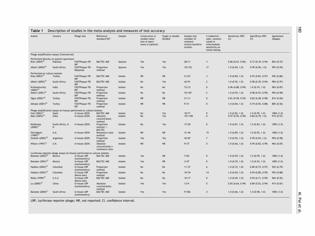
Table 1 Description of studies in the meta-analysis and measures of test accuracy
Author Country Phage test Referencestandard DST
Sample Consecutive orrandom selec-tion of speci-mens or patients
Single or doubleblinded
Sample size(number ofresistantstrains/sensitivestrains)
% indetermi-nate, contami-nated orintermediatesensitivity oninitial testing
Sensitivity (95%CI)
Specificity (95%CI)
Agreement(Kappa)
Phage amplification assays (Commercial)
Performed directly on sputum specimensButt (2004)29 Pakistan FASTPlaque-TB-
RIFBACTEC 460 Sputum Yes Yes 28/11 3 0.86 (0.67, 0.96) 0.73 (0.39, 0.94) 82% (0.57)
Albert (2004)24 South Africa FASTPlaque-TB-Response
Proportionmethod
Sputum Yes Yes 10/135 17 1.0 (0.69, 1.0) 0.99 (0.96, 1.0) 99% (0.95)
Performed on culture isolatesKisa (2003)33 Turkey FASTPlaque-TB-
RIFBACTEC 460 Isolate NR NR 21/67 3 1.0 (0.84, 1.0) 0.93 (0.83, 0.97) 94% (0.86)
Albert (2002)25 South Africa FASTPlaque-TB-RIF
BACTEC 460 Isolate No Yes 42/91 2 1.0 (0.92, 1.0) 0.98 (0.39, 0.94) 98% (0.97)
Krishnamurthy(2002)34
India FASTPlaque-TB-RIF
Proportionmethod
Isolate No No 73/12 0 0.96 (0.88, 0.99) 1.0 (0.74, 1.0) 96% (0.87)
Albert (2001)23 South Africa FASTPlaque-TB-RIF
Proportionmethod
Isolate No No 76/107 2 1.0 (0.95, 1.0) 0.98 (0.93, 0.99) 99% (0.98)
Oguz (2002)38 Turkey FASTPlaque-TB-RIF
Proportionmethod
Isolate NR NR 21/11 0 0.81 (0.58, 0.95) 0.82 (0.48, 0.98) 81% (0.60)
Aktepe (2001)22 Turkey FASTPlaque-TB-RIF
Proportionmethod
Isolate NR NR 9/31 0 1.0 (0.84, 1.0) 0.74 (0.55, 0.88) 80% (0.56)
Phage amplification assays (in-house) performed on culture isolatesGali (2003)31 Spain In-house (D29) BACTEC 460 Isolate No Yes 18/71 0 1.0 (0.82, 1.0) 1.0 (0.95, 1.0) 100% (1.0)Mani (2003)36 India In-house (D29) Absolute
concentrationmethod
Isolate No Yes 101/100 0 0.97 (0.92, 0.99) 0.84 (0.75, 1.0) 91% (0.91)
McNerney(2000)37
South Africa, U.K.
In-house (D29) Proportionmethod CBACTEC
Isolate No Yes 17/20 0 1.0 (0.81, 1.0) 1.0 (0.83, 1.0) 100% (1.0)
Eltringham(1999)30
U.K. In-house (D29) Resistance ratiomethod
Isolate NR NR 31/46 15 1.0 (0.89, 1.0) 1.0 (0.92, 1.0) 100% (1.0)
Simboli (2005)40 Argentina In-house (D29) Proportionmethod
Isolate Yes Yes 42/87 7 1.0 (0.92, 1.0) 0.99 (0.94, 1.0) 99% (0.98)
Wilson (1997)12 U.K. In-house (D29) AbsoluteconcentrationCresistance ratio
Isolate NR NR 9/37 0 1.0 (0.66, 1.0) 0.95 (0.82, 0.99) 96% (0.87)
Luciferase reporter phage assays (In-house) performed on culture isolatesBanaiee (2003)26 Mexico In-house LRP
(luminometry)BACTEC 460 Isolate Yes NR 7/65 0 1.0 (0.59, 1.0) 1.0 (0.95, 1.0) 100% (1.0)
Banaiee (2001)27 Mexico In-house LRP(luminometry)
BACTEC 460 Isolate Yes NR 3/47 0 1.0 (0.29, 1.0) 1.0 (0.93, 1.0) 100% (1.0)
Hazbon (2003)32 Colombia In-house LRP(luminometry)
Proportionmethod
Isolate No No 11/37 6 1.0 (0.72, 1.0) 0.89 (0.75, 0.97) 92% (0.79)
Hazbon (2003)32 Colombia In-house LRP(Bronx box)
Proportionmethod
Isolate No No 10/34 14 1.0 (0.69, 1.0) 0.94 (0.80, 0.99) 95% (0.88)
Riska (1999)39 U.S.A In-house LRP(Bronx box)
BACTEC 460 Isolate No No 10/17 0 1.0 (0.69, 1.0) 0.94 (0.71, 0.99) 96% (0.92)
Lu (2000)35 China In-house LRP(luminometry)
Absoluteconcentrationmethod
Isolate Yes Yes 13/9 0 0.92 (0.64, 0.99) 0.89 (0.52, 0.99) 91% (0.81)
Banaiee (2004)41 South Africa In-house LRP(luminometry)
BACTEC 460 Isolate Yes Yes 9/182 4 1.0 (0.66, 1.0) 1.0 (0.98, 1.0) 100% (1.0)
LRP, luciferase reporter phage; NR, not reported; CI, confidence interval.
M.
Pai
et
al.180
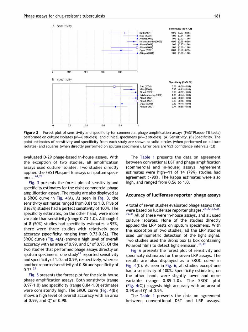
Figure 3 Forest plot of sensitivity and specificity for commercial phage amplification assays (FASTPlaque-TB tests)performed on culture isolates (NZ6 studies), and clinical specimens (NZ2 studies). (A) Sensitivity. (B) Specificity. Thepoint estimates of sensitivity and specificity from each study are shown as solid circles (when performed on cultureisolates) and squares (when directly performed on sputum specimens). Error bars are 95% confidence intervals (CI).
Phage assays for drug-resistant tuberculosis 181
evaluated D-29 phage-based in-house assays. Withthe exception of two studies, all amplificationassays used culture isolates. Two studies directlyapplied the FASTPlaque-TB assays on sputum speci-mens.24,29
Fig. 3 presents the forest plot of sensitivity andspecificity estimates for the eight commercial phageamplification assays. The results are also displayed asa SROC curve in Fig. 4(A). As seen in Fig. 3, thesensitivity estimates ranged from 0.81 to 1.0. Five of8 (63%) studies had a perfect sensitivity of 100%. Thespecificity estimates, on the other hand, were morevariable than sensitivity (range 0.73–1.0). Although 4of 8 (50%) studies had specificity estimates O95%,there were three studies with relatively pooraccuracy (specificity ranging from 0.73–0.82). TheSROC curve (Fig. 4(A)) shows a high level of overallaccuracy with an area of 0.99, and Q* of 0.95. Of thetwo studies that performed phage assays directly onsputum specimens, one study24 reported sensitivityand specificity of 1.0 and 0.99, respectively, whereasanother reported sensitivity of 0.86 and specificity of0.73.29
Fig. 5 presents the forest plot for the six in-housephage amplification assays. Both sensitivity (range0.97–1.0) and specificity (range 0.84–1.0) estimateswere consistently high. The SROC curve (Fig. 4(B))shows a high level of overall accuracy with an areaof 0.99, and Q* of 0.98.
The Table 1 presents the data on agreementbetween conventional DST and phage amplification(commercial and in-house) assays. Agreementestimates were high—11 of 14 (79%) studies hadagreement O90%. The kappa estimates were alsohigh, and ranged from 0.56 to 1.0.
Accuracy of luciferase reporter phage assays
A total of seven studies evaluated phage assays thatwere based on luciferase reporter phages.26,27,32,35,
39,41 All of these were in-house assays, and all usedculture isolates. None of the studies directlyapplied the LRP tests on sputum specimens. Withthe exception of two studies, all the LRP studiesused luminometric detection of the light signal.Two studies used the Bronx box (a box containingPolaroid film) to detect light emission.32,39
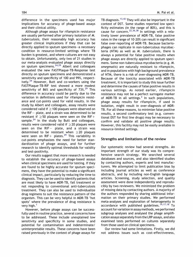
Fig. 6 presents the forest plot of sensitivity andspecificity estimates for the seven LRP assays. Theresults are also displayed as a SROC curve inFig. 4(C). As seen in Fig. 6, all studies except onehad a sensitivity of 100%. Specificity estimates, onthe other hand, were slightly lower and morevariable (range 0.89–1.0). The SROC plot(Fig. 4(C)) suggests high accuracy with an area of0.98 and Q* of 0.95.
The Table 1 presents the data on agreementbetween conventional DST and LRP assays.
Figure 4 Summary ROC plots for phage-based assays,stratified by type of assay. (A) Commercial amplificationassays (FASTPlaque-TB) (NZ8 studies). (B) In-houseamplification assays (D29 phage-based assays) (NZ6studies). (C) Luciferase reporter phage assays (NZ7studies). Each solid circle represents each study in themeta-analysis. The curve is the regression line thatsummarizes the overall diagnostic accuracy. SROC,summary receiver operating characteristic; AUC, areaunder the curve; SE (AUC), standard error of AUC; Q*, anindex defined by the point on the SROC curve where thesensitivity and specificity are equal, which is the pointclosest to the top-left corner of the ROC space; SE(Q*),standard error of Q* index.
M. Pai et al.182
Agreement estimates were exceptionally high—allseven studies had agreement O90%. The kappaestimates were ranged from 0.79 to 1.0.
Impact of assay methodology on accuracy
Overall, the SROC plots stratified by type of assay(Fig. 4(A)–(C)) were fairly similar for all three assays(commercial and in-house phage amplification, andLRP assays), although commercial assays appear toproduce lower and more variable accuracy esti-mates. The area under the SROC curve and Q*estimates were fairly similar, indicating compar-able diagnostic accuracy.
Isoniazid resistance among rifampicin-resistant isolates
Seven studies provided data on INH resistance byconventional DST methods among strains that wereresistant to rifampicin.12,24,25,29,31,37,40 Thesestudies showed that, on average, 96% of therifampicin-resistant isolates were also resistant toINH. Rifampicin resistance, therefore, was a fairlygood surrogate marker of MDR-TB in the populationsstudied.
Discussion
Principal findings and clinical implications
Our meta-analysis of 21 studies (with all but twostudies using culture isolates) on phage-basedassays for the detection of rifampicin resistancesuggests that phage assays are associated withfairly high sensitivity and specificity, when appliedto culture isolates. These results indicate that, as agroup, phage assays show promise for the detectionof rifampicin resistance in culture isolates whencompared to conventional agar-based drug suscep-tibility tests and rapid methods such as BACTEC 460.Once an isolate was drawn from a growing culture,the average turnaround time for phage-basedassays was 48–72 h, whereas with conventionalDST, the turnaround time might vary from 1–2weeks for BACTEC and MGIT, and 3–4 weeks for solidmedia-based methods. In addition to rapidity,phage-based assays have the advantage of beingless expensive and technologically simpler thanmolecular (e.g. PCR) and liquid-media based tests(e.g. MGIT, BACTEC). This offers some advantagesin resource-limited settings.
Although these results suggest that phage-basedassays are fairly sensitive and specific for rifampicin
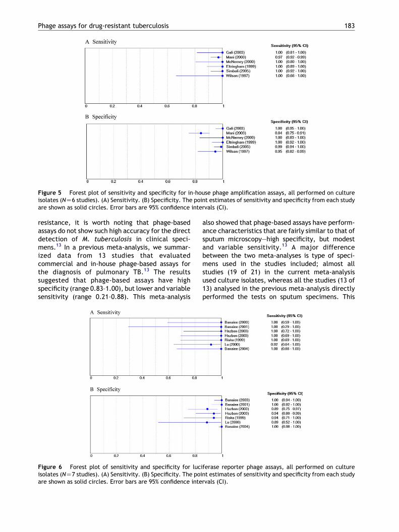
Figure 5 Forest plot of sensitivity and specificity for in-house phage amplification assays, all performed on cultureisolates (NZ6 studies). (A) Sensitivity. (B) Specificity. The point estimates of sensitivity and specificity from each studyare shown as solid circles. Error bars are 95% confidence intervals (CI).
Phage assays for drug-resistant tuberculosis 183
resistance, it is worth noting that phage-basedassays do not show such high accuracy for the directdetection of M. tuberculosis in clinical speci-mens.13 In a previous meta-analysis, we summar-ized data from 13 studies that evaluatedcommercial and in-house phage-based assays forthe diagnosis of pulmonary TB.13 The resultssuggested that phage-based assays have highspecificity (range 0.83–1.00), but lower and variablesensitivity (range 0.21–0.88). This meta-analysis
Figure 6 Forest plot of sensitivity and specificity for lucisolates (NZ7 studies). (A) Sensitivity. (B) Specificity. The poiare shown as solid circles. Error bars are 95% confidence inte
also showed that phage-based assays have perform-ance characteristics that are fairly similar to that ofsputum microscopy—high specificity, but modestand variable sensitivity.13 A major differencebetween the two meta-analyses is type of speci-mens used in the studies included; almost allstudies (19 of 21) in the current meta-analysisused culture isolates, whereas all the studies (13 of13) analysed in the previous meta-analysis directlyperformed the tests on sputum specimens. This
iferase reporter phage assays, all performed on culturent estimates of sensitivity and specificity from each studyrvals (CI).
M. Pai et al.184
difference in the specimens used has majorimplications for accuracy of phage-based assaysand their clinical utility.
Although phage assays for rifampicin resistanceare usually performed after primary isolation of M.tuberculosis, their reasonably high accuracy hasgreater clinical implications if they that can bedirectly applied to sputum specimens—a necessarycondition in resource-limited settings where TBburden is greatest, and where cultures are not easyto obtain. Unfortunately, only two of 21 studies inour meta-analysis evaluated phage assays directlyon sputum specimens.24,29 Albert and colleaguesevaluated the new FASTPlaque-TB-Response testdirectly on sputum specimens and demonstrated asensitivity and specificity of 100 and 99%, respect-ively.24 However, Butt and co-workers using theFASTPlaque-TB-RIF test showed a more modestsensitivity of 86% and specificity of 73%.29 Thisdifference in accuracy could be partly due to thevariation in definitions used for rifampicin resist-ance and cut-points used for valid results. In thestudy by Albert and colleagues, assay results wereconsidered valid if R100 plaques were observed onthe RIFK plate, and a strain was determined to beresistant if R50 plaques were seen on the RIFCsample.24 In the study by Butt and colleagues,results were considered valid if R20 plaques wereseen on the RIFK plate, and a strain wasdetermined to be resistant when R20 plaqueswere seen on RIFC plates.29 This variability incut-points emphasizes the need for better stan-dardization of phage assays, and for furtherresearch to identify optimal thresholds for validityand test-positivity.
Our results suggest that more research is neededto establish the accuracy of phage-based assayswhen clinical specimens are used for testing. If theyare found to be highly accurate for sputum speci-mens, they have the potential to make a significantclinical impact, particularly by reducing the time todiagnosis. They can be used to identify patients thatare most likely to have MDR-TB, fail treatment ornot responding to conventional anti-tuberculosistreatment. They can also be used to individualizedrug regimens to suit the resistance pattern of theorganism. This can be very helpful in MDR-TB ‘hotspots’ where the prevalence of drug resistance isvery high.1
However, before phage assays can be success-fully used in routine practice, several concerns haveto be addressed. These include unexplained lowsensitivity and specificity in some studies, andpotential for contamination and indeterminate/uninterpretable results. These concerns have beenraised previously in the context of phage assays for
TB diagnosis.13,42 They will also be important in thecontext of DST. Some studies reported low speci-ficity estimates (in the range of 80%), and this iscause for concern.22,36,38 In settings with a rela-tively lower prevalence of MDR-TB, false positiverates in the range of 10–20% can result in significantfalse over-reporting of MDR-TB. Because bacterio-phages can replicate in non-tuberculous mycobac-teria (NTM) as well as M. tuberculosis, there isalways a potential for false positive results whenphage assays are directly applied to sputum speci-mens. Some non-tuberculous mycobacteria (e.g. M.smegmatis) are naturally resistant to rifampicin,and in populations with high background prevalenceof NTM, there is a risk of over-diagnosing MDR-TB.Because of the toxicity associated with MDR-TBtreatment, it is important to study this issue furtherand determine the causes of false positive results invarious settings. As noted earlier, rifampicinresistance may not be a perfect surrogate markerof MDR-TB in all settings.2 In such populations,phage assay results for rifampicin, if used inisolation, might result in over-diagnosis of MDR-TB. For all these reasons, to minimize false positiveresults, a second confirmatory test (e.g. conven-tional DST for first line drugs) may be necessary toconfirm and validate all positive phage results.However, this facility may not be easily available inresource-limited settings.
Strengths and limitations of the review
Our systematic review had several strengths. Animportant strength of our study was its compre-hensive search strategy. We searched severaldatabases and sources, and also identified studiesby contacting authors, experts and test manufac-turers. We attempted to limit publication bias byincluding journal articles as well as conferenceabstracts, and by including non-English languagearticles. Screening, study selection, and qualityassessment were done independently and reprodu-cibly by two reviewers. We minimized the problemof missing data by contacting authors. A majority ofthe authors responded by sharing additional infor-mation on their primary studies. We performedmeta-analyses and exploration of heterogeneity inaccordance with published guidelines.14,17,18 Toaccount for variation inassaymethods,weperformedsubgroup analyses and analysed the phage amplifi-cationassays separately from theLRP assays, andalsoexamined tests performed on cultures separatelyfrom those used on clinical specimens.
Our review had some limitations. Firstly, we didnot address issues such as cost-effectiveness,

Phage assays for drug-resistant tuberculosis 185
reliability, the incremental benefit of adding phageassays to other conventional tests, and the neteffect of phage assays on clinical care and patientoutcomes. None of the studies in our meta-analysisreported data on the clinical impact of detection ofrifampicin resistance on long-term treatment out-comes such as cure rates, treatment failures andmortality. Another problem concerns the exclusionof phage assays that produced indeterminateand/or contaminated results, and intermediatesensitivity from the analyses in the primary studies.Such exclusions might have overestimated thesensitivity and specificity estimates in our meta-analysis. Lastly, despite our comprehensive litera-ture searches, it is still possible that the highaccuracy of phage-based assays in our meta-analysis could be due to publication bias. Althoughstatistical (e.g. Begg and Egger tests) and graphicalmethods (e.g. funnel plots) are available to detectpotential publication bias in meta-analyses ofrandomized controlled trials, such techniqueshave not been adequately evaluated for diagnosticdata.17 It is, therefore, difficult to rule outpublication bias in our meta-analysis.
Implications for research
Future studies on phage assays must include thefollowing design features: consecutive or randomselection of patients with suspected drug-resistantTB, blinded interpretation of both phage andconventional DST results, and an adequate numberof patients with confirmed rifampicin resistance.Studies should explicitly report the cut-points usedto determine test validity and drug resistance,concentrations of the drugs used, and the pro-portion of phage tests that are indeterminate orcontaminated. If indeterminate or contaminatedresults are excluded from the final analysis, theimpact of such exclusions on accuracy measuresshould be reported. In addition, more studies areneededon the accuracyofphage assayswhen directlyapplied to sputum specimens. The challenge in suchstudies would be to develop methods to increasethe sensitivity of phage-based assays withoutcompromising specificity. Another challengewould be to extend the scope of phage assays tocover other first-line anti-tuberculosis drugs. This isalready being attempted with some success.8,9
Lastly, studies should provide data on prevalenceof non-tuberculous mycobacteria and the effect ofNTM isolation on phage assay specificity.
Studies are also needed to establish the cost-effectiveness and operational, logistical (e.g. turn-around time), and clinical advantages of phage
assays over conventional DST. The advantage ingetting quicker results is clearly greater with directtesting of sputum specimens, as compared toperforming the phage assays after primary cultureisolation. Although only amplification-based assaysare currently available as commercial kits, it isnecessary to also simplify and develop LRP basedassays into a standardized, low-technology kit thatcould be made available commercially. This iscurrently being attempted on a small scale.32,39,41
Lastly, studies must evaluate the actual impact ofdetection of rifampicin resistance on patient careand clinical decision making. It will be necessary toshow that testing for rifampicin resistance usingphage assays can improve treatment outcomes,reduce morbidity and mortality among TB patients,and decrease transmission of MDR-TB.
Conclusions
Current evidence is largely restricted to the use ofphage assays for the detection of rifampicinresistance in culture isolates. When applied toisolates, these assays appear to have relatively highsensitivity and specificity. Specificity estimates,however, appear to be relatively lower and morevariable than sensitivity, and this can potentiallyresult in over-diagnosis of MDR-TB in low preva-lence settings. In contrast, evidence is lacking onthe accuracy of these assays when they are directlyapplied to sputum specimens. If phage-based assayscan be directly used on clinical specimens and ifthey are shown to have high accuracy, they have thepotential to improve the diagnosis and managementof MDR-TB. However, before phage assays can besuccessfully used in routine practice, severalconcerns have to be addressed, including unex-plained low sensitivity and specificity in somestudies, potential for contamination and indeter-minate results, and variability in cut-points used todetermine test validity and drug resistance. Betterstandardization of the assay procedure, therefore,is needed for widespread applicability.
Acknowledgements
This work was supported by the National Institutes ofHealth, Fogarty AIDS International Training Program(1-D43-TW00003-16), and NIH/NIAID (R01 AI 34238).We are grateful to the following authors who sentadditional information on their primary studies: TariqButt, Ozgul Kisa, Heidi Albert, Ruth McNerney,Cheruvu Mani, Viviana Ritacco, Shunqing Xu, Camilla
M. Pai et al.186
Rodriguez, Manzour Hazbon, Niaz Banaiee, and PaulRiska. We thank Heidi Albert, Richard Mole, PaulRiska, Ruth McNerney and Niaz Banaiee for theirassistance with identifying additional studies, andJavier Zamora for his support with the Meta-DiScsoftware. Lastly, we thank Ruth McNerney, PuneetDewan, and Ed Desmond for reviewing the draftversion of this manuscript.
References
1. Espinal MA. The global situation of MDR-TB. Tuberculosis(Edinb) 2003;83(1–3):44–51.
2. World Health Organization. Anti-tuberculosis drug resist-ance in the world. Third global report. Geneva: WorldHealth Organization; 2004.
3. Fisher M. Diagnosis of MDR-TB: a developing world problemon a developed world budget. Expert Rev Mol Diagn 2002;2(2):151–9.
4. Heifets LB, Cangelosi GA. Drug susceptibility testing ofMycobacterium tuberculosis: a neglected problem at theturn of the century. Int J Tuberc Lung Dis 1999;3(7):564–81.
5. Parsons LM, Somoskovi A, Urbanczik R, Salfinger M. Labora-tory diagnostic aspects of drug resistant tuberculosis. FrontBiosci 2004;9:2086–105.
6. Hazbon MH. Recent advances in molecular methods for earlydiagnosis of tuberculosis and drug-resistant tuberculosis.Biomedical 2004;24(Suppl 1):149–62.
7. Somoskovi A, Parsons LM, Salfinger M. The molecular basis ofresistance to isoniazid, rifampin, and pyrazinamide inMycobacterium tuberculosis. Respir Res 2001;2(3):164–8.
8. Mole RJ, Maskell T. Phage as a diagnostic—the use of phage inTB diagnosis. J Chem Technol Biotechnol 2001;76(7):683–8.
9. Trollip A, Albert H, Maskell T. Bacteriophage-based tech-nologies for the rapid diagnosis and drug susceptibilitytesting of tuberculosis. Am Clin Lab 2001;20(9):39–42.
10. Jacobs Jr WR, Barletta RG, Udani R, Chan J, Kalkut G,Sosne G, et al. Rapid assessment of drug susceptibilities ofMycobacterium tuberculosis by means of luciferase reporterphages. Science 1993;260(5109):819–22.
11. McNerney R. TB: the return of the phage. A review of fiftyyears of mycobacteriophage research. Int J Tuberc Lung Dis1999;3(3):179–84.
12. Wilson SM, Al-Suwaidi Z, McNerney R, Porter J,Drobniewski F. Evaluation of a new rapid bacteriophage-based method for the drug susceptibility testing ofMycobacterium tuberculosis. Nat Med 1997;3(4):465–8.
13. Kalantri SP. Bacteriophage-based tests for the detection ofMycobacterium tuberculosis in clinical specimens: a sys-tematic review and meta-analysis. Master’s in Public Health(MPH) Thesis, University of California, Berkeley; 2005.
14. Pai M, McCulloch M, Enanoria W, Colford Jr JM. Systematicreviews of diagnostic test evaluations: what’s behind thescenes? ACP J Club 2004;141(1):A11–A13.
15. Pai M, McCulloch M, Gorman JD, Pai N, Enanoria W,Kennedy G, et al. Systematic reviews and meta-analyses:an illustrated, step-by-step guide. Natl Med J India 2004;17(2):86–95.
16. Hadgu A. Discrepant analysis: a biased and an unscientificmethod for estimating test sensitivity and specificity. J ClinEpidemiol 1999;52(12):1231–7.
17. Deeks JJ. Systematic reviews in health care: systematicreviews of evaluations of diagnostic and screening tests. BMJ2001;323(7305):157–62.
18. Irwig L, Tosteson AN, Gatsonis C, Lau J, Colditz G,Chalmers TC, et al. Guidelines for meta-analyses evaluatingdiagnostic tests. Ann Intern Med 1994;120(8):667–76.
19. Littenberg B, Moses LE. Estimating diagnostic accuracy frommultiple conflicting reports: a new meta-analytic method.Med Decis Making 1993;13(4):313–21.
20. Walter SD. Properties of the summary receiver operatingcharacteristic (SROC) curve for diagnostic test data. StatMed 2002;21(9):1237–56.
21. Lijmer JG, Bossuyt PM, Heisterkamp SH. Exploring sources ofheterogeneity in systematic reviews of diagnostic tests. StatMed 2002;21:1525–37.
22. Aktepe OC, Altindis M, Esen N. A rapid method for rifampicinsusceptibility testing of Mycobacterium tuberculosis 11thEuropean Congress of Clinical Microbiology and InfectiousDiseases (ECCMID), 2001, Istanbul, Turkey.
23. Albert H, Heydenrych A, Mole R, Trollip A, Blumberg L.Evaluation of FASTPlaque TB-RIF, a rapid, manual test forthe determination of rifampicin resistance from Mycobac-terium tuberculosis cultures. Int J Tuberc Lung Dis 2001;5(10):906–11.
24. Albert H, Trollip A, Seaman T, Mole RJ. Simple, phage-based(FASTPplaque) technology to determine rifampicin resist-ance of Mycobacterium tuberculosis directly from sputum.Int J Tuberc Lung Dis 2004;8(9):1114–9.
25. Albert H, Trollip AP, Mole RJ, Hatch SJB, Blumberg L. Rapidindication of multidrug-resistant tuberculosis from liquidcultures using FASTPlaqueTB-RIF, a manual phage-basedtest. Int J Tuberc Lung Dis 2002;6(6):523–8.
26. Banaiee N, Bobadilla-del-Valle M, Riska PF, Bardarov S,Small PM, Ponce-de-Leon A, et al. Rapid identification andsusceptibility testing of Mycobacterium tuberculosis fromMGIT cultures with luciferase reporter mycobacteriophages.J Med Microbiol 2003;52(Pt 7):557–61.
27. Banaiee N, Bodadilla-del-Valle M, Bardarov S, Riska PF,Small PM, Ponce-de-Leon A, et al. Luciferase reportermycobacteriophages for detection, identification, andantibiotic susceptibility testing of Mycobacterium tubercu-losis in Mexico. J Clin Microbiol 2001;39(11):3883–8.
28. Bardarov S, Dou H, Eisenach K, Banaiee N, Ya S, Chan J, et al.Detection and drug-susceptibility testing of M-tuberculosisfrom sputum samples using luciferase reporter phage:comparison with the mycobacteria growth indicator tube(MGIT) system. Diagn Microbiol Infect Dis 2003;45(1):53–61.
29. Butt T, Ahmad RN, Afzal RK, Mahmood A, Anwar M. Rapiddetection of rifampicin susceptibility of Mycobacteriumtuberculosis in sputum specimens by mycobacteriophageassay. J Pak Med Assoc 2004;54(7):379–82.
30. Eltringham IJ, Drobniewski FA, Mangan JA, Butcher PD,Wilson SM. Evaluation of reverse transcription-PCR and abacteriophage-based assay far rapid phenotypic detection ofrifampin resistance in clinical isolates of Mycobacteriumtuberculosis. J Clin Microbiol 1999;37(11):3524–7.
31. Gali N, Dominguez J, Blanco S, Prat C, Quesada AD, Matas L,et al. Utility of an in-house mycobacteriophage-based assayfor rapid detection of rifampin resistance in Mycobacteriumtuberculosis clinical isolates. J Clin Microbiol 2003;41(6):2647–9.
32. Hazbon MH, Guarin N, Ferro BE, Rodriguez AL, Labrada LA,Tovar R, et al. Photographic and luminometric detection ofluciferase reporter phages for drug susceptibility testing ofclinical Mycobacterium tuberculosis isolates. J Clin Micro-biol 2003;41(10):4865–9.

Phage assays for drug-resistant tuberculosis 187
33. Kisa O, Albay A, Bedir O, Baylan O, Doganci L. Evaluation ofFASTPlaquetb-RIF for determination of rifampicin resistancein Mycobacterium tuberculosis complex isolates. IntJ Tuberc Lung Dis 2003;7(3):284–8.
34. Krishnamurthy A, Rodrigues C, Mehta AP. Rapid detection ofrifampicin resistance in M. tuberculosis by phage assay. IndJ Med Microbiol 2002;20(4):211–4.
35. Lu B, Fu Z, Xu S. Rapid rifampicin susceptibility test by usingrecombinant mycobacteriophages. Zhonghua Jie He He Hu XiZa Zhi 2000;23(8):480–4.
36. Mani C, Selvakumar N, Kumar V, Narayanan S, Narayanan PR.Comparison of DNA sequencing, PCR-SSCP and PhaB assayswith indirect sensitivity testing for detection of rifampicinresistance in Mycobacterium tuberculosis. Int J Tuberc LungDis 2003;7(7):652–9.
37. McNerney R, Kiepiela P, Bishop KS, Nye PM, Stoker NG. Rapidscreening of Mycobacterium tuberculosis for susceptibilityto rifampicin and streptomycin. Int J Tuberc Lung Dis 2000;4(1):69–75.
38. Oguz VA, Guneri S, Erdenizmenli M, Yapar N, Kuruuzum Z,Cakmak R, et al. Investigation of rifampin resistance in
Mycobacterium tuberculosis by two different methods.Toraks Dergisi 2002;3(2):173–7.
39. Riska PF, Su Y, Bardarov S, Freundlich L, Sarkis G, Hatfull G,et al. Rapid film-based determination of antibiotic suscep-tibilities of Mycobacterium tuberculosis strains by using aluciferase reporter phage and the Bronx box. J Clin Microbiol1999;37(4):1144–9.
40. Simboli N, Takiff H, McNerney R, Lopez B, Martin A,Palomino JC, et al. In-house phage amplification assay is asound alternative for detecting rifampin-resistant Mycobac-terium tuberculosis in low-resource settings. AntimicrobAgents Chemother 2005;49(1):425–7.
41. Banaiee N, January V, Barthus C, Lambride M, RoDiti D,Behr MA, et al. Semi-automation and evaluation ofluciferase reporter mycobacteriophages for susceptibilitytesting of Mycobacterium tuberculosis cultures in SouthAfrica. American Society for Microbiology 104th generalmeeting 2004, New Orleans, LA 2004 [Abstract U-018].
42. Takiff H, Heifets L. In search of rapid diagnosis and drug-resistance detection tools: is the FASTPlaqueTB test theanswer? Int J Tuberc Lung Dis 2002;6(7):560–1.


![BACTERIOPHAGE-RESISTANT AND BACTERIOPHAGE-SENSITIVE ...halsmith/phagemutantsubmitted_2.pdf · BACTERIOPHAGE-RESISTANT AND BACTERIOPHAGE-SENSITIVE BACTERIA IN A CHEMOSTAT ... [22],](https://img.pdfslide.us/doc/110x75/5b3839687f8b9a5a518d2ce1/bacteriophage-resistant-and-bacteriophage-sensitive-halsmithphagemutantsubmitted2pdf.jpg)