Embed Size (px)
Citation preview

Bacterial Tethering Analysis Reveals a “Run-Reverse-Turn”Mechanism for Pseudomonas Species Motility
Chen Qian,a Chui Ching Wong,a Sanjay Swarup,a,b,c,d Keng-Hwee Chiama,e
Mechanobiology Institute, National University of Singapore, Singapore, Singaporea; Department of Biological Sciences, National University of Singapore, Singapore,Singaporeb; NUS Environmental Research Institute (NERI), National University of Singapore, Singapore, Singaporec; Singapore Centre for Environmental Life SciencesEngineering (SCELSE), Nanyang Technological University, Singapore, Singapored; A*STAR Bioinformatics Institute, Singapore, Singaporee
We have developed a program that can accurately analyze the dynamic properties of tethered bacterial cells. The program worksespecially well with cells that tend to give rise to unstable rotations, such as polar-flagellated bacteria. The program has twonovel components. The first dynamically adjusts the center of the cell’s rotational trajectories. The second applies piecewise lin-ear approximation to the accumulated rotation curve to reduce noise and separate the motion of bacteria into phases. Thus, itcan separate counterclockwise (CCW) and clockwise (CW) rotations distinctly and measure rotational speed accurately. Usingthis program, we analyzed the properties of tethered Pseudomonas aeruginosa and Pseudomonas putida cells for the first time.We found that the Pseudomonas flagellar motor spends equal time in both CCW and CW phases and that it rotates with the samespeed in both phases. In addition, we discovered that the cell body can remain stationary for short periods of time, leading to theexistence of a third phase of the flagellar motor which we call “pause.” In addition, P. aeruginosa cells adopt longer run lengths,fewer pause frequencies, and shorter pause durations as part of their chemotactic response. We propose that one purpose of thepause phase is to allow the cells to turn at a large angle, where we show that pause durations in free-swimming cells positivelycorrelate with turn angle sizes. Taken together, our results suggest a new “run-reverse-turn” paradigm for polar-flagellated Pseu-domonas motility that is different from the “run-and-tumble” paradigm established for peritrichous Escherichia coli.
Flagellated bacteria swim in the aquatic environment by propel-ling their flagella (1). This swimming mechanism is best de-
scribed in the Escherichia coli model, where the peritrichous cellsare known to “run” and “tumble.” Flagella of a cell rotating coun-terclockwise (CCW) (when viewed from behind the cell) form abundle that propels the cell to run forward, while a transientswitch in the rotation direction of its flagellar motor causes theflagellar bundle to separate and the cell to tumble (2), allowing thecell to reorient its direction of motion.
In recent years, some other models have also been elucidated,including the three-step “run-reverse-flick” chemotactic responsefor the sodium-driven, monotrichous Vibrio alginolyticus (3, 4)and that of varying “run-and-stop” frequencies in monotrichousRhodobacter sphaeroides (5, 6). The diversity of flagellar arrange-ments, flagellar motor structures (7), and chemotactic gene clus-ters (8) across the bacterial kingdom likely accounts for the pres-ence of these different systems.
In the case of Pseudomonas spp., however, mechanisms of mo-tility and chemotaxis remain unclear. Current evidence suggeststhat the chemosensory system and flagellar apparatus arrange-ment in the strains belonging to this genus are more complex thanthose of other bacterial species. For example, Pseudomonas aerugi-nosa has five gene clusters involved in chemotaxis, with 26 methyl-accepting chemotaxis proteins (MCPs) and 20 chemotaxis (che)genes, compared to E. coli, which has one gene cluster, with fourMCPs and six che genes (9). Additionally, there are two sets offlagellar stators in Pseudomonas spp. compared to one set for E.coli and Salmonella enterica serovar Typhimurium (10, 11). AsPseudomonas spp. are polar flagellated, they are likely to possessesa “run-and-reverse” trajectory (12) rather than the typical “run-and-tumble” trajectory as well. Since both the Pseudomonas fla-gellar motor and chemosensory system present some unique fea-tures, it would therefore be interesting to study the motor
dynamics of Pseudomonas spp. Notably, many members of thisgenus play significant roles in their environment, such as in thedegradation of organic hydrocarbons, in plant growth promotion,and in nitrogen fixation. Other members, however, are patho-genic to humans, insects, or plants (13). Therefore, elucidating themotility and chemotactic mechanisms for Pseudomonas spp. canbe beneficial in many studies extending to bioremediation andhost-pathogen interactions. Additionally, across Pseudomonasspp., different species also exhibit dissimilar flagellar arrange-ments. In the plant growth-promoting rhizobium (PGPR) strainPseudomonas putida, the flagellum is arranged in a polar multi-trichous manner (14), whereas in the human pathogen P. aerugi-nosa, the flagellum is polar monotrichous (15). Hence, it wouldalso be interesting to determine if there are differences in the roleof each flagellum in contributing to Pseudomonas motility.
In order to study bacterial chemotaxis, various methods suchas the capillary (16) and agar plate (17) assays have been previ-ously developed to study the population movement in a macro-scopic view. Tracking of a single bacterium (18) or a group ofbacteria (19) in a three-dimensional environment has been usedto study the response of a single bacterium to chemoattractantsduring swimming. As the flagellar motor is directly linked to thischemotactic response, one can study the rotation of the motor byfixing the cell body to a surface so as to observe the rotation of a
Received 29 March 2013 Accepted 29 May 2013
Published ahead of print 31 May 2013
Address correspondence to Keng-Hwee Chiam, [email protected].
C.Q. and C.C.W. contributed equally to this work.
Copyright © 2013, American Society for Microbiology. All Rights Reserved.
doi:10.1128/AEM.01027-13
4734 aem.asm.org Applied and Environmental Microbiology p. 4734–4743 August 2013 Volume 79 Number 15
on January 14, 2020 by guesthttp://aem
.asm.org/
Dow
nloaded from
bead attached to the flagella (20, 21). Alternatively, this can also beachieved by fixing (tethering) the flagella to a surface to observethe rotation of the cell body (22). The latter approach, also knownas the cell-tethering method, is most widely used to study theresponse to stimuli of a large number of bacteria. It has been thekey technique to quantitatively reveal the fundamental propertiesand mechanisms of E. coli chemotaxis by measuring tumblingfrequency, run length, and kinetic response (23–25).
In this study, we have developed a program, which we call thebacterial tethering analysis program (BTAP), that can track largenumbers of tethered cells and extract accurate and reliable rota-tion data. Our program dynamically adjusts the centers of thecell’s rotational trajectories and applies piecewise linear approxi-mation to the accumulated rotation curve to reduce noise andseparate the motion of bacteria into phases. This is particularlyuseful for polar-flagellated bacteria, such as Pseudomonas spp., asthey tend to give rise to unstable rotation trajectories (26). Usingour program, we were therefore able to elucidate the flagellar mo-tor properties of two Pseudomonas strains, KT2440 and PAO1,belonging to P. putida and P. aeruginosa, respectively. We showthat, unlike E. coli, cells belonging to the two Pseudomonas strainsspend equal amounts of time rotating in the counterclockwise(CCW) and clockwise (CW) directions. Interestingly, the Pseu-domonas cells also have an additional “pause” phase that consti-tutes nearly 10% of the total observed time, and we propose thatthis pause phase allows the cells to vary their turn angle, adoptinga “run-reverse-turn” trajectory. In addition, BTAP analysis of acheY chemotaxis mutant in P. aeruginosa also revealed that Pseu-domonas cells vary their run lengths, pause frequencies, and pausedurations as part of their chemotactic response. By analyzing tra-jectories of free-swimming cells, we established a role for thepauses, where Pseudomonas cells vary their pause duration to ef-fect different turn angle sizes.
MATERIALS AND METHODSGrowth condition. Pseudomonas aeruginosa PAO1 wild-type and cheYcells (27) and Escherichia coli MG1655 cells from single colonies wereseparately cultured overnight in 10 ml Luria-Bertani (LB) broth at 37°C,250 rpm. Cultures were diluted to an optical density at 600 nm (OD600) of0.1 using LB broth and grown at 37°C, 250 rpm, until the late-exponentialgrowth phase was reached. For P. putida KT2440, cultures were grown at30°C, 250 rpm. Cell cultures were then diluted 1:10 prior to imaging usingvideo microscopy.
Cell tethering and video capture. For cell-tethering assays, standardmethods (22) were adapted as follows: glass coverslips were precoatedwith flagellar antibodies prior to use and cell chambers (2.0 cm by 1.0 cmby 150 �m) were created using three layers of double-sided tape betweenthe microscope slide and coverslips. Flagella were sheared off by passingthe bacterial cells through a 34-gauge blunt-end needle four times. Cellswere loaded into the cell chamber, and nontethered cells were rinsed awayusing LB broth. Cells were visualized using an inverted microscope(Nikon TE2000U) under a 100� objective. Videos of tethered bacteriawere taken at 120 frames per second (fps) for 1 to 5 min using a comple-mentary metal-oxide semiconductor (CMOS) camera (Thorlabs;DCC1645). Following the convention, cells are considered to be rotatingCW/CCW when viewed from the medium that they are tethered in (28).In the experiment with multitrichous P. putida, to eliminate the casewhere more than one filament is tethered and where the filaments do notrotate in synchrony, only cells that exhibited clear rotations were selectedfor analyses.
Free-swimming P. aeruginosa cells, with their intact flagellum, wereloaded in cell chambers and observed in the middle of the chamber depth
(away from the coverslip or microscope slide surface). Cells were visualizedunder a 40� objective, and videos of bacteria were taken at 25 fps. Bacterialswimming trajectories in two dimensions (2D) were captured using ImagePro Plus 6.3 (Media Cybernetics, Rockville, MD, USA), and images with celloutlines were obtained using ImageJ (NIH, Bethesda, MD, USA).
Pause detection for 2D swimming trajectories. The instantaneousspeed for free-swimming bacteria was calculated from the 2D trajectoriesframe by frame. Trajectories with fewer than 25 frames were discarded. Apause was identified when n (n � 3) consecutive instantaneous speedsthat were below 5 �m/s appeared. The moving direction of a bacterium ata certain time was defined as the vector connecting its current coordinatesto its subsequent coordinates. Accordingly, the change of angle was theangle difference between two such directions which ranged between ��and �. Additionally, to ensure that the cells swimming in the z directiondid not affect the angle size tracked, only cells that were in focus, and thusswimming within the x-y focal plane, were selected for analysis.
Image extraction and data preprocessing for tethered cells. For im-age processing, movies of single, tethered cells were converted to gray scaleand their contrasts were adjusted (saturated pixels � 0.4, histogram stack)using ImageJ (NIH, Bethesda, MD, USA). The image of the rotating cellbody was binarized to be isolated from the background for each frame inthe video. Next, using the Set Measurements function in ImageJ, the co-ordinates of the center of mass of the rotating cell body were measuredfrom the binarized image stack and imported into Matlab (Mathworks,Natick, MA, USA) for BTAP analyses.
RESULTSSignal processing in BTAP. BTAP is a code package in Matlab.BTAP has two essential components to reduce the noise from thevideo and separate the motion of the motor into phases. The firstcomponent of BTAP is to adjust for variations in the rotationalaxes of the moving trajectories of tethered cell bodies. For Pseu-domonas spp., we noticed that the rotation of the tethered cell wasnot stable: i.e., the rotational trajectories of the cell bodies did notcollapse onto a single circle but instead frequently collapsed as“clouds” (see Fig. 2B) or multiple partially overlapped circles (seeinset in Fig. 3D). As this instability was not observed in tethered E.coli cells (Fig. 1B), it is likely due to the inherent location of thePseudomonas flagellum at the cell pole, resulting in a rotating cellbody that is able to vary its axis of rotation (Fig. 2F). It was, there-fore, critical to adjust the axes of the rotational trajectories. If theinstantaneous rotational speeds were translated directly from thepositions of the cell body measured, and the rotational axes werenot adjusted, large biases would be observed in data. Therefore, toperform this adjustment, we first denoted the centroid coordi-nates of each tethered cell at each video frame i as (xi, yi). Toremove the impact of a changing axis on the rotational trajectoryof the tethered cell, the location of the rotational axis (ai, bi) wasidentified. This was performed by fitting a circle to the trajectoryas follow: a circle was fitted using the adjacent data points in themost recent 0.25 s of trajectories (30 points in our video samplestaken at a frame rate of 120 fps) with the modified least-squarederror method, which minimizes the sum of squared errors (SSE):
SSE �a, b, r� � �i � n � 15
i � n � 15
�r2 � �xi � a�2 � �yi � b�2�2 (1)
Here, xi and yi are coordinates of the cell body centroid at framei and a, b, and r are the centroid coordinates and radius of thebest-fitting circle, respectively. Next, the corresponding data point(xi, yi) was readjusted as (xi � a, yi � b).
The readjustment resulted in a more rounded scattering pat-tern for trajectories of the rotating cell body (Fig. 2D). Thus, the
Tethering Analysis Reveals Turns in Pseudomonas
August 2013 Volume 79 Number 15 aem.asm.org 4735
on January 14, 2020 by guesthttp://aem
.asm.org/
Dow
nloaded from

instantaneous rotational speeds were no longer subject to biasfrom the various positions of the rotation axis (Fig. 2A). In somecases, it was observed that the tethered cell body stopped rotatingand the centroid oscillated near the same position, leading to abiased circle fitting. For these cases, if the radius of the fitted circlewas smaller than a threshold (one-half of the global fitted circle),the center coordinates were reassigned to those of the global cen-ter of a circle (Fig. 2D, cross) fitted from all data points.
The second component of BTAP is translation of the read-justed rotational trajectories into the rotational angle �i � arctan(yi/xi) in order to measure the rotational speed (Fig. 2A). There aretwo considerations in this second component: to reduce the noiseinherent in the rotations and to obtain the rotational phase (i.e.,CCW or CW) of the motor. To accomplish these two tasks, wegenerated the cumulative rotations (CR) from the rotational an-gles of the cell bodies as CRi � �k�0
i �k (Fig. 2E). This curve wasthen smoothed using a piecewise linear approximation algorithm,where the cumulative rotation curve was fitted with line segments.We used the greedy bottom-up approach (29) for the linear fit-ting. The curve was initially divided into many small segments,where each line segment connects only two adjacent data points.During each iteration, BTAP merges two neighboring line seg-ments into a new segment if the benefit from the merge is thehighest among all possible neighboring pairs. It reduces the com-putational time from O(n3) to O(n2) without any significant com-
promise in finding the best linear approximation. (i) The cumu-lative rotation curve is separated into N/2 segments (N equals totaldata points rounded down to even number), defined as segi (i �1. . .N/2). So for each segment segi, it initially contains data points(x2i � 1, y2i � 1) and (x2i, y2i). (ii) For each iteration, two neighbor-ing linear segments with the lowest merging cost are merged intoone linear segment. The merging cost is defined as the increase ofsum of squared errors (SSE) resulting from such merge: the SSE ofthe newly formed line segment minus the sum of SSE of the twoneighboring segments before the merge. (iii) The greedy mergestops when the total number of segments reaches an arbitrarycut—in our case, this equals 4N/(frame rate).
A threshold was set at 0.5 Hz to distinguish the pause phase androtation phases: any segment with its slope between �0.5 Hz and0.5 Hz was considered pause; any segment with its slope higherthan 0.5 Hz was considered CW; any segment with its slope lowerthan �0.5 Hz was considered CCW.
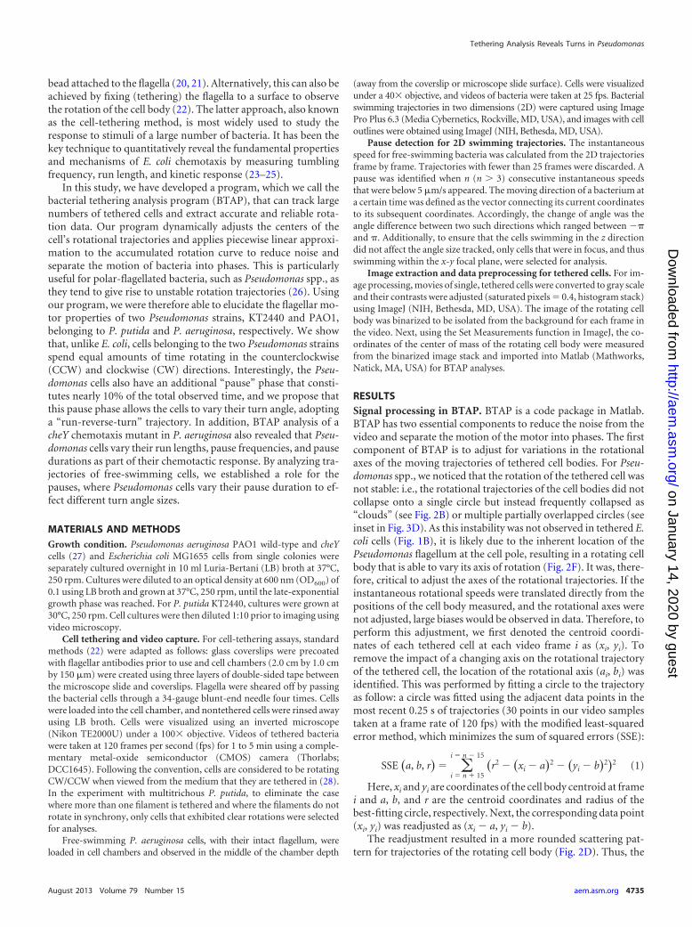
Speed analysis of peritrichous and monotrichous bacteria.To determine the effectiveness of BTAP, we first applied it toperitrichous E. coli, which has been extensively studied using cell-tethering analyses (22, 25, 30). The scattering raw data of E. colirotation follow an exact circle (Fig. 1B), which reflects the stablerotation of tethered E. coli. Because of this stability, BTAP does notdrastically change the measured results (Fig. 1A and C and 1B andD). In order to reduce the noise from the instantaneous rotation
FIG 1 Rotation profile of a sample tethered Escherichia coli cell. (A and C) Instantaneous rotational speed of the tethered E. coli cell before and after BTAPadjustment, respectively. (B and D) The scattering of the centroid positions of the tethered cell before and after the BTAP adjustment, respectively. The crossindicates the assumed rotation axis. Note that the points fall into a smooth circle and do not require refitting. (E) Cumulative rotation of the same cell (solid grayline) and the line fitted by BTAP algorithm (dashed black line). Top bar: labeling of rotational phases, where dark gray represents CW, light gray represents CCW,and white represents the pause phase. CW and CCW phases can be clearly separated. (F) Illustration of a tethered E. coli cell. The cell is tethered parallel to thesurface, and a small perturbation to the cell body (dashed line) does not change its centroid position by much as viewed from the top.
Qian et al.
4736 aem.asm.org Applied and Environmental Microbiology
on January 14, 2020 by guesthttp://aem
.asm.org/
Dow
nloaded from

speed (Fig. 1C), data were transformed into cumulative rotations,where the slopes of the curve indicate the rotational speeds (Fig.1E). This resulted in rotational trajectories with clear rotation di-rections. It is also noteworthy that for each rotation phase (CCWor CW), the corresponding segment in the cumulative curve wasvery close to a straight line, indicating that for each phase thebacterium keeps a relatively constant rotation speed. These resultsagree with previous studies using tethered E. coli strains (31).
Next, we tested BTAP on the monotrichous P. aeruginosa teth-ered cells. Compared to E. coli, the rotation speed translated di-rectly from the uncorrected coordinates appeared to have manypauses and fluctuations (Fig. 2A). The rotational trajectory of P.aeruginosa was also less stable (Fig. 2B). However, BTAP success-fully resolved these. After BTAP correction, the high/low spikeartifacts in the instantaneous speed (Fig. 2A) curve were elimi-nated and the true rotation of the cell was recovered, where coor-dinates fell into a clear circular shape (Fig. 2D), and the rotationspeed also showed less noise (Fig. 2C). The readjustment, there-fore, helped in representing the rotation phases in the cumulativespeed curve precisely (Fig. 2E). This algorithm can, thus, separatethe different rotational phases and calculate the average rotationalspeed for each rotation segment, yielding accurate statistics for theparticular cell (Fig. 2E).
Comparison of BTAP with previous methods. In a previousstudy, the moving average or weighted average method was used
to reduce the noise in data of instantaneous rotational speed (5).To demonstrate the performance of BTAP, we therefore com-pared BTAP to a simple moving average (MA) system, which usesa 30-point moving average window to smooth the instantaneousspeed. We saw that MA yielded results that were very noisy (Fig.3B) compared with the data treated with BTAP (Fig. 3C). Al-though by applying the moving average one can get a smoothspeed curve, it did not correct the systematic error rooted fromthe incorrect positioning of the rotational axis, as many of thespeed time series were near zero (Fig. 3B). It was also difficult todifferentiate the rotational phases (Fig. 3B and D) by usingeither instantaneous rotational speed or cumulative revolutioncurve from the MA system. However, using BTAP, cell phasesthat had been earlier incorrectly recognized to be in the pausephase were now identified to be in the CW rotation (Fig. 3C andE). The cumulative curve, therefore, had an increase in theCW/CCW contrast, allowing BTAP to accurately differentiatethe CW/CCW/pause phases and mark the precise moment atwhich the motor switched its rotation direction (Fig. 3E, topbar). As a result, BTAP measured the tethering data with cor-rection of the positions combined with noise reduction andcould better capture the statistical properties of the P. aerugi-nosa flagellar motor.
BTAP reveals the P. aeruginosa and P. putida flagellar motorto function with an additional pause phase. As the improved
FIG 2 Rotation profile of a sample tethered Pseudomonas aeruginosa cell. (A and C) Instantaneous rotational speed of the tethered P. aeruginosa cell before andafter BTAP adjustment, respectively. (B and D) The scattering of the centroid positions of the tethered cell before and after the BTAP adjustment, respectively.The cross indicates the assumed rotation axis. Note that the points do not fall into a smooth circle in panel B and require refitting. (E) Corresponding cumulativerotations (solid gray line) and the line fitted by BTAP algorithm (dashed black line). Top bar: labeling of rotational phases, where dark gray represents CW, lightgray represents CCW, and white represents the pause phase. (F) Illustration of a tethered P. aeruginosa cell. The cell is tethered at an angle to the surface, and asmall perturbation to the cell body (dashed line) largely changes its centroid positions as viewed from the top.
Tethering Analysis Reveals Turns in Pseudomonas
August 2013 Volume 79 Number 15 aem.asm.org 4737
on January 14, 2020 by guesthttp://aem
.asm.org/
Dow
nloaded from

noise reduction and center adjustment system in BTAP allows foranalyses of polar-flagellated bacteria, we next studied the flagellarmotor function of Pseudomonas spp. in detail. First, in the analy-ses, we tracked the CCW and CW speed distribution of both P.aeruginosa and P. putida and showed that for the two strains, thespeed distributions of both the CCW and CW rotation directionswere symmetric to each other (Fig. 4A and B). In comparison ofthe two, P. aeruginosa had a speed distribution similar to that of P.putida (Fig. 4A and B), where the average speed was not signifi-cantly different either (Fig. 4G). In addition, the interval distribu-tions for CCW/CW/pause followed an exponential distribution(Fig. 4D and E), with mean times of 1.30 s/1.15 s/0.85 s, respec-tively, for P. putida and 1.10 s/1.05 s/0.61 s, respectively, for P.aeruginosa (Fig. 4H). The average durations of CCW/CW/pausefor P. aeruginosa were all lower than those for P. putida (Fig. 4D, E,and H) while the speed distribution (Fig. 4A and B) and averagespeed (Fig. 4G) were comparable, indicating that P. putida exhib-its less switching frequency (Fig. 4I). Notably, the rotation speedsfor both Pseudomonas strains (5 to 6 Hz, Fig. 6G) were also com-parable to those previously reported for R. sphaeroides (�5 Hz)and E. coli (4 to 9 Hz) under similar, unstimulated growth condi-tions (3, 31, 32). The interval distributions for E. coli were alsoexponentially distributed, but the mean time for CCW phase is
considerably longer than that of CW phase (CCW/CW, 2.59s/1.38 s, respectively). This is similar to previous reports for E. coliwhere the CW/CCW rotation interval of distribution was shownto follow an exponential distribution (30, 31).
We observed that P. aeruginosa cells spent nearly equal dura-tions of time during CW (43%) and CCW (49%) rotation and, inaddition, a pause phase which consisted of 8% of the total timetracked (Fig. 5B). During this pause phase, the tethered cell bodystopped rotating for a short time interval before it resumed mo-tion. P. putida also exhibited three phases, CW (47%), CCW(41%), and pause (12%) (Fig. 5A). This rotational pattern is dif-ferent from that exhibited by E. coli cells, where the total timespent in CCW phase in E. coli is 50% longer than that in CW phaseand the total pause phase is short (6%) (31). There are three pos-sible transitions for P. aeruginosa and P. putida: CW↔CCW,CW↔pause, and CCW↔pause. The transition probabilities be-tween phases for the two strains were symmetric. For instance, inthe CW↔CCW transition, the chances of switching from CW toCCW were similar (37% for P. aeruginosa and 32 to 33% for P.putida) to those for switching from CCW to CW. The transitionbetween CW and CCW in E. coli was also shown to be symmetric(data not shown).
FIG 3 Comparison between a simple moving average system (MA) based on instant rotational speed (left panels) and BTAP (right panels). (A) Raw instanta-neous rotational speed signal before MA and BTAP processing. (B) The moving average of the raw signal (without BTAP adjustment) using a window of 30 pointsby the MA system. (C) Raw signal after BTAP correction. (D) Cumulative revolution measured by MA. (E) Cumulative revolution and fitting measured by BTAP.Top bar: labeling of rotational phases where dark gray represents CW, light gray represents CCW, and white represents the pause phase. (Insets in panels D andE) The scattering of the centroid positions of the tethered cell. The cross indicates the assumed rotation axis. BTAP is able to refit the shifting centers of rotationin a polar-flagellated, tethered cell, in order to recover the true rotational behavior of the motor.
Qian et al.
4738 aem.asm.org Applied and Environmental Microbiology
on January 14, 2020 by guesthttp://aem
.asm.org/
Dow
nloaded from

Positive correlation of pause phase with turn angles reveals anovel run-reverse-turn mechanism. It is noteworthy that al-though Pseudomonas cells pause at frequencies similar (0.12 to0.14 Hz versus 0.16 Hz) to those of E. coli, the Pseudomonas cellsspend a higher fraction of time pausing (8 to 12% versus 4.8%).While earlier reports for E. coli may have omitted this phase due tothe shorter duration (33), the pause phase may be a genuine andunique feature of the bacterial flagellar motor, being particularlypronounced for the polar-flagellated Pseudomonas spp. In orderto examine this phase further, we tested a cheY chemotaxis mutantof P. aeruginosa. The frequency of CW/CCW phases was lower
than that for the wild-type P. aeruginosa PAO1 (0.05 s�1 versus 0.4s�1) (Fig. 4I), and as expected, the duration for a CW/CCW phasewas longer (� 8.0 s versus �1.5 s) (Fig. 4E, F, and H). However, incontrast, the duration of pauses did not increase as in the CW/CCW phase but decreased instead (Fig. 4E and F). This suggeststhat the run length of a cell and both the frequency and duration ofa pause are regulated by CheY in P. aeruginosa. In addition, thecheY mutant has a suppressed CW rotation, such that the tetheredcells spend slightly more time in CCW rotation phase (53% versus43%) (Fig. 5C).
We propose that the pause phase allows the cell body to reori-
FIG 4 Speed distributions of Pseudomonas strains. (A to C) Rotational speed distributions of a P. putida (KT2440) strain and P. aeruginosa (PAO1) wild-type andcheY strains, respectively. (D to F) The corresponding cumulative distribution of interval durations from the strains in panels A to C, respectively. Solid linesdenote CCW rotation; dashed lines denote CW rotation; dotted lines denote pauses. Note that the wild-type P. aeruginosa has longer pause durations than doesthe cheY mutant. (G, to I) The average speed, average duration of intervals, and occurrence frequency of phases for the strains in panels A to C, respectively. Errorbars show standard deviations. Dark gray bars represent CW, light gray bars represent CCW, and white bars represent the pause phase. The frequencies forCW/CCW/pause in the cheY mutant were reduced compared to those for the wild type. Unlike the P. aeruginosa wild type, cheY mutants also have increased CCWand CW durations.
Tethering Analysis Reveals Turns in Pseudomonas
August 2013 Volume 79 Number 15 aem.asm.org 4739
on January 14, 2020 by guesthttp://aem
.asm.org/
Dow
nloaded from

ent and swim in a different direction, and we tested this by observ-ing if the pause periods correlated with turn angles in trajectoriesof free-swimming P. aeruginosa cells (Fig. 6A and B). Speed anal-yses revealed that the speed of the free-swimming cells (�5 to 40
�m/s) correlated with previous reports (�40 �m/s) (10), andadditionally, we noticed that cells also exhibited pauses in theirtrajectories. Notably, these cells that pause (Fig. 6C) also oftenturn at an angle (inset). In these examples, when cells are swim-
FIG 5 Time spent on different phases. (A) Times spent in CW/CCW/pause phases for P. putida are 47%/41%/12%, respectively. (B) Times spent in CW/CCW/pause phases for P. aeruginosa PAO1 are 43%/49%/8%, respectively. (C) Times spent in CW/CCW/pause phases for P. aeruginosa cheY mutant are 43%/53%/4%,respectively. The transition probabilities between phases are shown as the arrows indicate. Dark gray represents CW, light gray represents CCW, and whiterepresents the pause phase. Note the drastic decrease in time spent in pause frequency between P. aeruginosa cheY (4%) and wild-type (8%) strains.
FIG 6 Pause durations are positively correlated with turn angles. (A and D) Distribution of the turn angles (change of direction) during pauses for P. aeruginosawild-type and cheY strains, respectively. Angles between �/2 and � are subtracted from � as the change of cell body orientation during a pause followed by areversal. The densities for both strains decrease as turn angles become larger. The pauses are defined when the moving speed of the cell is below 5 �m/s for at least3 consecutive frames (frame rate � 25 fps). (B and E) The average turn angle sizes are positively correlated with pause durations. The line is a linear regressionfitting of the data. The y value of each point is the average of all the turning angles at certain duration (x value). If the number of turn angles at a certain durationmeasured is less than two, the point will not be counted to avoid bias from data scarcity. (C and F) Speed of two sample cells from P. aeruginosa wild-type andcheY strains, respectively. The insets are their corresponding trajectories. The circles mark the section of pauses.
Qian et al.
4740 aem.asm.org Applied and Environmental Microbiology
on January 14, 2020 by guesthttp://aem
.asm.org/
Dow
nloaded from

ming, they often do so in a clear direction, but when cells enter thepause phase, the cell bodies appear to be reorienting their posi-tion, allowing the cells to change direction when they resumeswimming. The level of reorienting can be defined as turn an-gles � angles of moving directions before and after pauses. As abacterium may follow (run) or change (reverse) its moving statusafter a turn, the according turn angle by definition is � and � � �,respectively; the turn angles from population analyses of the tra-jectories are folded into the range of (0, �/2) (Fig. 6A). The aver-age turn angle sizes are positively correlated with pause durations(Fig. 6B), and we hypothesize that this positive relation is due tothe rotational diffusion. This positive correlation was also ob-served in cheY cells (Fig. 6D and E), although the overall pausedurations were not as long as those in wild-type cells (Fig. 6B andE). Notably, the shorter pause durations in cheY free-swimmingcells also correlated with the shorter pause durations of tetheredcells than of wild-type cells.
DISCUSSION
Peritrichous bacteria such as E. coli have multiple flagella aroundtheir cell surface, while monotrichous bacteria such as P. aerugi-nosa have only one flagellum located at the pole. Therefore, incell-tethering experiments, when the flagella of peritrichousbacteria are sheared off and one flagellum is tethered to thesurface, the tethered flagellum stub is usually at the side of thecell body, such that the cell body is nearly parallel to the teth-ered surface, allowing a full rotation to be observed under themicroscope (Fig. 1F). However, when polar-flagellated bacteriasuch as P. aeruginosa are tethered to the surface, the cell body isusually not parallel to the tethered surface (Fig. 2F), allowingthe cell body to change its orientation easily because of theflexibility at the flagellar hook (34). This causes difficulty in theimage analysis process, as the observed tethered cells no longerrotate in a circular trajectory but often shift around, giving riseto a noisy rotational signal.
Our new program, therefore, has several advantages (1). It re-duces the position noise in the rotational tethered cell. Traditionalmethods translate only the centroid coordinate directly into therotational phases, a process which is prone to error because, whenthe actual rotation axis is slightly deviated from the presumed axis,a rotating rod with constant radial speed will show a changinginstantaneous rotational speed. By correcting the actual rotationalaxis, we are able to isolate the rotational data that best reflect theactual rotation of the motor from the experimental video (2). Bymeasuring the accumulative revolution instead of instantaneousrotation, we can further reduce the rotational noise from thepiecewise linear fitting. The previous method uses instantaneousspeed to measure the rotation i at frame i
�i ��i � �i � 1
ti � ti � 1(2)
where �i is rotational angle at frame i and ti is the correspond-ing time.
However, this approach results in a very noisy speed curve witha large variance. This is because tethered cells are not always ro-tating smoothly but exhibit fluctuations in speed within each ro-tation. In addition, the tethered cells in the fluidic environmentare affected by other surrounding factors, such as hydrodynamiceffects or Brownian force, causing the position of the cell body tohave a large variance (35). The problem is more significant when
the acquisition frame rate is high, such that an incidental largeangle change �i � �i � 1 between two frames can produce a spike inthe instantaneous speed curve. This can be partially alleviated byintroducing a (weighted) moving average window to smooth thedata, but the size of the window is subjective and can have a hugeimpact on the result. Conversely, our approach uses the cumula-tive revolution to fit the curve with multiple linear segments. Al-though the instantaneous speed curve may be noisy and havemany spikes, because the time intervals between two frames aresmall, the impact of such spikes on the cumulative curve would besmall and thus not affect the rotation trend (the slope of thecurve). These noises can be easily removed by a linear fitting in thecumulative curve (3). Through extracting the turning points onthe cumulative curve, we could separate clearly the rotation phasesof the tethered cell into clockwise, counterclockwise, and pause.This is difficult to achieve in the instantaneous curve because themoving average window will blur the boundary between differentphases. As the smoothing result improves (i.e., the the windowsize increases), the ability for phase separation, in turn, weakens.However, using our approach, we were able to achieve both noisereduction and phase separation without sacrificing the accuracy ofeither.
As the cell-tethering experiment is one of the key techniques inquantifying the motor properties and its response in the chemo-tactic environment, it is important to enhance the accuracy of thedata acquisition and analysis to reflect the change of motor phasesin chemotaxis. Our new program, therefore, helps image process-ing and data analysis in research on monotrichous bacteria andgives credible results for subsequent studies.
We used our program to distinguish novel properties of theflagellar motor in Pseudomonas species, where we were able tostudy its flagellar rotation in a high-throughput manner for thefirst time. First, the Pseudomonas flagellar motor spends equaltime in both CCW and CW rotations, which corroborates earlierreports for free-swimming cells, where trajectories adopt a run-and-reverse strategy (12). Additionally, speeds in the two direc-tions are similar, suggesting that the flagellar motor is symmetric.Notably, we observed an additional pause phase, into which about12 to 23% of cells in motion (either CCW or CW) will choose totransit. As there was a positive correlation between pause durationand turn angle sizes, our findings indicate that Pseudomonas canvary its pause duration in order to turn at different angles, result-ing in a run-reverse-turn trajectory. This novel mechanism, there-fore, allows the cell to swim and explore spaces more efficientlythan would a typical run-and-reverse trajectory for polar-flagel-lated bacteria.
In polar-flagellated bacteria, cells have a limited degree of free-dom in motility where they can swim only forward and backward.It appears now that there are diverse ways in which such types ofbacterial cells can compensate for their limited, bidirectionalmovement. In the case of R. sphaeroides, cells adopt a run-and-stop motility (6) with variable speed (5), whereas in the sodium-driven V. alginolyticus, cells adopt a three-step run-reverse-flickchemotactic response (3, 4) instead. Here, in the case of Pseu-domonas spp., we observe a run-reverse-turn trajectory for bothmonotrichous and multitrichous Pseudomonas cells. While earlierreports made brief references to these turns in free-swimmingtrajectories (12, 26), we have now shown that the additional pausephase in the flagellar motors allows the cells to turn at larger an-gles.
Tethering Analysis Reveals Turns in Pseudomonas
August 2013 Volume 79 Number 15 aem.asm.org 4741
on January 14, 2020 by guesthttp://aem
.asm.org/
Dow
nloaded from
In addition, it is known that chemotaxis mutants of P. aerugi-nosa are motile but rarely change their swimming directions (36,37). The question, therefore, arises whether the Pseudomonas che-motactic mechanism is similar to that of the peritrichous E. coli orwhether the Pseudomonas motor employs a different response ingeneral. We were able to elucidate this using BTAP, a chemotaxismutant of P. aeruginosa, which has reduced CCW and CW fre-quency, and the durations in both CCW and CW directions areincreased. Interestingly, there is a reduction in both the frequencyand the duration of pauses as well. This suggests that the cells“turn” at a smaller angle, therefore, giving rise to trajectories thattend to be straight. Taken together, the Pseudomonas motor un-dergoes chemotaxis by varying its pause and switch frequenciesand durations, resulting in cells that have longer runs and fewerturns. In comparison to other species, it is known that E. coliincreases its run length through a decrease in switch frequencyresulting in an increase in CCW rotation (28). In the case of P.aeruginosa, we show that unlike E. coli, run length is increasedthrough a decrease in pause frequency and duration and an in-crease in both CW and CCW durations. In the case of the three-step run-reverse-flick V. alginolyticus, cells have decreased flickingfrequency while retaining their turn angle sizes (4). Notably, whileV. alginolyticus uses a flick to change its turn angle, we show herethat P. aeruginosa adopts a pause mechanism to turn, and theseturn angles are decreased as part of the chemotactic response. Inthe case of R. sphaeroides, a decrease in stop frequency was ob-served (38), which was a response similar to that observed in thecheY P. aeruginosa mutant. Therefore, in comparison to thesethree species, we show that while the details of the control of theflagellar motor differ, the outcomes of longer run lengths andreduced turn (or tumbling) frequency remain broadly conservedin P. aeruginosa. The Pseudomonas motor, therefore, adopts acombination of different properties in its chemotactic response,once again revealing the complexity of the Pseudomonas chemo-sensory system (9).
ACKNOWLEDGMENTS
We thank Linda Carter for the P. aeruginosa strain; Liang Yang for the P.aeruginosa cheY strain; Fook Chiong Cheong and Chwee Teck Lim for theuse of the microscopy facility; and Sungsu Park, Linda Kenney, Yan Jie,and Boon Chuan Low for stimulating discussions.
This work was supported by the Seed Fund of the Mechanobiology Insti-tute, Research Centre of Excellence financed by the National Research Foun-dation of Singapore and the Singapore Ministry of Education.
REFERENCES1. Berg HC. 1973. Bacteria swim by rotating their flagellar filaments. Nature
245:380 –382.2. Turner L, Ryu WS, Berg HC. 2000. Real-time imaging of fluorescent
flagellar filaments. J. Bacteriol. 182:2793–2801.3. Kojadinovic M, Sirinelli A, Wadhams GH, Armitage JP. 2011. New
motion analysis system for characterization of the chemosensory responsekinetics of Rhodobacter sphaeroides under different growth conditions.Appl. Environ. Microbiol. 77:4082– 4088.
4. Xie L, Altindal T, Chattopadhyay S, Wu XL. 2011. Bacterial flagellum asa propeller and as a rudder for efficient chemotaxis. Proc. Natl. Acad. Sci.U. S. A. 108:2246 –2251.
5. Packer HL, Lawther H, Armitage JP. 1997. The Rhodobacter sphaeroidesflagellar motor is a variable-speed rotor. FEBS Lett. 409:37– 40.
6. Poole P, Sinclair DR, Armitage JP. 1988. Real time computer track-ing of free-swimming and tethered rotating cells. Anal. Biochem. 175:52–58.
7. Chen S, Beeby M, Murphy GE, Leadbetter JR, Hendrixson DR,
Briegel A, Li Z, Shi J, Tocheva EI, Müller A, Dobro MJ, Jensen GJ.2011. Structural diversity of bacterial flagellar motors. EMBO. J. 30:2972–2981.
8. Porter SL, Wadhams GH, Armitage JP. 2011. Signal processing in com-plex chemotaxis pathways. Nat. Rev. Microbiol. 9:153–165.
9. Kato J, Kim HE, Takiguchi N, Kuroda A, Ohtake H. 2008. Pseudomonasaeruginosa as a model microorganism for investigation of chemotacticbehaviors in ecosystem. J. Biosci. Bioeng. 106:1–7.
10. Doyle TB, Hawkins AC, McCarter LL. 2004. The complex flagellartorque generator of Pseudomonas aeruginosa. J. Bacteriol. 186:6341–6350.
11. Kanda E, Tatsuta T, Suzuki T, Taguchi F, Naito K, Inagaki Y, ToyodaK, Shiraishi T, Ichinose Y. 2011. Two flagellar stators and their roles inmotility and virulence in Pseudomonas syringae pv. tabaci 6605. Mol.Genet. Genomics 285:163–174.
12. Taylor BL, Koshland DE. 1974. Reversal of flagellar rotation inmonotrichous and peritrichous bacteria: generation of changes in direc-tion. J. Bacteriol. 119:640 – 642.
13. Silby MW, Winstanley C, Godfrey SA, Levy SB, Jackson RW. 2011.Pseudomonas genomes: diverse and adaptable. FEMS Microbiol. Rev. 35:652– 680.
14. Harwood CS, Fosnaugh K, Dispensa M. 1989. Flagellation of Pseu-domonas putida and analysis of its motile behavior. J. Bacteriol. 171:4063– 4066.
15. Macnab RM. 1976. Examination of bacterial flagellation by dark-fieldmicroscopy. J. Clin. Microbiol. 4:258 –265.
16. Adler J. 1973. A method for measuring chemotaxis and use of the methodto determine optimum conditions for chemotaxis by Escherichia coli. J.Gen. Microbiol. 74:77–91.
17. Adler J. 1966. Chemotaxis in bacteria. Science 153:708 –716.18. Berg HC, Brown DA. 1972. Chemotaxis in Escherichia coli analysed by
three-dimensional tracking. Nature 239:500 –504.19. Wu M, Roberts JW, Kim S, Koch DL, DeLisa MP. 2006. Collective
bacterial dynamics revealed using a three-dimensional population-scaledefocused particle tracking technique. Appl. Environ. Microbiol. 72:4987– 4994.
20. Yuan J, Berg HC. 2008. Resurrection of the flagellar rotary motor nearzero load. Proc. Natl. Acad. Sci. U. S. A. 105:1182–1185.
21. Scharf B, Fahrner KA, Turner L, Berg HC. 1998. Control of direction offlagellar rotation in bacterial chemotaxis. Proc. Natl. Acad. Sci. U. S. A.95:201–206.
22. Silverman M, Simon MI. 1974. Flagellar rotation and the mechanism ofbacterial motility. Nature 249:73–74.
23. Berg HC, Tedesco P. 1975. Transient response to chemotactic stimuli inEscherichia coli. Proc. Natl. Acad. Sci. U. S. A. 72:3235–3239.
24. Block SM, Segall JE, Berg HC. 1982. Impulse responses in bacterialchemotaxis. Cell 31:215–226.
25. Block SM, Segall JE, Berg HC. 1983. Adaptation kinetics in bacterialchemotaxis. J. Bacteriol. 154:312–323.
26. Ping L, Birkenbeil J, Monajembashi S. 24 January 2013. Swimmingbehavior of the monotrichous bacterium Pseudomonas fluorescensSBW25. FEMS Microbiol. Ecol. [Epub ahead of print.] doi:10.1111/1574-6941.12076.
27. Barken KB, Pamp SJ, Yang L, Gjermansen M, Bertrand JJ, Klausen M,Givskov M, Whitchurch CB, Engel JN, Tolker-Nielsen T. 2008. Roles oftype IV pili, flagellum-mediated motility and extracellular DNA in theformation of mature multicellular structures in Pseudomonas aeruginosabiofilms. Environ. Microbiol. 10:2331–2343.
28. Berg HC. 2003. The bacterial rotary motor, p 143–202. In Hackney DD,Tamanoi F (ed), The enzymes, 3rd ed, vol 23. Energy coupling and mo-lecular motors. Academic Press, New York, NY.
29. Keogh E, Chu S, Hart D, Pazzani M. 2003. Segmenting time series: asurvey and novel approach, p 1–21. In Last M, Kandel A, Bunke H (ed),Data mining in time series databases. World Scientific Publishing Com-pany, Hackensack, NJ.
30. Segall JE, Block SM, Berg HC. 1986. Temporal comparisons in bacterialchemotaxis. Proc. Natl. Acad. Sci. U. S. A. 83:8987– 8991.
31. Eisenbach M, Wolf A, Welch M, Caplan SR, Lapidus IR, Macnab RM,Aloni H, Asher O. 1990. Pausing, switching and speed fluctuation of thebacterial flagellar motor and their relation to motility and chemotaxis. J.Mol. Biol. 211:551–563.
32. Berg HC. 2003. The rotary motor of bacterial flagella. Biochemistry 72:19 –54.
Qian et al.
4742 aem.asm.org Applied and Environmental Microbiology
on January 14, 2020 by guesthttp://aem
.asm.org/
Dow
nloaded from

33. Kuo SC, Koshland DE. 1989. Multiple kinetic states for the flagellarmotor switch. J. Bacteriol. 171:6279 – 6287.
34. Hashimoto M, Mashimo T, Hirano T, Yamaguchi S, Aizawa S. 2008.Functional roles of the hook in a rotating tethered cell. J. Mol. Biol. 375:367–375.
35. Berg HC, Turner L. 1993. Torque generated by the flagellar motor ofEscherichia coli. Biophys. J. 65:2201–2216.
36. Masduki A, Nakamura J, Ohga T, Umezaki R, Kato J, Ohtake H. 1995.
Isolation and characterization of chemotaxis mutants and genes of Pseu-domonas aeruginosa. J. Bacteriol. 177:948 –952.
37. Taguchi K, Fukutomi H, Kuroda A, Kato J, Ohtake H. 1997. Geneticidentification of chemotactic transducers for amino acids in Pseudomonasaeruginosa. Microbiology 143:3223–3229.
38. Packer HL, Gauden DE, Armitage JP. 1996. The behavioural response ofanaerobic Rhodobacter sphaeroides to temporal stimuli. Microbiology 142:593–599.
Tethering Analysis Reveals Turns in Pseudomonas
August 2013 Volume 79 Number 15 aem.asm.org 4743
on January 14, 2020 by guesthttp://aem
.asm.org/
Dow
nloaded from