Embed Size (px)
Citation preview
9
Personal non-commercial use only EJENTAS copyright © 2019. All rights reserved DOI: 10.21608/ejentas.2018.4422.1026
Original Article
Bacterial Profile and Antimicrobial Resistance Patterns In Patients With Upper Respiratory Tract Infections In Debre Berhan Referral Hospital, Ethiopia
Tizazu Z. Zelelie, Yosef T. Mekonnen, T. Asfaw Abebe
Department of Medicine, Debre Berhan University, Ethiopia
ABSTRACTBackground: Upper respiratory tract infections have been regarded as the most frequent illnesses affecting people worldwide. Data about bacterial profile and current antibiotics resistance status for such infections is scarce in Ethiopia.Objective: This study was done to determine bacterial profile and their antimicrobial resistance patterns in patients with upper respiratory tract infections. Methods: A cross sectional study design was used and was conducted in Debre Berhan, Ethiopia from October 2016 to September 2017. Patients with upper respiratory infections presented to outpatient department were enrolled for the study. Socio-demographic and clinical data were collected using standardize questionnaires. Clinical samples of throat swabs were collected and transported to the microbiology laboratory for processing. Data was analyzed using SPSS version 20. Results: A total of 109 patients, almost equal number of male (49.5%) and female (50.5%) participants were involved in the study. Majority (62.4%) of the participants were with age of below 5 years old. The culture positivity was 36.7%. Among all bacterial isolates (n=40) the predominant isolate was Streptococcus pyogenes (42.5%), and followed by Streptococcus pneumoniae (22.5%), Staphylococcus aureus (15%), Haemophilus influenzae (12.5%), and Klebsiella pneumoniae (7.5%). Overall antimicrobial resistant pattern seen in all bacterial isolates ranges from 0 % to 66.6%. High resistances in all isolates have been seen. Cough and watery eye were found as predictor variables. Conclusion: High number of bacterial isolates and emergence of resistant strains have been found. We recommend to taken this in accounting during medical decision, guideline development and designing interventional strategies.Key Words: Antimicrobial resistance, bacterial profile, ethiopia, upper respiratory tract infection.Received: 12 July 2018, Accepted: 31 October 2018Corresponding Author: Tizazu Z. Zelelie, MD, Department of Medicine, Medical Microbiology Unit, Debre Berhan University, Ethiopia Tel.: +251 912 372 837, E-mail: [email protected]
ISSN: 2090-0740, March 2019 Vol.20, No.1
INTRODUCTION
Acute respiratory infections (ARI) are a major burden to child health and the leading cause of death in developing countries[1]. It is contagious which remain for few hours to 2-3 days of exposure[2]. Acute upper respiratory tract infections (URTIs) is an ARI involving the nose, paranasal sinuses, pharynx, larynx, trachea, and bronchi[3] and accounts 87.5% of ARI episodes occurred globally[4]. It can be characterized by various signs, symptoms[2] and group of disorders[5]. The prevalence of URTIs was estimated at 18.8 billion cases in 2013 with an estimated 4 million deaths[6].
And URTIs may be caused by viruses such as Rhinovirus, Parainfluenzae virus, Coronavirus, Adenovirus, Respiratory syncytial virus, Coxsackievirus, Epstein-Barr virus, and Influenza virus and bacteria such as Streptococcus pyogene, Corynebacterium diphtheriae, Neisseria gonorrhoeae, Chlamydia pneumoniae, Mycoplasma pneumoniae, Streptococcus pneumoniae,
Haemophilus influenzae, Bordetella pertussis, and Moraxella catarrhalis[3,5,7-10]. Different studies done elsewhere in the world showed varied bacterial profile of upper respiratory tract infections and antimicrobial resistance patterns[11-17]. In Ethiopia two old studies[18,19] showed the occurrence and the prevalence of bacterial isolates such as H. influenzae, Moraxella catarrhalis, and S. pneumoniae. Moreover a study that had done in other area of the country showed 11.3% prevalence of group A streptococcus among children with pharyngitis[20]. It is essential to differentiate viral and bacterial pictures in a particular setting to establish a satisfactory treatment. The frequent use of antibiotics, the easy dissemination of bacterial agents, and lack of empirical evidence may predispose to development of antimicrobial resistance. On the other hand selection of empirical treatment has been compromised due to the increase rates of resistance of bacterial isolates[21]. Like for example resistance levels for commonly used drugs of upper respiratory infections such as co-trimoxazole (84%), penicillin (52%), and ampicillin (25%) have been reported[22,23]. In addition because of
10
BACTERIAL PROFILE AND ANTIMICROBIAL RESISTANCE PATTERNS
overuse and misuse of antibiotics for the treatment of respiratory tract infection[24] there is high emergence of resistance of bacteria against different antibiotics[25].
Evidence on bacterial profile and their antimicrobial resistance patterns in upper respiratory tract infection is scarce in Ethiopia. Providing such evidence will have significance in improving patient treatment, developing different guidelines, designing interventional strategies and evidence based utilization of resource in the country in particular in the study area. This study was conducted to determine profiles of bacterial isolates in patients with upper respiratory tract infections and current sensitivity pattern of the isolates in Debre Berhan referral hospital, Ethiopia.
MATERIAL AND METHODS
Study setting
A health facility based cross sectional study design was conducted. The study was conducted in Debere Berhan referral hospital from October 2016 to September 2017. Debre Berhan is located in the Semen Shewa Zone of the Amhara Region, about 130 kilometers North East of the capital city Addis Ababa, Ethiopia. Patients with URTI presented to outpatient department (both adult and pediatric) of Debre Berhan referral hospital were enrolled for the study. All age groups of patients presented to outpatient departments due to URTI during data collection time as inclusion criteria and patients with history of antimicrobial therapy (treated in the last three months) as exclusion criteria were used. Convenient sampling technique was used for enrolment of study participants. Using prevalence of ARI of 92.3% of the study done in Gondar of Ethiopia [19], margin of error =5%, and 95% confidence level, a total of 109 sample size was determined.
Data collection
Socio-demographic and clinical data were collected using standardize questionnaire. The patients were fully examined and managed by the physicians and the data were transferred onto the standardized questionnaire.
Sample collection and transportation
Throat swabs were collected using sterile cotton swabs on wooden applicator sticks. The swabs collected was placed immediately in amies transport medium with charcoal to keep the sample stable for a prolonged period and transported to Debre Berhan University microbiology laboratory for laboratory analysis.
Culture
The clinical samples were inoculated on blood and chocolate agar. The inoculated plates were incubated at
37°C for 24-48 hour aerobically, and for chocolate agar the plates were incubated for 24-48h at 37°C in an atmosphere CO2 using candle jar. Microscopic and macroscopic examinations of the growing colonies on the plates were examined. Individual colonies were then sub cultured on a blood agar and chocolate agar for pure colony. Preliminary identification was performed based on gram staining, morphology and colony characteristics on selective and differential media[26].
Biochemical tests
Hemolytic characteristics of the bacterial isolates on blood agar, catalase test, coagulase test, Bacitracin test, Optochin test, and bile solubility test were used for identification of gram positive bacterial isolates. Similarly for identification of gram negative bacterial isolates Triple Sugar Iron Agar, Indole test, Citrate utilization, Urease test and Kovac’s oxidase test were used. And also satellitism test for identification of H. influenzae was used.
Antimicrobial susceptibility test
Antimicrobial susceptibility test was done using disk diffusion according to the guidelines of Clinical and Laboratory Standards Institute, CLSI[27] using antibiotics which included Ampicillin (10 μg), Amoxycillin (10 μg), Chloramphenicol (30 μg), Tetracycline (30μg), Gentamicin (10μg), Ciprofloxacin (5 μg), Erythromycin (15 μg), Trimethoprim+Sulphamethazole (1 μg) and ceftriaxone (30µg). A standardized inoculum of a bacterial isolate was swabbed onto the surface of a Mueller Hinton agar (MHA) plate, for Streptococcus pneumoniae and Streptococcus pyogenes MHA with 5% sheep blood was used instead. Filter paper disks impregnated with a standard amount of antimicrobial agents was placed on the agar, and then incubated overnight. The diameter of the zone of inhibition was measured around each disk using ruler. The zone sizes were compared to tables in the disc-diffusion tests. Antimicrobial susceptibility test for Haemophilus influenzae was not done due to shortage of Haemophilus Test Medium.
Quality assurance
Standard operating procedure (SOP) was developed and during procedures it was strictly followed. Sterility test for prepared culture media was done. Control organisms Streptococcus pneumonia (ATCC 49619), and Staphylococcus aureus (ATCC 25923) in each procedures were used.
Data analysis
After proper labeling and coding of the variables, the data were entered into SPSS version 20 for analysis. Chi-square test was used for testing statistical significance and results with less than 0.05 P-value was considered
11
Zelelie et al.
as significant. Logistic regression analysis was made for some variable to determine predictor variables.
RESULTS
Socio-demographic characteristics of study participants
A total of 109 patients with suspected of URTI were included for the analysis. Almost equal number of male (49.5%) and female (50.5%) participants were involved. Majority of the participants were with age of below 5 years old. The mean and median of the participant’s age were 8.3 and 3.0 respectively. About 72.5% of the participants were come from Debre Berhan town and the rest were from outside of the town (Sheno, Shewa Robbit, Chacha). Forty one (37.6%) of the participants were illiterate and the rest 18.3%[20], 17.4%[19] and 26.6%[29] were completed their primary, secondary and college level of their education. In (Table 1) other socio-demographic data have been presented.
Table 1: Socio-demographic characteristics of study participants from Oct 2016 to Sept 2017
S.N Variables Frequency Percentage
1 Sex Male 54 49.5
Female 55 50.5
2 Age 0-5 68 62.4
6-15 17 15.6
>15 24 22.0
3 Residents DebreBerhan 79 72.5
Othersa 30 27.5
4 Marital status Married 62 56.9
Unmarried 39 35.8
Divorced 8 7.3
5 Religion Orthodox 79 72.5
Muslim 23 21.1
Protestant 7 6.4
6 Ethnic group Amhara 81 74.3
Oromo 19 17.4
Tigray 9 8.3a outside Debre Berhan town
Clinical characteristics of study participants
Different clinical signs and symptoms of the patients were recorded. The primary signs and symptoms were fever (95%), cough (56%), dysphagia (51.4%), headache (44%) and vomiting (40.4%). Duration of symptoms was seen in three categories: before three days, three days to one week, and more than one week. And forty four (40.4%), forty nine (45%), and sixteen (14.7%) of the clinical symptoms were seen before three days, whining three days to one week
and more than one week respectively. Majority (85.4%) of the clinical symptoms were seen within one week. Only in twelve (11%) of the participants similar illness in the family have been reported.
Bacterial isolates
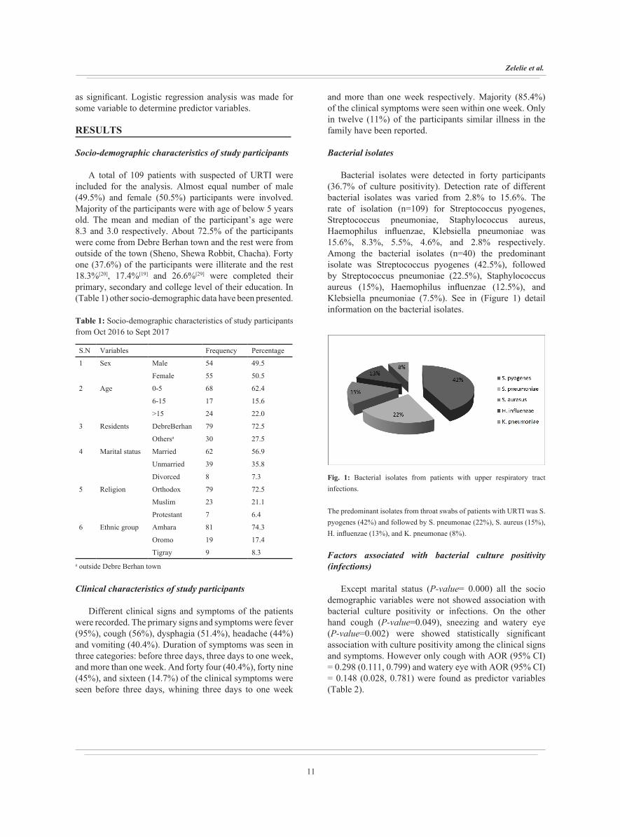
Bacterial isolates were detected in forty participants (36.7% of culture positivity). Detection rate of different bacterial isolates was varied from 2.8% to 15.6%. The rate of isolation (n=109) for Streptococcus pyogenes, Streptococcus pneumoniae, Staphylococcus aureus, Haemophilus influenzae, Klebsiella pneumoniae was 15.6%, 8.3%, 5.5%, 4.6%, and 2.8% respectively. Among the bacterial isolates (n=40) the predominant isolate was Streptococcus pyogenes (42.5%), followed by Streptococcus pneumoniae (22.5%), Staphylococcus aureus (15%), Haemophilus influenzae (12.5%), and Klebsiella pneumoniae (7.5%). See in (Figure 1) detail information on the bacterial isolates.
Fig. 1: Bacterial isolates from patients with upper respiratory tract infections.
The predominant isolates from throat swabs of patients with URTI was S. pyogenes (42%) and followed by S. pneumonae (22%), S. aureus (15%), H. influenzae (13%), and K. pneumonae (8%).
Factors associated with bacterial culture positivity (infections)
Except marital status (P-value= 0.000) all the socio demographic variables were not showed association with bacterial culture positivity or infections. On the other hand cough (P-value=0.049), sneezing and watery eye (P-value=0.002) were showed statistically significant association with culture positivity among the clinical signs and symptoms. However only cough with AOR (95% CI) = 0.298 (0.111, 0.799) and watery eye with AOR (95% CI) = 0.148 (0.028, 0.781) were found as predictor variables (Table 2).
12
BACTERIAL PROFILE AND ANTIMICROBIAL RESISTANCE PATTERNS
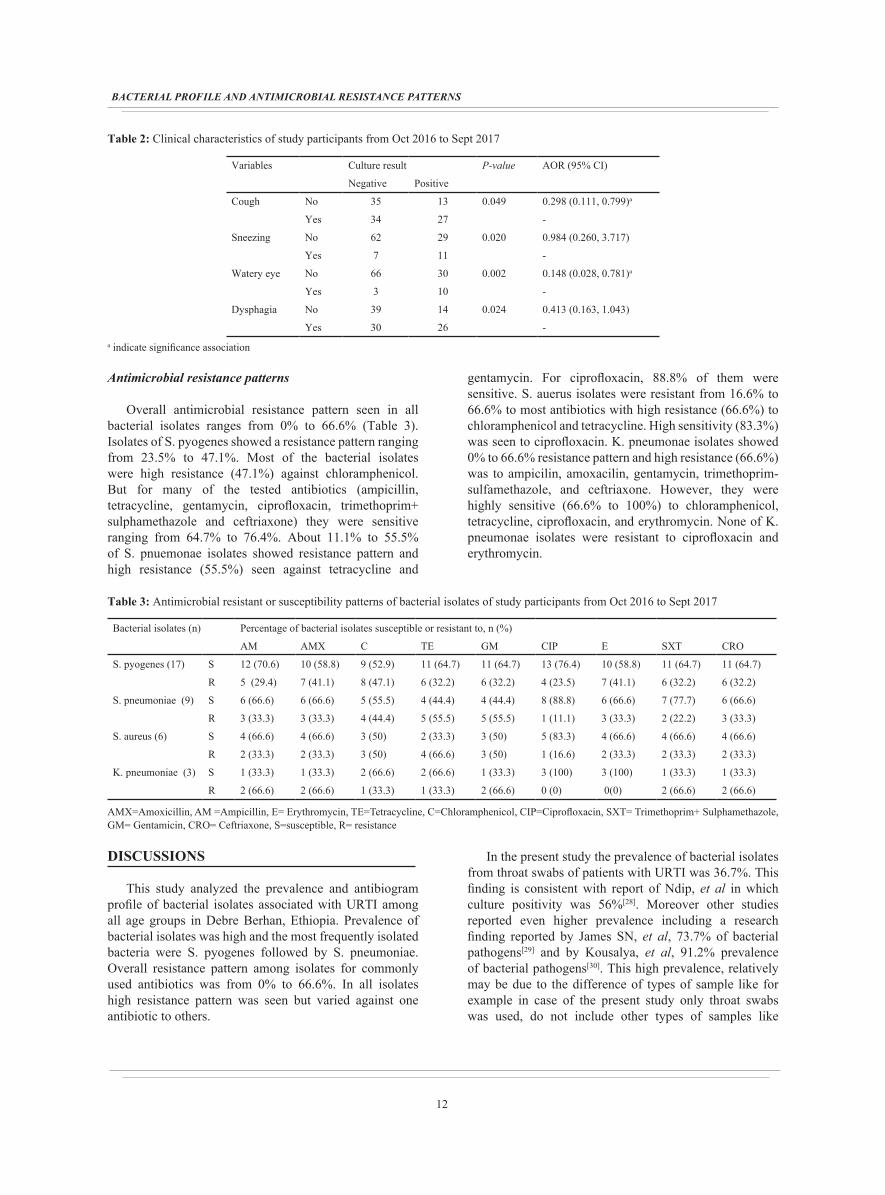
Antimicrobial resistance patterns
Overall antimicrobial resistance pattern seen in all bacterial isolates ranges from 0% to 66.6% (Table 3). Isolates of S. pyogenes showed a resistance pattern ranging from 23.5% to 47.1%. Most of the bacterial isolates were high resistance (47.1%) against chloramphenicol. But for many of the tested antibiotics (ampicillin, tetracycline, gentamycin, ciprofloxacin, trimethoprim+ sulphamethazole and ceftriaxone) they were sensitive ranging from 64.7% to 76.4%. About 11.1% to 55.5% of S. pnuemonae isolates showed resistance pattern and high resistance (55.5%) seen against tetracycline and
gentamycin. For ciprofloxacin, 88.8% of them were sensitive. S. auerus isolates were resistant from 16.6% to 66.6% to most antibiotics with high resistance (66.6%) to chloramphenicol and tetracycline. High sensitivity (83.3%) was seen to ciprofloxacin. K. pneumonae isolates showed 0% to 66.6% resistance pattern and high resistance (66.6%) was to ampicilin, amoxacilin, gentamycin, trimethoprim-sulfamethazole, and ceftriaxone. However, they were highly sensitive (66.6% to 100%) to chloramphenicol, tetracycline, ciprofloxacin, and erythromycin. None of K. pneumonae isolates were resistant to ciprofloxacin and erythromycin.
DISCUSSIONS
This study analyzed the prevalence and antibiogram profile of bacterial isolates associated with URTI among all age groups in Debre Berhan, Ethiopia. Prevalence of bacterial isolates was high and the most frequently isolated bacteria were S. pyogenes followed by S. pneumoniae. Overall resistance pattern among isolates for commonly used antibiotics was from 0% to 66.6%. In all isolates high resistance pattern was seen but varied against one antibiotic to others.
In the present study the prevalence of bacterial isolates from throat swabs of patients with URTI was 36.7%. This finding is consistent with report of Ndip, et al in which culture positivity was 56%[28]. Moreover other studies reported even higher prevalence including a research finding reported by James SN, et al, 73.7% of bacterial pathogens[29] and by Kousalya, et al, 91.2% prevalence of bacterial pathogens[30]. This high prevalence, relatively may be due to the difference of types of sample like for example in case of the present study only throat swabs was used, do not include other types of samples like
Table 2: Clinical characteristics of study participants from Oct 2016 to Sept 2017
Variables Culture result P-value AOR (95% CI)
Negative Positive
Cough No 35 13 0.049 0.298 (0.111, 0.799)a
Yes 34 27 -
Sneezing No 62 29 0.020 0.984 (0.260, 3.717)
Yes 7 11 -
Watery eye No 66 30 0.002 0.148 (0.028, 0.781)a
Yes 3 10 -
Dysphagia No 39 14 0.024 0.413 (0.163, 1.043)
Yes 30 26 -a indicate significance association
Table 3: Antimicrobial resistant or susceptibility patterns of bacterial isolates of study participants from Oct 2016 to Sept 2017
Bacterial isolates (n) Percentage of bacterial isolates susceptible or resistant to, n (%)
AM AMX C TE GM CIP E SXT CRO
S. pyogenes (17) S 12 (70.6) 10 (58.8) 9 (52.9) 11 (64.7) 11 (64.7) 13 (76.4) 10 (58.8) 11 (64.7) 11 (64.7)
R 5 (29.4) 7 (41.1) 8 (47.1) 6 (32.2) 6 (32.2) 4 (23.5) 7 (41.1) 6 (32.2) 6 (32.2)
S. pneumoniae (9) S 6 (66.6) 6 (66.6) 5 (55.5) 4 (44.4) 4 (44.4) 8 (88.8) 6 (66.6) 7 (77.7) 6 (66.6)
R 3 (33.3) 3 (33.3) 4 (44.4) 5 (55.5) 5 (55.5) 1 (11.1) 3 (33.3) 2 (22.2) 3 (33.3)
S. aureus (6) S 4 (66.6) 4 (66.6) 3 (50) 2 (33.3) 3 (50) 5 (83.3) 4 (66.6) 4 (66.6) 4 (66.6)
R 2 (33.3) 2 (33.3) 3 (50) 4 (66.6) 3 (50) 1 (16.6) 2 (33.3) 2 (33.3) 2 (33.3)
K. pneumoniae (3) S 1 (33.3) 1 (33.3) 2 (66.6) 2 (66.6) 1 (33.3) 3 (100) 3 (100) 1 (33.3) 1 (33.3)
R 2 (66.6) 2 (66.6) 1 (33.3) 1 (33.3) 2 (66.6) 0 (0) 0(0) 2 (66.6) 2 (66.6)
AMX=Amoxicillin, AM =Ampicillin, E= Erythromycin, TE=Tetracycline, C=Chloramphenicol, CIP=Ciprofloxacin, SXT= Trimethoprim+ Sulphamethazole, GM= Gentamicin, CRO= Ceftriaxone, S=susceptible, R= resistance
13
Zelelie et al.
nasopharyngeal swabs which was included in these studies. The prevalence of S. pyogenes in this study was 15.6% which agree with studies done elsewhere including in Ethiopia. S. pyogenes prevalence of 16.3% in Tanzania, 14.7% in America, 8.7% in Jordan[1, 14, 16] and 11.3% and 0.8% in Ethiopia[19, 20] have been reported. On the contrary Kousalya, et al reported 22.8% of S. pyogenes (30) which is higher compared to the present finding. This discrepancy may be because of difference in study population and true difference of the etiological profile in the area. The S. pneumoniae prevalence in the present study (8.3%) agreed with other reports such as 15% prevalence by Ndip, and 13% prevalence by Wang, et al[28]. But James SN, et al[29] and El-Mahmood, et al[18] reported high prevalence of S. pneumoniae, 35% and 57%, respectively which are inconsistent with the present finding. This difference may be due to utilization of different diagnostic method, study period, and true difference of the etiological profile.
Low isolation of S. aureus have been reported in a study done in India (4.9%) and in Camerron (10%)[28, 30]. However James et al reported 55.3% prevalence of S. aureus in Tanzania[29] which disagreed with the present study. This inconsistency may be due to difference in including nasopharyngeal sampling (in addition to throat swabs) and also it may be due to difference in geographical factors. The low prevalence of H. influenzae in the present study agreed with the report of 1.32% prevalence by Kousalya et al[30]. However higher prevalence of H. influenza in other area have been reported such as 17.1% by James, et al[29], 20% by Ndip, et al[28] and 23% by Wang, et al[31]. Similarly low and high (14.9%) prevalence of K. pneumnae have been reported[28, 30].
Bacterial isolates showed varied antimicrobial resistance pattern against different antibiotics. S. pyogens showed resistant against all antibiotics but highly to amoxicillin and chloramphenicol. In other studies[1, 29] it has been indicated that S. pyogenes was highly sensitive to gentamycin and ceftriaxone contrary to the present finding in which more than 32% resistant against these antibiotics have been seen. Similarly resistance pattern in the other isolates varied from one another. But it seems comparative with the other studies[16, 29-31]. High resistance of S. pneumoniae to tetracycline and gentanmycin was seen in the present study and is consistent with the report of Wasihun, et al[32]. High resistance of S. aureus to β-lactam antibiotics such as ampicillin (65%), marcolide antibiotics such as erythromycin (54%) was reported by Wang, et al[31] in contrary to the present finding of 33.3% resistant to ampicillin and erythromycin. This difference may be due to difference in geographical area. But high resistance to chloramphenicol, gentamycin, and tetracycline by S. aureus strains was seen in the present study. Similarly high resistant of K. pneumoniae to ampicilin, amoxicillin, gentamycin, Trimethoprim+ Sulphamethazole and Ceftriaxone was seen in the present study.
Resistant to penicillin and third generation groups of antibiotics seen by all bacterial isolates especially by K. pneumoniae in the present study (Table 3) may be an indication for developments of extended spectrum beta lactamase producing strains. This may lead to lack of treatment options sometime later due to the emergence of resistance bacterial isolates against all the commonly used antibiotics. However in the present study S. pyogenes to ampicilin, S. pneumoniae to ciprofloxacin, S. aureus to ciprofloxacin, K. pneumoniae to ciprofloxacin and erythromycin showed relatively high sensitivity. Ciprofloxacin was also relatively good choice of drug for all the bacterial isolates. This may be used as evidence for empirical choice of treatment at least in the study area. Moreover cough and watery eye may be used as predicting variables when physician make a clinical decision for upper respiratory tract infection.
CONCLUSIONS
There was high prevalence of bacterial profile in patients with URTI. Relatively S. pyogenes was the most frequently isolated bacteria. High antimicrobial resistance patterns were seen in all the bacterial isolates. Relatively Ciprofloxacin was found as good choice of drug. Cough and watery eye were predictor variables for culture positivity. Generally the finding concluded the occurrence of bacterial isolates and drug resistance emerging strains associated with URTI. Considerations of occurrence of potential public health risk emerging bacterial strains in association with URTI during patient management, revising treatment guidelines and establishing continuous surveillance programs for detection of bacterial isolates and assessment of resistance bacterial strains is recommended for better management of the patients, prevention and control of such infections and resistance problems.
ABBREVIATIONS
ARI: Acute respiratory infections, ATCC=American Type Culture Collection, CLSI: Clinical and Laboratory Standards Institute, MHA: Mueller Hinton agar, SOP: Standard operating procedure, URTI: upper respiratory tract infections, SPSS= Statistical Package for social Science
DECLARATIONS
Ethics approval
Ethical clearance was obtained from Debre Berhan ethical review committee and official letter was given to Debre Berhan referral hospital for creating smooth environment of the research tasks. Written consent form and information sheet was used to obtain consent from study subjects and explain about study respectively. Both the written consent form and information sheet was translated and written in the local language, Amharic for use.
14
BACTERIAL PROFILE AND ANTIMICROBIAL RESISTANCE PATTERNS
Consent for publication
Not applicable
Availability of data and material
All relevant data are included in the manuscript and supplementary information files
Competing interests
The authors declare that they have no competing interests.
Funding
This work was funded from Debre Berhan University core budget
Author contributions
TZ and YT participated in its design and performed the laboratory activities. TZ analyzed the data and wrote the manuscript. YT and TA reviewed the manuscript. All authors read and approved the final manuscript.
ACKNOWLEDGMENT
We would like to thank Debre Berhan University for giving this opportunity and for funding the research. We also thank staffs and head or director of Debre Berhan referral hospital for their help during data collection process. At last but not least our thank goes to technical assistants of microbiology laboratory of college of medicine for their support and cooperation during laboratory investigations.
CONFLICT OF INTEREST
The authors show no Conflict of interest.
REFERENCE
1. Nawal SF. Respiratory tract bacterial infection etiological agents and susceptibility testing. European Scientific Journal. 2014; 10(30): 204-211.
2. Lozano R, Naghavi M, Foreman K, Lim S, Shibuya K, Aboyans V, Abraham J, Adair T, Aggarwal R, Ahn SY, AlMazroa MA. Global and regional mortality from 235 causes of death for 20 age groups in 1990 and 2010: a systematic analysis for the Global Burden of Disease Study 2010. The lancet. 2012; 380(9859): 2095-2128.
3. Mossad SB. Upper respiratory tract infections. Clevel and Clinic. Department of Infectious Disease. 2013.
4. Jain N, Lodha R, Kabra SK. Upper respiratory tract infections. Indian journal of paediatrics. 2001; 68 (12):1135-1138.
5. Mäkelä M.J, Puhakka T, Ruuskanen O, Leinonen M, Saikku P, Kimpimäki M, Blomqvist S, Hyypiä T, Arstila P. Viruses and bacteria in the etiology of the common cold. Journal of clinical microbiology. 1998; 36(2): 539-542.
6. Global Burden of Disease Study 2013 Collaborators. (22 August 2015). "Global, regional, and national incidence, prevalence, and years lived with disability for 301 acute and chronic diseases and injuries in 188 countries, 1990-2013: a systematic analysis for the Global Burden of Disease Study 2013. Lancet. 2015; 22; 386(9995):743-800.
7. Kho BP, Ong CM, Tan FT, Wee CY. Antibiotic prescribing for upper respiratory tract infections in sarawak district hospitals. Med J Malaysia. 2013; 68: 136-40.
8. Poole MD, Portugal LG. Treatment of rhinosinusitis in the outpatient setting. Am J Med. 2005; 118: 45-50.
9. Musher DM. How contagious are common respiratory tract pathogens? N Engl J Med. 2003; 348: 1256-1266.
10. Wilson JF. In the clinic: acute sinusitis. Ann Intern Med. 2010; 153: 3-14.
11. Wessels MR. Clinical practice: streptococcal pharyngitis. N Engl J Med. 2011; 364:648–655.
12. Piccirillo JF. Acute bacterial sinusitis. N Engl J Med. 2004; 351: 902-910.
13. Macfarlane J, van Weel C, Holmes W, Gard P, Thornhill D, Macfarlane R, Hubbard R. Reducing antibiotic use for acute bronchitis in primary care: blinded, randomised controlled trial of patient information leaflet Commentary: More self-reliance in patients and fewer antibiotics: still room for improvement. Bmj. 2002; 324(7329): 91-94.
14. Manikandan C, Amsath A. Antibiotic susceptibility of bacterial strains isolated from patients with respiratory tract infections. International Journal of Pure and Applied Zoology. 2013; 1(1): 61-69.
15. El-Mahmood AM, Isa H, Mohammed A, Tirmidhi AB. Antimicrobial susceptibility of some respiratory tract pathogens to commonly used antibiotics at the Specialist Hospital, Yola, Adamawa State, Nigeria. Journal of Clinical Medicine and Research. 2010; 2(8),135-142.
15
Zelelie et al.
16. Amilton M, Luci B Tabacow H, Marines D, Valle Martino, Jacyr P. Prevalence of upper respiratory tract infections at a tertiary care hospital in the city of São Paulo. Einstein. 2010; 8(2):197-199.
17. Matthew PK, Chuan Z, Rita MS. Bacterial Prevalence and Antimicrobial Prescribing Trends for Acute Respiratory Tract Infections. American Academy of Pediatrics. 2014; 134 (4):956-965.
18. Lulu Muhe. Child health and acute respiratory infections in Ethiopia: Epidemiology for Prevention and Control. Unpublished thesis, 1994:1-79.
19. Mohammed E, Muhe L, Geyid A, Asmelash T, Tesema T, Dejene A, Mekonnen Y, Mammo K, Afework A, Muzein R. Prevalence of acute respiratory bacterial pathogens in children in Gondar. Ethiopian Journal of Health Development. 2000; 14(2): 191-197.
20. Getnet T, Gebru K, Demeke M, Alemseged A. Prevalence of group A β-haemolytic streptococcus among children with pharyngitis in Jimma town, southwest Ethiopia. Egyptian Journal of Ear, Nose, Throat and Allied science. 2015; 16:35-40.
21. Gonzalo C.R. What is the importance of bacterial eradication in the treatment of respiratory tract infections? An. Pediatr. (Barc). 2004; 60:459–467.
22. Nyandiko WM, Greenberg D, Shany E, Yiannoutsos CT, Musick B, Mwangi AW. Nasopharygeal Streptococcus pneumoniae among under-five year children at the MoiTeachning and Referral Hospital, Eldoret, Kenya. East Afr Med J. 2007; 84:156–62.
23. Kacou-N'douba A, Guessennd-Kouadio N, Kouassi-M'bengue A, Dosso M. Evolution of S. pneumoniae antibiotic resistance in Abidjan: update on nasopharygeal carriage from 1997–2001. Med Mal Infect. 2004; 34:83–85.
24. Gnzles R, Steiner J, Sande M. Antibiotics prescribing for adults with colds, upper rspiratiry infection, and bronchitis by ambulatory care physicians. JIMA. 1997; 278: 901-904.
25. Upki AE, Ngozi ME, Adam M. Enzymatic inactivation of pencillins: an emergiing threat toglobal public health. Int J Pharm SciRs. 2015; 6:3276-3284.
26. Cheesbrough M. Medical Laboratory Manual forTropicalCountries 2nd ed. London, UK. 2006.
27. Clinical and Laboratory Standards Institute. Performance standards for antimicrobial susceptibility testing. Twenty-Fifth Informational Supplement. 2015; 35(3): 13-18.
28. Ndip RN, Ntiege EA., Ndip LM, Nkwelang G., Akoachere JT, Nkuo AT. Antimicrobial resistance of bacterial agents of the upper respiratory tract of school children in Buea, Cameroon. Journal of health, population, and nutrition. 2008; 26(4): 397-404.
29. James SN, Caroline A, Margaretha S, Blandina TM, Gibson SK. Bacterial Etiology of Respiratory Tract Infections among Ambulatory School Children in Moshi Municipality, Tanzania. Science Journal of Public Health. 2015; 3(5): 625-632.
30. Kousalya K, Thirumurugu S, Arumainayagam DC, Manavalan R, Vasantha, J, Reddy, C.U.M. Antimicrobial resistance of bacterial agents of the upper respiratory tract in south Indian population. Journal of advanced pharmaceutical technology & research. 2010; 1(2):.207-215.
31. Wang L, Qiao X, Ai L, Zhai J, Wang X. Isolation of antimicrobial resistant bacteria in upper respiratory tract infections of patients. 3 Biotech. 2016; 6(2): 166.
32. Wasihun AG, Zemene Y. Bacterial profile and antimicrobial susceptibility patterns of otitis media in Ayder Teaching and Referral Hospital, Mekelle University, Northern Ethiopia. Springerplus. 2015; 4(1): 701.