Embed Size (px)
Citation preview
PAPER • OPEN ACCESS
Bacterial biofilm mechanical properties persistupon antibiotic treatment and survive cell deathTo cite this article: K Zrelli et al 2013 New J. Phys. 15 125026
View the article online for updates and enhancements.
You may also likeFrom molecules to multispeciesecosystems: the roles of structure inbacterial biofilmsVernita Gordon, Layla Bakhtiari and KristinKovach
-
Local Current Variation by Depth inGeobacter Sulfurreducens BiofilmsJerome T. Babauta and Haluk Beyenal
-
Material properties of biofilms—a review ofmethods for understanding permeabilityand mechanicsNicole Billings, Alona Birjiniuk, Tahoura SSamad et al.
-
This content was downloaded from IP address 186.236.4.226 on 23/02/2022 at 06:30
Bacterial biofilm mechanical properties persist uponantibiotic treatment and survive cell death
K Zrelli1, O Galy1, P Latour-Lambert2, L Kirwan3, J M Ghigo2,C Beloin2 and N Henry1,3,4
1 Laboratoire Physico-Chimie Curie (CNRS UMR 168), Institut Curie,F-75231 Paris, France2 Institut Pasteur, Unite de Genetique des Biofilms, Departement deMicrobiologie, F-75015 Paris, France3 Laboratoire Jean Perrin (CNRS FRE 3231), UPMC, F-75252 Paris, FranceE-mail: [email protected]
New Journal of Physics 15 (2013) 125026 (17pp)Received 8 June 2013Published 20 December 2013Online at http://www.njp.org/doi:10.1088/1367-2630/15/12/125026
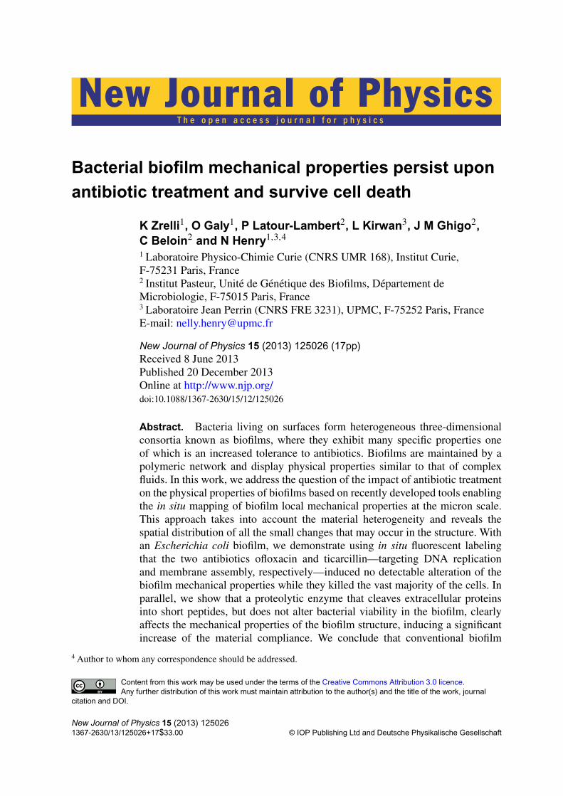
Abstract. Bacteria living on surfaces form heterogeneous three-dimensionalconsortia known as biofilms, where they exhibit many specific properties oneof which is an increased tolerance to antibiotics. Biofilms are maintained by apolymeric network and display physical properties similar to that of complexfluids. In this work, we address the question of the impact of antibiotic treatmenton the physical properties of biofilms based on recently developed tools enablingthe in situ mapping of biofilm local mechanical properties at the micron scale.This approach takes into account the material heterogeneity and reveals thespatial distribution of all the small changes that may occur in the structure. Withan Escherichia coli biofilm, we demonstrate using in situ fluorescent labelingthat the two antibiotics ofloxacin and ticarcillin—targeting DNA replicationand membrane assembly, respectively—induced no detectable alteration of thebiofilm mechanical properties while they killed the vast majority of the cells. Inparallel, we show that a proteolytic enzyme that cleaves extracellular proteinsinto short peptides, but does not alter bacterial viability in the biofilm, clearlyaffects the mechanical properties of the biofilm structure, inducing a significantincrease of the material compliance. We conclude that conventional biofilm
4 Author to whom any correspondence should be addressed.
Content from this work may be used under the terms of the Creative Commons Attribution 3.0 licence.Any further distribution of this work must maintain attribution to the author(s) and the title of the work, journal
citation and DOI.
New Journal of Physics 15 (2013) 1250261367-2630/13/125026+17$33.00 © IOP Publishing Ltd and Deutsche Physikalische Gesellschaft
2
control strategy relying on the use of biocides targeting cells is missing akey target since biofilm structural integrity is preserved. This is expected toefficiently promote biofilm resilience, especially in the presence of persistercells. In contrast, the targeting of polymer network cross-links—among whichextracellular proteins emerge as major players—offers a promising route for thedevelopment of rational multi-target strategies to fight against biofilms.
S Online supplementary data available from stacks.iop.org/NJP/15/125026/mmedia
Contents
1. Introduction 22. Materials and methods 4
2.1. Chemicals, media, strains . . . . . . . . . . . . . . . . . . . . . . . . . . . . . 42.2. Biofilm growth and magnetic probe seeding . . . . . . . . . . . . . . . . . . . 42.3. Biofilm treatments and labeling . . . . . . . . . . . . . . . . . . . . . . . . . . 42.4. Magnetic force setup . . . . . . . . . . . . . . . . . . . . . . . . . . . . . . . 52.5. Particle imaging and tracking . . . . . . . . . . . . . . . . . . . . . . . . . . . 52.6. Microrheology analysis . . . . . . . . . . . . . . . . . . . . . . . . . . . . . . 52.7. Confocal imaging . . . . . . . . . . . . . . . . . . . . . . . . . . . . . . . . . 6
3. Results 63.1. Biofilm mechanical profiles . . . . . . . . . . . . . . . . . . . . . . . . . . . . 63.2. Antibiotic treatment . . . . . . . . . . . . . . . . . . . . . . . . . . . . . . . . 83.3. Protease effect on biofilm mechanics . . . . . . . . . . . . . . . . . . . . . . . 11
4. Discussion and conclusions 13References 16
1. Introduction
Although our understanding of bacterial biofilm molecular biology has markedly increasedrecently, the development of these complex and fascinating living assemblages still raise anumber of fundamental and practical questions. Rational strategies to control their development,maintenance or removal are still rare, calling for a deeper understanding of the causalrelationships linking their physical, chemical and biological properties. For this purpose, anintegrated and comprehensive description of biofilm mechanical properties is necessary. Thechallenge is not only to bring about new ideas for the physical control of biofilms, but alsoto find the mainspring of the material’s mechanical behavior and eventually clarify the roleof the physical cues in biofilm specific lifestyle. The heterogeneity and the highly dynamicalnature of the biofilm material seriously hinders this enterprise. During the last decade, thequestion of bacterial biofilm mechanical properties has been addressed by several groups usinga variety of methods such as the analysis of biofilm streamer deformations due to variationsin fluid flow rates (Stoodley et al 1999, Klapper et al 2002), uniaxial compression of biofilmpieces lifted from agar medium or grown on cover slides (Korstgens et al 2001, Cense et al2006), shearing of biofilm collected from the environment and transferred to a parallel platerheometer (Towler et al 2003, Shaw et al 2004), atomic force microscopy using a glass
New Journal of Physics 15 (2013) 125026 (http://www.njp.org/)

3
bead coated with a bacterial biofilm attached to an AFM cantilever (Lau et al 2009) ora dedicated microcantilever method for measuring the tensile strength of detached biofilmfragments (Poppele and Hozalski 2003, Aggarwal et al 2010). These approaches have produceda large range of elastic moduli, viscosities or cohesion forces that differ by several orders ofmagnitude. Discrepancies observed in the literature originate in part from important differencesin the biological material investigated across the various studies and in the experimentalapproaches involving disparate force and time scales as well as calculation methods (Aravas andLaspidou 2008). In addition, authors have also recognized large variations in data sets stemmingfrom the same consistent investigations (Brindle et al 2011) and conceded to both technicaldifficulties and biofilm inherent variability. In this context, a general agreement about the visco-elastic nature of bacterial biofilms has emerged but the large dispersion of the values usuallyobtained on biofilms removed from their native environment compromised further insightsinto the understanding of the fundamental components underpinning biofilm mechanicalproperties. Most investigators have considered biofilms as homogeneous materials and thereforeanalyzed mechanical responses averaged over the whole material, whereas almost all biofilmcharacteristic properties such as biomass, concentration of chemicals or gene expressionmeasured at the micron scale were shown to exhibit strongly heterogeneous spatial distribution(Stewart and Franklin 2008). To address the question of the biofilm mechanical heterogeneity,we recently introduced a biofilm-microrheology experiment directed to measuring in situ thebiofilm mechanical properties at the micron scale and mapping out their spatial distribution.The principle of the experiment consists of remote actuation of micrometric magnetic particlesseeded in a growing biofilm using a dedicated magnetic tweezer setup. Using this new toolwith Escherichia coli biofilms, we were able to measure the three-dimensional (3D) spatialdistribution of their visco-elastic parameters and to demonstrate their heterogeneity, collectingvalues spreading over almost three orders of magnitude in the same biofilm. Thereby, we havedemonstrated the clear-cut effect of cell surface appendages and environmental conditions onthe mechanical properties of biofilms (Galy et al 2012).
Here, we raise the question of the effect of antibiotics on biofilm mechanical properties.This problem is not only relevant to the design of biofilm control strategies but also toa better understanding of the molecular features supporting biofilm mechanical properties.Indeed, finding effectors altering these properties should help to identify their bases. Moreover,biofilms have been shown to build up significant strong multifactorial resistance to antibiotictreatment (Stewart and Costerton 2001, Anderson and O’Toole 2008, Hoiby et al 2010) but thepotential role of physical factors in these processes is still poorly understood. Mainly based onsemi-quantitative evaluation, previous studies addressing this question have not brought out adefinitive picture of the effects of antibiotics on biofilm mechanics. On the basis of macroscopicrheometry data, Lieleg and co-workers examined several Pseudomonas biofilms and haveconcluded to the absence of the effect of antibiotics (Lieleg et al 2011), while in anotherinvestigation, the authors found that ciprofloxacin and rifampicin weakened P. aeruginosa andS. epidermidis biofilms (Jones et al 2011). However, there is a concern that scrapping andpooling of the biofilm before transfer to the rheometer could have blurred potentially inducedchanges. The purpose of the work reported here is to take advantage of the detailed informationprovided by the remote actuation of magnetic particles in a biofilm maintained in its nativeenvironment, to reconsider the question of the effect of antibiotics on biofilm physical propertiesand to collect information useful to the recognition of the molecular factors contributing tobiofilm physical properties.
New Journal of Physics 15 (2013) 125026 (http://www.njp.org/)

4
We conducted this study in a model biofilm formed by an E. coli strain carrying aderepressed conjugative plasmid F and producing F pilus, a surface appendage that promotesbacterial adhesion and biofilm formation (Ghigo 2001). In order to test the effect of twoantibiotics holding different mechanisms of action we used ofloxacin, known to inhibit DNAgyrase, and ticarcillin, known to prevent cross-linking of peptidoglycan during cell wallsynthesis. We describe here the mechanical profile of a reference biofilm grown under acontrolled nutrient flow before and after antibiotic treatment. In parallel, we probed in situbacterial mortality induced by the antibiotic using a cell death fluorescent marker. To endorsethe antibiotic results and gain further insight into the understanding of the biofilm mechanics,we also assessed the effect on biofilm mechanics induced by the proteolytic enzyme, trypsin.
We compare the impact of antibiotic and protease on biofilm physical characteristics andconclude that biofilm mechanics are strongly resistant to antibiotic treatment. We also propose acrucial role for extracellular proteins in the E. coli biofilm structural organization. These resultssupport the idea already proposed by others of a distinction between cell killing power andbiofilm elimination efficiency of antibiotics. Eventually, we analyze the consequences of ourfindings for the development of new biofilm control strategies.
2. Materials and methods
2.1. Chemicals, media, strains
Tetracycline, ticarcillin (Ticarpen®) was from GlaxoSmithKline (Marly-le-Roi, France) andofloxacin from Sigma-Aldrich (France). Propidium iodide (PI) and magnetic beads (DynabeadsM-270 Amine) were purchased from Life Technologies (France).
Bacteria were grown in lysogeny broth medium and in defined M63B1 medium with0.4% glucose (M63B1Glu). We used isogenic E. coli bacterial strains carrying a derivative ofthe F-conjugative plasmid (F’tet) (Ghigo 2001) and constitutively producing green fluorescentprotein (GFP), MG1655 ampg f p F′tet (TetR, AmpR) and MG1655 kmg f p F′tet (TetR, KmR).Minimum inhibitory concentration (MIC) values of ticarcillin and ofloxacin were taken equalto be 1 and 0.0625 µg ml−1, respectively (Bernier et al 2013).
2.2. Biofilm growth and magnetic probe seeding
Bacteria grown in the presence of tetracycline 7.5 µg ml−1 at 37 ◦C, taken in the exponentialphase were introduced at OD = 0.05 in a 800 µm side length internal side and 160 µm wallthickness capillaries (Composite Metal Services, Shipley, UK) at the same time as the magneticparticles, 2.8 µm in diameter, at a final concentration of 2.5 × 10⁶ ml−1. The mixed suspensionwas allowed to sediment under static conditions for 1 h before starting the flow for the entiregrowth period. Continuous flow was applied using a push–pull syringe pump which delivered a0.3 ml h−1 laminar flow (Reynolds number below 2) and an approximate wall shear 10−3 Pa.
2.3. Biofilm treatments and labeling
2.3.1. Enzymatic and antibiotic treatment. Antibiotics—ofloxacin and ticarcillin—and en-zyme were soaked through the biofilm by adding the required concentrations in the mediumflow—M63B1 medium containing glucose—at 37 ◦C from time ti, generally 24 h after biofilminitiation, to final incubation time, tf.
New Journal of Physics 15 (2013) 125026 (http://www.njp.org/)
5
2.3.2. Propidium iodide. PI 20 µM was introduced in the biofilm from flow circuit outletenabling flow inversion during the time necessary for the counterflow to reach the capillary.Then the flow was stopped for 5 min and the capillary was imaged before re-starting the nutrientflow forward.
2.4. Magnetic force setup
Magnetic tweezers were set up as detailed in a previous paper (Galy et al 2012). Briefly, twomagnetic poles, each made of a copper coil with 2120 turns of 0.56 mm in diameter copper wireand soft magnetic alloy cores (Supra50-Arcelor Mittal, France) were mounted on an invertedNikon TE-300 microscope, north pole facing south pole, in order to generate a magnetic force inone direction along the length of the capillary. In order to determine the absolute force acting onthe beads embedded in the biofilm, we measured the velocity of beads dispersed in a purelyviscous mixture of glycerol and water (39.8 g in 200 µl water). We derived the force fromStokes’ law neglecting the inertia of the particles and checked linear dependence between forceand current. The variation in the force with the distance to the poles was taken into account byrecording the particle trajectories in the entire volume of interest, and storing the velocities withtheir coordinates (xi, yi, zi) in a calibration file which was used to derive visco-elastic parametersfrom particle displacement curves in the biofilm. The amplitude of force in the zone of interestvaried from 29 pN in microvolumes most distal from the poles at the center of the capillary to104 pN at the side walls of the capillaries near the pole pieces. The linearity of the visco-elasticresponse at applied forces ranging from 20 to 100 pN was verified. As well, superimposed creepcurves were obtained when the same force was applied successively on the same particle.
2.5. Particle imaging and tracking
Particles in the biofilm were imaged in the capillary using a Nikon S Fluor ×40 objective (NA0.9, WD 0.3) and an electron multiplying charge coupled device (EMCCD) camera (C 9100-02,Hamamatsu Photonics). Particles were imaged using their large-spectrum intrinsic fluorescencesignal (filters Exc 540/25 nm; DM 565; Em 605/55). To monitor particle motion upon magneticforce application, image sequences were recorded at a frequency of 30 Hz over a period of20 s and further analyzed using an ImageJ particle tracker, as developed by Sbalzarini andKoumoutsakos (2005), that yielded particle trajectories from which individual particle creepcurves giving material strain versus time could be plotted. The error made on the particleposition was evaluated by monitoring the position of the resting beads and found to be equal to0.02 µm.
2.6. Microrheology analysis
Material compliance was derived from particle motion as previously established for a probeparticle of radius R embedded in an incompressible, homogeneous visco-elastic medium(Schnurr et al 1997), which gives the time-dependent creep compliance of the network J (t)(equal to the reciprocal macroscopic shear modulus), knowing probe deflection d(t) and appliedforce f as follows:
J (t) = d(t) ×6π R
f. (1)
New Journal of Physics 15 (2013) 125026 (http://www.njp.org/)
6
Next, we extracted the visco-elastic moduli by fitting the creep curves to the time-dependent visco-elastic behavior of Burger’s model—an equivalent mechanical circuit madeof a spring and a dashpot combined in parallel and a second spring and dashpot added inseries—as classically done to quantify visco-elastic materials, but also more complex andbiological polymer rheological properties (Bausch et al 1998, Jones et al 2011). The resultswere analyzed according to the corresponding analytical solution as follows:
J (t) = J0 + J1
(1 − e−t /τ
)+
t
η0, (2)
where J0 is the elastic instantaneous compliance, τ is the relaxation time required for thetransition from the elastic to the viscous regime, J1 gives the amplitude of elastic relaxationand η0 measures the effective viscosity of the material.
Boundary conditions were evaluated using the theoretical approach of Perkins and Jones1991, 1992) for both hard wall and free surface effects. The correction function was calculatedto the fifth order and taken into account to correct particle velocity in the capillary in the limit of20% correction, i.e. from 4 µm from the bottom of the capillary to 3 µm from the free surface.Particles located outside of these limits were not taken into account in the analysis.
2.7. Confocal imaging
Confocal microscopy images were acquired using a Leica TCS SP5 AOBS inverted confocallaser scanning microscope equipped with HCX PL APo 63x/1.4-0.6 Oil immersion objectivelens. We monitored cell GFP and PI fluorescence using 488 and 561 nm excitation wavelength,respectively, and collecting emission band-pass filters centered at 520/20 nm (GFP) and640/60 nm (PI).
3. Results
3.1. Biofilm mechanical profiles
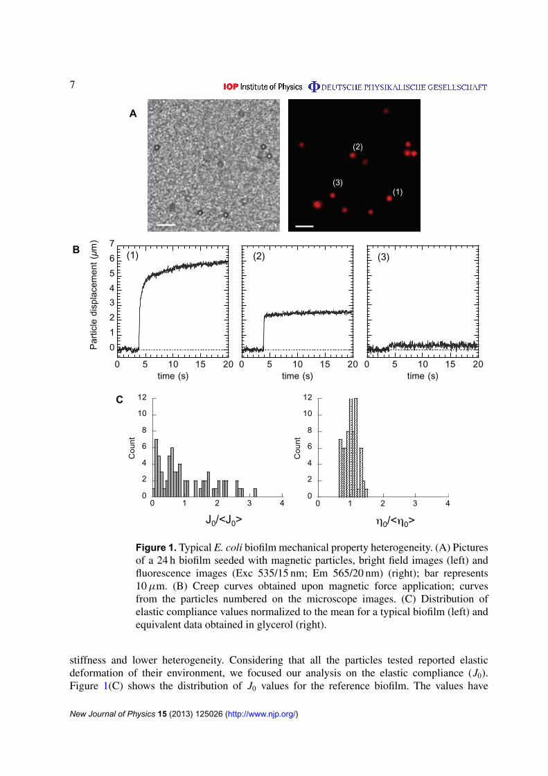
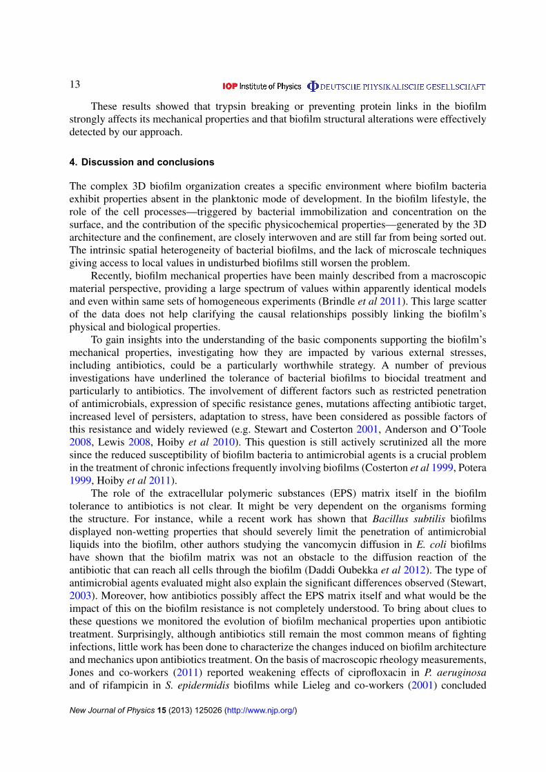
To establish a reference mechanical profile, we grew a typical biofilm using F pilus producingE. coli bacteria seeded with magnetic particles during 24 h under a continuous nutrient flowof 0.3 ml h−1 providing an adherent bacterial layer of 30–40 µm height. Examples of particlespatial distribution in a plane are shown in figure 1(A). Local mechanical properties of thematerial were then probed by applying a 16 s force step on the particles dispersed in thethree dimensions of the biofilm. The induced particle deflection recorded versus time wasused to extract the visco-elastic parameters as previously shown (Galy et al 2012). Particledisplacements reported heterogeneous local mechanical environments exhibiting responsesdiffering both in shape and amplitude as shown in figure 1(B). Local responses werepredominantly of a visco-elastic nature displaying elastic compliance (J0), viscosity (η0) andrelaxation (J1, τ ) (see panel 1 in figure 1(B)). However, in 30 ± 15% of the cases, the particlesreported purely elastic behavior (see panel 2 in figure 1(B)). The spatial distribution of themechanical parameters indicated that environments with elasticity and viscosity values differingfrom two orders of magnitude coexisted laterally and in the depth of the biofilm (see compliancevalues spatial distribution in figure S1 (available from stacks.iop.org/NJP/15/125026/mmedia)).A bottom layer extending to about 10 µm thickness close to the adhesive substrate exhibitedlower values of compliance as well as lower spreading of the values or in other words higher
New Journal of Physics 15 (2013) 125026 (http://www.njp.org/)
7
A
0
1
2
3
4
5
6
7
0 5 10 15 20
Par
ticle
dis
plac
emen
t (
m)
time (s)
0
2
3
4
5
6
7
0 5 10 15 20time (s)
(1) (2) (3) B
(1)
(2)
(3)
0
2
4
6
8
10
12
0 1 2 3 4
Cou
nt
0/< 0>
0
2
4
6
8
10
12
0 1 2 3 4
Cou
nt
J0/<J0>
C
0 5 10 15 20time (s)
Figure 1. Typical E. coli biofilm mechanical property heterogeneity. (A) Picturesof a 24 h biofilm seeded with magnetic particles, bright field images (left) andfluorescence images (Exc 535/15 nm; Em 565/20 nm) (right); bar represents10 µm. (B) Creep curves obtained upon magnetic force application; curvesfrom the particles numbered on the microscope images. (C) Distribution ofelastic compliance values normalized to the mean for a typical biofilm (left) andequivalent data obtained in glycerol (right).
stiffness and lower heterogeneity. Considering that all the particles tested reported elasticdeformation of their environment, we focused our analysis on the elastic compliance (J0).Figure 1(C) shows the distribution of J0 values for the reference biofilm. The values have
New Journal of Physics 15 (2013) 125026 (http://www.njp.org/)
8
Table 1. Parameters of the value distributions normalized to ensemble-averagefor glycerol—amplitude of the viscous flow over (1/η0)—and for the referencebiofilm—elastic compliance (J0).
Material Median Standard deviation
Glycerol 1.01 ± 0.005 0.038 ± 0.002F pilus biofilm 0.3 ml h−1—24 h 0.7 ± 0.16 0.82 ± 0.1
± Stdt error.
been normalized to the ensemble average, enabling distribution characterization independentlyof the parameter itself and evaluation of the degree of heterogeneity of the material to becompared with the distribution of the values obtained in glycerol by the same technique ofparticle actuation (only viscous contribution as expected). In this case, the distribution displayeda completely different symmetrical shape centered on unit as expected from a typicallyhomogeneous viscous liquid and a much lower standard deviation (see table 1). The spreadingof the data obtained in glycerol also gave the experimental error of the measurements—muchlower than the standard deviation of the data obtained in the biofilm. In this approach standarddeviation of the data is directly related to the degree of heterogeneity of the sample.
Thus, the reference biofilm exhibited an the overall mechanical profile reporting aheterogeneous response dominated by an elastic deformation differing by two orders ofmagnitude in the three dimensions of the material, exhibiting a mean elastic compliance valueof 0.4 m2 N−1 and a standard deviation of 0.33 m2 N−1 in good agreement with previous resultswe have obtained on similar biofilms grown at slightly different flow rates (Galy et al 2012).
3.2. Antibiotic treatment
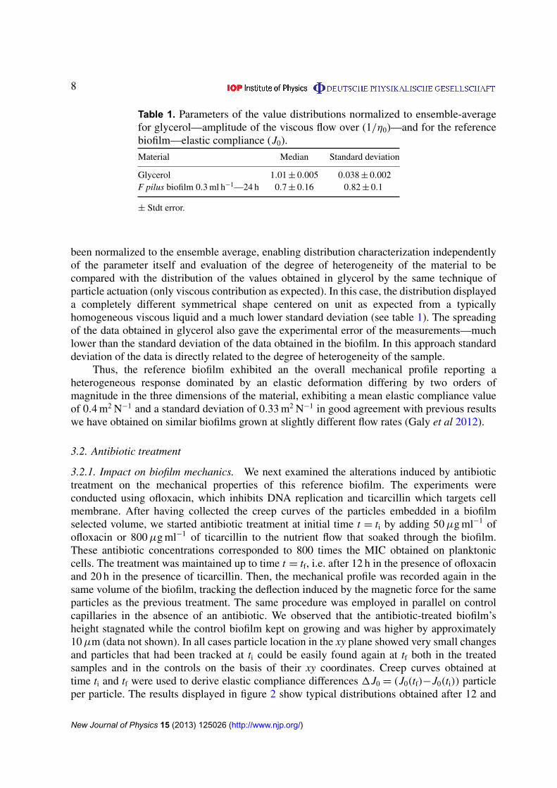
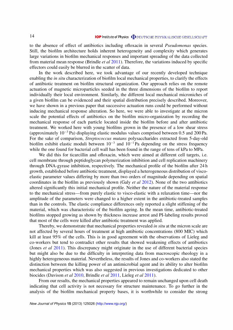
3.2.1. Impact on biofilm mechanics. We next examined the alterations induced by antibiotictreatment on the mechanical properties of this reference biofilm. The experiments wereconducted using ofloxacin, which inhibits DNA replication and ticarcillin which targets cellmembrane. After having collected the creep curves of the particles embedded in a biofilmselected volume, we started antibiotic treatment at initial time t = ti by adding 50 µg ml−1 ofofloxacin or 800 µg ml−1 of ticarcillin to the nutrient flow that soaked through the biofilm.These antibiotic concentrations corresponded to 800 times the MIC obtained on planktoniccells. The treatment was maintained up to time t = tf, i.e. after 12 h in the presence of ofloxacinand 20 h in the presence of ticarcillin. Then, the mechanical profile was recorded again in thesame volume of the biofilm, tracking the deflection induced by the magnetic force for the sameparticles as the previous treatment. The same procedure was employed in parallel on controlcapillaries in the absence of an antibiotic. We observed that the antibiotic-treated biofilm’sheight stagnated while the control biofilm kept on growing and was higher by approximately10 µm (data not shown). In all cases particle location in the xy plane showed very small changesand particles that had been tracked at ti could be easily found again at tf both in the treatedsamples and in the controls on the basis of their xy coordinates. Creep curves obtained attime ti and tf were used to derive elastic compliance differences 1J0 = (J0(tf)−J0(ti)) particleper particle. The results displayed in figure 2 show typical distributions obtained after 12 and
New Journal of Physics 15 (2013) 125026 (http://www.njp.org/)
9
-0,4
-0,2
0
0,2
0,4
Ofloxacin20h
control 12h
Ticarcillin 20h
control 20h
B A C
J 0 (
m2 /
N)
J 0 (
m2 /
N)
J 0 (
m2 /
N)
-0,8
-0,4
0
0,4
0,8
Ofloxacin Control
-0,8
-0,4
0
0,4
0,8
Ticarcillin Control
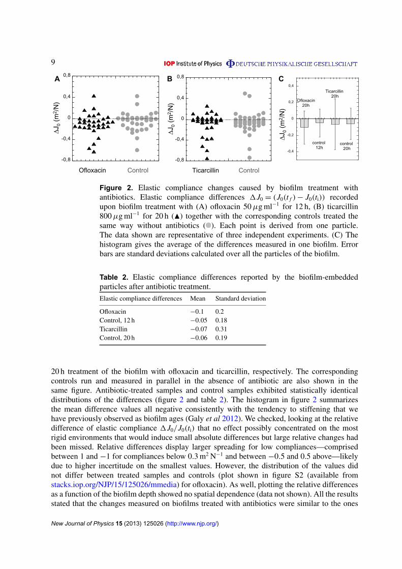
Figure 2. Elastic compliance changes caused by biofilm treatment withantibiotics. Elastic compliance differences 1J0 = (J0(t f ) − J0(ti)) recordedupon biofilm treatment with (A) ofloxacin 50 µg ml−1 for 12 h, (B) ticarcillin800 µg ml−1 for 20 h (N) together with the corresponding controls treated thesame way without antibiotics ( ). Each point is derived from one particle.The data shown are representative of three independent experiments. (C) Thehistogram gives the average of the differences measured in one biofilm. Errorbars are standard deviations calculated over all the particles of the biofilm.
Table 2. Elastic compliance differences reported by the biofilm-embeddedparticles after antibiotic treatment.
Elastic compliance differences Mean Standard deviation
Ofloxacin −0.1 0.2Control, 12 h −0.05 0.18Ticarcillin −0.07 0.31Control, 20 h −0.06 0.19
20 h treatment of the biofilm with ofloxacin and ticarcillin, respectively. The correspondingcontrols run and measured in parallel in the absence of antibiotic are also shown in thesame figure. Antibiotic-treated samples and control samples exhibited statistically identicaldistributions of the differences (figure 2 and table 2). The histogram in figure 2 summarizesthe mean difference values all negative consistently with the tendency to stiffening that wehave previously observed as biofilm ages (Galy et al 2012). We checked, looking at the relativedifference of elastic compliance 1J0/J0(ti) that no effect possibly concentrated on the mostrigid environments that would induce small absolute differences but large relative changes hadbeen missed. Relative differences display larger spreading for low compliances—comprisedbetween 1 and −1 for compliances below 0.3 m2 N−1 and between −0.5 and 0.5 above—likelydue to higher incertitude on the smallest values. However, the distribution of the values didnot differ between treated samples and controls (plot shown in figure S2 (available fromstacks.iop.org/NJP/15/125026/mmedia) for ofloxacin). As well, plotting the relative differencesas a function of the biofilm depth showed no spatial dependence (data not shown). All the resultsstated that the changes measured on biofilms treated with antibiotics were similar to the ones
New Journal of Physics 15 (2013) 125026 (http://www.njp.org/)
10
0
20
40
60
80
100
Bef
ore
trea
tmen
t
12h
Oflo
xaci
n
12h
cont
rol
PI
Mea
n flu
ores
ecnc
e in
tens
ity p
er p
ixel
(a.
u.)
Before treatment (ti)
12h Ofloxacin (tf)
A
B
D
12h control (tf)
C
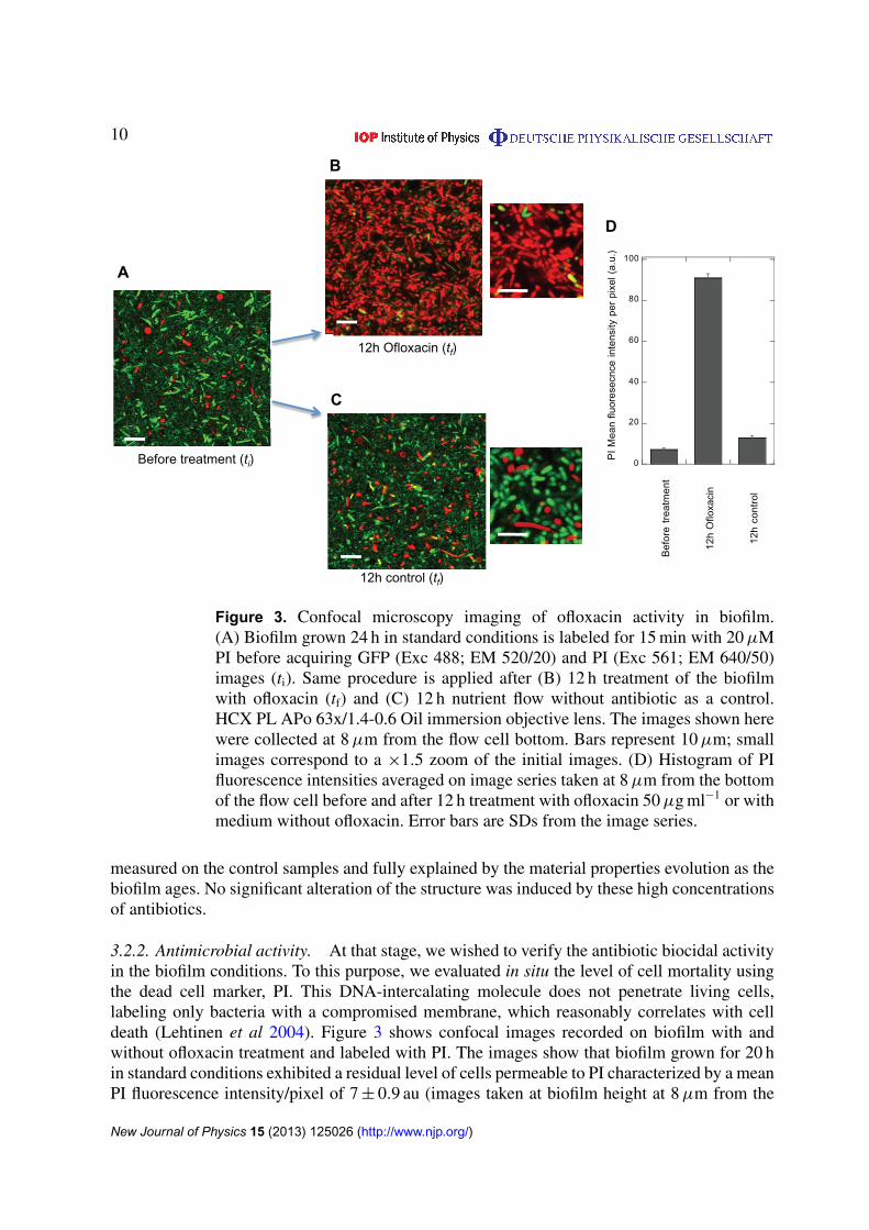
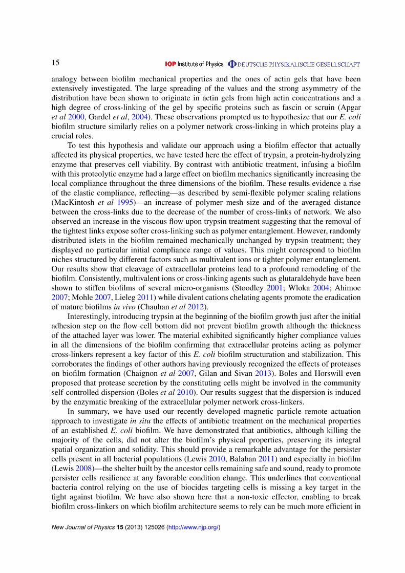
Figure 3. Confocal microscopy imaging of ofloxacin activity in biofilm.(A) Biofilm grown 24 h in standard conditions is labeled for 15 min with 20 µMPI before acquiring GFP (Exc 488; EM 520/20) and PI (Exc 561; EM 640/50)images (ti). Same procedure is applied after (B) 12 h treatment of the biofilmwith ofloxacin (tf) and (C) 12 h nutrient flow without antibiotic as a control.HCX PL APo 63x/1.4-0.6 Oil immersion objective lens. The images shown herewere collected at 8 µm from the flow cell bottom. Bars represent 10 µm; smallimages correspond to a ×1.5 zoom of the initial images. (D) Histogram of PIfluorescence intensities averaged on image series taken at 8 µm from the bottomof the flow cell before and after 12 h treatment with ofloxacin 50 µg ml−1 or withmedium without ofloxacin. Error bars are SDs from the image series.
measured on the control samples and fully explained by the material properties evolution as thebiofilm ages. No significant alteration of the structure was induced by these high concentrationsof antibiotics.
3.2.2. Antimicrobial activity. At that stage, we wished to verify the antibiotic biocidal activityin the biofilm conditions. To this purpose, we evaluated in situ the level of cell mortality usingthe dead cell marker, PI. This DNA-intercalating molecule does not penetrate living cells,labeling only bacteria with a compromised membrane, which reasonably correlates with celldeath (Lehtinen et al 2004). Figure 3 shows confocal images recorded on biofilm with andwithout ofloxacin treatment and labeled with PI. The images show that biofilm grown for 20 hin standard conditions exhibited a residual level of cells permeable to PI characterized by a meanPI fluorescence intensity/pixel of 7 ± 0.9 au (images taken at biofilm height at 8 µm from the
New Journal of Physics 15 (2013) 125026 (http://www.njp.org/)
11
bottom). This fluorescence significantly increased upon 12 h ofloxacin 50 µg ml−1 treatment,reaching 91 ± 2 au, which corresponded to almost all the cells exhibiting PI labeling. However,it should be mentioned that even in these conditions of high antibiotic concentration (800 MIC),a small amount of cells remained free of PI and still exhibiting GFP content. By counting locallyon the PI and the GFP confocal images the number of GFP and PI labeled cells, we evaluatedthat 96 ± 3% of the cells were killed by ofloxacin treatment, showing that although the vastmajority of the cells were killed, a small fraction of the cells remained alive. Similar results wereobtained with ticarcillin (figure S3 (available from stacks.iop.org/NJP/15/125026/mmedia))although PI fluorescence pattern exhibited an additional fuzzy fluorescent pattern suggestingthat cells have released their internal content upon lysis. This is consistent with the ticarcillinmechanism of action targeting cell membrane. The percentage of dead cells was then moredifficult to evaluate but very few cell still containing GFP were detected qualitatively indicatinghigh cell mortality.
3.3. Protease effect on biofilm mechanics
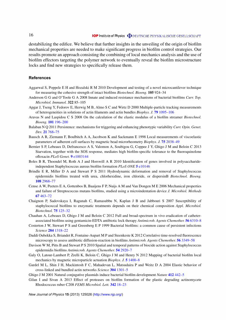
To demonstrate the ability of our approach to evidence local mechanical changes induced byan external effector, we tested the effects of trypsin, an enzyme that cleaves proteins intopieces but does not enter into the cells preserving their viability over the time scale of afew hours. This experiment was inspired by our previous results indicating that extracellularpolymer matrix cross-linking might essentially support biofilm mechanical properties (Galyet al 2012). The mechanical profile of the biofilm was determined before and after 1 h 30 mintreatment with 500 µg ml−1 of trypsin. Figure 4 shows the variation of the elastic complianceinduced by this treatment in a typical experiment. In contrast with what had been observedwith antibiotics, trypsin-treatment-induced a significant increase of compliance in the biofilm.In addition, 20% of the particles initially recorded disappeared from the field of analysis whichreported an environment of compliance greater than 0.1 Pa−1, which had never been detectedin untreated samples. These long-trajectory particles appeared to be randomly distributed in thebiofilm and stemmed from initial environment with compliance values greater than 0.3 Pa−1.Conversely, 25% of the particles reported small alterations remaining in the range of control’ssmall changes. As for the particles removed from the field of observation, neither a definedlocation nor a given initial compliance value range were found for these environments protectedfrom trypsin effects. The creep curve pairs recorded before and after trypsin treatment usuallyexhibited quasi-homothetic shapes (see examples in figure 4(C)) indicating that both elasticityand viscosity were jointly affected by trypsin treatment. Similarly, purely elastic signature wasalso conserved after trypsin treatment but with much higher elastic compliance values.
Bacterial viability was tested using PI labeling which remained at the level of thecontrol—PI intensity found equal to 8 ± 1 au on the images taken at 8 µm from thebiofilm bottom—confirming as expected that trypsin did not induce bacterial cell deathat the concentration and at the time scale of our observations (figure S4 (available fromstacks.iop.org/NJP/15/125026/mmedia)).
In a different set of experiments, we grew the biofilm in the presence of trypsin from thevery beginning of the biofilm formation, just after initial surface colonization as we startednutrient flow. This treatment did not prevent biofilm formation but limited its height that wasfound equal to 20 ± 2 µm in the presence of trypsin versus 38 ± 4 µm in its absence. The biofilmformed in the presence of trypsin displayed much higher compliance arising from the increase
New Journal of Physics 15 (2013) 125026 (http://www.njp.org/)
12
-0,5
0
0,5
1
1,5
2
Control
Ofloxacin
Ticarcillin
Trypsin
J 0 (
m2/N
)
A
C
time (s)
Dis
plac
emen
t (m
) D
ispl
acem
ent (
m)
Before After
0
5
10
15
20
25
0 5 10 15 20
0
1
2
3
4
5
6
0 5 10 15 20
-0,5
0
0,5
1
1,5
2
2,5
3
0 5 10 15 20
0
1
2
3
4
5
6
7
0 5 10 15 20
B
-2
0
2
4
6
J 0 (
m2/N
)
Trypsin Control
time (s)
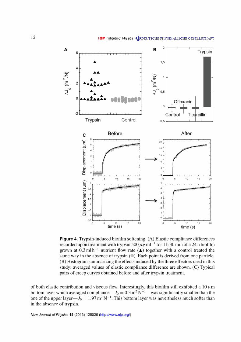
Figure 4. Trypsin-induced biofilm softening. (A) Elastic compliance differencesrecorded upon treatment with trypsin 500 µg ml−1 for 1 h 30 min of a 24 h biofilmgrown at 0.3 ml h−1 nutrient flow rate (N) together with a control treated thesame way in the absence of trypsin ( ). Each point is derived from one particle.(B) Histogram summarizing the effects induced by the three effectors used in thisstudy; averaged values of elastic compliance difference are shown. (C) Typicalpairs of creep curves obtained before and after trypsin treatment.
of both elastic contribution and viscous flow. Interestingly, this biofilm still exhibited a 10 µmbottom layer which averaged compliance—J0 = 0.3 m2 N−1—was significantly smaller than theone of the upper layer—J0 = 1.97 m2 N−1. This bottom layer was nevertheless much softer thanin the absence of trypsin.
New Journal of Physics 15 (2013) 125026 (http://www.njp.org/)
13
These results showed that trypsin breaking or preventing protein links in the biofilmstrongly affects its mechanical properties and that biofilm structural alterations were effectivelydetected by our approach.
4. Discussion and conclusions
The complex 3D biofilm organization creates a specific environment where biofilm bacteriaexhibit properties absent in the planktonic mode of development. In the biofilm lifestyle, therole of the cell processes—triggered by bacterial immobilization and concentration on thesurface, and the contribution of the specific physicochemical properties—generated by the 3Darchitecture and the confinement, are closely interwoven and are still far from being sorted out.The intrinsic spatial heterogeneity of bacterial biofilms, and the lack of microscale techniquesgiving access to local values in undisturbed biofilms still worsen the problem.
Recently, biofilm mechanical properties have been mainly described from a macroscopicmaterial perspective, providing a large spectrum of values within apparently identical modelsand even within same sets of homogeneous experiments (Brindle et al 2011). This large scatterof the data does not help clarifying the causal relationships possibly linking the biofilm’sphysical and biological properties.
To gain insights into the understanding of the basic components supporting the biofilm’smechanical properties, investigating how they are impacted by various external stresses,including antibiotics, could be a particularly worthwhile strategy. A number of previousinvestigations have underlined the tolerance of bacterial biofilms to biocidal treatment andparticularly to antibiotics. The involvement of different factors such as restricted penetrationof antimicrobials, expression of specific resistance genes, mutations affecting antibiotic target,increased level of persisters, adaptation to stress, have been considered as possible factors ofthis resistance and widely reviewed (e.g. Stewart and Costerton 2001, Anderson and O’Toole2008, Lewis 2008, Hoiby et al 2010). This question is still actively scrutinized all the moresince the reduced susceptibility of biofilm bacteria to antimicrobial agents is a crucial problemin the treatment of chronic infections frequently involving biofilms (Costerton et al 1999, Potera1999, Hoiby et al 2011).
The role of the extracellular polymeric substances (EPS) matrix itself in the biofilmtolerance to antibiotics is not clear. It might be very dependent on the organisms formingthe structure. For instance, while a recent work has shown that Bacillus subtilis biofilmsdisplayed non-wetting properties that should severely limit the penetration of antimicrobialliquids into the biofilm, other authors studying the vancomycin diffusion in E. coli biofilmshave shown that the biofilm matrix was not an obstacle to the diffusion reaction of theantibiotic that can reach all cells through the biofilm (Daddi Oubekka et al 2012). The type ofantimicrobial agents evaluated might also explain the significant differences observed (Stewart,2003). Moreover, how antibiotics possibly affect the EPS matrix itself and what would be theimpact of this on the biofilm resistance is not completely understood. To bring about clues tothese questions we monitored the evolution of biofilm mechanical properties upon antibiotictreatment. Surprisingly, although antibiotics still remain the most common means of fightinginfections, little work has been done to characterize the changes induced on biofilm architectureand mechanics upon antibiotics treatment. On the basis of macroscopic rheology measurements,Jones and co-workers (2011) reported weakening effects of ciprofloxacin in P. aeruginosaand of rifampicin in S. epidermidis biofilms while Lieleg and co-workers (2001) concluded
New Journal of Physics 15 (2013) 125026 (http://www.njp.org/)
14
to the absence of effect of antibiotics including ofloxacin in several Pseudomonas species.Still, the biofilm architecture holds inherent heterogeneity and complexity which generateslarge variations in biofilm mechanical responses and important spreading of the data collectedfrom material mean response (Brindle et al 2011). Therefore, the variations induced by specificeffectors could easily be blurred in the scatter of data.
In the work described here, we took advantage of our recently developed techniqueenabling the in situ characterization of biofilm local mechanical properties, to clarify the effectsof antibiotic treatment on biofilm structural organization. Our approach relies on the remoteactuation of magnetic microparticles seeded in the three dimensions of the biofilm to reportindividually their local environment. Similarly, the different local mechanical microniches ofa given biofilm can be evidenced and their spatial distribution precisely described. Moreover,we have shown in a previous paper that successive actuation runs could be performed withoutinducing mechanical response alteration. So here, we were able to investigate at the micronscale the potential effects of antibiotics on the biofilm micro-organization by recording themechanical response of each particle located inside the biofilm before and after antibiotictreatment. We worked here with young biofilms grown in the presence of a low shear stress(approximately 10−3 Pa) displaying elastic modulus values comprised between 0.5 and 200 Pa.For the sake of comparison, Streptococcus mutans polysaccharides extracted from 5-day-oldbiofilm exhibit elastic moduli between 10−3 and 10−1 Pa depending on the stress frequencywhile the one found for bacterial cell wall has been found in the range of tens of kPa to MPa.
We did this for ticarcillin and ofloxacin, which were aimed at different cell targets, i.e.cell membrane through peptidoglycan polymerization inhibition and cell replication machinerythrough DNA-gyrase inhibition, respectively. The mechanical profile of the biofilm after 24 hgrowth, established before antibiotic treatment, displayed a heterogeneous distribution of visco-elastic parameter values differing by more than two orders of magnitude depending on spatialcoordinates in the biofilm as previously shown (Galy et al 2012). None of the two antibioticsaltered significantly this initial mechanical profile. Neither the nature of the material responseto the mechanical stress—from purely elastic to visco-elastic with a relaxation time—nor theamplitude of the parameters were changed to a higher extent in the antibiotic-treated samplesthan in the controls. The elastic compliance differences only reported a slight stiffening of thematerial, which was characteristic of the biofilm ageing. In the mean time, antibiotic-treatedbiofilms stopped growing as shown by thickness increase arrest and PI-labeling results provedthat most of the cells were killed after antibiotic treatment was applied.
Thereby, we demonstrate that mechanical properties revealed in situ at the micron scale arenot affected by several hours of treatment at high antibiotic concentrations (800 MIC) whichkill at least 95% of the cells. This is in good agreement with the observations of Lieleg andco-workers but tend to contradict other results that showed weakening effects of antibiotics(Jones et al 2011). This discrepancy might originate in the use of different bacterial speciesbut might also be due to the difficulty in interpreting data from macroscopic rheology in ahighly heterogeneous material. Nevertheless, the results of Jones and co-workers also stated thedistinction between the killing power of an antimicrobial agent and its ability to alter biofilmmechanical properties which was also suggested in previous investigations dedicated to otherbiocides (Davison et al 2010, Brindle et al 2011, Lieleg et al 2011).
From our results, the mechanical properties appeared to remain unchanged upon cell deathindicating that cell activity is not necessary for structure maintenance. To go further in theanalysis of the biofilm mechanical property bases, it is worthwhile to consider the strong
New Journal of Physics 15 (2013) 125026 (http://www.njp.org/)
15
analogy between biofilm mechanical properties and the ones of actin gels that have beenextensively investigated. The large spreading of the values and the strong asymmetry of thedistribution have been shown to originate in actin gels from high actin concentrations and ahigh degree of cross-linking of the gel by specific proteins such as fascin or scruin (Apgaret al 2000, Gardel et al, 2004). These observations prompted us to hypothesize that our E. colibiofilm structure similarly relies on a polymer network cross-linking in which proteins play acrucial roles.
To test this hypothesis and validate our approach using a biofilm effector that actuallyaffected its physical properties, we have tested here the effect of trypsin, a protein-hydrolyzingenzyme that preserves cell viability. By contrast with antibiotic treatment, infusing a biofilmwith this proteolytic enzyme had a large effect on biofilm mechanics significantly increasing thelocal compliance throughout the three dimensions of the biofilm. These results evidence a riseof the elastic compliance, reflecting—as described by semi-flexible polymer scaling relations(MacKintosh et al 1995)—an increase of polymer mesh size and of the averaged distancebetween the cross-links due to the decrease of the number of cross-links of network. We alsoobserved an increase in the viscous flow upon trypsin treatment suggesting that the removal ofthe tightest links expose softer cross-linking such as polymer entanglement. However, randomlydistributed islets in the biofilm remained mechanically unchanged by trypsin treatment; theydisplayed no particular initial compliance range of values. This might correspond to biofilmniches structured by different factors such as multivalent ions or tighter polymer entanglement.Our results show that cleavage of extracellular proteins lead to a profound remodeling of thebiofilm. Consistently, multivalent ions or cross-linking agents such as glutaraldehyde have beenshown to stiffen biofilms of several micro-organisms (Stoodley 2001; Wloka 2004; Ahimoe2007; Mohle 2007, Lieleg 2011) while divalent cations chelating agents promote the eradicationof mature biofilms in vivo (Chauhan et al 2012).
Interestingly, introducing trypsin at the beginning of the biofilm growth just after the initialadhesion step on the flow cell bottom did not prevent biofilm growth although the thicknessof the attached layer was lower. The material exhibited significantly higher compliance valuesin all the dimensions of the biofilm confirming that extracellular proteins acting as polymercross-linkers represent a key factor of this E. coli biofilm structuration and stabilization. Thiscorroborates the findings of other authors having previously recognized the effects of proteaseson biofilm formation (Chaignon et al 2007, Gilan and Sivan 2013). Boles and Horswill evenproposed that protease secretion by the constituting cells might be involved in the communityself-controlled dispersion (Boles et al 2010). Our results suggest that the dispersion is inducedby the enzymatic breaking of the extracellular polymer network cross-linkers.
In summary, we have used our recently developed magnetic particle remote actuationapproach to investigate in situ the effects of antibiotic treatment on the mechanical propertiesof an established E. coli biofilm. We have demonstrated that antibiotics, although killing themajority of the cells, did not alter the biofilm’s physical properties, preserving its integralspatial organization and solidity. This should provide a remarkable advantage for the persistercells present in all bacterial populations (Lewis 2010, Balaban 2011) and especially in biofilm(Lewis 2008)—the shelter built by the ancestor cells remaining safe and sound, ready to promotepersister cells resilience at any favorable condition change. This underlines that conventionalbacteria control relying on the use of biocides targeting cells is missing a key target in thefight against biofilm. We have also shown here that a non-toxic effector, enabling to breakbiofilm cross-linkers on which biofilm architecture seems to rely can be much more efficient in
New Journal of Physics 15 (2013) 125026 (http://www.njp.org/)
16
destabilizing the edifice. We believe that further insights in the unveiling of the origin of biofilmmechanical properties are needed to make significant progress in biofilm control strategies. Ourresults promote an approach consisting the combining of local mechanics analysis and the use ofbiofilm effectors targeting the polymer network to eventually reveal the biofilm microstructurelocks and find new strategies to specifically release them.
References
Aggarwal S, Poppele E H and Hozalski R M 2010 Development and testing of a novel microcantilever techniquefor measuring the cohesive strength of intact biofilms Biotechnol. Bioeng. 105 924–34
Anderson G G and O’Toole G A 2008 Innate and induced resistance mechanisms of bacterial biofilms Curr. Top.Microbiol. Immunol. 322 85–105
Apgar J, Tseng Y, Fedorov E, Herwig M B, Almo S C and Wirtz D 2000 Multiple-particle tracking measurementsof heterogeneities in solutions of actin filaments and actin bundles Biophys. J. 79 1095–106
Aravas N and Laspidou C S 2008 On the calculation of the elastic modulus of a biofilm streamer Biotechnol.Bioeng. 101 196–200
Balaban N Q 2011 Persistence: mechanisms for triggering and enhancing phenotypic variability Curr. Opin. Genet.Dev. 21 768–75
Bausch A R, Ziemann F, Boulbitch A A, Jacobson K and Sackmann E 1998 Local measurements of viscoelasticparameters of adherent cell surfaces by magnetic bead microrheometry Biophys. J. 75 2038–49
Bernier S P, Lebeaux D, Defrancesco A S, Valomon A, Soubigou G, Coppee J Y, Ghigo J M and Beloin C 2013Starvation, together with the SOS response, mediates high biofilm-specific tolerance to the fluoroquinoloneofloxacin PLoS Genet. 9 e1003144
Boles B R, Thoendel M, Roth A J and Horswill A R 2010 Identification of genes involved in polysaccharide-independent Staphylococcus aureus biofilm formation PLoS ONE 5 e10146
Brindle E R, Miller D A and Stewart P S 2011 Hydrodynamic deformation and removal of Staphylococcusepidermidis biofilms treated with urea, chlorhexidine, iron chloride, or dispersinB Biotechnol. Bioeng.108 2968–77
Cense A W, Peeters E A, Gottenbos B, Baaijens F P, Nuijs A M and Van Dongen M E 2006 Mechanical propertiesand failure of Streptococcus mutans biofilms, studied using a microindentation device J. Microbiol. Methods67 463–72
Chaignon P, Sadovskaya I, Ragunah C, Ramasubbu N, Kaplan J B and Jabbouri S 2007 Susceptibility ofstaphylococcal biofilms to enzymatic treatments depends on their chemical composition Appl. Microbiol.Biotechnol. 75 125–32
Chauhan A, Lebeaux D, Ghigo J M and Beloin C 2012 Full and broad-spectrum in vivo eradication of catheter-associated biofilms using gentamicin-EDTA antibiotic lock therapy Antimicrob. Agents Chemother. 56 6310–8
Costerton J W, Stewart P S and Greenberg E P 1999 Bacterial biofilms: a common cause of persistent infectionsScience 284 1318–22
Daddi Oubekka S, Briandet R, Fontaine-Aupart M P and Steenkeste K 2012 Correlative time-resolved fluorescencemicroscopy to assess antibiotic diffusion-reaction in biofilms Antimicrob. Agents Chemother. 56 3349–58
Davison W M, Pitts B and Stewart P S 2010 Spatial and temporal patterns of biocide action against Staphylococcusepidermidis biofilms Antimicrob. Agents Chemother. 54 2920–7
Galy O, Latour-Lambert P, Zrelli K, Beloin C, Ghigo J M and Henry N 2012 Mapping of bacterial biofilm localmechanics by magnetic microparticle actuation Biophys. J. 5 1400–8
Gardel M L, Shin J H, Mackintosh F C, Mahadevan L, Matsudaira P and Weitz D A 2004 Elastic behavior ofcross-linked and bundled actin networks Science 304 1301–5
Ghigo J M 2001 Natural conjugative plasmids induce bacterial biofilm development Nature 412 442–5Gilan I and Sivan A 2013 Effect of proteases on biofilm formation of the plastic degrading actinomycete
Rhodococcus ruber C208 FEMS Microbiol. Lett. 342 18–23
New Journal of Physics 15 (2013) 125026 (http://www.njp.org/)
17
Hoiby N, Bjarnsholt T, Givskov M, Molin S and Ciofu O 2010 Antibiotic resistance of bacterial biofilms. Int. J.Antimicrob. Agents 35 322–32
Hoiby N, Ciofu O, Johansen H K, Song Z J, Moser C, Jensen P O, Molin S, Givskov M, Tolker-Nielsen T andBjarnsholt T 2011 The clinical impact of bacterial biofilms Int. J. Oral Sci. 3 55–65
Jones W L, Sutton M P, Mckittrick L and Stewart P S 2011 Chemical and antimicrobial treatments change theviscoelastic properties of bacterial biofilms Biofouling 27 207–15
Klapper I, Rupp C J, Cargo R, Purvedorj B and Stoodley P 2002 Viscoelastic fluid description of bacterial biofilmmaterial properties. Biotechnol. Bioeng. 80 289–96
Korstgens V, Flemming H C, Wingender J and Borchard W 2001 Uniaxial compression measurement device forinvestigation of the mechanical stability of biofilms J. Microbiol. Methods 46 9–17
Lau P C, Dutcher J R, Beveridge T J and Lam J S 2009 Absolute quantitation of bacterial biofilm adhesion andviscoelasticity by microbead force spectroscopy Biophys. J. 96 2935–48
Lehtinen J, Nuutila J and Lilius E M 2004 Green fluorescent protein-propidium iodide (GFP-PI) based assay forflow cytometric measurement of bacterial viability Cytometry A 60 165–72
Lewis K 2008 Multidrug tolerance of biofilms and persister cells Curr. Top. Microbiol. Immunol. 322 107–31Lewis K 2010 Persister cells Annu. Rev. Microbiol. 64 357–72Lieleg O, Caldara M, Baumgartel R and Ribbeck K 2011 Mechanical robustness of Pseudomonas aeruginosa
biofilms Soft Matter 7 3307–14Mackintosh F C, Kas J and Janmey P A 1995 Elasticity of semiflexible biopolymer networks Phys. Rev. Lett.
75 4425–8Perkins G S and Jones R B 1991 Hydrodynamic interaction of a spherical particle with a planar boundary: I. Free
surface Physica A 171 575–604Perkins G S and Jones R B 1992 Hydrodynamic interaction of a spherical-particle with a planar boundary:
2. Hard-wall Physica A 189 447–77Poppele E H and Hozalski R M 2003 Micro-cantilever method for measuring the tensile strength of biofilms and
microbial flocs J. Microbiol. Methods 55 607–15Potera C 1999 Forging a link between biofilms and disease Science 283 1837–9Sbalzarini I F and Koumoutsakos P 2005 Feature point tracking and trajectory analysis for video imaging in cell
biology J. Struct. Biol. 151 182–95Schnurr B, Gittes F, Mackintosh F C and Schmidt C F 1997 Determining microscopic viscoelasticity in flexible
and semiflexible polymer networks from thermal fluctuations Macromolecules 30 7781–92Shaw T, Winston M, Rupp C J, Klapper I and Stoodley P 2004 Commonality of elastic relaxation times in biofilms
Phys. Rev. Lett. 93 0988102Stewart P S 2003 Diffusion in biofilms J. Bacteriol. 185 1485–91Stewart P S and Costerton J W 2001 Antibiotic resistance of bacteria in biofilms Lancet 358 135–8Stewart P S and Franklin M J 2008 Physiological heterogeneity in biofilms Nature Rev. Microbiol. 6 199–210Stoodley P, Lewandowski Z, Boyle J D and Lappin-Scott H M 1999 Structural deformation of bacterial biofilms
caused by short-term fluctuations in fluid shear: an in situ investigation of biofilm rheology Biotechnol. Bioeng.65 83–92
Towler B W, Rupp C J, Cunningham A B and Stoodley P 2003 Viscoelastic properties of a mixed culture biofilmfrom rheometer creep analysis Biofouling 19 279–85
New Journal of Physics 15 (2013) 125026 (http://www.njp.org/)