Embed Size (px)
Citation preview

Bacteria

Bacteria of Ophthalmic ImportanceDiane Hendrix, DVM, DACVOProfessor of Ophthalmology

BacteriaProkaryotic organisms
– cell membrane
– cytoplasm
– RNA
– DNA
– often a cell wall
– +/- specialized surface structures such as capsules or pili.
– lack a nuclear membrane or mitotic apparatus, but the DNA is organized into a single circular chromosome
www.norcalblogs.com/.../GeneralBacteria.jpg

Bacteria+/- smaller molecules of DNA termed plasmids that carry information for drug resistance or code for toxins that can affect host cellular functions
www.fairscience.org
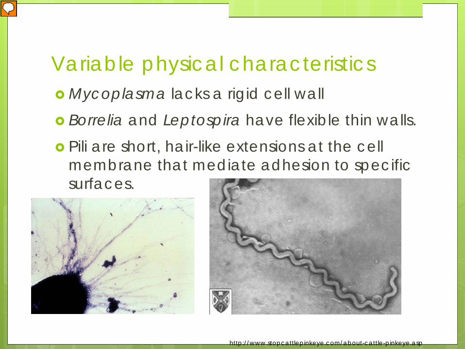
Variable physical characteristics Mycoplasma lacks a rigid cell wall Borrelia and Leptospira have flexible thin walls. Pili are short, hair-like extensions at the cell
membrane that mediate adhesion to specific surfaces.
http://www.stopcattlepinkeye.com/about-cattle-pinkeye.asp

Bacteria reproductionAsexual binary fissionThe bacterial growth cycle includes: the lag phase the logarithmic growth phase the stationery growth phase the decline phase
Iron is essential for bacteria

Opportunistic bacteria Staphylococcus epidermidisBacillus sp. Corynebacterium sp. Escherichia coliKlebsiella sp. Enterobacter sp. Serratia sp. Pseudomonas sp.
(other than P aeruginosa).
Bacteria

Infectivity Adhesins are protein determinates of
adherence. Some are expressed in bacterial pili. Virulent factors increase the capacity of an
organism to cause tissue inflammation and destruction. (Proteses, elastases, hemolysins, cytoxins)
Secretomes and lipopolysaccharide core biosynthetic genes inhibit corneal epithelial cell migration
Pseudomonas aeruginosa and Moraxella bovis


Normal bacterial and fungal floraBacteria can be cultured from 50 to 90% of normal dogs.
Gram + aerobes are most common. Gram - bacteria have been recovered from
8% of normal dogs. Anaerobes are rarely isolated.
Normal flora varies with the season and the breed of dog. Fungi have been isolated from 22% of dogs in one study.
Bacteria

Conjunctival flora in dogs with ulcerative keratitis. Bacteria are more commonly isolated. Malassezia pachydermatitis is present in 23% of
eyes with corneal ulceration
Bacteria

Equine flora
Normal bacterial floraCorynebacterium spp., beta-hemolytic Streptococcus, Staphylococcus spp., Klebsiellaspp., Bacillus cereus and Moraxella spp.
Fungal floraUnidentifiable molds, dematiaceous molds, Chrysosporuim spp., Cladosporium spp., Aspergillus spp. and Penicillium spp.

Bovine flora
Cladosporiumspp. and Penicillium spp.
No seasonal or housing difference.
May represent transient seeding from the environment, including the hay, as suspected in other species.

Normal flora Bats Alpacas Chelonians

Staphylococcus spp.
Ubiquitous and are part of the microflora of the skin and mucous membranes.
Gram + organisms that appear cytologically as individuals, pairs, small groups or grapelike clusters.
Facultative anaerobes and fermentative.
Isolates commonly recovered from ocular sources are coagulase-positive species.
ww
w.b
iology4kids.com

Staphylococcus spp.
Infectious Keratitis S aureus is isolated from about 5% of horses S intermedius is isolated from 2% of horses and
29% of dogs Coagulase-negative species include
S epidermidis (isolated from 6% of affected horses).

Canine isolates are sensitive to cefazolin, ciprofloxacin, and chloramphenicol.
Of 4 equine isolates all were sensitive to bacitracin, chloramphenicol, neomycin, and enrofloxacin.
Resistance
upload.w
ikimed
ia.orgStaphylococcus spp.

Ubiquitous, suppurative bacteria
Enterococci are opportunists Streptococcal keratitis is
relatively common
Most pathogenic Streptococci produce varying hemolysins.
Streptococcus spp.and related cocci

β-hemolytic Strep spp -17% of dogs
S. equi subsp zoo - 12% and 22% of the isolates from horses.
Streptococcus spp.

UT – Equine and canine all isolates were susceptible to ciprofloxacin, cephalothin
and chloramphenicol
> 80% resistance to neomycin, polymixin B and tobramycin
UF - Equine All susceptible to chloramphenicol, bacitracin
An increase in resistance of S.equi subsp zooepidemicus to gentamicin was found over time
Australia>80% of isolates were resistant to ciprofloxacin but
remained susceptible to chloramphenicol and cephalexin
Streptococcus spp.
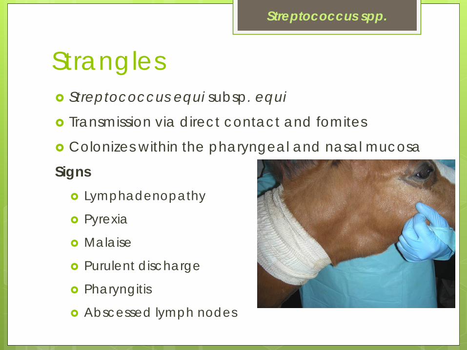
Streptococcus equi subsp. equi
Transmission via direct contact and fomites
Colonizes within the pharyngeal and nasal mucosa
Signs Lymphadenopathy
Pyrexia
Malaise
Purulent discharge
Pharyngitis
Abscessed lymph nodes
Strangles
Streptococcus spp.

Cases involving any area other than the pharyngeal area.
Ocular abnormalities
Serous then mucopurulent discharge
Panophthalmitis
Chorioretinitis
Central blindness
Dx via culture or PCR
Bastard strangles
Streptococcus spp.

Corynebacterium spp. Gram + rods
Appear singly or in pairs
+/- clubbed ends
Flora of normal skin and mucous membranes

Bacillus spp. Gram + rods found singly, in pairs or chains. May have a single endospore More pathogenic organisms usually present
as co-infections.
http://content.answers.com/main/content/wp/en-commons/thumb/4/42/260px-Bacillus_subtilis_Spore.jpg
Most common organism isolated from endophthalmitis in humans.

Listeriosis Rod-shaped, Gram + bacterium
L monocytogenes most common in animals
Spoiled or incompletely fermented corn or hay silage is the main source of infection in outbreaks.
VCNA Food AnimPract. 2010 Nov;26(3):487-503

CNS disease is most likely to be associated with ocular signs in food animal species. vestibular ataxia
cranial nerve deficits
brain stem involvement facial nerve paralysis
Kcs
Keratitis Anterior uveitis w/hypopyon Purulent endophthalmitis
VCNA Food AnimPract. 2010 Nov;26(3):487-503

Other species Dog
Conjunctivitis, neurologic signs, and pancytopenia with generalized infection.
Sheep & goats Scleral hyperemia
Unilateral keratitis +/- ulceration
CNS signs
en.wikipedia.org/wiki/Listeriosis_in_animals

Swine listeriosis Septicemia
Encephalitis
Diagnosis clinical signs
culture and identification of the organism from body fluids

Pseudomonas spp. Gram - rods Widely distributed. Found in the skin and
mucous membranes. Cytologically
indistinguishable from other rods
Antibiotic susceptibility testing is especially important

Pseudomonas aeruginosa Isolated from about 15% of horses with bacterial
keratitis Isolated from 21% of dogs

Innate resistance
Evans 2013

Pathogenic mechanisms
www.cdc.gov www.asylumresearch.co.uk/.../Bacteria/Cell!.jpgsciencephoto.com
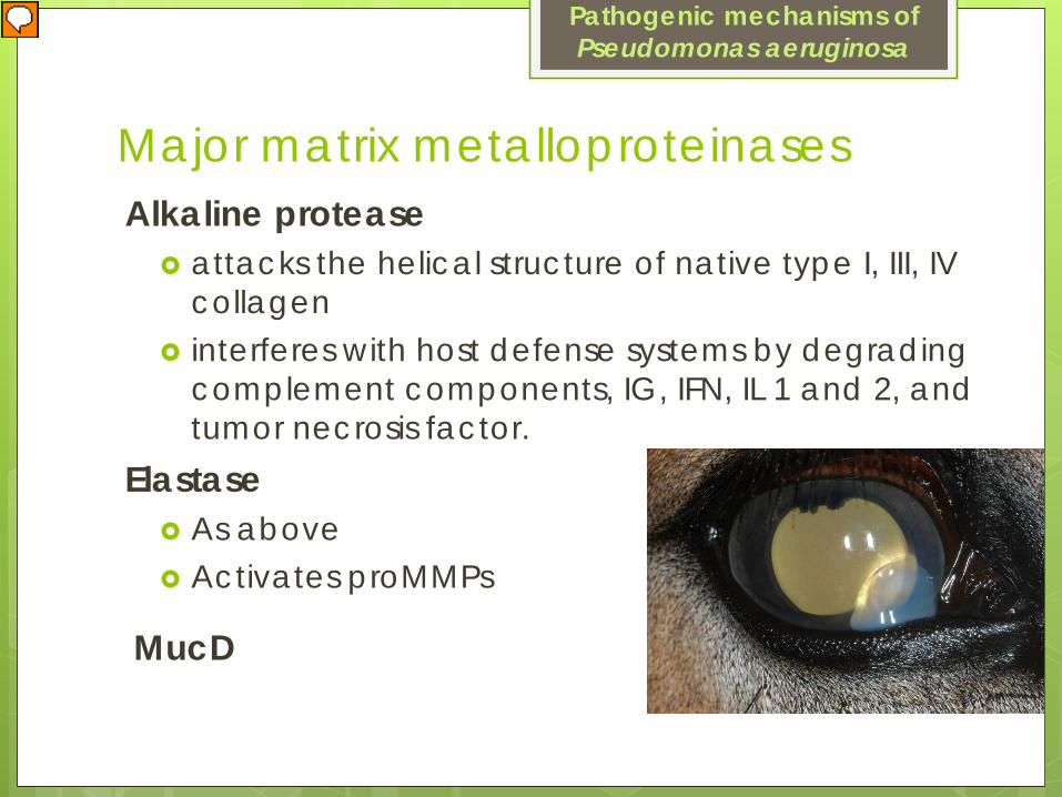
Major matrix metalloproteinasesAlkaline protease
attacks the helical structure of native type I, III, IV collagen
interferes with host defense systems by degrading complement components, IG, IFN, IL 1 and 2, and tumor necrosis factor.
Pathogenic mechanisms of Pseudomonas aeruginosa
Elastase As above Activates proMMPs
MucD

Cytotoxic and invasive strains Cytotoxic strains remain mostly extracellular Invasive strains enter cells and replicate within
them. Tobramycin vs ofloxacin Steroids? Both antibiotics hastened disease resolution infections caused by either strain.
Pathogenic mechanisms of Pseudomonas aeruginosa

IOVS 2011 March; 52(3): 1368–1377

Resistance No resistance to gentamicin, tobramycin,
ciprofloxacin, neomycin and polymixin B in horses at UT
At UF a statistically significant increase in resistance to gentamicin and tobramycin was found in horses between the isolates from 1992 to 1998 and those from 1999 to 2000.
Isolates of P. aeruginosa from dogs were susceptible to tobramycin and gentamicin and had limited resistance to ciprofloxacin (0.07%).
Pseudomonas aeruginosa
Resistance 27 P aeruginosa isolates from dogs
7 fluoroquinolones
24/ 27 isolates were susceptible to all fluoroquinolones evaluated
Susceptibility ranged from 88.9% to 100%
No significant differences among isolate susceptibilities to the individual antimicrobials or among generations of fluoroquinolones
Pseudomonas aeruginosa

Multi-drug resistant, extensively drug resistant, and pan-drug resistant strains of P aeruginosa
Risk factors: bandage contact lens, topical steroids, previous therapeutic graft, preservative-free lubricant ointment and ocular surface disorders.
Of 15 isolates, 6 were sensitive only to imipenem, 3 to colistin, 2 to neomycin, 1 each to imipenem and colistin, imipenem and ceftazidime, and azithromycin. One isolate was resistant to all antibiotics.
Success with medical therapy alone was not common. These cases are more likely to require the use of tissue adhesives and keratoplasty and are likely to have treatment failure.
Another study compared the efficacy of topical 1.5% and 0.5% levofloxacin.

Bacteriophages/Predatory Prokaryotes
AJVR 2011 Aug;72(8):1079-86
Pseudomonas aeruginosa

Moraxella spp. A large, plump, Gram - coccobacillus
Primary cause of infectious bovine keratoconjunctivitis (IBK) “pinkeye”
Highly contagious ocular infection of cattle
Monetary losses caused by: decreased weight gain
decreased milk production
devaluation because of eye disfigurement
cost of treatment

Transmission Opportunistic pathogen Environmental factors
Exposure to UV light
Irritants- face fly
Host factors Genetic
Nutritional
Immune status
Current infections
Moraxella bovis

Nonpiliated, nonpathogenic forms can exist in a carrier state in the host.
Carrier animals are asymptomatic, but shed the organism.
Harbored in nasal, ocular, and vaginal secretions
Transmitted by direct contact, aerosol, or fomites. Cattle are the primary natural reservoir for M bovisand have a high nasal carrier rate.
Transmission of Moraxella bovis

The face fly Musca autumnalis is a primary mechanical vector and serves as an irritant.
www.forestryimages.org bugguide.net popgen.unimaas.nl
Transmission of Moraxella bovis

UV light causes direct conversion of nonhemolytic, nonpiliated organisms to pathogenic forms in carrier animals.
Then a rapid logarithmic growth phase of the organism begins.
Transmission of Moraxella bovis

Bos taurus is more susceptible to IBK than is Bos indicus (such as Zebu and Brahman)
Calves are more prone to disease than adults.
IBK is more common in summer and fall
Transmission of Moraxella bovis
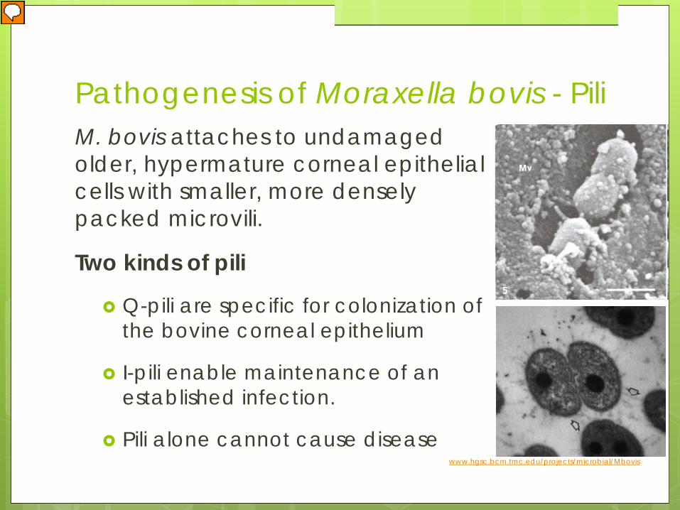
Pathogenesis of Moraxella bovis - PiliM. bovis attaches to undamaged older, hypermature corneal epithelial cells with smaller, more densely packed microvili.
Two kinds of pili
Q-pili are specific for colonization of the bovine corneal epithelium
I-pili enable maintenance of an established infection.
Pili alone cannot cause disease www.hgsc.bcm.tmc.edu/projects/microbial/Mbovis
β-hemolysin of M bovis is also required to cause disease.
RTX family of toxins
causes clinical signs directly as a result of damaged ocular cells or indirectly through lysis of the WBCs attracted to the site.
Pathogenesis of Moraxella bovis

Roge
rs D
G, C
hevi
lle N
F, P
ugh
GW
J. P
ath
ogen
esis
of c
orne
al le
sions
ca
used
by
Mor
axe
lla b
ovis
in g
noto
biot
ic c
alv
es. V
et P
ath
1987
; 24
(4):2
87-2
95.

Pink eye – Infectious Bovine Keratitis
Summer and fall
Younger cattle
Incubation period 2-3 days.
Moraxella bovis
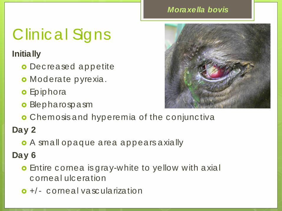
Clinical Signs Initially
Decreased appetite Moderate pyrexia. Epiphora Blepharospasm Chemosis and hyperemia of the conjunctiva
Day 2 A small opaque area appears axially
Day 6 Entire cornea is gray-white to yellow with axial
corneal ulceration +/- corneal vascularization
Moraxella bovis

OutcomeTypically Complete recovery in 3-5 weeks Some with persistent scar
Moraxella bovis
Infrequent outcomes Severe ulceration
Corneal rupture with iris prolapse
Conical bulging of the eye
Blindness

Treatment - Parenteral
Oxytetracycline (LA200 2 injections, 20 mg/kg IM at 72 hour intervals)
Oxytetracycline (Tetradur, 300 mg/ml, 1-2 ml IM, lasts 7-10 days.)
Florfenicol (2 IM dosages of 20 mg/kg 48 hours apart or a single 40 mg/kg SC dosage)
Moraxella bovis

Treatment - Subconjunctival Procaine penicillin (1-2 ml) +/- subconjunctival
dexamethasone (1-2 ml)
Oxytetracycline (100 mg/ml subconjunctival 1-2 ml)
Moraxella bovis

Treatment - other
Ceftiofur crystalline-free acid (CCFA)(6.6 mg of ceftiofur equivalents/kg, SC)****
Tulathromycin 1 dose SC Tilmicosin SC (5 or 10 mg/kg)
Moraxella bovis

Treatment Nictitating membrane flaps or temporary
tarsorrhaphies
Decreasing the fly population
Decreasing UV radiation
Autogenous vaccines
Cytokines with inactivated bacteria
Intranasal vaccines
Moraxella bovis

Moraxella bovoculi Isolated from IBK cases Blepharitis and conjunctivitis Respond to IBK treatments Association with M bovis?
Other diseases by Moraxella spp. Moraxella spp. cause other ocular infections of
small ruminants and horses. Moraxella ovis
Gram - diplococcus
cultured from normal small ruminants
Cultured from sheep and goats with keratoconjunctivitis.
May occur as a co-infection with chlamydial or mycoplasmal conjunctivitis
May complicate other ocular diseases
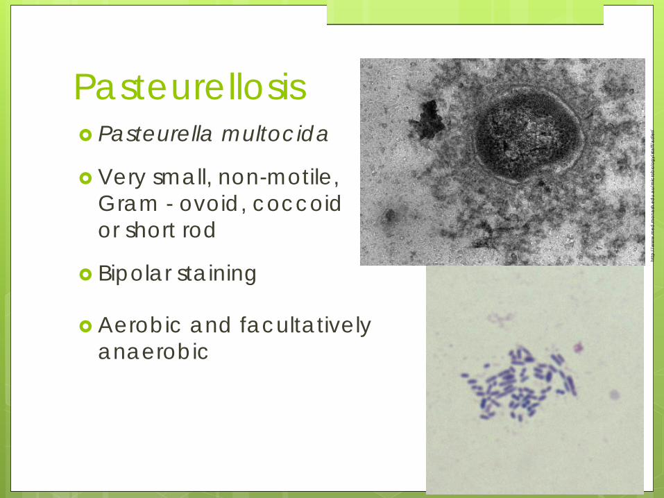
Pasteurellosis Pasteurella multocida
Very small, non-motile, Gram - ovoid, coccoid or short rod
Bipolar staining
http
://w
ww
.med
.mon
ash
.ed
u.a
u/m
icro
biol
ogy/
sta
ff/a
dler
/
Aerobic and facultatively anaerobic

Opportunistic bacteria
Virulent factorsendotoxin adhesins filamentous appendages help P. multocida
colonize mucous membranes
Pasteurellosis

Rhinitis (or snuffles) Pneumonia Genital infections Wound infections Abscesses Otitis media
Clinical signs in rabbits
ww
w.m
ri.sari.a
c.uk/%5C
jpg%
5Cb
act-rep10-fig2b.jpg
Pasteurellosis
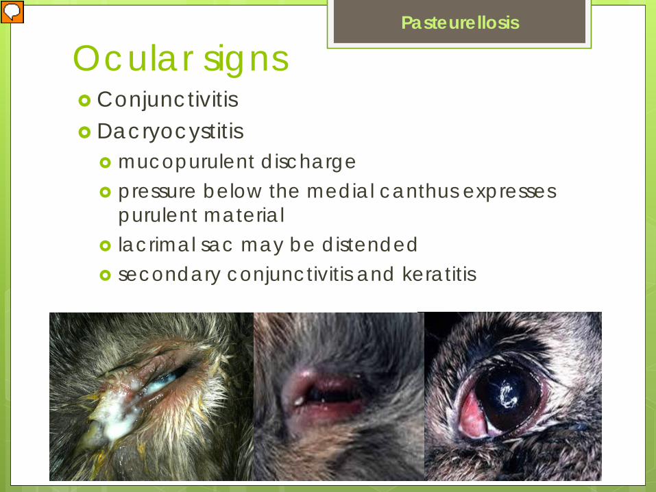
Ocular signs Conjunctivitis Dacryocystitis
mucopurulent discharge pressure below the medial canthus expresses
purulent material lacrimal sac may be distended secondary conjunctivitis and keratitis
http://www.merckvetmanual.com/mvm/index.jsp?cfile=htm/bc/171317.htm
Pasteurellosis

Brucellosis Brucella canis Zoonotic aerobic Gram-coccobacillus Survives in mononuclear cells. Penetrates mucous membranes. Ingestion and venereal transmission are
common. Can be transmitted via fomites, such as cages or
equipment.

Ocular signs Occur in ~ 14 % of dogs with brucellosis.
Endophthalmitis
Chronic uveitis
Hyphema
Chorioretinitis
Brucellosis

Systemic signs Diskospondylitis Glomerulopathy Meningoencephalitis Abortion and infertility are common in breeding
dogs Dogs not used for breeding may have
undetected disease for long periods of time due to the prolonged bacteremia and secondary localization
Brucellosis

Diagnosis Isolation and identification Serologic screening involves the rapid slide
agglutination test with and without 2-mercaptoethanol.
Tube agglutination, ELISA or IFA tests or the cytoplasmic protein agar gel immunodiffusion test have greater specificity
Zoonotic potential
Brucellosis

Case Report 3 neutered dogs Chronic recurrent uveitis Blood culture or PCR Responded to therapy
Brucellosis
Vet Ophthalmol. 2009 May-Jun;12(3):183-91.

Haemophilus spp. Haemophilus spp. requires factors from blood for
growth.
Normal flora of the oral cavity and nasopharynx
Also primary etiology for acute mucopurulent conjunctivitis in humans.
Little affinity for the avascular cornea, and corneal involvement is a rare complication of conjunctivitis.

ThromboembolicMeningoencephalitisHaemophilus somnusAcute septic TEME most commonly occurs in
feedlot cattle
Can Vet J. 1971 September; 12(9): 180–182.

Zoonotic Disease Acid-fast bacterium,
Mycobacterium bovis Ocular tuberculosis is
rare in cattle and swine subretinal exudation
retinal hemorrhage
anterior uveitis
endophthalmitis with a granulomatous response
Bovine tuberculosis
Basic Science Guides Design of New TB Vaccine Candidates M. J. Friedrich JAMA. 2005;293:2703-2705.http://jama.ama-assn.org/cgi/content/extract/293/22/2703
Affected small animals are in farm settings and are drinking unpasteurized milk.

Mycoplasma Smallest prokaryotic cells capable of self-
replication
Ubiquitous free-living saprophytes (eg. members of the Genus Acholeplasma)
Animal pathogens include the genera Mycoplasma and Ureaplasma

Lack a true cell wall, but have a plasma membrane. accounts for their plasticity and pleomorphism,
including cocci, spiral filament, and ring-like structures
http://www.malp-research.de/malp_history.html, webmedia.unmc.edu/.../2SL31-mycoplasma.jpg
Mycoplasma

stain poorly with Gram stain Giemsa and other Romanowsky stains are better fragility, pleomorphism and weak staining
characteristics make direct examination of stained smears of limited value in making a diagnosis
Mycoplasma
Vet Clin Pathol 41/2 (2012) 283–290

Pathogenesis Adhere to host mucous membranes where they
remain extracellular Produce
hemolysins proteases nucleases other toxic factors
Latency can occur Host specific Eye infections are characterized by serous
discharge and conjunctival hyperemia
Mycoplasma
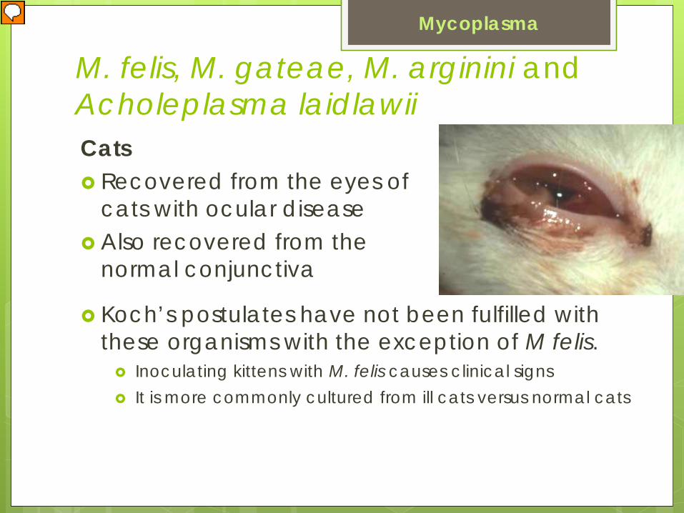
M. felis, M. gateae, M. arginini and Acholeplasma laidlawiiCats Recovered from the eyes of
cats with ocular disease Also recovered from the
normal conjunctiva
Mycoplasma
Koch’s postulates have not been fulfilled with these organisms with the exception of M felis. Inoculating kittens with M. felis causes clinical signs It is more commonly cultured from ill cats versus normal cats

M conjunctivae and M agalactiaeSheep
keratoconjunctivitis (with corneal vascularization but not corneal ulcers)
Goats (and maybe sheep)
M. agalactiae causes systemic disease including arthritis, mastitis or abortion
M. mycoides subsp. mycoides (large-colony type) causes septicemia, mastitis, and arthritis

Ophthalmic signs in sheep and goatsMycoplasma keratoconjunctivitis anterior uveitis choroiditis hyalitis
M. mycoides in goats keratoconjuctivitis with perilimbal corneal
opacities eye lesions may occur without systemic signs

M. bovoculi, Ureaplasma spp.,M. laidlawii and M. bovirhinisCattle bovine conjunctivitis association with M bovis has not been
confirmed. tends to occur in the summer mild and self-limiting

Mycoplasma bovisCattle
Pneumonia
Arthritis
Mastitis
Meningitis
Infertility
Subcutaneous abscesses in cattle
Keratoconjunctivitis
Mycoplasma

Chlamydophila sp.
Obligate intracellular organisms
Cell walls similar to those of other Gram-bacteria
Lack the machinery that allow autonomous survival and replication http://www.merckvetmanual.com/mvm/index.jsp?cfile=htm/bc/201700.htm
(Chlamydiae)

The cell replication cycle involves:
Extracellular (elementary body)
0.2-0.6 µm in size with rigid cell walls
Intracellular (initial body, reticulate body)
0.5 – 1.5 µm lack cell walls
http
://u
ploa
d.w
ikim
edia
.org
/wik
iped
ia/e
n/th
umb/
1/1a
/Chl
amyd
ophi
la_p
neum
onia
e.jp
g/20
0px-
Chl
amyd
ophi
la_p
neum
onia
e.jp
gw
ww
.vet
.uga
.edu
/.../
turn
eran
drob
bins
/Fig
1.jp
gFig
ure
1.El
emen
tary
bod
ies o
f Chl
amyd
ophi
la p
sitta
ci
(pre
viou
sly C
hlam
ydia
psit
taci
) in
the
cyto
plas
m o
f hep
atoc
ytes
(Par
rot,
liver
, hem
atox
ylin
and
eos
in st
ain)
.

Previously all members of the family Chlamydiaceae were known as one species Chlamydia psittaci
Currently there are 2 genera, Chlamydia and Chlamydophila, and multiple species within each

Chlamydophila felis Transmission
direct contact or aerosols.
Only survives a few days in the environment. Cellular and hormonal mechanisms play a role in
immunity. Cats under < 8 weeks and > 5 years are unlikely
to become infected. In general chlamydiae are considered to have a
restricted host range.
Pathogensis Highly contagious Spreads rapidly by direct contact A low dose incites unilateral disease and a high
dose incites bilateral disease. Spread internally to colonize many tissues
including the tonsil, lung, liver, spleen and kidney.
Shed in the tears and nasal secretions May persist in the ocular tissues for months
following remission of ocular signs.
Chlamydophila felis

Clinical signs initial chemosis
suppurative conjunctivitis
petechial hemorrhages
conjunctival lymphoid follicles
+/- respiratory signs
Chlamydophila felis

Clinical signs
Serous ocular discharge becomes mucoid or mucopurulent within 3-5 days
Cats that become bilaterally affected have clinical signs persist for 22-25 days.
Recovery can result in persistent infections with conjunctival shedding for up to 8 months.
Chronic shedding of organisms from the urogenital and GI tract has been documented.
Chlamydophila felis
Experimental infection unilateral conjunctivitis within 5-
10 days of exposure

Diagnosis C. felis, in contrast to Chlamydial infections in
other species, is not associated with keratitis. Isolation from conjunctival cotton swabs (without
wooden sticks) ELISA PCR FA
Chlamydophila felis
Intracytoplasmic elementary bodies
Vet Clin Pathol 41/2 (2012) 283–290

Treatment Topical oxytetracycline QID for 2 weeks past
resolution of clinical signs
Erythromycin and chloramphenicol are also effective
Pradofloxacin vs doxycycline
Chlamydophila felis

Disease in Farm Animals Chlamydophila pecorum
Chlamydophila abortus
Chlamydia suis

C. pecorum Cattle, sheep and swine Encephalomyelitis Enteritis Polyarthritis Metritis Pneumonia Conjunctivitis Typically young animals are affected in sporadic
outbreaks.

C. abortus Sheep
Conjunctivitis
Keratitis
Polyarthritis
Pneumonia
Orchitis
Epididymitis
Abortion
Chlamyodophilosis among lambs and kids may produce both ocular signs and polyarthritis.
C. abortus

Ocular signs Bilateral in 80% Conjunctiva lesions
Conjunctivitis
Petechial hemorrhages
Epiphora and purulent exudation
Lymphoid follicle proliferation (which may become confluent, producing folds)
Cornea Peripheral edema and neovascularization
Ulceration is rare
C. abortus
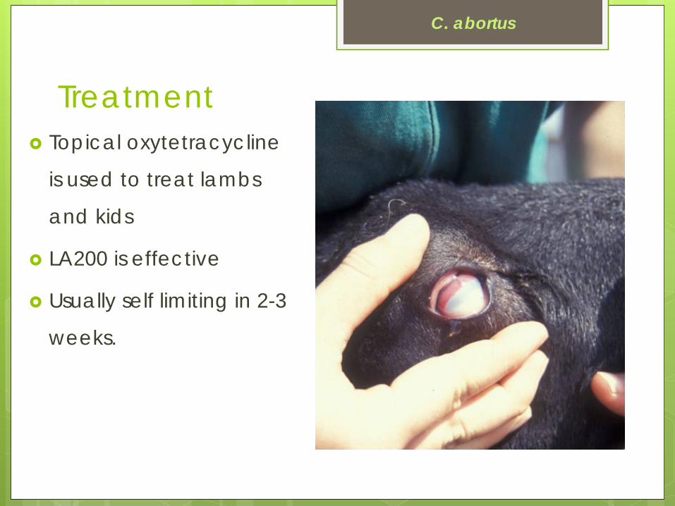
Treatment Topical oxytetracycline
is used to treat lambs
and kids
LA200 is effective
Usually self limiting in 2-3
weeks.
C. abortus

Diagnosis Conjunctival cytology
Culture
ELISA
PCR
C. abortus
Persistent infection with intermittent shedding is common among ovine chlamydial diseases.
The elementary bodies are relatively resistant and may remain viable for several days.

C suisSwine Conjunctivitis Keratoconjunctivitis Enteritis Pneumonia Lymphofollicular hyperplasia of the palpebral
conjunctiva Isolated from the conjunctiva of healthy pigs.
Journal of Zoo and Wildlife Medicine 44(1): 159–162, 2013

Chlamydophila psittaci
Avian chlamydiosis
Subclinical, acute, subacute, or chronic infection
Worldwide at least 150 avian species
Respiratory, digestive, or systemic infection.
10-30% of surveyed avian populations may be found positive.
Serotypes All share an identical genus-specific antigen in
their lipopolysaccharide Currently, 8 serotypes are recognized The same strain may cause mild disease or
asymptomatic infection in one species, but severe or fatal disease in another species.
Avian serotypes are capable of infecting people and other mammals.
Chlamydophila psittaci

Transmission Airborne elementary bodies are resistant to
drying. Also spread by fecal oral transmission. Stress can initiate shedding and cause
recurrence. Carriers can shed the organism for extended
periods. The incubation period typically is 3-10 days.
Chlamydophila psittaci

Clinical signs Nasal and ocular discharge Conjunctivitis Sinusitis Green to yellow-green
droppings Fever Inactivity Ruffled feathers Weakness Inappetence, and weight loss Asymptomatic infections are common
Chlamydophila psittaci

Diagnostics clinical findings
hematology
clinical chemistries
radiology
organism can be seen in impression smears of affected tissues stained by Giemsa, Gimenez, or Macchiavello’s method.
IFA, ELISA, PCR
ww
w.vet.uga
.edu/vpp/clerk/Bockino/Fig1.jpg
Chlamydophila psittaci
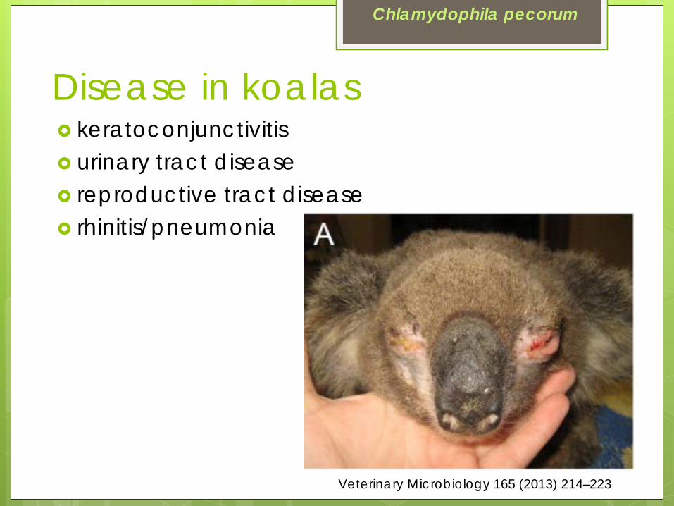
Disease in koalas
Veterinary Microbiology 165 (2013) 214–223
Chlamydophila pecorum
keratoconjunctivitis urinary tract disease reproductive tract disease rhinitis/pneumonia
Treatment
Chlamydophila pecorum
Response to treatment is variable Decreased palpebral fissure, entropion
and corneal scarring may result. Enrofloxacin, chloramphenicol Not tetracyclines and macrolides

Bartonella Gram-negative
Facultative intracellular rod or coccobacillus
Bartonellaceae family
Transmitted by arthropods Infection is suspected to be primarily in RBCs, but
infection of vascular endothelium also occurs.

In Cats Bartonella henselae is the most frequently
reported species to infect cats Naturally occurring infection is mild and transient Clinical findings: pyrexia, lymphadenopathy,
lethargy, anorexia, CNS disorders, urologic diseases and endocarditis
Ocular disease: uveitis, keratitis and chorioretinitis
Bartonella

In DogsB henselae, B vinsonii, B clarridgeiae, &
B elizbethaeAnterior uveitisHyphemaChorioretinitisRetinal detachment due to systemic
hypertensionMultiple systemic signsDiagnosis is based on serology
Bartonella
Diagnosis in cats Serology Blood culture PCR In the United States, 12-67% of the cats are
seropositive for Bartonella. A higher prevalence of affected cats occurs in
warm, humid areas which have more fleas.
Bartonella

Neonatal septicemia/bacteremia
Commonly isolated bacteria in foals E coli Klebsiella spp Actinobacillus spp Enterobacter spp Pseudomonas spp Rhodococcus equi

In foalsRoutes of entry for bacteria
Placenta, umbilicus, lungs & GI tract. The major risk factor for septicemia is failure of passive transfer of colostral antibodies.Other factors include
unsanitary environmental conditions gestational age of the foal (prematurity) health and condition of the mare difficulty of parturition new pathogens in the environment
Neonatal septicemia/bacteremia
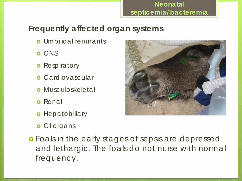
Frequently affected organ systems Umbilical remnants
CNS
Respiratory
Cardiovascular
Musculoskeletal
Renal
Hepatobiliary
GI organs
Foals in the early stages of sepsis are depressed and lethargic. The foals do not nurse with normal frequency.
Neonatal septicemia/bacteremia

Ophthalmic findings Fibrin in the anterior chamber Hypopyon Severe miosis Entropion (if the foal is dehydrated)
Neonatal septicemia/bacteremia

Neonatal septicemia/bacteremiaCommonly isolated bacteria in farm animals E coli Klebsiella spp Actinobacillus spp Streptococcus spp Arcanobacterium
pyogenes Salmonella spp Pasteurella spp

Disease in farm animals Calves, piglets, kids and
lambs
Umbilical infections or ingesting bacteria.
Clinical signs include polyarthritis, meningitis and/or diarrhea.
Ocular lesions: fibrin clots in the AC, hypopyon or hyphema, miosis, and chorioretinal embolic lesions
Neonatal septicemia/bacteremia
Anaerobic Pathogens Anaerobic bacteria possess complex species-
dependent virulent mechanisms. Direct corneal damage
Elaboration of toxins, metabolites, enzymes, and degradation products
Indirect corneal damage Stimulation of corneal immune responses.

Anaerobic Bacteria Isolated from dogs, cats, horses and alpacas
with ulcerative keratitis.
Isolated from 13% of corneal samples.
Genera Clostridium, Peptostreptococcus, Actinomyces, Fusobacterium, and Bacteroides.
Positive correlation between isolation and ocular trauma, preexisting corneal disease and chronic dermatologic disease.
Anaerobic Pathogens

Concurrent or prior facultative aerobic bacterial multiplication
Aerobic bacteria may also produce essential nutrients, growth factors, energy substrates and protective enzymes.
Mixed infections provide mutual protection from phagocytosis and intracellular killing.
Anaerobic Pathogens

Most antimicrobials have limited to no microbial action against anaerobic pathogens.
Antimicrobial susceptibility patterns are relatively predictable (except Bacteriodes spp.)
Elimination of synergistic aerobic bacteria and disruption of low oxygen corneal microenvironment may be mechanism.
Anaerobic Pathogens
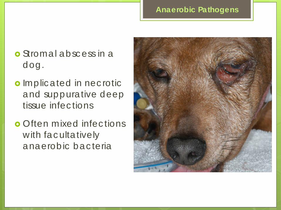
Stromal abscess in a dog.
Implicated in necrotic and suppurative deep tissue infections
Often mixed infections with facultatively anaerobic bacteria
Anaerobic Pathogens

Other Bacterial Infections
Rickettsia
Ehrlichia
Borrelia
Leptospira

Rickettsial species minute, obligate intracellular bacteria transmitted by ticks rod-shaped or coccobacilli 0.3 to 0.6 µm in length
•users.wfu.edu/derkls4/images/attachment%20of%...
contain both RNA and DNA replicate primarily within the
cytosol of target cells. nonmotile aerobic

Rocky Mountain Spotted Fever R rickettsii An acute febrile illness in dogs and humans. USA, Western Canada and Central and South
America.

Tick vectors are D variabilis and D andersoni. Methods for tick infection Transmission does not occur for 5-20 hours post
attachment
stri.discoverlife.org/IM/I_GA/0000/640/Dermac...
Rocky MountainSpotted Fever

Pathogenesis Vasculitis is the primary lesion Pathogenesis relates directly to the vascular lesions
which initiate platelet activation and activation of the coagulation system.
Rocky MountainSpotted Fever

Clinical signs in dogs FeverNeurologic dysfunction Polyarthritis Petechial and ecchymotic hemorrhages ThrombocytopeniaNonregenerative anemia
Signs begin within 3 days of tick attachment.
Hemorrhages most commonly occur on mucous membranes, but epistaxis, melenaand hematuria may be present in severely affected animals.
Rocky MountainSpotted Fever

Ocular signs Altered vascular permeability in the conjunctiva,
uvea, and retina results in ocular signs.
Rocky MountainSpotted Fever

Conjunctivitis conjunctivitis chemosis petechial hemorrhages mucopurulent to purulent
ocular discharge Anterior segment iris stromal petechiations anterior uveitis hyphema
Rocky MountainSpotted Fever

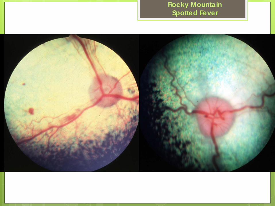
Posterior segment retinitis characterized by perivasculitis,
focal areas of edema, and petechiation.
Unilateral or bilateral optic neuritis
Ocular disease may be confined to the retina
Ophthalmic lesions are usually mild
Rocky MountainSpotted Fever
Rocky MountainSpotted Fever

Experimental infectionFluorescein angiography ↑permeability in retinal
vessels beginning 6 days post infection and 2 days after onset of pyrexia.
Venules are more permeable than arterioles
Rocky MountainSpotted Fever

Histopathology Necrotizing vasculitis with perivascular
accumulations of PMNs and lymphoreticular cells.
Organs with endarterial circulation such as skin, brain, heart, kidney and retina are affected.
Rocky MountainSpotted Fever

Diagnosis
rising serum titers on micro IFA test
isolation
•ww
w.answ
ers.com/.../rocky-m
ountain-spotted-feverw
ww
.cdc.gov/ncid
od/dvrd/rmsf/IM
AG
ES/rick-IFA.jp
gRocky Mountain
Spotted Fever

Treatment Doxycycline Tetracycline Chloramphenicol Enrofloxacin 14-21 days is usually effective +/- inflammatory doses of oral prednisone in
conjunction with antibiotic therapy.
Rocky MountainSpotted Fever

Ehrlichia canis
Causes canine monocytic ehrlichiosis
Acute, subclinical and chronic disease.
Gram - bacteria
Lack peptidoglycan and lipopolysaccharide components.

Transmission Obligate intracellular parasite transmitted by the
brown dog tick, Rhipicephalus sanguineus. The tick can transmit the disease more than 5
months after detaching from the canine host.
E. canis
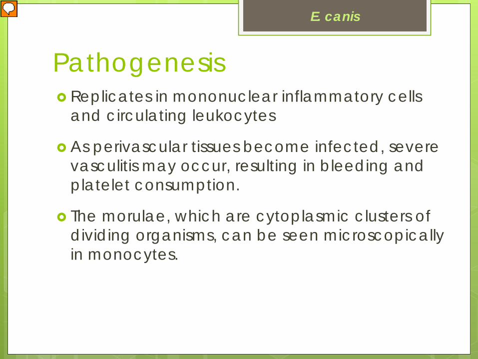
Pathogenesis Replicates in mononuclear inflammatory cells
and circulating leukocytes
As perivascular tissues become infected, severe vasculitis may occur, resulting in bleeding and platelet consumption.
The morulae, which are cytoplasmic clusters of dividing organisms, can be seen microscopically in monocytes.
E. canis

Disease in dogs Acute phase occurs 8-20 days post infection and
lasts 2-4 weeks. Common clinical signs include fever and depression +/- neurologic signs petechial and ecchymotic hemorrhages,
epistaxis lymphadenopathy limb edema vomiting
Ticks are found on 40% of dogs.
E. canis

Subclinical phase The subclinical phase lasts for weeks to months.
Clinical signs may regress.
E. canis

Chronic phase May persist for years Signs may include:
depression
weight loss
pale mucous membranes
abdominal tenderness
bleeding episodes
secondary infections
limb edema
E. canis

Associated with:
acute vasculitis
perivasculitis
thrombocytopenia
platelet dysfunction
hyperviscosity
Ocular signs
E. canis

Ocular signs Conjunctival hyperemia
and hemorrhages Corneal edema Deep corneal
vascularization Anterior uveitis Chorioretinitis Panuveitis Optic neuritis
E. canis

Chronic ocular signs
hyperproteinemia and hyperviscosity syndrome lead to retinal vascular engorgement
E. canis
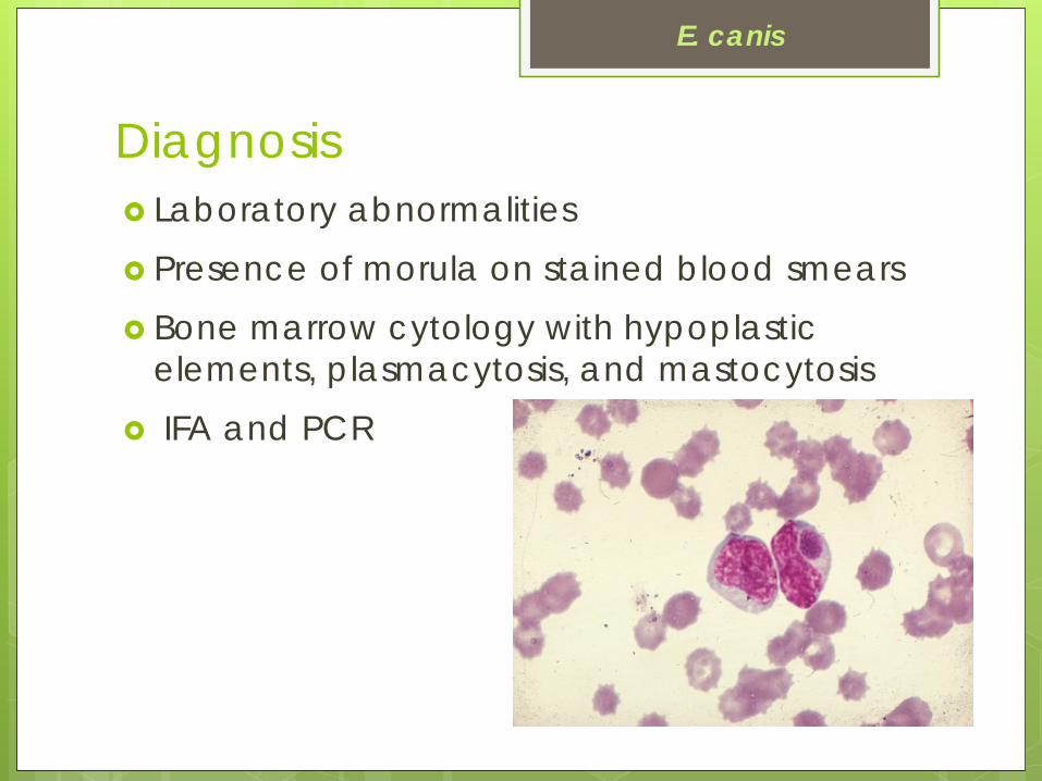
Laboratory abnormalities Presence of morula on stained blood smears Bone marrow cytology with hypoplastic
elements, plasmacytosis, and mastocytosis IFA and PCR
Diagnosis
E. canis

Histopathology of E. canis The most consistent histopathologic finding is a
predominantly monocytic or lymphocytic cell infiltrate of the uveal tract, retina and optic nerve.
E. canis

Anaplasma platys Rickettsial parasite
Causes infectious thrombocytopenia in dogs
Reported to cause uveitis
E. chaffeensis, E. ewingii, and E. equi (in dogs) may also cause anterior uveitis.

Borreliosis or Lyme’s Disease Tick-borne spirochetosis Borrelia burgdorferi Small corkscrew shaped
motile microaerophilic bacteria.
Do not survive free living in the environment.
www.medicalecology.org/diseases/lyme/em.jpg
Highest incidence of disease remains in the northeastern US.

Transmission of B. burgdorferi Ticks of the Ixodes sp. Primary reservoirs are
small rodents and birds. Transmission relates to
contact time of the tick on the host 48 to 72 hours for a
38-92% transmission rate.
www.cdc.gov/.../images/TickMaster4_12_w452.gif

Pathogenesis Once inside the host, the
organisms use their specialized endoflagella to move through the connective tissues.
Organisms can survive for years in skin, connective tissue, joints and nervous system.
Borreliosis

Disease in dogs and cats Systemic signs in dogs and cats include
lameness, joint pain, pyrexia and lymphadenopathy.
Ocular lesions include conjunctivitis, corneal edema, anterior uveitis, retinal petechia, chorioretinitis, and retinal detachment
Definitive proof is lacking…..
Borreliosis

Borreliosis in other species Organisms were found in the anterior chamber
of a pony and 2 horses.
Humans exhibit conjunctivitis, keratitis, panuveitis, chorioretinitis, retinal detachment, optic neuritis and periorbital edema.
Borreliosis
VO (2012) 15, 6, 398–405

Diagnosis There is a high prevalence of serum + antibody
titers (75%) with actual disease in ~ 5-10% of dogs.
www.lersus.de/.../1/res/files/borrelia_0_.jpg
Current lab tests employ the specific c6 lipoprotein antigen in ELISA and western blot
Borreliosis

Leptospirosis Motile spirochetal bacteria The predominant serovars responsible for
causing disease in dogs are canicola, icterohemorrhagica, grippotyphosa, pomona, and bratislava.
Maintained in host adapted species that act as reservoir hosts and is shed in the urine.
Direct transmission through contact with infected urine, bites, ingestion of affected material and contact with contaminated water.
http://www.med.monash.edu.au/microbiology/staff/adler
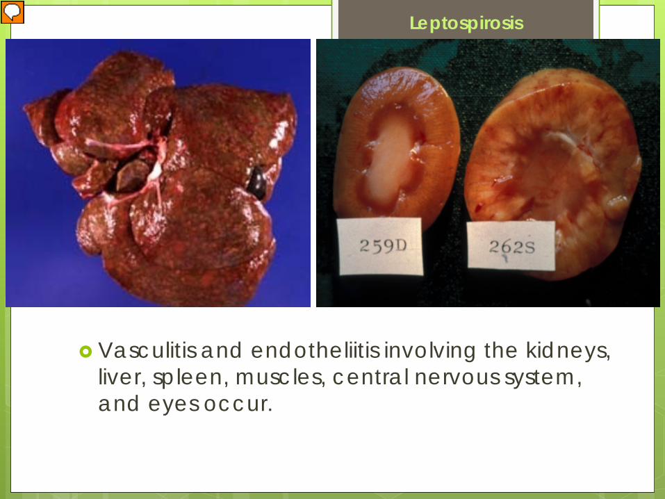
Vasculitis and endotheliitis involving the kidneys, liver, spleen, muscles, central nervous system, and eyes occur.
Leptospirosis

Lesions in dogsInfrequent
conjunctivitis with mucopurulent oculonasal discharge
scleritis
anterior uveitis
Systemic signs renal or hepatic failure or dysfunction.
Leptospirosis

Disease in horsesAcute signs Transient depression, fever, icterus, anemia
and anorexia. Serologic surveys of horses have shown that
exposure to Leptospira is common, but variable, according to the geographic location or climate.
Horses positive for leptospirosis are common in the Ohio, Delaware, Tennessee, and Mississippi river valleys.
Leptospirosis

Leptospira interrogansmost likely plays a role in many cases of ERU.
20 serovars L interrogans serovar
pomona is most often associated with ERU.
Usually horses develop ERU 18 to 24 months after the initial infection.
Leptospirosis

Diagnosis
Clinical signs Microscopic agglutination test or ELISA Histology PCR Cultured from the aqueous humor of dogs Can be shed in the urine up to 3 months
www.buddycom.com/bacteria/nongram/leptofa1346.jpg
Sequestered in kidneys, liver, spleen, CNS and eyes.


Proteases and toxins Increase virulence by damaging tissue and
interfering with host defense systems. Exo-products contribute directly to keratitis
through toxic effects on corneal cells and degradation of corneal proteins and indirectly through activation of corneal proteases.