Embed Size (px)
Citation preview

RESEARCH Open Access
Bacteremia in critical care units at BugandoMedical Centre, Mwanza, Tanzania: the roleof colonization and contaminated cots andmothers’ hands in cross-transmission ofmultidrug resistant Gram-negative bacteriaVitus Silago1,2* , Dory Kovacs3, Delfina R. Msanga4, Jeremiah Seni1, Louise Matthews3, Katarina Oravcová3,Ruth N. Zadoks3,5, Athumani M. Lupindu6†, Abubakar S. Hoza2† and Stephen E. Mshana1†
Abstract
Background: Multidrug resistance (MDR) is a major clinical problem in tertiary hospitals in Tanzania and jeopardizes the lifeof neonates in critical care units (CCUs). To better understand methods for prevention of MDR infections, this study aimed todetermine, among other factors, the role of MDR-Gram-negative bacteria (GNB) contaminating neonatal cots and hands ofmothers as possible role in transmission of bacteremia at Bugando Medical Centre (BMC), Mwanza, Tanzania.
Methods: This cross-sectional, hospital-based study was conducted among neonates and their mothers in a neonatalintensive care unit and a neonatology unit at BMC from December 2018 to April 2019. Blood specimens (n= 200) were sub-cultured on 5% sheep blood agar (SBA) and MacConkey agar (MCA) plates. Other specimens (200 neonatal rectal swabs, 200maternal hand swabs and 200 neonatal cot swabs) were directly inoculated on MCA plates supplemented with 2 μg/mlcefotaxime (MCA-C) for screening of GNB resistant to third generation cephalosporins, r-3GCs. Conventional biochemicaltests, Kirby-Bauer technique and resistance to cefoxitin 30 μg were used for identification of bacteria, antibiotic susceptibilitytesting and detection of MDR-GNB and screening of potential Amp-C beta lactamase producing GNB, respectively.
(Continued on next page)
© The Author(s). 2020 Open Access This article is licensed under a Creative Commons Attribution 4.0 International License,which permits use, sharing, adaptation, distribution and reproduction in any medium or format, as long as you giveappropriate credit to the original author(s) and the source, provide a link to the Creative Commons licence, and indicate ifchanges were made. The images or other third party material in this article are included in the article's Creative Commonslicence, unless indicated otherwise in a credit line to the material. If material is not included in the article's Creative Commonslicence and your intended use is not permitted by statutory regulation or exceeds the permitted use, you will need to obtainpermission directly from the copyright holder. To view a copy of this licence, visit http://creativecommons.org/licenses/by/4.0/.The Creative Commons Public Domain Dedication waiver (http://creativecommons.org/publicdomain/zero/1.0/) applies to thedata made available in this article, unless otherwise stated in a credit line to the data.
* Correspondence: [email protected]†Athumani M. Lupindu, Abubakar S. Hoza and Stephen E. Mshanacontributed equally to this work.1Department of Microbiology and Immunology, Weill Bugando School ofMedicine, Catholic University of Health and Allied Sciences, P. O. Box 1464,Bugando, Mwanza, Tanzania2Department of Veterinary Microbiology, Parasitology and Biotechnology,College of Veterinary Medicine and Biomedical Sciences, Sokoine Universityof Agriculture, P. O. Box 3000, Morogoro, TanzaniaFull list of author information is available at the end of the article
Silago et al. Antimicrobial Resistance and Infection Control (2020) 9:58 https://doi.org/10.1186/s13756-020-00721-w

(Continued from previous page)
Results: The prevalence of culture confirmed bacteremia was 34.5% of which 85.5% were GNB. Fifty-five (93.2%) of GNBisolated from neonatal blood specimens were r-3GCs. On the other hand; 43% of neonates were colonized with GNB r-3GCs, 32% of cots were contaminated with GNB r-3GCs and 18.5% of hands of neonates’mothers were contaminated withGNB r-3GCs. The prevalences of MDR-GNB isolated from blood culture and GNB r-3GCs isolated from neonatal colonization,cots and mothers’ hands were 96.6, 100, 100 and 94.6%, respectively. Significantly, cyanosis (OR[95%CI]: 3.13[1.51–6.51], p=0.002), jaundice (OR[95%CI]: 2.10[1.07–4.14], p= 0.031), number of invasive devices (OR[95%CI]: 2.52[1.08–5.85], p = 0.031) andcontaminated cot (OR[95%CI]: 2.39[1.26–4.55], p= 0.008) were associated with bacteremia due to GNB. Use of tap water only(OR[95%CI]: 2.12[0.88–5.09], p= 0.040) was protective for bacteremia due to GNB.
Conclusion: High prevalence of MDR-GNB bacteremia and intestinal colonization, and MDR-GNB contaminating cotsand mothers’ hands was observed. Improved cots decontamination strategies is crucial to limit the spread of MDR-GNB. Further, clinical presentations and water use should be considered in administration of empirical therapy whilstawaiting culture results.
Keywords: Antimicrobial resistance, Hand hygiene, Hospital surfaces contamination, Multidrug resistant bacteria,Bacteremia
BackgroundMultidrug resistance (MDR) is defined as acquired resistanceto at least one agent in three or more antimicrobial classes[1–3]. MDR is a growing global concern which is estimatedto cause 10 million deaths and cost US$100 trillion annuallyby 2050 [4, 5]. Improper use of antibiotics in human and vet-erinary medicine, counterfeit antibiotics and non-compliantuse of rationally prescribed antibiotics are among factorsdriving the emergence and spread of MDR bacteria [6]. Highantibiotic pressure (empirically prescribed and administered)in the critical care units (intensive care units and neonat-ology units) results in the selection and emergence of MDRbacteria [7, 8]. The spread of MDR bacteria in healthcare set-tings presents a challenge, as treating infected patients be-comes increasingly difficult with poorer outcomes [6]. MDRGram-negative bacteria (MDR-GNB) such as beta-lactamase(extended spectrum beta-lactamase (ESBL), Amp-C beta-lactamase and carbapenemases) producing Enterobacteria-ceae, Acinetobacter baumannii and Pseudomonas aeruginosaare frequently reported, causing infections in critical careunits globally [9–12]. These organisms are responsible forbloodstream infections (BSIs), urinary tract infections (UTIs),pneumonia, and skin and soft tissue infections, resulting inhigh morbidity and mortality [13].In critical care units, infections due to MDR-GNB bac-
teria may be acquired endogenously or exogenously [14].Endogenous acquisition occurs from the patient’s ownbody flora colonizing a certain body surface, for ex-ample, MDR-GNB colonizing patient’s gastrointestinaltract (such as MDR-E. coli) may cause an extra-intestinal infection (e.g., BSI and UTI) which may evenresult to mortality from treatment failure [14, 15]. Ex-ogenous acquisition occurs due to contact with otherpeople e.g., healthcare workers (HCWs), patients or caregivers (CGs) i.e., mothers; and/or contaminated surfaces,such as ventilators, beds, side tables and infusion stands,
and water sources [14, 16]. Contaminated patients’ envi-ronments with MDR-GNB increases the risk of exogen-ous acquire of healthcare associate infections (HCAIs)from MDR-GNB which are mostly cross-transmitted bycontaminated hands of HCWs and CGs [17]. Hands ofHCWs and CGs become contaminated when touchingcontaminated surfaces and even colonized patients dur-ing provision of medical care [18].In Mwanza, Tanzania, 10.5 to 49% of bacteremia cases
due to GNB are caused by MDR-GNB with mortality rateranging from 34.4 to 52% as compared to mortality withnone MDR-GNB which ranging from 16.2 to 25% [11, 19,20]. Rectal colonization of neonates with extended-spectrum beta-lactamase producing Enterobacteriaceae(ESBL-PE) is common (25.4 to 54.6%) [19, 20], as is con-tamination of the hospital’s inanimate surfaces (33.5%) [21].In Tanzania, it is common that, mothers play an importantrole in feeding and caring for hospitalized neonates. Todate, their role in infection transmission or prevention hasnot been considered. To reduce the incidence and improvethe management of MDR-GNB cases in the neonatal ICUand neonatology unit at BMC, we explored exogenous andendogenous risk factors for neonatal MDR-GNB sepsis, in-cluding potential exogenous exposures in the household oforigin, in the hospital or from mothers, and endogenous ex-posure (neonatal carriage). Results can be used to informcase management, and to target infection prevention andcontrol measures to reduce case incidence.
MethodsThe aim, design and setting of the studyA cross-sectional hospital-based study was conductedbetween December 2018 and July 2019 aimed to deter-mine, among other factors, the role of MDR-Gram-negative bacteria (GNB) contaminating neonatal cotsand hands of mothers as possible role in transmission of
Silago et al. Antimicrobial Resistance and Infection Control (2020) 9:58 Page 2 of 14

bacteremia among neonates admitted to the neonatalICU (NICU) and neonatology unit at Bugando MedicalCentre (BMC), Mwanza, Tanzania. BMC is a tertiary, teach-ing, consultancy and zonal referral hospital with an estimated1000-bed capacity, serving Lake Zone regions (Mwanza,Simiyu, Kagera, Shinyanga, Musoma, Tabora, Geita andKigoma) and a catchment population of 13 million people(https://www.bugandomedicalcentre.go.tz/index.php). TheNICU was equipped with 15 neonatal cots (with no walkingspace between cots), 15 trained nurses and 2 pediatricians.In the neonatology unit, there were 36 cots about 0.5mapart, 11 trained nurses and 4 pediatricians. When operatingat or above capacity, two neonates may share a cot (in bothunits). In both units, the cots are irregularly disinfected be-fore new occupancy by using 1:50 Dettol in water.
Sample size calculation and selection criteriaA minimum sample size for this study was 144 partici-pants, which was calculated using Kish Leslie formula of1965 [22], using an MDR-GNB prevalence of 10.5% [20].Neonates admitted to NICU and neonatology unit withsigns and symptoms of infections as previous reportedby “WHO Young infants Study group” [23] and theirmothers were enrolled in this study. Neonates with signsand symptoms of infection but either missing socio-demographic information or a complete set of specimenswere excluded from the final analysis (n = 15). Partici-pants (neonates and mothers) moving between the neo-natal ICU and neonatology units were not re-enrolled.
Data and specimen collectionStructured questionnaires were used to obtain socio-demographic and clinical information from study partici-pants after the mother or guardian consented to partici-pation. Neonatal blood samples, neonatal rectal swabs,cot swabs and maternal hand swabs were collected.About 1 ml of venous blood was collected into an in-house made tryptone soy broth (TSB, 10 ml) by paedia-trician; rectal swabs were collected by a trained medicaldoctor; and bed swabs (in every new occupancy) andmothers’ hand swabs specimens were collected. All swabsamples were collected using sterile cotton swabs pre-moistened in sterile 0.85% physiological saline. All swabspecimens were transported to the laboratory in Amiestransport media (Amies, UK). In total, 800 specimens(200 blood, 200 rectal swabs, 200 bed swabs and 200mothers’ hands swabs) were collected. All specimenswere sent to the microbiology laboratory of the Cath-olic University of Health and Allied Sciences for isola-tion, identification, antibiotic susceptibility testing anddetection of MDR-GNB following in-house standardoperating procedures and international guidelinessuch as Clinical and Laboratory Standard Institute(CLSI, 2018) [24].
DefinitionsIn this study, GNB isolated from blood with resistanceto ceftriaxone and/or ceftazidime and GNB isolated fromrectal, bed and hand swabs grown on MacConkey agarplates supplemented with 2 μg/ml cefotaxime (MCA-C)were considered resistant to third generation cephalo-sporins (r-3GCs) [11]. All GNB isolated from neonates’blood, rectal, cots and mothers’ hands swab specimensshowing resistance to at least one antibiotic agent inthree different classes of antibiotics i.e., penicillins: ampi-cillin (AMP), amoxicillin/clavulanate (AMC), piperacil-lin/tazobactam (TZP); third generation cephalosporins(3GCs): ceftriaxone (CRO), ceftazidime (CAZ) and/orisolated on MCA-C; carbapenems: meropenem (MEM);trimethoprim-sulfamethoxazole (SXT); aminoglycosides:gentamicin (CN), amikacin (AK); fluoroquinolones: cip-rofloxacin (CIP); tetracyclines: tetracycline (TET); and/or polymyxins: colistin (CT), were termed as MDR-GNB as previously reported [1, 2]. In this paper, isolatesexhibiting intermediate activities against antibiotics werealso termed as resistant.
Laboratory procedures
Bacterial isolation, identification and antibioticsusceptibility testing Clinical specimens (blood):Blood specimens in TSB bottles were incubated aerobic-ally at 37 °C for 18–24 h upon receipt in the laboratory,and before being inoculated onto in-house prepared 5%sheep blood agar (SBA) and MacConkey agar (MCA)plates (Oxoid, UK). SBA and MCA plates were incu-bated aerobically at 37 °C for 18–24 h. However isolationof Gram positive bacteria was not the objective of thisstudy, we purposely isolated and identified them andtheir antibiotic susceptibility testing were performed toguide rational antibiotic therapy for proper patients’management only.Isolated bacteria were identified by in-house prepared
conventional biochemical identification tests includingsugars fermentation, CO2 gas production and sulfur pro-duction by triple sugar iron (TSI) test; sulfur production,indole production and motility by sulfur-indole-motility(SIM) test, urease production by urease test; utilizationof citrate as the sole source of energy by Simmons’ cit-rate test; and oxidase production by oxidase test stripsas reported previously [25]. Kirby-Bauer disc diffusionmethod was used for antibiotics susceptibility testing(AST) on MHA plates [26]. Briefly, bacterial suspensionsequivalent to 0.5 McFarland turbidity standard solutionwere prepared from a MacConkey subculture (arisingfrom a cultured clinical specimen and one isolated col-ony from cefotaxime-supplemented MacConkey agar)into sterile 0.85% physiological saline and then swabbedon entire plates of MHA (Oxoid, UK). Ampicillin (AMP)
Silago et al. Antimicrobial Resistance and Infection Control (2020) 9:58 Page 3 of 14
10 μg, trimethoprim-sulfamethoxazole (SXT) 25 μg, ami-kacin (AK) 30 μg, tetracycline (TE) 30 μg, piperacillin-tazobactam (TZP) 110 μg, gentamicin (CN) 10 μg, cipro-floxacin (CIP) 5 μg, amoxicillin-clavulanic acid (AMC)30 μg, ceftriaxone (CRO) 30 μg, ceftazidime (CAZ) 30 μg,meropenem (MEM) 10 μg and colistin sulfate (CT) 10 μgantibiotic discs (Oxoid, UK) were seeded onto inoculatedMHA plates within 15 min. Interpretation of zones of in-hibitions was done according to CLSI, 2018 [27]. Cefoxi-tin (FOX) 30 μg discs were also included in ASTpurposely for screening of potential Amp-C beta lacta-mase producing GNB. Isolates exhibiting zone diameters≤18mm were considered potential Amp-C beta lacta-mase producers as reported previous [28, 29]. Zone di-ameters for CT were interpreted as previous reported byGalani et al. 2008 [30].Colonization and contamination specimens (rectal,
cot and hand swabs): Immediately upon receipt of swabspecimens in the laboratory, these were inoculated on MCA-C (Medochemie Ltd., Cyprus) for isolation of MDR-GNB.Plates were incubated aerobically at 37 °C for 18–24 h. Con-ventional biochemical identification tests were used for char-acterisation of isolates to species levels as described earlier.For AST, the antimicrobial panels and concentrations wereas described above, but beta-lactam antibiotic discs were ex-cluded as isolation of resistant GNB involved the use of cefo-taxime (beta-lactam) 2 μg/ml supplemented MCA plates.CLSI (2018) [27] and Galani et al. 2008 [30] guidelines wereused for interpretation of zones of inhibitions.
Statistical analysisSTATA software version 13.0 was used for data analysis.Continuous data were presented as median (interquartilerange) whereby categorical data were presented as per-centages and fractions. Logistic regression and a step-wise backwards model selection analysis was used todetermine risk factors and clinical symptoms for neo-natal bacteremia in critical care units. A p value lessthan 0.05 at 95% confidence interval was consideredstatistical significant.
ResultsSocio-demographic and clinical characteristics ofneonates admitted in neonatal ICU and neonatology unitat BMCTwo-hundred neonates with median age (interquartilerange) of 1 (1–2) days were enrolled during this studyperiod, including 52.5% males and 47.5% females. Justover half of the neonates (58%) were enrolled from theneonatology unit. The median duration (interquartilerange) of a hospital stay was 7 (1–22.5) days. The major-ity of neonates (73%), were enrolled after > 48 h of ad-mission and 87.5% were on antibiotic treatment at thetime of clinical sampling and 24.5 and 84% had fever
and invasive devices during enrolment, respectively. In-unit mortality was 9% in either unit (Table 1).
Culture results; blood, rectal, neonatal cots and mothers’hands specimensThe prevalence of culture confirmed bacteremia was34.5% of which 85.5% were GNB. About 93.2% of theGNB isolated from positive blood cultures were r-3GCs.The prevalence of GNB r-3GCs (grown MCA-C) colon-izing neonates, contaminating neonates’ cots andmothers’ hands was 43, 32 and 18.5%, respectively. K.pneumoniae, Acinetobacter spp., E. coli and C. freundiiwere frequently isolated from neonates’ blood and rectalswab specimens suggesting that rectal colonization maybe the source of bacteremia. On the other hand, K.pneumoniae, Acinetobacter spp. and E. aerogenes werefrequently isolated from neonates’ cots and mothers’hands suggesting possibilities of mothers’ hands get con-taminated when touching contaminated neonates’ cots.The incidence of potential Amp-C beta lactamase pro-ducers was higher among isolates contaminating neo-nates’ cots and mothers’ hands, respectively (Table 2).
Percentage resistance of GNB isolated from blood cultureand GNB r-3GCs isolated from rectal, cots and handsswabs specimens and respective magnitude of MDR-GNBMore than 90% of GNB isolated from blood exhibitedresistance to AMP, SXT, AMC and CRO. Isolates colon-izing neonates and contaminating their cots had similarfrequencies of antibiotics resistance. Both exhibitedmore than 95 and 70% resistance to STX and TE, re-spectively. GNB contaminating mothers’ hands werehighly resistant to SXT (> 90%) and CN (> 85%). GNBcontaminating cots were more resistant to CT (67.2%)compared to GNB isolated from blood (47.5%), rectalswabs (52.6%) and mothers’ hand swabs (40.5%). Com-parison of common antibiotic agents tested against allisolates is reported below in Fig. 1. Over 90% of GNBisolated from blood, rectal swabs, neonates’ cots andhands of neonates’ mothers were MDR-GNB (resistantto one or more antibiotic agents in three different clas-ses of antibiotics), Fig. 2.
Factors associated with bacteremia in critical care unitsOn multivariate regression analysis, cyanosis(OR[95%CI]: 3.13[1.51–6.51], p = 0.002), jaundice(OR[95%CI]: 2.10[1.07–4.14], p = 0.031), number of inva-sive devices (OR[95%CI]: 2.52[1.08–5.85], p = 0.031), ma-ternal fever during pregnancy (OR[95%CI]: 2.17[1.17-4.05], p = 0.014) and contaminated cot with MDR-GNB(OR[95%CI]: 2.39[1.26–4.55], p = 0.008) found to be sig-nificantly associated with bacteremia due to GNB. Theuse of tap water only (OR[95%CI]: 2.12[0.88–5.09], p =0.040) was protective for bacteremia due to GNB
Silago et al. Antimicrobial Resistance and Infection Control (2020) 9:58 Page 4 of 14

Table 1 Socio-demographic and clinical characteristics of neonates admitted in neonatal ICU and neonatology unit at BMC
Characteristics Frequency (n) Percentage (%)
Sex (N = 200) Females 95 47.5
Males 105 52.5
Unit (N = 200) Neonatology unit 116 58
Neonatal ICU 84 42
Keeping livestock at home (N = 200) Yes 37 18.5
No 163 81.5
Keeping pet* at home (N = 200) Yes 74 37
No 126 63
Water sources (N = 200) Open sources 14 7
Tap water 175 87.5
Both 11 5.5
Drinking water treatment (boiling) (N = 200) Yes 123 61.5
No 77 38.5
Fever during sampling (N = 200) Yes 35 17.5
No 165 82.5
Type of fever (N = 35) Hypothermia 16 45.7
Hyperthermia 19 54.3
Heart rate (N = 192) Normal 145 75.5
Abnormal 47 24.5
Breathing/respiration rate (N = 191) Normal 145 75.9
Abnormal 46 24.1
Oxygen saturation (N = 192) Normal 142 73.9
Abnormal 50 26.1
Prematurity status (N = 200) Yes 144 72.0
No 56 28.0
Length of hospital stay at enrollment (N = 200) < 48 h 143 71.5
> 48 h 57 28.5
On antibiotics at the time of clinical sampling (N = 200) Yes 175 87.5
No 25 12.5
Type of antibiotic (N = 175) Ceftriaxone 3 1.7
Gentamicin 166 94.9
Ampicillin/ampiclox 171 97.7
Presence of invasive device at sampling (N = 200) Yes 166 83.0
No 34 17.0
Type of invasive device (N = 166) Urinary catheter (UC) 5 3.0
Nasogastric tube (NT) 125 75.3
Intravenous line (IV line) 161 96.9
IV line + NT 117 70.5
IV line + NT + UC 5 3.0
Convulsion (N = 192) Yes 7 3.6
No 185 96.4
Paleness (N = 200) Yes 22 11.0
No 178 89.0
Meconium stained (N = 200) Yes 33 16.5
Silago et al. Antimicrobial Resistance and Infection Control (2020) 9:58 Page 5 of 14
(Table 3). In addition, neonates colonized with MDR-GNB, their cots were also significantly contaminatedwith MDR-GNB (OR[95%CI]: 2.43[1.33–4.47], p =0.004).
Phenotypic similarities of MDR-GNB between blood isolatesand rectal colonization or bed contamination or mother’shand contaminationA proportion of 11.7% (7/59), 8.5% (5/59) and 6.8% (4/59) MDR-GNB isolates causing bacteremia had identicalbacteria species with MDR-GNB colonizing neonates,contaminating neonates’ beds and contaminating handsof neonates’ mothers, respectively (Table 4).
DiscussionSlightly majority (52.5%) of neonates enrolled in thisstudy were males with overall median duration of stay inthe respective unit of 7 days however 1 day was theshortest stay and about 23 days was the longest stay. Themajority (73%) were enrolled in this study after 48 h ofbeing admitted in the respective unit, suggesting thatthese neonates developed HCAIs however this was notstatistically significant. The majority (87.5%) of neonateswere also on antibiotics use during clinical sampling,which may have reduced the sensitivity of culture baseddiagnostic tests mainly blood culture [31, 32].In the current study, about one third of neonates had
positive culture confirmed bacteremia, despite the factthat a large proportion of neonates were already
receiving treatment, which may reduce recover of bac-teria from blood culture [31, 32]. Over three quarters ofthe isolated bacteria from blood cultures were Gram-negative bacteria, of which K. pneumoniae, Acinetobacterspp. and E. coli were frequently isolated. Similar resultswere reported previously in the same setting, BMC [11]and elsewhere [33].Significantly large proportion of GNB isolated from
blood culture were resistant to 3GCs. In addition, almost95% of GNB isolated from blood culture and GNB r-3GCs isolated from rectal, cots and mothers’ handsswabs were found to be MDR-GNB. Generally, all MDR-GNB isolated from blood, rectal swabs, bed swabs andhand swabs were more frequently resistant to commonlyused antibiotics than uncommon antibiotics. Commonlyused antibiotics, such as ampicillin, trimethoprim-sulfamethoxazole, tetracycline, gentamicin, ciprofloxacin,amoxicillin-clavulanate and ceftriaxone, are used as first-and second-line treatment options and as prophylaxis[34]. The MDR-GNB showed low prevalences of resist-ance against amikacin and meropenem. Regulated use ofthese antibiotics in Tanzania, as meropenem is reservedfor treatment of infections with MDR bacteria and ami-kacin for treatment of tuberculosis and actinemycetoma,may explain the low bacterial resistance against them[34]. Despite the fact that colistin sulfate is not regis-tered and available for clinical use in Tanzania [34],GNB isolated in our settings exhibited higher percent-ages of resistance against it. In the same region
Table 1 Socio-demographic and clinical characteristics of neonates admitted in neonatal ICU and neonatology unit at BMC(Continued)
Characteristics Frequency (n) Percentage (%)
No 167 83.5
Resuscitation (N = 192) Yes 128 66.7
No 64 33.3
Poor feeding (N = 192) No 59 30.7
Yes 133 69.3
Jaundice (N = 200) Yes 64 32.0
No 136 68.0
Cyanosis (N = 200) Yes 53 26.5
No 147 73.5
Nasal flaring (N = 200) Yes 111 55.5
No 89 44.5
Chest indrawing (N = 200) Yes 78 39.0
No 122 61.0
Discharging umbilical cord (N = 200) Yes 60 30.0
No 140 70.0
Outcomes (N = 200) Death 18 9.0
Discharge 182 91.0
Notes: IQR interquartile range; Median age (IQR) in days: 1 (1–2) days; Median days (IQR) of hospital stay: 7 (1–22.5) days and *pet = dog and/or cat
Silago et al. Antimicrobial Resistance and Infection Control (2020) 9:58 Page 6 of 14
(Tanzania), one study reported a 66.1% resistance to co-listin sulfate among Enterobacteriaceae colonizing hotelemployees [35] and another study reported a 95.6% re-sistance to colistin sulfate among Campylobacter spp.isolated from humans [36]. The use of colistin sulfate inveterinary medicine in Tanzania [37], suggests that, vet-erinary use of antimicrobials may be a key driver of theAMR problems in environment as well as clinical set-tings as observed in this study.This current study examined risk factors of bacteremia
due to GNB based on pre-admission history, neonatalclinical presentation and potential transmission in theunit; neonatal ICU and/or neonatology unit. Therefore,this study found that, domestic use of tap water only aspre-admission history is protective factor (p = 0.040) forbacteremia. Treatment of water for domestic use by sandfiltrations at water treatment plant in Mwanza [38], mayhave been played an effective role of reducing the abso-lute concentrations of MDR-bacteria and antibiotic re-sistance genes (ARGs) from contaminated source [39] asreported by Zhang et al, 2016 [40]. Thus, admitted
neonates with parents’ domestic use of water from opensources such as dams and lake, should be screened forpossibilities of bacteremia due to GNB. Maternal feverduring pregnancy is the manifestation of systemic in-flammations which may be due to infections such asBSIs, UTIs, infections of the amniotic fluid, or foetalmembranes or placenta. Apart from causing maternalcomplications, these infections may be associated withearly onset of neonatal complications such as bacteremia,pneumonia and meningitis [54]. Neonates with clinicalpresentations of jaundice (p = 0.031) and cyanosis (p =0.002) were significantly culture confirmed positive forbacteremia due to GNB. Sepsis induces host productionof cytokines (interleukin-1β, tumor necrosis factor-α, ni-tric oxide and reactive oxygen species), which result indysregulated systemic inflammatory response associatedwith multiple organ damage and shock e.g., cardiac dys-function and hepatocellular injury [41]. Cardiac dysfunc-tion, a cardiopulmonary condition, causes shortage supplyof oxygenated haemoglobin (blood) reaching body partsresulting to cyanosis [42]. Further, hepatocellular injury
Table 2 Culture results: blood, rectal swab, cot swab and mothers’ hands swab specimens
Variables Blood culture Rectal culture Cots culture CGs’ hands culture
n % n % n % n %
Culture results Positive 69 34.5 86 43 64 32 37 18.5
Negative 131 65.5 114 57 136 68 163 81.5
Classification of positive blood culture Gram-positive 10 14.5 NA NA NA NA NA NA
Gram-negative 59 85.5 NA NA NA NA NA NA
Genus and species of isolated bacteria# K. pneumoniae 28 47.5 49 45.4 18 28.1 17 45.9
Acinetobacter spp 19 32.2 23 21.3 35 54.7 8 21.6
E. coli 5 8.5 14 12.9 1 1.6 2 5.4
C. freundii 3 5.1 10 9.3 3 4.7 1 2.7
E. aerogenes 1 1.7 4 3.7 6 9.4 5 13.5
Others* 3 5.1 8 7.4 1 1.6 4 10.8
Resistant to 3GCs (blood culture only) Positive 55 93.2 NA NA NA NA NA NA
Negative 4 6.8 NA NA NA NA NA NA
Amp-C beta lactamase (FOX≤18 mm) Positive 23 38.9 50 46.3 48 75 22 59.5
Negative 36 61.1 58 53.7 16 25 15 40.5
Genus and species of potential Amp-C beta lactamase producers Acinetobacter spp 13 56.5 22 44 34 70.8 8 36.4
C. freundii 3 13.0 7 14 3 6.3 1 4.5
K. pneumoniae 2 8.7 12 24 4 8.3 5 22.7
E. coli 2 8.7 2 4 1 2.1 1 4.5
Others** 3 12.9 7 14 6 12.5 7 31.8#Blood culture: GNB only*Blood culture: E. cloacae (n = 1), Salmonella spp. (n = 1) and unidentified GNB (n = 1)*Rectal swabs: E. cloacae (n = 2), Shigella spp. (n = 2), P. aeruginosa (n = 1), Salmonella spp. (n = 1), K. oxytoca (n = 1) and P. agglomerans (n = 1)*Neonatal cot swabs: A. hydrophila (n = 1)*Mothers’ hands swabs: E. cloacae (n = 3), K. oxytoca (n = 1)**Blood culture: E. aerogenes (n = 1), Salmonella spp. (n = 1) and unidentified GNB (n = 1)**Rectal swabs: E. aerogenes (n = 3), E. cloacae (n = 2), P. aeruginosa (n = 1) and Salmonella spp. (n = 1)**Neonatal cot swabs: E. aerogenes (n = 6)**Mothers’ hands swabs: E. aerogenes (n = 4), E. cloacae (n = 2) and K. oxytoca (n = 1)
Silago et al. Antimicrobial Resistance and Infection Control (2020) 9:58 Page 7 of 14

and bacterial products which causes hemolysis e.g., cytoly-sins, promotes elevation of serum bilirubin leading tojaundice [43]. Therefore, cyanosis and jaundice can be usedas accompanying markers in diagnosis of sepsis among neo-nates in critical care units. Empirical antibiotic therapy, mayalso be initiated after blood sample collection if neonate pre-sents clinical signs and symptoms of cyanosis and jaundiceand whilst awaiting for microbiological culture results. How-ever, third line antibiotic therapy is recommended at this set-ting as significant higher proportion of GNB isolated fromblood culture are resistant to 3GCs and are MDR-GNB,respectively.Contaminated cots (p = 0.008) and multiple invasive de-
vices (p = 0.031) suggests potential transmission in theunits as they significantly associated with bacteremia.Similar findings were reported elsewhere [44–46]. Invasivedevices e.g., intravascular lines required for venous accessfor administration of medications among critically ill mayalso provide portal of entry of potential pathogenic bac-teria if inserted through contaminated skin [45]. Contami-nated inanimate surfaces in the patient’s zone (patient’simmediate surroundings) such as cots increases the risk ofhealthcare associated infections (HCAIs) mostly amongpatients with multiple invasive devices [46]. Contaminatedhands of HCWs and/or CGs play the major role in cross-
transmitting pathogens from contaminated inanimate sur-faces to patients resulting to HCAIs [46].As previously reported [20], rectal colonization with
MDR-GNB among neonates in critical care units is highat BMC. This study (43%) and a another study (54.6%)in 2016 [20] found a higher prevalence of neonatal rectalcolonization with MDR-GNB than a study (25.4%) con-ducted in 2013 [19] at BMC. Trends towards increasingprevalence of MDR colonization likely reflect increasingrates of antibiotic resistance. A study conducted from2013 to 2015 [47] observed high resistance of GNB to3GCs causing infections at the same setting, Mwanza,Tanzania. Similarly to other studies [19, 48, 49], MDR-K. pneumoniae and Acinetobacter spp. were the mostcommon GNB r-3GCs and potential Amp-C beta-lactamase producers, respectively, predominantly colon-izing neonates in critical care units in our study.A large proportion (32%) of neonates’ cots were con-
taminated with MDR-GNB, significantly (p = 0.004) as-sociated with rectal colonization of the currentneonates occupying the cots. Similar observation, largeproportion of inanimate surfaces contamination, wasreported previous in similar hospital in Mwanza,Tanzania [21]. The capacity for biofilm formation andmultiple mechanisms of resistance to antibiotics, heavy
Fig. 1 Comparison of percentage resistance of isolates from blood, rectal swab, cot swabs and mothers’ hands swab specimens against antibioticagents tested in common
Silago et al. Antimicrobial Resistance and Infection Control (2020) 9:58 Page 8 of 14

metals and detergents/disinfectants enables long dur-ation survival of contaminating bacteria on inanimatesurfaces including cots [46, 50, 51]. Patients’ immediateinanimate surfaces, such as neonatal cots, can be dir-ectly contaminated by microorganisms shedded frominfected and/or colonized patients as observed in thisstudy that, contamination of neonatal cots is signifi-cantly associated with neonate’s rectal colonization.Microorganisms, may also be cross-transmitted to con-taminate inanimate surfaces through contaminatedhands of healthcare workers (HCWs) and caregivers(CGs) [46]. Overcrowding of neonates, unacceptablysmall distances between cots and infrequent decontam-ination of neonates’ cots as observed by this study maylead to increased contamination of neonates’ cots inthis settings. Furthermore, other factors including con-centration of decontaminant, types of surface contam-inating bacteria, contact time with surfaces, and care ofcleaning cloth are reported associated with high levelsof contamination of inanimate surfaces [39]. CDCrecommends regularly decontamination of reusablecleaning cloths and mops [40]. Further, surfaces con-taminated with MDR-GNB were found a significant riskfactor for bacteremia in critical care units as reported
previously [52]. A patient occupying a bed or roomafter an MDR colonized or infected patient, which wasimproperly (or not) disinfected, has an increased risk ofacquiring infection due to MDR bacteria [52].Almost one fifth (18.5%) of mothers’ hands were
contaminated with GNB r-3GCs in this setting. Highproportion (94.6%) of GNB -3GCs, were MDR-GNB.Before touching and breastfeeding their neonates,mothers wash their hands with running tap water anddetergents. It is possible that handwashing practicesare insufficient or they acquired contamination whentouching contaminated surfaces such as beds and/orduring other contact with their baby such as diaperchanging, as significant number of neonates and bedswere colonized and contaminated, respectively. Thehands of healthcare workers (HCWs) or caregivers(CGs) after touching contaminated inanimate surfacessuch as beds act as vehicles in cross-transmittingMDR bacteria to patients [53]; consequently resultingto patients’ acquisition of infections due to MDRbacteria.This study observed seven, five and four pairs out
of 59 pairs of MDR-GNB isolated from neonatalblood having similar species with MDR-GNB isolated
Fig. 2 Proportion of MDR-GNB isolated from neonates’ blood, rectal, cots and mothers’ hands. The number of isolates in indicated in brackets
Silago et al. Antimicrobial Resistance and Infection Control (2020) 9:58 Page 9 of 14
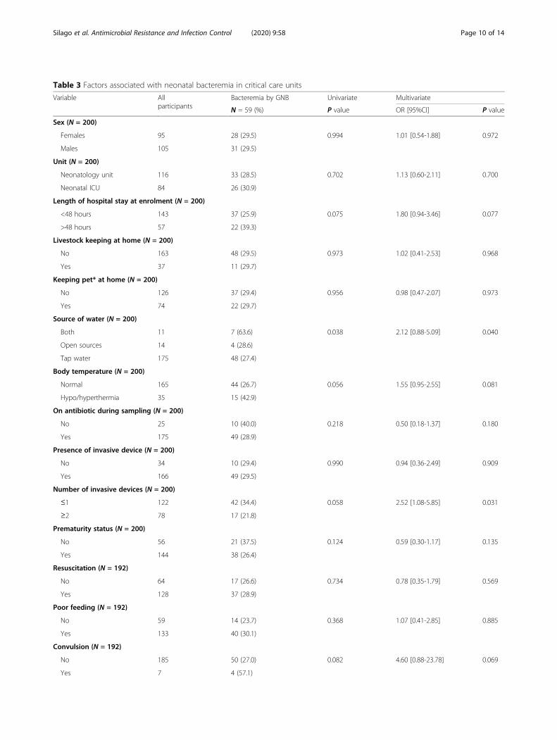
Table 3 Factors associated with neonatal bacteremia in critical care units
Variable Allparticipants
Bacteremia by GNB Univariate Multivariate
N = 59 (%) P value OR [95%CI] P value
Sex (N = 200)
Females 95 28 (29.5) 0.994 1.01 [0.54-1.88] 0.972
Males 105 31 (29.5)
Unit (N = 200)
Neonatology unit 116 33 (28.5) 0.702 1.13 [0.60-2.11] 0.700
Neonatal ICU 84 26 (30.9)
Length of hospital stay at enrolment (N = 200)
<48 hours 143 37 (25.9) 0.075 1.80 [0.94-3.46] 0.077
>48 hours 57 22 (39.3)
Livestock keeping at home (N = 200)
No 163 48 (29.5) 0.973 1.02 [0.41-2.53] 0.968
Yes 37 11 (29.7)
Keeping pet* at home (N = 200)
No 126 37 (29.4) 0.956 0.98 [0.47-2.07] 0.973
Yes 74 22 (29.7)
Source of water (N = 200)
Both 11 7 (63.6) 0.038 2.12 [0.88-5.09] 0.040
Open sources 14 4 (28.6)
Tap water 175 48 (27.4)
Body temperature (N = 200)
Normal 165 44 (26.7) 0.056 1.55 [0.95-2.55] 0.081
Hypo/hyperthermia 35 15 (42.9)
On antibiotic during sampling (N = 200)
No 25 10 (40.0) 0.218 0.50 [0.18-1.37] 0.180
Yes 175 49 (28.9)
Presence of invasive device (N = 200)
No 34 10 (29.4) 0.990 0.94 [0.36-2.49] 0.909
Yes 166 49 (29.5)
Number of invasive devices (N = 200)
≤1 122 42 (34.4) 0.058 2.52 [1.08-5.85] 0.031
≥2 78 17 (21.8)
Prematurity status (N = 200)
No 56 21 (37.5) 0.124 0.59 [0.30-1.17] 0.135
Yes 144 38 (26.4)
Resuscitation (N = 192)
No 64 17 (26.6) 0.734 0.78 [0.35-1.79] 0.569
Yes 128 37 (28.9)
Poor feeding (N = 192)
No 59 14 (23.7) 0.368 1.07 [0.41-2.85] 0.885
Yes 133 40 (30.1)
Convulsion (N = 192)
No 185 50 (27.0) 0.082 4.60 [0.88-23.78] 0.069
Yes 7 4 (57.1)
Silago et al. Antimicrobial Resistance and Infection Control (2020) 9:58 Page 10 of 14

from rectal colonization, cots contamination andmothers’ hands contamination, respectively. This ob-servation may suggests possible cross-transmission ofMDR-GNB between these niches [46]. Further,screening of multiple isolates per sample and molecu-lar typing techniques with greater resolution, e.g.multi-locus sequence typing, pulse-field gel electro-phoresis (PFGE) or, ideally, whole genome sequencing(WGS) will be important in determining clonal simi-larities of these isolates.
ConclusionOur study found high prevalence of antimicrobial resistantGram-negative bacteria in sepsis patients in neonatal ICUand neonatology unit. Additionally, high prevalence ofMDR-GNB colonizing neonates, contaminating hands ofneonates’ mothers and contaminating neonates’ immedi-ate environment, their cots, is extremely concerning. As aresult, this study provides evidence for immediate recom-mendation for: better and frequently (e.g., weekly) decon-tamination on neonates’ cots; information campaign for
Table 3 Factors associated with neonatal bacteremia in critical care units (Continued)
Variable Allparticipants
Bacteremia by GNB Univariate Multivariate
N = 59 (%) P value OR [95%CI] P value
Paleness (N = 200)
No 178 52 (29.2) 0.801 1.29 [0.48-3.53] 0.607
Yes 22 7 (31.8)
Jaundice (N = 200)
Negative 136 34 (25.0) 0.043 2.10 [1.07-4.14] 0.031
Positive 64 25 (39.1)
Cyanosis (N = 200)
Negative 147 34 (23.1) <0.001 3.13 [1.51-6.51] 0.002
Positive 53 25 (47.2)
Nasal flaring (N = 200)
Negative 89 24 (26.9) 0.482 0.86 [0.42-1.76] 0.688
Positive 111 35 (31.5)
Chest indrawing (N = 200)
Negative 122 31 (25.4) 0.114 1.83 [0.94-3.57] 0.076
Positive 78 28 (35.9)
Discharging umbilicus (N = 200)
Negative 140 40 (28.6) 0.660 1.55 [0.755-3.18] 0.232
Positive 60 19 (31.7)
Rectal colonization (N = 200)
Negative 114 28 (24.6) 0.079 1.82 [0.93-3.57] 0.079
Positive 86 31 (36.1)
Cot contamination (N = 200)
Negative 136 32 (23.5) 0.008 2.39 [1.26-4.55] 0.008
Positive 64 27 (42.2)
Mother’s hand contamination (N = 200)
Negative 163 49 (30.1) 0.715 0.84 [0.36-1.93] 0.684
Positive 37 10 (27.0)
Maternal fever during pregnancy (N = 200)
No 105 23 (21.9%)
Yes 95 36 (37.9%) 0.013 2.17 [1.17-4.05] 0.014
Outcome (N = 200)
Discharge 182 52 (28.6) 0.363 1.63 [0.57-4.57] 0.355
Death 18 7 (38.9)
Silago et al. Antimicrobial Resistance and Infection Control (2020) 9:58 Page 11 of 14

mothers on potential cross-transmission of MDR bacteriain causing bacteremia through contaminated hands; andprioritization of 3rd line treatments based on clinical(cyanosis and jaundice) and pre-admission history (do-mestic use of open water sources) in neonatal intensivecare and neonatology units at this setting. Furthermore, afollow-up study is recommended to determine the inci-dence of bacteremia after proper decontamination proto-cols are followed up and mothers are educated oninfection control practices as recommended.
AbbreviationsAMC: Amoxicillin-clavulanic acid; AMP: Ampicillin; ARGs: Antibiotic resistantgenes; AST: Antibiotic susceptibility testing; BMC: Bugando Medical Centre;CAZ: Ceftazidime; CG: Care giver; CIP: Ciprofloxacin; CLSI: Clinical andLaboratory Standards Institute; CN: Gentamicin; CRO: Ceftriaxone; CT: Colistinsulfate; DDS: Double Disk Synergy; GNB: Gram-negative bacteria;HCAIs: Healthcare associated infections; HCW: Health care worker;ICU: Intensive care unit; IPC: Infection prevention and control; MCA-C: MacConkey Agar supplemented with cefotaxime; MDR: Multidrugresistance; NICU: Neonatal ICU; r-3GCs: resistant to third generationcephalosporins; SBA: Sheep blood agar; TSI: Triple sugar iron; UTI: Urinarytract infection; 3GC: Third generation cephalosporin
Table 4 AST profiles as a measure of phenotypic similarities between pairs of isolates of MDR-GNB isolated from blood and MDR-GNB isolated from rectal, bed and mothers’ hands swabs
Phenotypic pairs ID Isolates Sources Comparisons and interpretations of inhibition zones (mm)
SXT TE CN CIP MEM CT
Blood vs rectal colonization11.9% (7/59)
068CL K. pneumoniae Blood 6 (R) 20 (S) 10 (R) 34 (S) 30 (S) 13 (I)
Rectal 6 (R) 20 (S) 10 (R) 32 (S) 30 (S) 12 (I)
233CL E. aerogenes Blood 6 (R) 8 (R) 15 (S) 20 (I) 32 (S) 13 (I)
Rectal 6 (R) 12 (I) 17 (S) 22 (S) 28 (S) 13 (I)
275CL K. pneumoniae Blood 6 (R) 24 (S) 14 (I) 28 (S) 28 (S) 13 (I)
Rectal 6 (R) 22 (S) 14 (I) 28 (S) 32 (S) 14 (S)
285CL K. pneumoniae Blood 6 (R) 6 (R) 6 (R) 17 (I) 30 (S) 15 (S)
Rectal 6 (R) 6 (R) 8 (R) 20 (I) 30 (S) 15 (S)
185CL Acinetobacter spp Blood 22 (S) 18 (S) 20 (S) 22 (S) 32 (S) 13 (I)
Rectal 6 (R) 6 (R) 10 (R) 10 (R) 23 (S) 14 (S)
083CL K. pneumoniae Blood 6 (R) 20 (S) 10 (R) 34 (S) 30 (S) 13 (I)
Rectal 6 (R) 20 (S) 8 (R) 32 (S) 30 (S) 11 (R)
282CL K. pneumoniae Blood 6 (R) 22 (S) 15 (S) 27 (S) 30 (S) 16 (S)
Rectal 6 (R) 23 (S) 16 (S) 27 (S) 28 (S) 16 (S)
Blood vs bed contamination8.5% (5/59)
249CL K. pneumoniae Blood 6 (R) 23 (S) 15 (S) 26 (S) 12 (R) 11 (R)
Bed 6 (R) 20 (S) 14 (I) 27 (S) 29 (S) 12 (I)
241CL Acinetobacter spp Blood 6 (R) 6 (R) 16 (S) 6 (R) 10 (R) 13 (I)
Bed 6 (R) 6 (R) 10 (R) 13 (R) 8 (R) 14 (S)
187CL Acinetobacter spp Blood 6 (R) 8 (R) 15 (S) 25 (S) 27 (S) 13 (I)
Bed 6 (R) 6 (R) 24 (S) 6 (R) 6 (R) 13 (I)
242CL Acinetobacter spp Blood 24 (S) 18 (S) 14 (I) 30 (S) 28 (S) 14 (S)
Bed 6 (R) 6 (R) 14 (I) 28 (S) 6 (R) 13 (I)
243CL Acinetobacter spp Blood 6 (R) 25 (S) 24 (S) 30 (S) 15 (I) 13 (I)
Bed 6 (R) 6 (R) 15 (S) 27 (S) 6 (R) 15 (S)
Blood vs mother contaminated hand6.8% (4/59)
068CL K. pneumoniae Blood 6 (R) 20 (S) 10 (R) 34 (S) 30 (S) 13 (I)
Hand 6 (R) 11 (R) 6 (R) 22 (S) 26 (S) 15 (S)
083CL K. pneumoniae Blood 6 (R) 12 (I) 12 (R) 32 (S) 30 (S) 13 (I)
Hand 6 (R) 22 (S) 6 (R) 28 (S) 28 (S) 14 (S)
186CL K. pneumoniae Blood 6 (R) 18 (S) 10 (R) 28 (S) 28 (S) 14 (S)
Hand 6 (R) 6 (R) 8 (R) 17 (I) 28 (S) 12 (I)
294CL K. pneumoniae Blood 6 (R) 6 (R) 6 (R) 32 (S) 32 (S) 14 (S)
Hand 6 (R) 22 (S) 6 (R) 15 (R) 28 (S) 16 (S)
Notes: SXT trimethoprim-sulfamethoxazole, TE tetracycline, CN gentamicin, CIP ciprofloxacin, MEM meropenem and CT colistin sulfate, S sensitive, I intermediateand R resistant
Silago et al. Antimicrobial Resistance and Infection Control (2020) 9:58 Page 12 of 14

AcknowledgementsAuthors would like to acknowledge the Department of MicrobiologyLaboratory of the Catholic University of Health and Allied Sciences, Bugando;Healthcare workers in neonatal ICU and neonatology unit; and neonates’mothers for their participation in this study.
Authors’ contributionsVS, AML, ASH and SEM designed this study. VS and DRM collected researchdata. VS performed laboratory procedures. VS, DK, LM and RNZ analysed andinterpreted data. VS prepared the manuscript which was read and approvedby all authors.
FundingThis work was funded by the Antimicrobial Resistance Cross-Council Initiativethrough a grant from the Medical Research Council, a Council of UK Re-search and Innovation, and the National Institute for Health Research (Awardno: MR/S004815/1).
Availability of data and materialsThe datasets generated and/or analysed during the current study areavailable in the Microbiology Laboratory Department at Catholic Universityof Health and Allied Sciences, Bugando, Mwanza-Tanzania.
Ethics approval and consent to participateProtocols and procedures in this study were approved by Code of Conductfor Research Ethics of the Sokoine University of Agriculture with certificatenumber: SUA/CVMBS/R.1/2018/8 and ethically cleared by the joint CUHAS/BMC Research Ethics and Review Committee (CREC) with certificate number:CREC/298/2018. All participants were asked to sign in informed consentforms before their enrolment in this study except for participants aged < 18years their consent of participation were provided by their parents orguardians. Detailed microbiological reports of clinical specimens were timelyshared with attending doctors in respective units for proper neonates’management.
Consent for publicationNot applicable.
Competing interestsAuthors declare no competing interests.
Author details1Department of Microbiology and Immunology, Weill Bugando School ofMedicine, Catholic University of Health and Allied Sciences, P. O. Box 1464,Bugando, Mwanza, Tanzania. 2Department of Veterinary Microbiology,Parasitology and Biotechnology, College of Veterinary Medicine andBiomedical Sciences, Sokoine University of Agriculture, P. O. Box 3000,Morogoro, Tanzania. 3Institute of Biodiversity, Animal Health andComparative Medicine, University of Glasgow, Glasgow, UK. 4Department ofPediatrics and Child Health, Weill Bugando School of Medicine, CatholicUniversity of Health and Allied Sciences, P. O. Box 1464, Bugando, Mwanza,Tanzania. 5Sydney School of Veterinary Science, University of Sydney, Sydney,Australia. 6Department of Veterinary Medicine and Public Health, College ofVeterinary Medicine and Biomedical Sciences, Sokoine University ofAgriculture, P. O. Box 3000, Morogoro, Tanzania.
Received: 29 January 2020 Accepted: 22 April 2020
References1. Basak S, Singh P, Rajurkar M. Multidrug resistant and extensively drug
resistant bacteria: a study. Journal of pathogens. 2016;2016:1–5.2. Sweeney MT, Lubbers BV, Schwarz S, Watts JL. Applying definitions for
multidrug resistance, extensive drug resistance and pandrug resistance toclinically significant livestock and companion animal bacterial pathogens. JAntimicrob Chemother. 2018;73(6):1460–3.
3. Sievert DM, Ricks P, Edwards JR, Schneider A, Patel J, Srinivasan A, Kallen A,Limbago B, Fridkin S. Antimicrobial-resistant pathogens associated withhealthcare-associated infections summary of data reported to the NationalHealthcare Safety Network at the Centers for Disease Control andPrevention, 2009–2010. Infect Control Hosp Epidemiol. 2013;34(1):1–14.
4. OBI J, Berthe A, Jean FC, Le Gall FG, Marquez PV. Drug-resistant infections : athreat to our economic future (Vol. 2) : final report (English). In: HNP/Agriculture Global Antimicrobial Resistance Initiative. Washington, D.C: TheWorld Bank; 2017.
5. O’neill J. Review on antimicrobial resistance: tackling a crisis for the healthand wealth of nations. 2014. London: HM Government; 2016.
6. Ndihokubwayo JB, Yahaya AA, Desta AT, Ki-Zerbo G, Odei E, Keita B, PanaAP, Nkhoma W. Antimicrobial resistance in the African region: issues,challenges and actions proposed. African Health Monitor. 2013;16:27–30.
7. Albrich W, Angstwurm M, Bader L, Gärtner R. Drug resistance in intensivecare units. Infection. 1999;27(2):S19–23.
8. Karam G, Chastre J, Wilcox MH, Vincent J-L. Antibiotic strategies in the eraof multidrug resistance. Crit Care. 2016;20(1):136.
9. Vincent J-L, Rello J, Marshall J, Silva E, Anzueto A, Martin CD, Moreno R,Lipman J, Gomersall C, Sakr Y. International study of the prevalence andoutcomes of infection in intensive care units. Jama. 2009;302(21):2323–9.
10. Tosi M, Roat E, De Biasi S, Munari E, Venturelli S, Coloretti I, Biagioni E,Cossarizza A, Girardis M. Multidrug resistant bacteria in critically ill patients: astep further antibiotic therapy. J Emerg Crit Care Med. 2018;2:1–9.
11. Kayange N, Kamugisha E, Mwizamholya DL, Jeremiah S, Mshana SE.Predictors of positive blood culture and deaths among neonates withsuspected neonatal sepsis in a tertiary hospital, Mwanza-Tanzania. BMCPediatr. 2010;10(1):39.
12. Choudhuri AH, Khurana P, Biswas PS, Uppal R. Epidemiology and risk factorsfor multidrug-resistant bacteria in critically ill patients with liver disease.Saudi J Anaesth. 2018;12(3):389.
13. WHO: WHO publishes list of bacteria for which new antibiotics are urgentlyneeded. 2017.
14. WHO. Prevention of hospital-acquired infections : a practical guide. In:JFaLN GD, editor. . 2nd ed. Geneva: World Health Organization; 2002.
15. Seni J. Characterization of Escherichia coli involved in extraintestinalinfections among patients in North-Western Tanzania: circulating sequencetypes, risk factors and antimicrobial resistance profiles; 2018.
16. Choi WS, Kim SH, Jeon EG, Son MH, Yoon YK, Kim J-Y, Kim MJ, Sohn JW,Kim MJ, Park DW. Nosocomial outbreak of carbapenem-resistantAcinetobacter baumannii in intensive care units and successful outbreakcontrol program. J Korean Med Sci. 2010;25(7):999–1004.
17. La Fauci V, Costa GB, Genovese C, Palamara MAR, Alessi V, Squeri R. Drug-resistant bacteria on hands of healthcare workers and in the patient area:an environmental survey in southern Italy’s hospital. Revista Española deQuimioterapia. 2019;32(4):303.
18. Morgan DJ, Rogawski E, Thom KA, Johnson JK, Perencevich EN, Shardell M,Leekha S, Harris AD. Transfer of multidrug-resistant bacteria to healthcareworkers’ gloves and gowns after patient contact increases withenvironmental contamination. Crit Care Med. 2012;40(4):1045.
19. Nelson E, Kayega J, Seni J, Mushi MF, Kidenya BR, Hokororo A, Zuechner A,Kihunrwa A, Mshana SE. Evaluation of existence and transmission ofextended spectrum beta lactamase producing bacteria from post-deliverywomen to neonates at Bugando medical center, Mwanza-Tanzania. BMCRes Notes. 2014;7(1):279.
20. Marando R, Seni J, Mirambo MM, Falgenhauer L, Moremi N, Mushi MF,Kayange N, Manyama F, Imirzalioglu C, Chakraborty T. Predictors of theextended-spectrum-beta lactamases producing Enterobacteriaceae neonatalsepsis at a tertiary hospital, Tanzania. Int J Med Microbiol. 2018;308(7):803–11.
21. Moremi N, Claus H, Silago V, Kabage P, Abednego R, Matee M, Vogel U,Mshana S. Hospital surface contamination with antimicrobial-resistant gram-negative organisms in Tanzanian regional and tertiary hospitals: the need toimprove environmental cleaning. J Hosp Infect. 2019;102(1):98–100.
22. Kish L. Survey sampling; 1965.23. Group WYIS. Clinical prediction of serious bacterial infections in young
infants in developing countries. Pediatr Infect Dis J. 1999;18(10):S23–31.24. In C. Performance standards for antimicrobial susceptibility testing. In:
Wayne PA, editor. Clinical and Laboratory Standards Institute; 2018.25. Winn WC. Koneman's color atlas and textbook of diagnostic microbiology:
Lippincott Williams & wilkins; 2006.26. Bauer A, Kirby W, Sherris JC, Turck M. Antibiotic susceptibility testing by a
standardized single disk method. Am J Clin Pathol. 1966;45(4_ts):493–6.27. CLSI: Performance Standards for Antimicrobial Susceptibility Testing. 28th
ed. CLSI supplement M100. Wyne, PA. Pennsylvania: Clinical and LaboratoryStandards Institute; 2018. 2018.
28. Jacoby GA. AmpC β-lactamases. Clin Microbiol Rev. 2009;22(1):161–82.
Silago et al. Antimicrobial Resistance and Infection Control (2020) 9:58 Page 13 of 14

29. Polsfuss S, Bloemberg GV, Giger J, Meyer V, Böttger EC, Hombach M.Practical approach for reliable detection of AmpC beta-lactamase-producingEnterobacteriaceae. J Clin Microbiol. 2011;49(8):2798–803.
30. Galani I, Kontopidou F, Souli M, Rekatsina P-D, Koratzanis E, Deliolanis J,Giamarellou H. Colistin susceptibility testing by Etest and disk diffusionmethods. Int J Antimicrob Agents. 2008;31(5):434–9.
31. Harris AM, Bramley AM, Jain S, Arnold SR, Ampofo K, Self WH, Williams DJ,Anderson EJ, Grijalva CG, McCullers JA. Influence of antibiotics on thedetection of bacteria by culture-based and culture-independent diagnostictests in patients hospitalized with community-acquired pneumonia. In:Open forum infectious diseases: Oxford University Press; 2017, 2017.
32. Mubito EP, Shahada F, Kimanya ME, Buza JJ. Antimicrobial use in the poultryindustry in Dar-es-Salaam, Tanzania and public health implications; 2014.
33. Breurec S, Bouchiat C, Sire J-M, Moquet O, Bercion R, Cisse MF, Glaser P,Ndiaye O, Ka S, Salord H. High third-generation cephalosporin resistantEnterobacteriaceae prevalence rate among neonatal infections in Dakar,Senegal. BMC Infect Dis. 2016;16(1):587.
34. Ministry of Health T: Standard Treatment Guidelines & National EssentialMedicines List-Tanzania Mainland. 2017.
35. Büdel T, Kuenzli E, Clément M, Bernasconi OJ, Fehr J, Mohammed AH,Hassan NK, Zinsstag J, Hatz C, Endimiani A. Polyclonal gut colonization withextended-spectrum cephalosporin- and/or colistin-resistantEnterobacteriaceae: a normal status for hotel employees on the island ofZanzibar, Tanzania. J Antimicrob Chemother. 2019;74(10):2880–90.
36. Komba EV, Mdegela RH, Msoffe P, Nielsen LN, Ingmer H. Prevalence,antimicrobial resistance and risk factors for thermophilic campylobacterinfections in symptomatic and asymptomatic humans in Tanzania.Zoonoses Public Health. 2015;62(7):557–68.
37. Mubito EP, Shahada F, Kimanya ME, Buza JJ. Antimicrobial use in the poultryindustry in Dar-es-salaam, Tanzania and public health implications. Am J ResCommun. 2014;2:51–63.
38. Mwanza Urban Water and Sanitation Authority (MWAUWASA): Cleaning andFiltering [http://mwauwasa.go.tz/?page_id=80]. Accessed 18 Jan 2020.
39. Moremi N, Manda EV, Falgenhauer L, Ghosh H, Imirzalioglu C, Matee M,Chakraborty T, Mshana SE. Predominance of CTX-M-15 among ESBLproducers from environment and fish gut from the shores of Lake Victoriain Mwanza, Tanzania. Front Microbiol. 2016;7:1862.
40. Zhang S, Lin W, Yu X. Effects of full-scale advanced water treatment onantibiotic resistance genes in the Yangtze Delta area in China. FEMSMicrobiol Ecol. 2016;92(5):fiw065.
41. Suzuki T, Suzuki Y, Okuda J, Kurazumi T, Suhara T, Ueda T, Nagata H,Morisaki H. Sepsis-induced cardiac dysfunction and β-adrenergic blockadetherapy for sepsis. J Intensive Care. 2017;5(1):22.
42. Adeyinka A, Kondamudi NP. Cyanosis; 2019.43. Chand N, Sanyal AJ. Sepsis-induced cholestasis. Hepatology. 2007;45(1):230–41.44. Schechner V, Nobre V, Kaye KS, Leshno M, Giladi M, Rohner P, Harbarth S,
Anderson DJ, Karchmer AW, Schwaber MJ. Gram-negative bacteremia uponhospital admission: when should Pseudomonas aeruginosa be suspected?Clin Infect Dis. 2009;48(5):580–6.
45. Bullard KM, Dunn DL. Bloodstream and intravascular catheter infections. In:Surgical Treatment: Evidence-Based and Problem-Oriented. edn.:Zuckschwerdt; 2001.
46. Russotto V, Cortegiani A, Raineri SM, Giarratano A. Bacterial contaminationof inanimate surfaces and equipment in the intensive care unit. J IntensiveCare. 2015;3(1):54.
47. Moremi N, Claus H, Mshana SE. Antimicrobial resistance pattern: a report ofmicrobiological cultures at a tertiary hospital in Tanzania. BMC Infect Dis.2016;16(1):756.
48. Gramatniece A, Silamikelis I, Zahare I, Urtans V, Zahare I, Dimina E, Saule M,Balode A, Radovica-Spalvina I, Klovins J. Control of Acinetobacter baumanniioutbreak in the neonatal intensive care unit in Latvia: whole-genomesequencing powered investigation and closure of the ward. AntimicrobResist Infect Control. 2019;8(1):84.
49. Singla P, Sikka R, Deeep A, Gagneja D, Chaudhary U. Co-production of ESBLand AmpC β-lactamases in clinical isolates of A. baumannii and A. lwoffii ina tertiary care hospital from northern India. J Clin Diagn Res. 2014;8(4):DC16.
50. Gupta G, Tak V, Mathur P. Detection of AmpC β lactamases in gram-negative bacteria. J Lab Physicians. 2014;6(1):1.
51. Espinal P, Marti S, Vila J. Effect of biofilm formation on the survival ofAcinetobacter baumannii on dry surfaces. J Hosp Infect. 2012;80(1):56–60.
52. Hewitt KM, Mannino FL, Gonzalez A, Chase JH, Caporaso JG, Knight R, KelleyST. Bacterial diversity in two neonatal intensive care units (NICUs). PLoSOne. 2013;8(1):e54703.
53. Weinstein RA, Hota B. Contamination, disinfection, and cross-colonization:are hospital surfaces reservoirs for nosocomial infection? Clin Infect Dis.2004;39(8):1182–9.
54. Chen KTC. Intrapartum fever. Webpage "UpToDate", 45.0. 2012. https://www.uptodate.com/contents/intrapartum-fever.
Publisher’s NoteSpringer Nature remains neutral with regard to jurisdictional claims inpublished maps and institutional affiliations.
Silago et al. Antimicrobial Resistance and Infection Control (2020) 9:58 Page 14 of 14