Embed Size (px)
Citation preview

ORIGINAL RESEARCHpublished: 04 November 2016
doi: 10.3389/fncel.2016.00255
Baclofen Protects Primary RatRetinal Ganglion Cells from ChemicalHypoxia-Induced Apoptosis Throughthe Akt and PERK PathwaysPingping Fu 1, Qiang Wu 1,2*, Jianyan Hu 1, Tingting Li 1 and Fengjuan Gao 1
1 Department of Ophthalmology, Shanghai Jiao Tong University Affiliated Sixth People’s Hospital, Shanghai, China, 2 ShanghaiKey Laboratory of Diabetes Mellitus, Shanghai Jiao Tong University Affiliated Sixth People’s Hospital, Shanghai, China
Edited by:James Francis Curtin,
Dublin Institute of Technology, Ireland
Reviewed by:Nicola Berretta,
Fondazione Santa Lucia (IRCCS),Italy
Ulkan Kilic,Istanbul Medipol University, Turkey
*Correspondence:Qiang Wu
Received: 28 March 2016Accepted: 18 October 2016
Published: 04 November 2016
Citation:Fu P, Wu Q, Hu J, Li T and Gao F
(2016) Baclofen Protects Primary RatRetinal Ganglion Cells from ChemicalHypoxia-Induced Apoptosis Through
the Akt and PERK Pathways.Front. Cell. Neurosci. 10:255.
doi: 10.3389/fncel.2016.00255
Retinal ganglion cells (RGCs) consume large quantities of energy to convert lightinformation into a neuronal signal, which makes them highly susceptible to hypoxicinjury. This study aimed to investigate the potential protection by baclofen, a GABAB
receptor agonist of RGCs against hypoxia-induced apoptosis. Cobalt chloride (CoCl2)was applied to mimic hypoxia. Primary rat RGCs were subjected to CoCl2 with orwithout baclofen treatment, and RNA interference techniques were used to knockdown the GABAB2 gene in the primary RGCs. The viability and apoptosis of RGCswere assessed using cell viability and terminal deoxynucleotidyl transferase-mediateddUTP nick end-labeling (TUNEL) assays, Hoechst staining, and flow cytometry. Theexpression of cleaved caspase-3, bcl-2, bax, Akt, phospho-Akt, protein kinase RNA(PKR)-like ER kinase (PERK), phospho-PERK, eIF2α, phospho-eIF2α, ATF-4 andCCAAT/enhancer-binding protein homologous protein (CHOP) were measured usingwestern blotting. GABAB2 mRNA expression was determined using quantitativereal-time polymerase chain reaction (qRT-PCR) analysis. Our study revealed that CoCl2significantly induced RGC apoptosis and that baclofen reversed these effects. CoCl2-induced reduction of Akt activity was also reversed by baclofen. Baclofen preventedthe activation of the PERK pathway and the increase in CHOP expression induced byCoCl2. Knockdown of GABAB2 and the inactivation of the Akt pathway by inhibitorsreduced the protective effect of baclofen on CoCl2-treated RGCs. Taken together,these results demonstrate that baclofen protects RGCs from CoCl2-induced apoptosisby increasing Akt activity and by suppressing the PERK pathway and CHOP activation.
Keywords: apoptosis, retinal hypoxia disease, retinal ganglion cells, baclofen, GABAB receptor, cobalt, ER stress
INTRODUCTION
The retina is the most metabolically active tissue in human body (Ames, 1992; Caprara andGrimm, 2012). In retinal disease such as retinopathy of prematurity, diabetic retinopathy,age-related macular degeneration and glaucoma, an insufficient supply of oxygen or nutrients,may occur during conditions of disturbed hemodynamics or vascular defects, results in highlyimpaired cellular oxygen balance, and retinal neurons become hypoxic (Kaur et al., 2009, 2012;Munemasa and Kitaoka, 2012). Among the retinal neurons, retinal ganglion cells (RGCs) are the
Frontiers in Cellular Neuroscience | www.frontiersin.org 1 November 2016 | Volume 10 | Article 255

Fu et al. Baclofen Protects RGCs from CoCl2-Mimicked Hypoxia
predominant components that undergo hypoxic injury (Abu-El-Asrar et al., 2004; Caprara and Grimm, 2012; Munemasa andKitaoka, 2012), for they are the ultimate neurons in retina thatconsume large quantities of energy to convey visual informationto the brain (Sanes and Masland, 2015). Hypoxia-relateddisorders enhance or induce the death of RGCs in the retina,resulting in irreversible vision loss, most of which throughapoptosis (Kaur et al., 2009; Yang et al., 2013).
Several hypoxic chemicals have been used to explore themolecular events that underlie the hypoxic neuronal death (Kimet al., 2015). Among all the divalent cations that act as hypoxicmimetics, Cobalt chloride (CoCl2) has shown to induce hypoxia(Yan et al., 2005; Cascio et al., 2008; Law et al., 2012; Lopez-Sánchez et al., 2014) by increasing HIF1α stability and degradingHIF1α (Carbajo-Pescador et al., 2013; Kim et al., 2015; Masoudand Li, 2015). In the present study, RGCswere treated with CoCl2to stimulate hypoxia.
Gamma-aminobutyric acid (GABA) is the main inhibitoryneurotransmitter involved in neuronal information processingwithin the retina (Koulen et al., 1998), and ganglion cells,along with other types of retinal neurons receiving inputfrom GABAergic cells (Nag and Wadhwa, 1997; Koulen et al.,1998). GABA mediates its actions via distinct receptor systems,including the ionotropic GABAA and GABAC receptors and themetabotropic GABAB receptor. The GABAB receptor belongs tothe class C G-protein-coupled receptors and is composed of twosubunits—GABAB1 and GABAB2—both of which are requiredfor normal receptor function (Bettler et al., 2004; Liu et al., 2004;Pin et al., 2004).
Recent advances indicate that GABA can act beyond itsclassical role in synaptic communication and may modulatenearly all critical steps of neuronal network formation includingneurodegenerative, neuroinflammatory and neuromigration(Gaiarsa and Porcher, 2013; Crowley et al., 2015). Accumulatingevidences have shown that specific activation of GABAB receptorcan protect neurons from apoptosis under metabolic stress (Tuet al., 2010; Naseer et al., 2013; Shilpa et al., 2013). Baclofenis a stereoselective GABAB receptor agonist that reduces therelease of excitatory neurotransmitters and substances (Albright,1996; Krach, 2001). It is a drug approved by the Food andDrug Administration that has been used to control spasticity(Furr-Stimming et al., 2014; Crowley et al., 2015) and in thetreatment of pain management (Ward and Kadies, 2002) andalcohol addiction (Howland, 2012). Studies have demonstrateda neuroprotective effect of baclofen through the activation ofGABAB receptor (Babcock et al., 2002; Tyurenkov et al., 2012;Naseer et al., 2013). It has been reported that the upregulationof GABAB receptor activity could inhibit NMDA-receptor-mediated nitric oxide (NO) production by neuronal NO synthase(nNOS) in brain ischemic injury (Zhou et al., 2008). Recentstudies have revealed that, under chronic cerebral hypoperfusion,the effects of GABAB receptors activated by baclofen couldreverse neuronal damage by increasing the activation of Akt,GSK-3β and ERK and upregulating the bcl-2/bax ratio, whichsuppress cytodestructive autophagy (Liu et al., 2015). Anotherstudy has shown that baclofen markedly improves memoryimpairment and alleviates neuronal damage in chronic ischemic
rats by increasing the expression and regulating the function ofthe TPR-containing Rab8b-interacting protein (TRIP8b; Li et al.,2014b).
Given previous reports linking protective effects of baclofenand neurons with hypoxic injuries, particularly with relevanceto neuron apoptosis, it is therefore rational to hypothesizedthat baclofen might have similar neuroprotective effects onRGCs undergo hypoxic stress. In the present investigation, weexplored the impact of baclofen on the cobalt-induced hypoxicRGCs, using primary rat ganglion cells isolated from neonatalSprague-Dawley (SD) rats. In addition, the relevant molecularpathways associated with the neuroprotective effects of baclofenwere evaluated.
MATERIALS AND METHODS
Purified Rat Retinal Ganglion Cell CultureAll animal experiments were approved by the AnimalExperimentation Ethics Committee of the Sixth People’sHospital, Shanghai Jiao Tong University and were carried outin accordance with the approved guidelines for the Care andUse of Laboratory Animals of the National Institutes of Health(Bethesda, MD, USA). Primary RGC cultures were established aspreviously described (Yamagishi and Aihara, 2014), with somemodification. Briefly, retinas were dissected from the eyes of1- to 4-day-old SD rats (SIPPR/BK Lab Animal Ltd, Shanghai,China). This was followed by a two-step immunopanningprocedure, as described below (Yamagishi and Aihara, 2014).Retinal tissues were incubated at 37◦C for 30 min in a solutioncontaining 5 mg/ml papain (all reagents were obtained fromSigma Aldrich, St Louis, MO, USA, unless otherwise stated)in minimum essential medium (MEM; Gibco, Grand Island,NY, USA). To obtain a single cell suspension, tissues werethen sequentially triturated through a narrow-bore Pasteurpipette in an ovomucoid solution. After centrifugation at400× g for 10 min, cells were resuspended in 0.1% bovineserum albumin (BSA) in MEM. Antibodies were then removed,and the cell suspension was incubated in an anti-macrophageantibody-coated (Abcam, Cambridge, MA, USA) flask for 1 hat room temperature and in an anti-Thy1.1 antibody-coated(Abcam, Cambridge, MA, USA) flask for 1 h at 37◦C. Cellsadhering to the flask (RGCs) were resuspended in a basemedium (Neurobasal medium (Gibco, Grand Island, NY, USA)containing 2% B27, 0.1 mg/ml BSA, 0.1 mg/ml transferrin, 1 mML-glutamine, 5 µg/ml insulin, 1 mM sodium pyruvate, 40 ng/mltriiodothyronine, 40 ng/mL thyroxine, 60 ng/ml progesterone, 16µg/ml putrescine, 40 ng/ml sodium selenite, 60 µg/ml N-acetylcysteine, 50 ng/ml brain derived neurotrophic factor (BDNF),10 ng/ml basic fibroblast growth factor (bFGF), 10 ng/mlciliary-derived neurotrophic factor (CNTF), 5 mM Forskolin,100 units/mL penicillin, and 100 mg/mL streptomycin), seededonto plates that had been coated with 0.05 mg/ml poly-L-lysineovernight, and rinsed twice with sterile deionized water. RGCswere cultured for 48 h at 37◦C in a humidified incubator with5% CO2 in base medium before each experiment. Cultures werewashed once with phosphate-buffered saline (PBS; Gibco, Grand
Frontiers in Cellular Neuroscience | www.frontiersin.org 2 November 2016 | Volume 10 | Article 255

Fu et al. Baclofen Protects RGCs from CoCl2-Mimicked Hypoxia
Island, NY, USA) and pre-incubated at 37◦C in neurobasalmedium for 60 min before drug treatment. Drugs were freshlyprepared and dissolved in serum-free neurobasal mediumcontaining 2% B27. Drug treatments were as follows: MK-22062HCl (5 µM, 24 h; Selleckchem, Houston, TX, USA); CoCl2(for the indicated concentrations and times); baclofen (for theindicated concentrations and times).
Cell Viability AssayCell viability was measured using Cell Counting Kit-8 (CCK-8; Dojindo, Japan) according to the manufacturer’s instructions.The CCK-8 assay was performed in a 96-well culture plate withthree replicates for each condition at an absorbance of 450 nm.The number of living cells in each well was expressed as the valuerelative to the control.
Small Interfering RNA TransfectionSmall interfering RNA (siRNA) targeted against GABAB2 andscrambled small hairpin RNA (shRNA) were designed andpackaged by Genechem Co., Ltd (Shanghai, China). Thesequences used were as follows: rat GABAB2 siRNA1 (5′-CCAAGG ACA AGA CCA UCA UTT-3′), siRNA2 (5′-CCA AACAAA UCA AGA CCA UTT -3′), siRNA3 (5′-CCG AGU GUGACA AUG CAA ATT-3′), and negative control (5′-UUC UCCGAA CGU GUC ACG UTT -3′). Transfection was performedwith Lipofectamine 2000 (Thermo Fisher Scientific, Shanghai,China) according to the manufacturer’s instruction. Forty-eighthours after transfection, the medium was replaced and the cellswere treated with CoCl2 and baclofen as described above.
TUNEL AssayTerminal deoxynucleotidyl transferase-mediated dUTPnick end-labeling (TUNEL) staining was used to detectapoptosis-specific nuclear DNA fragmentation. Cells were fixedin 4% paraformaldehyde for 20 min at room temperature beforeTUNEL staining. The fixed cells were then stained using acommercial TUNEL kit (In Situ Cell Death Detection Kit; Boster,Wuhan, China) according to the manufacturer’s recommendedinstructions. TUNEL-positive cell nuclei were visualized basedon green fluorescence and imaging was performed under40× magnification. The percentage of TUNEL-positive cellswas calculated in five microscopic fields from each slide. Thepopulation of TUNEL-positive cells was also quantified by flowcytometry.
Hoechst StainAfter being fixed with 4% paraformaldehyde at 4◦C for 30 min,cells were incubated with the DNA stain Hoechst 33,342(Beyotime, Shanghai, China) for 10 min at room temperatureand mounted with a fluorescent mounting medium (Beyotime,Shanghai, China). Images were acquired using a fluorescencemicroscope (Olympus, Shinjuku, Japan).
Apoptosis Detection by Flow CytometryApoptosis was measured using a fluorescein isothiocyanate(FITC) Annexin V Apoptosis Detection Kit (BD Biosciences, San
Diego, CA, USA) according to the manufacturer’s instructions.Cells were incubated in trypsin-EDTA, collected and suspendedin 1× annexin V binding buffer. One hundred microlitersof each cell suspension was incubated with 5 µl annexinV-FITC and 5 µl propidium iodide (PI) at room temperaturein the dark for 15 min. Following this incubation, 400 µlof 1× binding buffer was added to each tube, and sampleswere analyzed using FACS. Each experiment was repeated intriplicate.
Western Blot AnalysisAfter each treatment, RGCs were harvested and lysed inRIPA buffer (1% Nonidet P-40, 0.5% sodium deoxycholate,0.1% SDS in PBS) and centrifuged at 12,000 rpm for20 min at 4◦C. Protein was extracted, and equal amountsof protein (30 µg per lane) were separated by 8% or10% SDS-PAGE and transferred onto polyvinylidene fluoridemembranes (Millipore). The membranes were blocked with 5%nonfat milk for 1 h and then incubated overnight at 4◦C withthe following primary antibodies (all antibodies were obtainedfrom Cell Signaling Technology Inc., Danvers, MA, USA,unless otherwise stated): rabbit anti-cleaved caspase-3 (1:1000),rabbit anti-bcl-2 (1:1000), rabbit anti-bax (1:1000), rabbitanti-Akt (1:1000), mouse anti-phospho-Akt (1:1000), rabbitanti-protein kinase RNA (PKR)-like ER kinase (PERK) (1:1000),rabbit anti-phospho-PERK (1:1000), rabbit anti-eIF2α (1:1000),rabbit anti-phospho-eIF2α (1:1000), rabbit anti-ATF-4 (1:1000),and rabbit anti-CHOP (1:1000; Abcam, Cambridge, MA, USA).After the membranes were washed, a goat anti-rabbit orgoat anti-mouse horseradish peroxidase-conjugated secondaryantibody (1:2000; Proteintech Group, Chicago, IL, USA) wasapplied, and proteins were visualized using an enhancedchemiluminescence detection system (Bio-Rad, Hercules, CA,USA). Band intensity was analyzed with Quantity One version4.6.2 software (Bio-Rad) and comparedwith the α-tubulin (1:200;Boster, Wuhan, China) internal standard.
RT-PCRHarvested cells were washed with PBS, and total RNA wasextracted from cells using Trizol reagent (Invitrogen, Carlsbad,CA, USA). The primer sequences used were as follows: ratGABAB2 (forward: 5′-CGG AGG ACA GTG GAG AGGTA-3′, reverse: 5′-AGA CAA TGC CAA GCC AGA TG-3′),and rat β-actin (forward: 5′-CAC CCG CGA GTA CAACCT TC-3′, reverse: 5′-CCC ATA CCC ACC ATC ACACC-3′). Quantitative real-time polymerase chain reaction (qRT-PCR) was performed using the SYBR Green quantitativePCR (qPCR) Super Mixture (Takara, Tokyo, Japan) andthe ABI Prism 7500 Sequence Detection System (AppliedBiosystems, Foster City, CA, USA). All reactions were performedin triplicate. The data were analyzed using the 2−11CT
method.
Statistical AnalysisAll experiments were repeated at least three times, andrepresentative results are shown. The differences between two
Frontiers in Cellular Neuroscience | www.frontiersin.org 3 November 2016 | Volume 10 | Article 255

Fu et al. Baclofen Protects RGCs from CoCl2-Mimicked Hypoxia
groups were compared with an independent-sample t-test (SPSS19.0 software, Chicago, IL, USA). A two-tailed P < 0.05 wasconsidered to indicate statistical significance. Data are presentedas the means± standard deviation (SD).
RESULTS
CoCl2 Induces RGC ApoptosisTo establish a cell model for this study, we first assessedthe apoptotic response of RGCs to hypoxia-mimetic agentcobalt. Different concentrations of CoCl2 (0, 100, 200, 400,800 µM) were added to the RGC culture medium. TheCCK-8 assay was used to determine cell viability, and theresults showed that cell viability was significantly decreasedafter 24 h of exposure to CoCl2 in a dose-dependent manner(Figure 1A). Western blot analysis indicated that CoCl2stimulation not only increased the level of cleaved caspase-3and bax proteins, but also decreased the level of bcl-2 proteinin a dose-dependent manner (Figures 1B,C). We noticed that,at a concentration of 200 µM of CoCl2, RGC viability was notsignificantly impaired even though the level of apoptosis-relatedproteins had been obviously changed. Furthermore, annexinV/PI double-stained flow cytometry was used to evaluate theCoCl2-induced apoptosis in RGCs. Annexin V-positive andPI-negative cells were considered to be the apoptotic cells.After treatment with 200 µM CoCl2 for 24 h, the percentageof apoptotic cells increased significantly compared with the
percentage following treatment with 0 µM CoCl2 (P < 0.01,Figures 1D,E). Once our experimental conditions were setup and the use of CoCl2 as an effective apoptosis inductorconfirmed, the 200 µM concentration was chosen for furtherinvestigations.
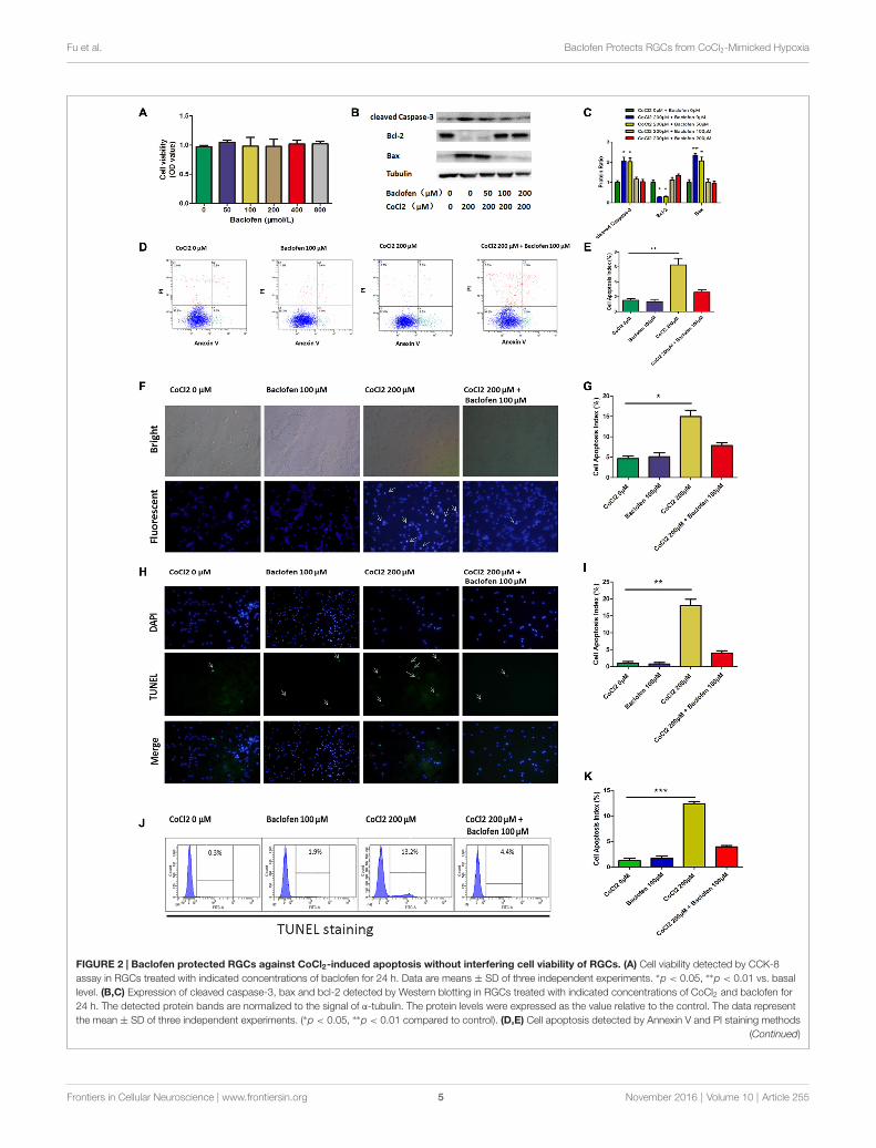
Baclofen Protected RGCs AgainstCoCl2–Induced ApoptosisFirst, to determine whether baclofen could affect the survivalof RGCs, CCK-8 assays were performed, and the resultsshowed that cell viability was not significantly altered after24 h of exposure to baclofen at concentrations up to 800 µM,which indicates that, under certain conditions, baclofen has nosignificant influence on RGC viability (Figure 2A). Furthermore,we explored the effects of baclofen on cobalt-challengedRGCs. We found that baclofen decreased the levels of cleavedcaspase-3 and bax and increased the expression of bcl-2compared with those of hypoxic RGCs without baclofenand that the effect was dose-dependent (Figures 2B,C). Ata concentration of 100 µM, the levels of apoptosis-relatedproteins were changed significantly. The levels of cellapoptosis detected by annexin V/PI double-stained flowcytometry also showed that baclofen significantly counteractedhypoxia-induced apoptosis in RGCs (P < 0.01, Figures 2D,E).To further confirm the protective effects of baclofen againsthypoxia-induced cell death, we used Hoechst staining to
FIGURE 1 | Cobalt chloride (CoCl2) induces retinal ganglion cells (RGCs) apoptosis. (A) Cell viability detected by Cell Counting Kit-8 (CCK-8) assay in RGCstreated with indicated concentrations of CoCl2 for 24 h. Data are means ± SD of three independent experiments. ∗p < 0.05, ∗∗p < 0.01 vs. basal level. (B,C)Expression of cleaved caspase-3, bax and bcl-2 detected by Western blotting in RGCs treated with indicated concentrations of CoCl2 for 24 h. The correspondingdensitometric analyses of the protein bands detected in the immunoblots and normalized to the signal of α-tubulin are also shown. The level of protein in each groupwas expressed as the value relative to the control. Data are means ± SD of three independent experiments. ∗∗p < 0.01, compared with the control. (D,E) Cellapoptosis detected by Annexin V and propidium iodide (PI) staining methods in RGCs treated with indicated concentrations of CoCl2 for 24 h. The C3 quadrant(Annexin V−/PI−), C4 quadrant (Annexin V + /PI−) and C2 quadrant (Annexin V + /PI+) indicate the percentage of viable cells, apoptotic cells and necrotic cells,respectively. The percentage of apoptotic cells following CoCl2 treatment compared with the control group. Values represent the mean ± SD of three independentexperiments. ∗∗P < 0.01.
Frontiers in Cellular Neuroscience | www.frontiersin.org 4 November 2016 | Volume 10 | Article 255
Fu et al. Baclofen Protects RGCs from CoCl2-Mimicked Hypoxia
FIGURE 2 | Baclofen protected RGCs against CoCl2-induced apoptosis without interfering cell viability of RGCs. (A) Cell viability detected by CCK-8assay in RGCs treated with indicated concentrations of baclofen for 24 h. Data are means ± SD of three independent experiments. ∗p < 0.05, ∗∗p < 0.01 vs. basallevel. (B,C) Expression of cleaved caspase-3, bax and bcl-2 detected by Western blotting in RGCs treated with indicated concentrations of CoCl2 and baclofen for24 h. The detected protein bands are normalized to the signal of α-tubulin. The protein levels were expressed as the value relative to the control. The data representthe mean ± SD of three independent experiments. (∗p < 0.05, ∗∗p < 0.01 compared to control). (D,E) Cell apoptosis detected by Annexin V and PI staining methods
(Continued)
Frontiers in Cellular Neuroscience | www.frontiersin.org 5 November 2016 | Volume 10 | Article 255
Fu et al. Baclofen Protects RGCs from CoCl2-Mimicked Hypoxia
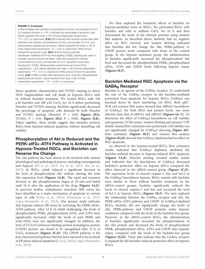
FIGURE 2 | Continuedin RGCs treated with indicated concentrations of CoCl2 and baclofen for 24 h.C4 quadrant (Annexin V + /PI−) indicates the percentage of apoptotic cells.Values represent the mean ± SD of three independent experiments.∗∗P < 0.01 vs. basal level. (F,G) RGCs stained with Hoechst nuclear stain aftertreatment with indicate concentrations of CoCl2 and baclofen for 24 h, andrepresentative experiments are shown. Values represent the mean ± SD ofthree independent experiments. ∗P < 0.05 vs. basal level. White arrowsindicate the apoptotic nuclei. (H,I) Terminal deoxynucleotidyltransferase-mediated dUTP nick end-labeling (TUNEL) staining was used toevaluate hypoxia-induced cell death. Cells were exposed to indicateconcentrations of CoCl2 and baclofen for 24 h. Apoptotic nuclei werevisualized by TUNEL. Representative experiments are shown (Finalmagnification, ×40). Values represent the mean ± SD of three independentexperiments. ∗∗P < 0.01 vs. basal level. White arrows indicate the apoptoticnuclei. (J,K) TUNEL-positive cells detected by flow cytometry. Representativeexperiments are shown. Values represent the mean ± SD of threeindependent experiments. ∗∗∗P < 0.001 vs. basal level.
detect apoptotic characteristics and TUNEL staining to detectDNA fragmentation and cell death in hypoxia RGCs withor without baclofen treatment. We treated RGCs with 100µM baclofen and 200 µM CoCl2 for 24 h before performingHoechst and TUNEL staining. Baclofen significantly decreasedthe percentage of apoptotic cells detected by both Hoechstand TUNEL staining (Hoechst: P < 0.05, Figures 2F,G;TUNEL: P < 0.01, Figures 2H,I; P < 0.001, Figures 2J,K).Taken together, these results suggest that baclofen protectsRGCs from hypoxia-induced apoptosis without disturbing cellviability.
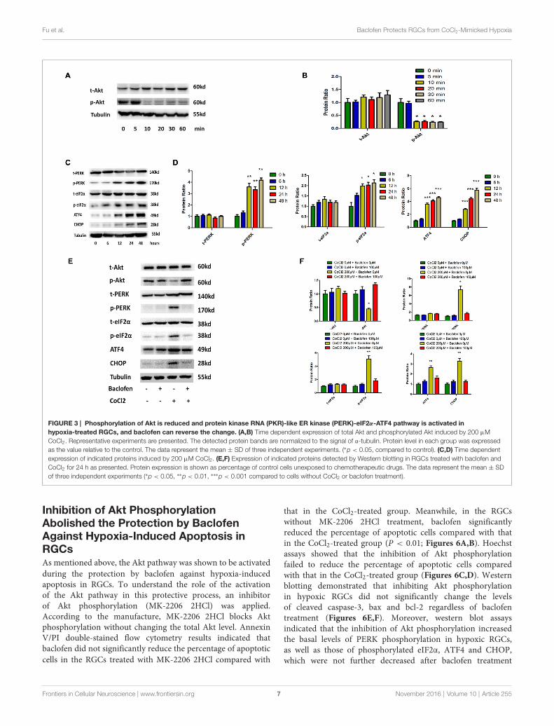
Phosphorylation of Akt is Reduced and thePERK-eIF2α-ATF4 Pathway is Activated inHypoxia-Treated RGCs, and Baclofen canReverse the ChangeThe Akt pathway has been shown to be involved with variousphysiological and pathological process, including tumorigenesisand hypoxia (Di et al., 2015; Liu et al., 2015; Zhu et al.,2015). In RGCs, cobalt induced a significant decrease inthe level of phosphorylated Akt without altering the totalAkt expression level (Figures 3A,B). The rapid and transientdecrease in Akt phosphorylation began at 10 min and lasteduntil 24 h after the application of the drug (Figures 3A,E).In previous studies, endoplasmic reticulum (ER) stress hasbeen identified as a major mechanism of apoptosis in varioustypes of cells (Chen et al., 2015a; Hiramatsu et al., 2015;López-Hernández et al., 2015). Our present study indicatesthat hypoxia induces ER stress by activating the PERK-eIF2α-ATF4 pathway. After 12 h of cobalt challenging, the levels ofphosphorylated PERK, phosphorylated eIF2α, and ATF4 weresignificantly increased, while the levels of total PERK andtotal eIF2α were not significantly changed. In addition, thelevel of CCAAT/enhancer-binding protein homologous protein(CHOP) protein was found to be upregulated after 12 h ofCoCl2 treatment (Figures 3C,D). The CHOP pathway is thebest-characterized pathway that has been reported to be involvedin ER stress-induced apoptosis (Li et al., 2014a; López-Hernándezet al., 2015).
We then explored the beneficial effects of baclofen onhypoxia-mediated stress in RGCs. We pretreated RGCs withbaclofen and with or without CoCl2 for 24 h and thendetermined the levels of the relevant proteins using westernblot analysis. As described above, baclofen had no negativeeffect on RGC survival, and western blotting indicatedthat baclofen did not change the Akt, PERK-pathway orCHOP protein levels compared with those of the controlgroup. In the hypoxia treatment group, the administrationof baclofen significantly increased the phosphorylated Aktlevel and decreased the phosphorylated PERK, phosphorylatedeIF2α, ATF4 and CHOP levels following hypoxic injury(Figures 3E,F).
Baclofen Mediated RGC Apoptosis via theGABAB ReceptorBaclofen is an agonist of the GABAB receptor. To understandthe role of the GABAB receptor in the baclofen-mediatedprotection from apoptosis for hypoxic RGCs, GABAB2 wasknocked down by short interfering (si) RNA. Both qRT-PCR and western blot assays showed that siRNA2 knockdownof GABAB2, for both RNA and protein levels, was moreeffective than that of siRNA1 and siRNA3 (Figures 4A–C). Todetermine the effect of GABAB2 knockdown on cell viabilityand apoptosis, CCK8 assays, western blotting and annexin V/PIdouble-stained flow cytometry were performed. Cell viability wasnot significantly changed by GABAB2 silencing (Figure 4D).Flow cytometry (Figures 4E,F) and western blot analysis(Figures 4G,H) showed that GABAB2 depletion had no effect onRGC apoptosis.
As observed in the hypoxia-treated RGCs, flow cytometryresults indicated that GABAB2 depletion abolished thebaclofen-induced decrease in hypoxia-induced RGC apoptosis(Figures 5A,B). Hoechst staining revealed similar resultsand indicated that the knockdown of GABAB2 decreasedbaclofen’s protective effect on hypoxic RGCs compared theeffect observed in the siRNA-control group (Figures 5C,D).The expression levels of cleaved caspase-3, bax and bcl-2 inthe GABAB2-knockdown hypoxic RGCs treated with baclofenwere similar to those without baclofen treatment. In thesiRNA-control groups, baclofen significantly reduced thelevels of cleaved caspase-3 and bax and increased the levelof bcl-2 in hypoxic RGCs (Figures 5E,F). We next exploredthe relationship between the GABAB receptor and Akt, thePERK-eIF2α-ATF4 pathway and CHOP. In GABAB2-depletedRGCs, baclofen did not significantly change the levels ofAkt, PERK-pathway and CHOP proteins under hypoxicconditions compared with the levels in the baclofen-free group.However, in the siRNA-control RGCs, the administrationof baclofen significantly increased the phosphorylation ofthe Akt protein and decreased the levels of phosphorylatedPERK, phosphorylated eIF2α, ATF4 and CHOP after hypoxicinjury, compared with the levels of the baclofen-free group(Figures 5G,H). These data indicate that the GABAB receptoris required for the baclofen-induced protective effect on hypoxicRGCs.
Frontiers in Cellular Neuroscience | www.frontiersin.org 6 November 2016 | Volume 10 | Article 255
Fu et al. Baclofen Protects RGCs from CoCl2-Mimicked Hypoxia
FIGURE 3 | Phosphorylation of Akt is reduced and protein kinase RNA (PKR)-like ER kinase (PERK)-eIF2α-ATF4 pathway is activated inhypoxia-treated RGCs, and baclofen can reverse the change. (A,B) Time dependent expression of total Akt and phosphorylated Akt induced by 200 µMCoCl2. Representative experiments are presented. The detected protein bands are normalized to the signal of α-tubulin. Protein level in each group was expressedas the value relative to the control. The data represent the mean ± SD of three independent experiments. (∗p < 0.05, compared to control). (C,D) Time dependentexpression of indicated proteins induced by 200 µM CoCl2. (E,F) Expression of indicated proteins detected by Western blotting in RGCs treated with baclofen andCoCl2 for 24 h as presented. Protein expression is shown as percentage of control cells unexposed to chemotherapeutic drugs. The data represent the mean ± SDof three independent experiments (∗p < 0.05, ∗∗p < 0.01, ∗∗∗p < 0.001 compared to cells without CoCl2 or baclofen treatment).
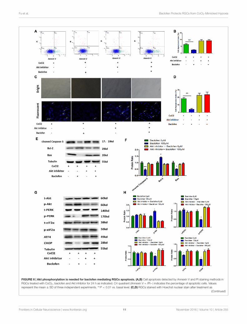
Inhibition of Akt PhosphorylationAbolished the Protection by BaclofenAgainst Hypoxia-Induced Apoptosis inRGCsAs mentioned above, the Akt pathway was shown to be activatedduring the protection by baclofen against hypoxia-inducedapoptosis in RGCs. To understand the role of the activationof the Akt pathway in this protective process, an inhibitorof Akt phosphorylation (MK-2206 2HCl) was applied.According to the manufacture, MK-2206 2HCl blocks Aktphosphorylation without changing the total Akt level. AnnexinV/PI double-stained flow cytometry results indicated thatbaclofen did not significantly reduce the percentage of apoptoticcells in the RGCs treated with MK-2206 2HCl compared with
that in the CoCl2-treated group. Meanwhile, in the RGCswithout MK-2206 2HCl treatment, baclofen significantlyreduced the percentage of apoptotic cells compared with thatin the CoCl2-treated group (P < 0.01; Figures 6A,B). Hoechstassays showed that the inhibition of Akt phosphorylationfailed to reduce the percentage of apoptotic cells comparedwith that in the CoCl2-treated group (Figures 6C,D). Westernblotting demonstrated that inhibiting Akt phosphorylationin hypoxic RGCs did not significantly change the levelsof cleaved caspase-3, bax and bcl-2 regardless of baclofentreatment (Figures 6E,F). Moreover, western blot assaysindicated that the inhibition of Akt phosphorylation increasedthe basal levels of PERK phosphorylation in hypoxic RGCs,as well as those of phosphorylated eIF2α, ATF4 and CHOP,which were not further decreased after baclofen treatment
Frontiers in Cellular Neuroscience | www.frontiersin.org 7 November 2016 | Volume 10 | Article 255

Fu et al. Baclofen Protects RGCs from CoCl2-Mimicked Hypoxia
FIGURE 4 | Small interfering RNA (siRNA) silenced GABAB2 expression without interfering RGCs survival. (A–C) GABAB2 mRNA expression (A) andGABAB2 protein level (B,C). Up on transfected with siRNAs against GABAB2 and control siRNA. ∗∗p < 0.01, ∗∗∗p < 0.001 vs. basal level. (D) Time course of cellviability detected by CCK-8 assay in RGCs transfected with indicated siRNAs. Cell viability was quantified based on three independent experiments (mean ± SD).(E,F) Expression of cleaved caspase-3, bax and bcl-2 detected by Western blotting in RGCs transfected with indicated siRNAs. The detected protein bands arenormalized to the signal of α-tubulin. The level of protein in each group was expressed as the value relative to the control. The data represent the mean ± SD of threeindependent experiments. (∗p < 0.05, vs. control). (G,H) Cell apoptosis detected by Annexin V and PI staining methods in RGCs transfected with indicated siRNAs.C4 quadrant (Annexin V + /PI−) indicates the percentage of apoptotic cells. Representative experiments are shown. Values represent the mean ± SD of threeindependent experiments.
(Figures 6G,H). Together, these results indicate that Aktphosphorylation is necessary for baclofen’s protective effectagainst hypoxia-induced apoptosis and the activation of ERstress in RGCs.
DISCUSSION
Hypoxia, in vitro and in vivo, triggers mixed cell deathdue to both necrosis and apoptosis. Previously, studies haveindicated that baclofen has pharmacological effects againstcell apoptosis in various types of cells (Naseer et al., 2013;Tian et al., 2013; Liu et al., 2015). However, no data haveyet been reported in an in vitro experiment related toRGCs. Thus, we investigated the use of an in vitro modelof purified rat RGC cultures to examine whether baclofenhas neuroprotective effects against RGC apoptosis induced byhypoxic stress.
It has been reported that ganglion cells in diabetic retinasexpress several proapoptosis molecules, suggesting that thesecells are among the most vulnerable neurons (Abu-El-Asraret al., 2004). Following study revealed that in superoxide
dismutase 1 (SOD1)-deficient mice, RGC degeneration precedesthe degeneration of other layers in retina, indicating the greatervulnerability of RGCs to oxidative stress compared with otherneurons (Yuki et al., 2011).
While it is difficult to control oxygen levels precisely tosimulate hypoxic condition in cell culture, various in vitromodels of neuronal hypoxia have been provided using divalentcations such as cobalt (Montel et al., 2007; Hang et al.,2009; Lopez-Sánchez et al., 2014; Yang et al., 2016). Cobalthave been applicable to mimic hypoxic conditions in culturedcells because they activate hypoxic signals by stabilizingthe expression of hypoxia-inducible factor-1 alpha (HIF-1α), an O2-regulated transcriptional activator. Studied haveverified that incubation of cells with CoCl2 mimics thehypoxia situation confirmed by the observed increase ofVEGF protein expression and secretion vs. normoxia (Carbajo-Pescador et al., 2013). Increased HIF-1α leval in cells culturedin hypoxia-mimetic agent treated cells are similar to thoseobserved in a hypoxia chamber (1% O2; Liu et al., 2014).Thus in this study, CoCl2 was added at a final concentrationof 200 mM to mimic hypoxia. We demonstrated that
Frontiers in Cellular Neuroscience | www.frontiersin.org 8 November 2016 | Volume 10 | Article 255

Fu et al. Baclofen Protects RGCs from CoCl2-Mimicked Hypoxia
FIGURE 5 | Baclofen mediated RGCs apoptosis via GABAB receptor. (A,B) RGCs were transfected with GABAB2 siRNA2 or control siRNA before treatmentwith CoCl2 and baclofen for 24 h as indicated. Cell apoptosis detected by Annexin V and PI staining methods. C2, C4 quadrant (Annexin V + /PI−) indicate thepercentage of necrotic or apoptotic cells, respectively. Values represent the mean ± SD of three independent experiments. ∗∗P < 0.01 vs. basal level. (C,D) RGCsstained with Hoechst nuclear stain after treatment as described above. White arrows indicate the apoptotic nuclei. Values represent the mean ± SD of threeindependent experiments. ∗P < 0.05 vs. basal level. (E–H) RGCs were transfected with GABAB2 siRNA2 or control siRNA before treatment with CoCl2 and baclofen.
(Continued)
Frontiers in Cellular Neuroscience | www.frontiersin.org 9 November 2016 | Volume 10 | Article 255
Fu et al. Baclofen Protects RGCs from CoCl2-Mimicked Hypoxia
FIGURE 5 | ContinuedExpression of apoptosis related proteins (E,F) and pathway related proteins(G,H) detected by Western blotting treated with baclofen and CoCl2 for 24 has presented. α-tubulin served as a loading control. The level of protein in eachgroup was expressed as the value relative to the control. The data representthe mean ± SD of three independent experiments. (∗p < 0.05, vs. control).
mimicking hypoxia in vitro by using CoCl2 is capable ofinducing apoptosis in primary RGC cell culture in a dosedependent way.
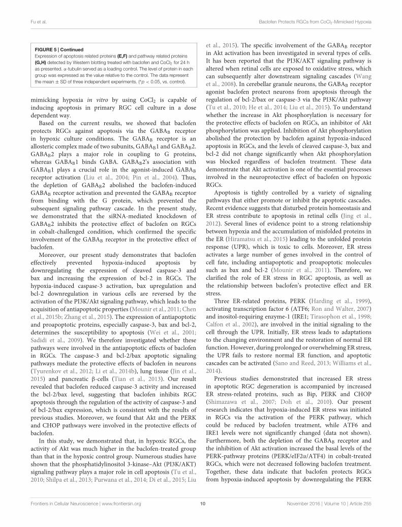
Based on the current results, we showed that baclofenprotects RGCs against apoptosis via the GABAB receptorin hypoxic culture conditions. The GABAB receptor is anallosteric complex made of two subunits, GABAB1 and GABAB2.GABAB2 plays a major role in coupling to G proteins,whereas GABAB1 binds GABA. GABAB2’s association withGABAB1 plays a crucial role in the agonist-induced GABABreceptor activation (Liu et al., 2004; Pin et al., 2004). Thus,the depletion of GABAB2 abolished the baclofen-inducedGABAB receptor activation and prevented the GABAB receptorfrom binding with the G protein, which prevented thesubsequent signaling pathway cascade. In the present study,we demonstrated that the siRNA-mediated knockdown ofGABAB2 inhibits the protective effect of baclofen on RGCsin cobalt-challenged condition, which confirmed the specificinvolvement of the GABAB receptor in the protective effect ofbaclofen.
Moreover, our present study demonstrates that baclofeneffectively prevented hypoxia-induced apoptosis bydownregulating the expression of cleaved caspase-3 andbax and increasing the expression of bcl-2 in RGCs. Thehypoxia-induced caspase-3 activation, bax upregulation andbcl-2 downregulation in various cells are reversed by theactivation of the PI3K/Akt signaling pathway, which leads to theacquisition of antiapoptotic properties (Mounir et al., 2011; Chenet al., 2015b; Zhang et al., 2015). The expression of antiapoptoticand proapoptotic proteins, especially caspase-3, bax and bcl-2,determines the susceptibility to apoptosis (Wei et al., 2001;Sadidi et al., 2009). We therefore investigated whether thesepathways were involved in the antiapoptotic effects of baclofenin RGCs. The caspase-3 and bcl-2/bax apoptotic signalingpathways mediate the protective effects of baclofen in neurons(Tyurenkov et al., 2012; Li et al., 2014b), lung tissue (Jin et al.,2015) and pancreatic β-cells (Tian et al., 2013). Our resultrevealed that baclofen reduced caspase-3 activity and increasedthe bcl-2/bax level, suggesting that baclofen inhibits RGCapoptosis through the regulation of the activity of caspase-3 andof bcl-2/bax expression, which is consistent with the results ofprevious studies. Moreover, we found that Akt and the PERKand CHOP pathways were involved in the protective effects ofbaclofen.
In this study, we demonstrated that, in hypoxic RGCs, theactivity of Akt was much higher in the baclofen-treated groupthan that in the hypoxic control group. Numerous studies haveshown that the phosphatidylinositol 3-kinase–Akt (PI3K/AKT)signaling pathway plays a major role in cell apoptosis (Tu et al.,2010; Shilpa et al., 2013; Purwana et al., 2014; Di et al., 2015; Liu
et al., 2015). The specific involvement of the GABAB receptorin Akt activation has been investigated in several types of cells.It has been reported that the PI3K/AKT signaling pathway isaltered when retinal cells are exposed to oxidative stress, whichcan subsequently alter downstream signaling cascades (Wanget al., 2008). In cerebellar granule neurons, the GABAB receptoragonist baclofen protect neurons from apoptosis through theregulation of bcl-2/bax or caspase-3 via the PI3K/Akt pathway(Tu et al., 2010; He et al., 2014; Liu et al., 2015). To understandwhether the increase in Akt phosphorylation is necessary forthe protective effects of baclofen on RGCs, an inhibitor of Aktphosphorylation was applied. Inhibition of Akt phosphorylationabolished the protection by baclofen against hypoxia-inducedapoptosis in RGCs, and the levels of cleaved caspase-3, bax andbcl-2 did not change significantly when Akt phosphorylationwas blocked regardless of baclofen treatment. These datademonstrate that Akt activation is one of the essential processesinvolved in the neuroprotective effect of baclofen on hypoxicRGCs.
Apoptosis is tightly controlled by a variety of signalingpathways that either promote or inhibit the apoptotic cascades.Recent evidence suggests that disturbed protein homeostasis andER stress contribute to apoptosis in retinal cells (Jing et al.,2012). Several lines of evidence point to a strong relationshipbetween hypoxia and the accumulation of misfolded proteins inthe ER (Hiramatsu et al., 2015) leading to the unfolded proteinresponse (UPR), which is toxic to cells. Moreover, ER stressactivates a large number of genes involved in the control ofcell fate, including antiapoptotic and proapoptotic moleculessuch as bax and bcl-2 (Mounir et al., 2011). Therefore, weclarified the role of ER stress in RGC apoptosis, as well asthe relationship between baclofen’s protective effect and ERstress.
Three ER-related proteins, PERK (Harding et al., 1999),activating transcription factor 6 (ATF6; Ron and Walter, 2007)and inositol-requiring enzyme-1 (IRE1; Tirasophon et al., 1998;Calfon et al., 2002), are involved in the initial signaling to thecell through the UPR. Initially, ER stress leads to adaptationsto the changing environment and the restoration of normal ERfunction. However, during prolonged or overwhelming ER stress,the UPR fails to restore normal ER function, and apoptoticcascades can be activated (Sano and Reed, 2013; Williams et al.,2014).
Previous studies demonstrated that increased ER stressin apoptotic RGC degeneration is accompanied by increasedER stress-related proteins, such as Bip, PERK and CHOP(Shimazawa et al., 2007; Doh et al., 2010). Our presentresearch indicates that hypoxia-induced ER stress was initiatedin RGCs via the activation of the PERK pathway, whichcould be reduced by baclofen treatment, while ATF6 andIRE1 levels were not significantly changed (data not shown).Furthermore, both the depletion of the GABAB receptor andthe inhibition of Akt activation increased the basal levels of thePERK-pathway proteins (PERK/eIF2α/ATF4) in cobalt-treatedRGCs, which were not decreased following baclofen treatment.Together, these data indicate that baclofen protects RGCsfrom hypoxia-induced apoptosis by downregulating the PERK
Frontiers in Cellular Neuroscience | www.frontiersin.org 10 November 2016 | Volume 10 | Article 255
Fu et al. Baclofen Protects RGCs from CoCl2-Mimicked Hypoxia
FIGURE 6 | Akt phosphorylation is needed for baclofen mediating RGCs apoptosis. (A,B) Cell apoptosis detected by Annexin V and PI staining methods inRGCs treated with CoCl2, baclofen and Akt inhibitor for 24 h as indicated. C4 quadrant (Annexin V + /PI−) indicates the percentage of apoptotic cells. Valuesrepresent the mean ± SD of three independent experiments. ∗∗P < 0.01 vs. basal level. (C,D) RGCs stained with Hoechst nuclear stain after treatment as
(Continued)
Frontiers in Cellular Neuroscience | www.frontiersin.org 11 November 2016 | Volume 10 | Article 255
Fu et al. Baclofen Protects RGCs from CoCl2-Mimicked Hypoxia
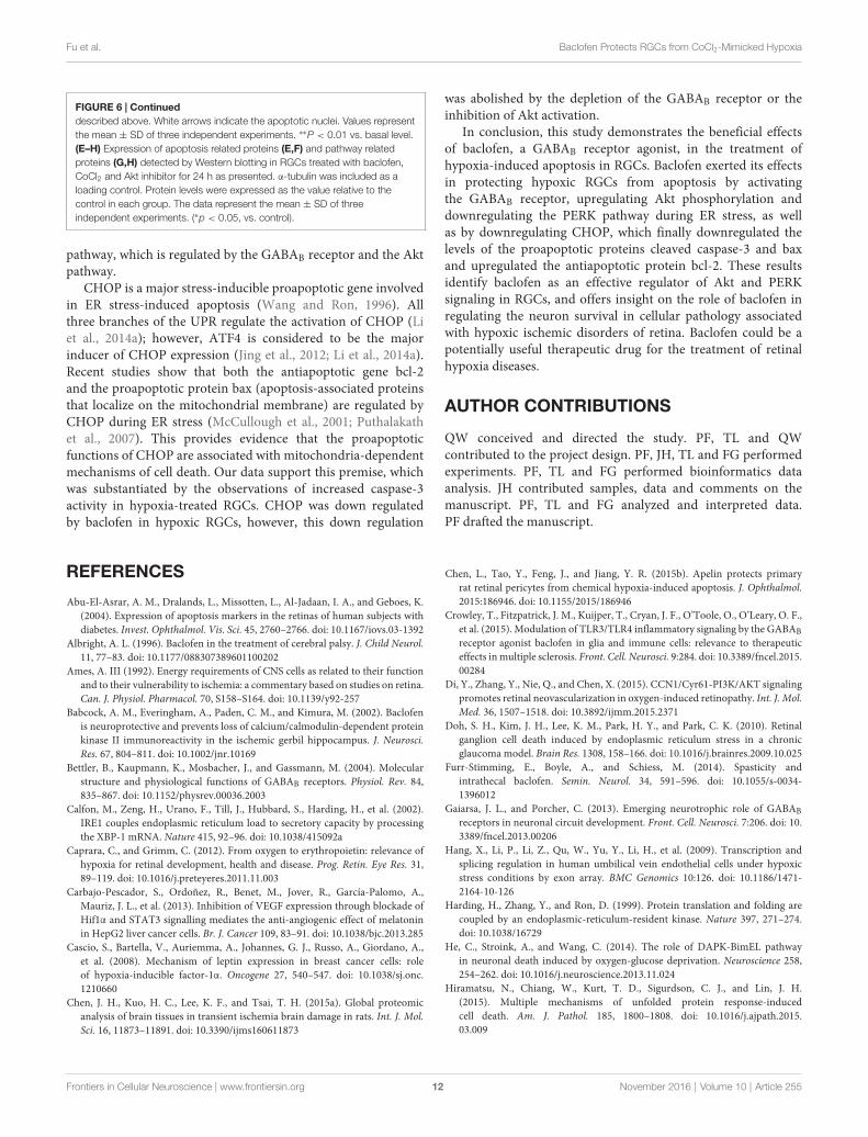
FIGURE 6 | Continueddescribed above. White arrows indicate the apoptotic nuclei. Values representthe mean ± SD of three independent experiments. ∗∗P < 0.01 vs. basal level.(E–H) Expression of apoptosis related proteins (E,F) and pathway relatedproteins (G,H) detected by Western blotting in RGCs treated with baclofen,CoCl2 and Akt inhibitor for 24 h as presented. α-tubulin was included as aloading control. Protein levels were expressed as the value relative to thecontrol in each group. The data represent the mean ± SD of threeindependent experiments. (∗p < 0.05, vs. control).
pathway, which is regulated by the GABAB receptor and the Aktpathway.
CHOP is a major stress-inducible proapoptotic gene involvedin ER stress-induced apoptosis (Wang and Ron, 1996). Allthree branches of the UPR regulate the activation of CHOP (Liet al., 2014a); however, ATF4 is considered to be the majorinducer of CHOP expression (Jing et al., 2012; Li et al., 2014a).Recent studies show that both the antiapoptotic gene bcl-2and the proapoptotic protein bax (apoptosis-associated proteinsthat localize on the mitochondrial membrane) are regulated byCHOP during ER stress (McCullough et al., 2001; Puthalakathet al., 2007). This provides evidence that the proapoptoticfunctions of CHOP are associated with mitochondria-dependentmechanisms of cell death. Our data support this premise, whichwas substantiated by the observations of increased caspase-3activity in hypoxia-treated RGCs. CHOP was down regulatedby baclofen in hypoxic RGCs, however, this down regulation
was abolished by the depletion of the GABAB receptor or theinhibition of Akt activation.
In conclusion, this study demonstrates the beneficial effectsof baclofen, a GABAB receptor agonist, in the treatment ofhypoxia-induced apoptosis in RGCs. Baclofen exerted its effectsin protecting hypoxic RGCs from apoptosis by activatingthe GABAB receptor, upregulating Akt phosphorylation anddownregulating the PERK pathway during ER stress, as wellas by downregulating CHOP, which finally downregulated thelevels of the proapoptotic proteins cleaved caspase-3 and baxand upregulated the antiapoptotic protein bcl-2. These resultsidentify baclofen as an effective regulator of Akt and PERKsignaling in RGCs, and offers insight on the role of baclofen inregulating the neuron survival in cellular pathology associatedwith hypoxic ischemic disorders of retina. Baclofen could be apotentially useful therapeutic drug for the treatment of retinalhypoxia diseases.
AUTHOR CONTRIBUTIONS
QW conceived and directed the study. PF, TL and QWcontributed to the project design. PF, JH, TL and FG performedexperiments. PF, TL and FG performed bioinformatics dataanalysis. JH contributed samples, data and comments on themanuscript. PF, TL and FG analyzed and interpreted data.PF drafted the manuscript.
REFERENCES
Abu-El-Asrar, A. M., Dralands, L., Missotten, L., Al-Jadaan, I. A., and Geboes, K.(2004). Expression of apoptosis markers in the retinas of human subjects withdiabetes. Invest. Ophthalmol. Vis. Sci. 45, 2760–2766. doi: 10.1167/iovs.03-1392
Albright, A. L. (1996). Baclofen in the treatment of cerebral palsy. J. Child Neurol.11, 77–83. doi: 10.1177/088307389601100202
Ames, A. III (1992). Energy requirements of CNS cells as related to their functionand to their vulnerability to ischemia: a commentary based on studies on retina.Can. J. Physiol. Pharmacol. 70, S158–S164. doi: 10.1139/y92-257
Babcock, A. M., Everingham, A., Paden, C. M., and Kimura, M. (2002). Baclofenis neuroprotective and prevents loss of calcium/calmodulin-dependent proteinkinase II immunoreactivity in the ischemic gerbil hippocampus. J. Neurosci.Res. 67, 804–811. doi: 10.1002/jnr.10169
Bettler, B., Kaupmann, K., Mosbacher, J., and Gassmann, M. (2004). Molecularstructure and physiological functions of GABAB receptors. Physiol. Rev. 84,835–867. doi: 10.1152/physrev.00036.2003
Calfon, M., Zeng, H., Urano, F., Till, J., Hubbard, S., Harding, H., et al. (2002).IRE1 couples endoplasmic reticulum load to secretory capacity by processingthe XBP-1 mRNA. Nature 415, 92–96. doi: 10.1038/415092a
Caprara, C., and Grimm, C. (2012). From oxygen to erythropoietin: relevance ofhypoxia for retinal development, health and disease. Prog. Retin. Eye Res. 31,89–119. doi: 10.1016/j.preteyeres.2011.11.003
Carbajo-Pescador, S., Ordoñez, R., Benet, M., Jover, R., García-Palomo, A.,Mauriz, J. L., et al. (2013). Inhibition of VEGF expression through blockade ofHif1α and STAT3 signalling mediates the anti-angiogenic effect of melatoninin HepG2 liver cancer cells. Br. J. Cancer 109, 83–91. doi: 10.1038/bjc.2013.285
Cascio, S., Bartella, V., Auriemma, A., Johannes, G. J., Russo, A., Giordano, A.,et al. (2008). Mechanism of leptin expression in breast cancer cells: roleof hypoxia-inducible factor-1α. Oncogene 27, 540–547. doi: 10.1038/sj.onc.1210660
Chen, J. H., Kuo, H. C., Lee, K. F., and Tsai, T. H. (2015a). Global proteomicanalysis of brain tissues in transient ischemia brain damage in rats. Int. J. Mol.Sci. 16, 11873–11891. doi: 10.3390/ijms160611873
Chen, L., Tao, Y., Feng, J., and Jiang, Y. R. (2015b). Apelin protects primaryrat retinal pericytes from chemical hypoxia-induced apoptosis. J. Ophthalmol.2015:186946. doi: 10.1155/2015/186946
Crowley, T., Fitzpatrick, J. M., Kuijper, T., Cryan, J. F., O’Toole, O., O’Leary, O. F.,et al. (2015). Modulation of TLR3/TLR4 inflammatory signaling by the GABABreceptor agonist baclofen in glia and immune cells: relevance to therapeuticeffects inmultiple sclerosis. Front. Cell. Neurosci. 9:284. doi: 10.3389/fncel.2015.00284
Di, Y., Zhang, Y., Nie, Q., and Chen, X. (2015). CCN1/Cyr61-PI3K/AKT signalingpromotes retinal neovascularization in oxygen-induced retinopathy. Int. J. Mol.Med. 36, 1507–1518. doi: 10.3892/ijmm.2015.2371
Doh, S. H., Kim, J. H., Lee, K. M., Park, H. Y., and Park, C. K. (2010). Retinalganglion cell death induced by endoplasmic reticulum stress in a chronicglaucoma model. Brain Res. 1308, 158–166. doi: 10.1016/j.brainres.2009.10.025
Furr-Stimming, E., Boyle, A., and Schiess, M. (2014). Spasticity andintrathecal baclofen. Semin. Neurol. 34, 591–596. doi: 10.1055/s-0034-1396012
Gaiarsa, J. L., and Porcher, C. (2013). Emerging neurotrophic role of GABABreceptors in neuronal circuit development. Front. Cell. Neurosci. 7:206. doi: 10.3389/fncel.2013.00206
Hang, X., Li, P., Li, Z., Qu, W., Yu, Y., Li, H., et al. (2009). Transcription andsplicing regulation in human umbilical vein endothelial cells under hypoxicstress conditions by exon array. BMC Genomics 10:126. doi: 10.1186/1471-2164-10-126
Harding, H., Zhang, Y., and Ron, D. (1999). Protein translation and folding arecoupled by an endoplasmic-reticulum-resident kinase. Nature 397, 271–274.doi: 10.1038/16729
He, C., Stroink, A., and Wang, C. (2014). The role of DAPK-BimEL pathwayin neuronal death induced by oxygen-glucose deprivation. Neuroscience 258,254–262. doi: 10.1016/j.neuroscience.2013.11.024
Hiramatsu, N., Chiang, W., Kurt, T. D., Sigurdson, C. J., and Lin, J. H.(2015). Multiple mechanisms of unfolded protein response-inducedcell death. Am. J. Pathol. 185, 1800–1808. doi: 10.1016/j.ajpath.2015.03.009
Frontiers in Cellular Neuroscience | www.frontiersin.org 12 November 2016 | Volume 10 | Article 255

Fu et al. Baclofen Protects RGCs from CoCl2-Mimicked Hypoxia
Howland, R. H. (2012). Baclofen for the treatment of alcohol dependence.J. Psychosoc. Nurs. Ment. Health Serv. 50, 11–14. doi: 10.3928/02793695-20120906-92
Jin, S., Merchant, M. L., Ritzenthaler, J. D., McLeish, K. R., Lederer, E. D.,Torres-Gonzalez, E., et al. (2015). Baclofen, a GABABR agonist, amelioratesimmune-complexmediated acute lung injury bymodulating pro-inflammatorymediators. PLoS One 10:e0121637. doi: 10.1371/journal.pone.0121637
Jing, G., Wang, J., and Zhang, S. (2012). ER stress and apoptosis: a newmechanismfor retinal cell death. Exp. Diabetes Res. 2012:589589. doi: 10.1155/2012/589589
Kaur, C., Sivakumar, V., Foulds, W. S., Luu, C. D., and Ling, E. A. (2009). Cellularand vascular changes in the retina of neonatal rats after an acute exposureto hypoxia. Invest. Ophthalmol. Vis. Sci. 50, 5364–5374. doi: 10.1167/iovs.09-3552
Kaur, C., Sivakumar, V., Foulds,W. S., Luu, C. D., and Ling, E. A. (2012). Hypoxia-induced activation of N-methyl-D-aspartate receptors causes retinal ganglioncell death in the neonatal retina. J. Neuropathol. Exp. Neurol. 71, 330–347.doi: 10.1097/NEN.0b013e31824deb21
Kim, S., Seo, J. W.W., Oh, S. B., Kim, S. H., Kim, I., Suh, N., et al. (2015). Disparateroles of zinc in chemical hypoxia-induced neuronal death. Front. Cell. Neurosci.9:1. doi: 10.3389/fncel.2015.00001
Koulen, P., Malitschek, B., Kuhn, R., Bettler, B., Wässle, H., and Brandstätter, J.(1998). Presynaptic and postsynaptic localization of GABAB receptors inneurons of the rat retina. Eur. J. Neurosci. 10, 1446–1456. doi: 10.1046/j.1460-9568.1998.00156.x
Krach, L. (2001). Pharmacotherapy of spasticity: oral medications andintrathecal baclofen. J. Child Neurol. 16, 31–36. doi: 10.1177/088307380101600106
Law, P. C., Auyeung, K. K., Chan, L. Y., and Ko, J. K. (2012). Astragalussaponins downregulate vascular endothelial growth factor under cobaltchloride-stimulated hypoxia in colon cancer cells. BMC Complement. Altern.Med. 12:160. doi: 10.1186/1472-6882-12-160
Li, Y., Guo, Y., Tang, J., Jiang, J., and Chen, Z. (2014a). New insights into theroles of CHOP-induced apoptosis in ER stress. Acta Biochim. Biophys. Sin.(Shanghai) 46, 629–640. doi: 10.1093/abbs/gmu048
Li, C., Lu, Y., Zhou, M., Zong, X., Li, C., Xu, X., et al. (2014b). Activation ofGABAB receptors ameliorates cognitive impairment via restoring the balanceof HCN1/HCN2 surface expression in the hippocampal CA1 area in ratswith chronic cerebral hypoperfusion. Mol. Neurobiol. 50, 704–720. doi: 10.1007/s12035-014-8736-3
Liu, L., Li, C. J., Lu, Y., Zong, X. G., Luo, C., Sun, J., et al. (2015). Baclofen mediatesneuroprotection on hippocampal CA1 pyramidal cells through the regulationof autophagy under chronic cerebral hypoperfusion. Sci. Rep. 5:14474. doi: 10.1038/srep14474
Liu, J., Maurel, D., Etzol, S., Brabet, I., Ansanay, H., Pin, J. P., et al. (2004).Molecular determinants involved in the allosteric control of agonist affinity inthe GABAB receptor by the GABAB2 subunit. J. Biol. Chem. 279, 15824–15830.doi: 10.1074/jbc.M313639200
Liu, H., Wang, Z., Yu, S., and Xu, J. (2014). Proteasomal degradationof O-GlcNAc transferase elevates hypoxia-induced vascular endothelialinflammatory responsedagger. Cardiovasc. Res. 103, 131–139. doi: 10.1093/cvr/cvu116
López-Hernández, B., Ceña, V., and Posadas, I. (2015). The endoplasmic reticulumstress and the HIF-1 signalling pathways are involved in the neuronal damagecaused by chemical hypoxia. Br. J. Pharmacol. 172, 2838–2851. doi: 10.1111/bph.13095
Lopez-Sánchez, L. M., Jimenez, C., Valverde, A., Hernandez, V., Peñarando, J.,Martinez, A., et al. (2014). CoCl2, a mimic of hypoxia, induces formationof polyploid giant cells with stem characteristics in colon cancer. PLoS One9:e99143. doi: 10.1371/journal.pone.0099143
Masoud, G. N., and Li, W. (2015). HIF-1α pathway: role, regulation andintervention for cancer therapy. Acta Pharm. Sin. B 5, 378–389. doi: 10.1016/j.apsb.2015.05.007
McCullough, K. D., Martindale, J. L., Klotz, L. O., Aw, T. Y., andHolbrook, N. J. (2001). Gadd153 sensitizes cells to endoplasmicreticulum stress by down-regulating Bcl2 and perturbing the cellularredox state. Mol. Cell. Biol. 21, 1249–1259. doi: 10.1128/mcb.21.4.1249-1259.2001
Montel, V., Gaultier, A., Lester, R. D., Campana, W. M., and Gonias, S. L.(2007). The low-density lipoprotein receptor-related protein regulates cancercell survival and metastasis development. Cancer Res. 67, 9817–9824. doi: 10.1158/0008-5472.can-07-0683
Mounir, Z., Krishnamoorthy, J. L., Wang, S., Papadopoulou, B., Campbell, S.,Muller, W. J., et al. (2011). Akt determines cell fate through inhibitionof the PERK-eIF2α phosphorylation pathway. Sci. Signal. 4:ra62. doi: 10.1126/scisignal.2001630
Munemasa, Y., and Kitaoka, Y. (2012). Molecular mechanisms of retinalganglion cell degeneration in glaucoma and future prospects for cell bodyand axonal protection. Front. Cell. Neurosci. 6:60. doi: 10.3389/fncel.2012.00060
Nag, T., and Wadhwa, S. (1997). Expression of GABA in the fetal, postnataland adult human retinas: an immunohistochemical study. Vis. Neurosci. 14,425–432. doi: 10.1017/s0952523800012104
Naseer, M. I., Ullah, I., Al-Qahtani, M. H., Karim, S., Ullah, N., Ansari, S. A.,et al. (2013). Decreased GABABR expression and increased neuronal cell deathin developing rat brain after PTZ-induced seizure. Neurol. Sci. 34, 497–503.doi: 10.1007/s10072-012-1083-0
Pin, J. P., Kniazeff, J., Binet, V., Liu, J., Maurel, D., Galvez, T., et al. (2004).Activation mechanism of the heterodimeric GABAB receptor. Biochem.Pharmacol. 68, 1565–1572. doi: 10.1016/j.bcp.2004.06.035
Purwana, I., Zheng, J., Li, X., Deurloo, M., Son, D. O., Zhang, Z.,et al. (2014). GABA promotes human beta-cell proliferation andmodulates glucose homeostasis. Diabetes 63, 4197–4205. doi: 10.2337/db14-0153
Puthalakath, H., O’Reilly, L. A., Gunn, P., Lee, L., Kelly, P. N., Huntington, N. D.,et al. (2007). ER stress triggers apoptosis by activating BH3-only protein Bim.Cell 129, 1337–1349. doi: 10.1016/j.cell.2007.04.027
Ron, D., and Walter, P. (2007). Signal integration in the endoplasmic reticulumunfolded protein response. Nat. Rev. Mol. Cell Biol. 8, 519–529. doi: 10.1038/nrm2199
Sadidi, M., Lentz, S. I., and Feldman, E. L. (2009). Hydrogen peroxide-inducedAkt phosphorylation regulates Bax activation. Biochimie 91, 577–585. doi: 10.1016/j.biochi.2009.01.010
Sanes, J. R., and Masland, R. H. (2015). The types of retinal ganglion cells: currentstatus and implications for neuronal classification. Annu. Rev. Neurosci. 38,221–246. doi: 10.1146/annurev-neuro-071714-034120
Sano, R., and Reed, J. C. (2013). ER stress-induced cell deathmechanisms. Biochim.Biophys. Acta 1833, 3460–3470. doi: 10.1016/j.bbamcr.2013.06.028
Shilpa, J., Anitha, M., and Paulose, C. S. (2013). Increased neuronal survival inthe brainstem during liver injury: role of γ-aminobutyric acid and serotoninchitosan nanoparticles. J. Neurosci. Res. 91, 1203–1214. doi: 10.1002/jnr.23243
Shimazawa, M., Ito, Y., Inokuchi, Y., and Hara, H. (2007). Involvement ofdouble-stranded RNA-dependent protein kinase in ER stress-induced retinalneuron damage. Invest. Ophthalmol. Vis. Sci. 48, 3729–3736. doi: 10.1167/iovs.06-1122
Tian, J., Dang, H., Chen, Z., Guan, A., Jin, Y., Atkinson, M., et al. (2013).γ-Aminobutyric acid regulates both the survival and replication of humanβ-cells. Diabetes 62, 3760–3765. doi: 10.2337/db13-0931
Tirasophon, W., Welihinda, A. A., and Kaufman, R. J. (1998). A stress responsepathway from the endoplasmic reticulum to the nucleus requires a novelbifunctional protein kinase/endoribonuclease (Ire1p) in mammalian cells.Genes Dev. 12, 1812–1824. doi: 10.1101/gad.12.12.1812
Tu, H., Xu, C., Zhang, W., Liu, Q., Rondard, P., Pin, J. P., et al. (2010).GABAB receptor activation protects neurons from apoptosis via IGF-1 receptortransactivation. J. Neurosci. 30, 749–759. doi: 10.1523/JNEUROSCI.2343-09.2010
Tyurenkov, I. N., Borodkina, L. E., and Bagmetova, V. V. (2012). Functionalaspects of neuroprotective effects of new salts and compositions of baclofenin the convulsive syndrome caused by electroshock. Bull. Exp. Biol. Med. 153,710–713. doi: 10.1007/s10517-012-1806-5
Wang, L., Chen, Y., Sternberg, P., and Cai, J. (2008). Essential roles of thePI3 kinase/Akt pathway in regulating Nrf2-dependent antioxidant functionsin the RPE. Invest. Ophthalmol. Vis. Sci. 49, 1671–1678. doi: 10.1167/iovs.07-1099
Wang, X. Z., and Ron, D. (1996). Stress-induced phosphorylation and activation ofthe transcription factor CHOP (GADD153) by p38 MAP Kinase. Science 272,1347–1349. doi: 10.1126/science.272.5266.1347
Frontiers in Cellular Neuroscience | www.frontiersin.org 13 November 2016 | Volume 10 | Article 255

Fu et al. Baclofen Protects RGCs from CoCl2-Mimicked Hypoxia
Ward, A. B., and Kadies, M. (2002). The management of pain in spasticity.Disabil.Rehabil. 24, 443–453. doi: 10.1080/09638280110108878
Wei, M. C., Zong, W. X., Cheng, E. H., Lindsten, T., Panoutsakopoulou, V.,Ross, A. J., et al. (2001). Proapoptotic BAX and BAK: a requisite gatewayto mitochondrial dysfunction and death. Science 292, 727–730. doi: 10.1126/science.1059108
Williams, B., Verchot, J., and Dickman, M. B. (2014). When supply does not meetdemand-ER stress and plant programmed cell death. Front. Plant Sci. 5:211.doi: 10.3389/fpls.2014.00211
Yamagishi, R., and Aihara, M. (2014). Neuroprotective effect of astaxanthinagainst rat retinal ganglion cell death under various stresses that induceapoptosis and necrosis.Mol. Vis. 20, 1796–1805.
Yan, H., Peng, Z.-G., Wu, Y. L., Jiang, Y., Yu, Y., Huang, Y., et al. (2005). Hypoxia-simulating agents and selective stimulation of arsenic trioxide-inducedgrowth arrest and cell differentiation in acute promyelocytic leukemic cells.Haematologica 90, 1607–1616.
Yang, J. H., Kwak, H. W., Kim, T. G., Han, J., Moon, S. W., and Yu, S. Y. (2013).Retinal neurodegeneration in type II diabetic otsuka long-evans tokushimafatty rats. Invest. Ophthalmol. Vis. Sci. 54, 3844–3851. doi: 10.1167/iovs.12-11309
Yang, G., Xu, S., Peng, L., Li, H., Zhao, Y., andHu, Y. (2016). The hypoxia-mimeticagent CoCl2 induces chemotherapy resistance in LOVO colorectal cancer cells.Mol. Med. Rep. 13, 2583–2589. doi: 10.3892/mmr.2016.4836
Yuki, K., Ozawa, Y., Yoshida, T., Kurihara, T., Hirasawa, M., Ozeki, N., et al.(2011). Retinal ganglion cell loss in superoxide dismutase 1 deficiency. Invest.Ophthalmol. Vis. Sci. 52, 4143–4150. doi: 10.1167/iovs.10-6294
Zhang, W., Neo, S. P., Gunaratne, J., Poulsen, A., Boping, L., Ong, E. H.,et al. (2015). Feedback regulation on PTEN/AKT pathway by the ER stresskinase PERK mediated by interaction with the Vault complex. Cell. Signal. 27,436–442. doi: 10.1016/j.cellsig.2014.12.010
Zhou, C., Li, C., Yu, H. M., Zhang, F., Han, D., and Zhang, G. Y. (2008).Neuroprotection of gamma-aminobutyric acid receptor agonists via enhancingneuronal nitric oxide synthase (Ser847) phosphorylation through increasedneuronal nitric oxide synthase and PSD95 interaction and inhibited proteinphosphatase activity in cerebral ischemia. J. Neurosci. Res. 86, 2973–2983.doi: 10.1002/jnr.21728
Zhu, C., Wang, S., Wang, B., Du, F., Hu, C., Li, H., et al. (2015). 17β-Estradiolup-regulates Nrf2 via PI3K/AKT and estrogen receptor signaling pathways tosuppress light-induced degeneration in rat retina. Neuroscience 304, 328–339.doi: 10.1016/j.neuroscience.2015.07.057
Conflict of Interest Statement: The authors declare that the research wasconducted in the absence of any commercial or financial relationships that couldbe construed as a potential conflict of interest.
Copyright © 2016 Fu, Wu, Hu, Li and Gao. This is an open-access articledistributed under the terms of the Creative Commons Attribution License (CC BY).The use, distribution and reproduction in other forums is permitted, providedthe original author(s) or licensor are credited and that the original publicationin this journal is cited, in accordance with accepted academic practice. Nouse, distribution or reproduction is permitted which does not comply with theseterms.
Frontiers in Cellular Neuroscience | www.frontiersin.org 14 November 2016 | Volume 10 | Article 255