Embed Size (px)
Citation preview
Journey into the world of pathology
Back to important events: historical landmarks in pathology
C. Laboisse Professor of Pathology
Nantes University School of Medicine
Prerequisite knowledge
• In order to understand the topic of this lecture, you should:– know the basics of histology and
histophysiology– know the basics of immunology
Learning objectives• After watching this lecture, you should be able
to:– describe briefly the pathologist’s job– define the basis of disease nomenclature– define tissue homeostasis– give examples of loss of tissue homeostasis– describe the pre-analytical procedure for a
microscopic evaluation of a tissue– describe the Koch’s postulates– give a simple account of immunofluorescence
methods– define the « clusters of differentiation »– decribe a method to detect gene amplification at the
tissue level
Pathological Anatomy in 2014• The Pathologist’s job:
– diagnosis of diseases based on the morphological interpretation of lesions (gross examination, microscopy, immunohistochemistry, molecular biology…)
– determination of prognostic clues as well as eligibility (or non eligibility) of patients for targeted therapies (neoplastic diseases)
• How to become a Pathologist (in France)?– get a M.D. degree– perform successfully a 5-Y training (internship) in
pathology departments
The basis of « general pathology »
• General pathology is concerned with the loss of tissue homeostasis in relation with stimuli or stresses , that underlie all diseases.
• Tissue homeostais is defined as the maintenance of structural and functional tissue integrity
Chapter 1: Diagrammatic representation of general pathology
• Space-time diagram of an individual’s life• Developmental pathology• physiological tissue alterations: aging• tissue homeostasis• loss of tissue homeostasis• illustrations of loss of tissue homeostasis
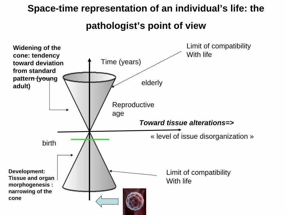
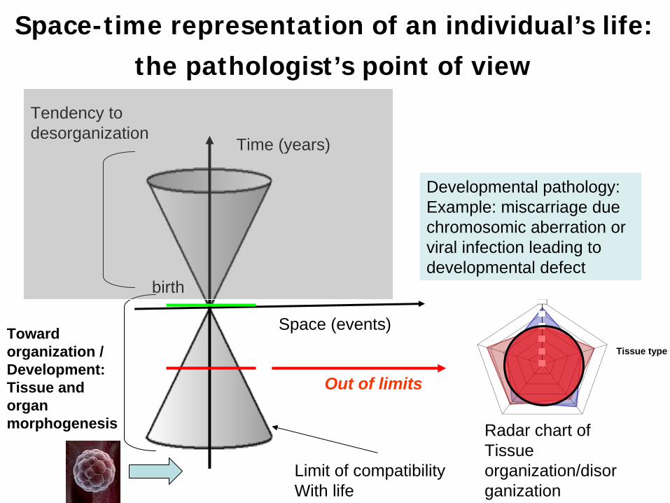
Space-time representation of an individual’s life: the
pathologist’s point of view
« level of issue disorganization »
Time (years)
birth
Development:Tissue and organmorphogenesis : narrowing of the cone
Widening of the cone: tendency toward deviation from standard pattern (young adult)
Reproductive age
Limit of compatibility With life
Limit of compatibility With life
Toward tissue alterations=>
elderly
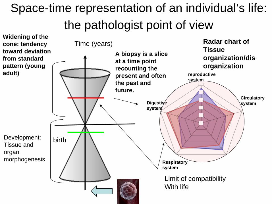
Space-time representation of an individual’s life: the pathologist point of view
Time (years)
birthDevelopment:Tissue and organmorphogenesis
Limit of compatibility With life
Digestive system
reproductive system
Respiratory system
A biopsy is a slice at a time point recounting the present and often the past and future.
Circulatory system
Widening of the cone: tendency toward deviation from standard pattern (young adult)
Radar chart ofTissue organization/dis organization
Space-time representation of an individual’s life: the pathologist’s point of view
Space (events)
Time (years)
birth
Toward organization /Development:Tissue and organmorphogenesis
Tendency to desorganization
Limit of compatibility With life
Developmental pathology:Example: miscarriage due chromosomic aberration or viral infection leading to developmental defect
Out of limits
Tissue type
Radar chart ofTissue organization/disor ganization
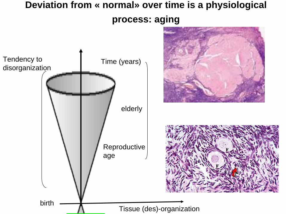
Deviation from « normal» over time is a physiological process: aging
Tissue (des)-organization
Time (years)
birth
Tendency to disorganization
Reproductive age
elderly
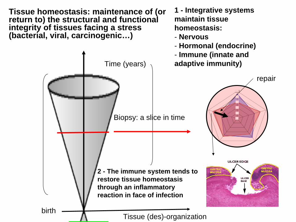
Tissue homeostasis: maintenance of (or return to) the structural and functional integrity of tissues facing a stress (bacterial, viral, carcinogenic…)
Tissue (des)-organization
Time (years)
birth
1 - Integrative systems maintain tissue homeostasis:- Nervous- Hormonal (endocrine)- Immune (innate and adaptive immunity)
Biopsy: a slice in time
2 - The immune system tends to restore tissue homeostasis through an inflammatory reaction in face of infection
repair
Aging (senescence) affects the integrative systems and contributes
to the loss of homeostasis• Senescence of the immune system
(immunosenescence) weakens the immunosurveillance of tumors. Explains the increased frequencies of tumors over time
• Hormonal senescence (monopause)
• Definition: immunosurveillance of tumors is the ability of the immune system to detect and destroy nascent tumors
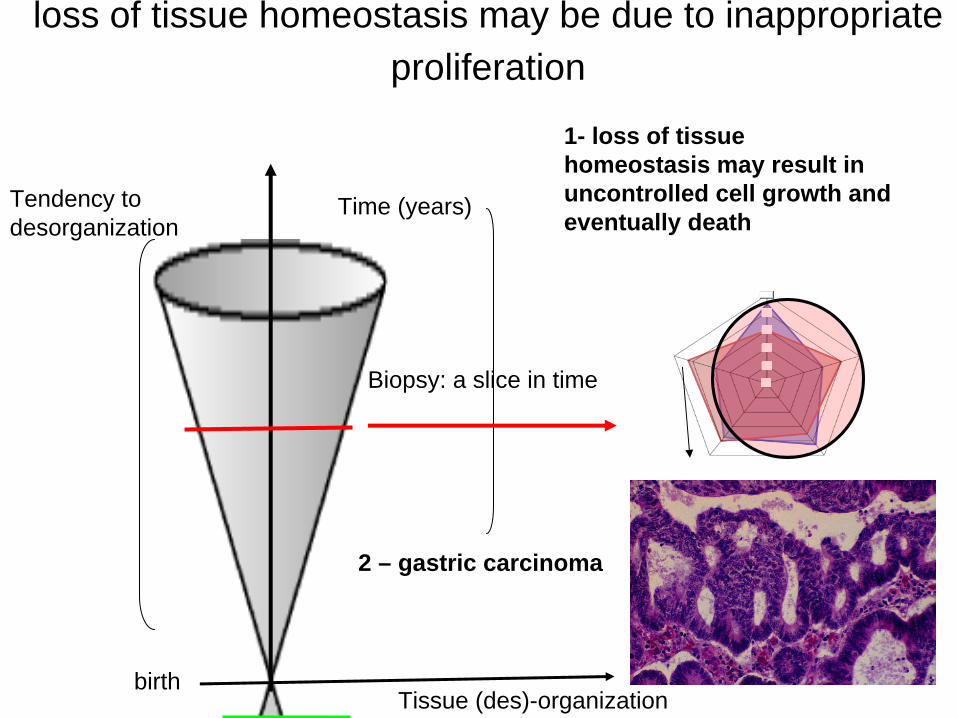
loss of tissue homeostasis may be due to inappropriate proliferation
Tissue (des)-organization
Time (years)
birth
Tendency to desorganization
1- loss of tissue homeostasis may result in uncontrolled cell growth and eventually death
Biopsy: a slice in time
2 – gastric carcinoma
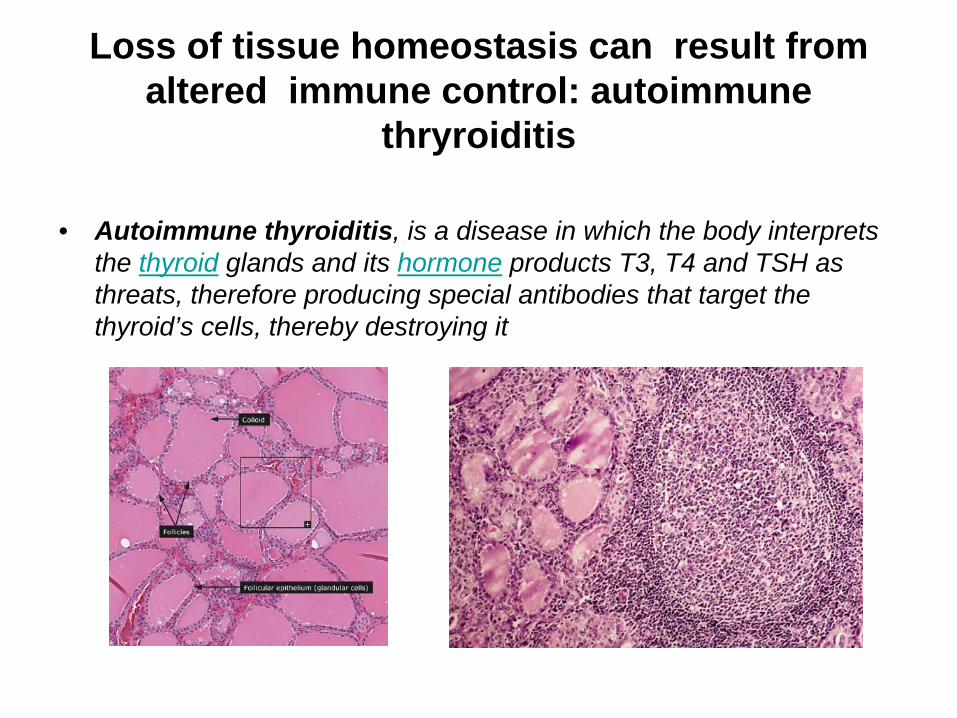
Loss of tissue homeostasis can result from altered immune control: autoimmune
thryroiditis
• Autoimmune thyroiditis, is a disease in which the body interprets the thyroid glands and its hormone products T3, T4 and TSH as threats, therefore producing special antibodies that target the thyroid’s cells, thereby destroying it
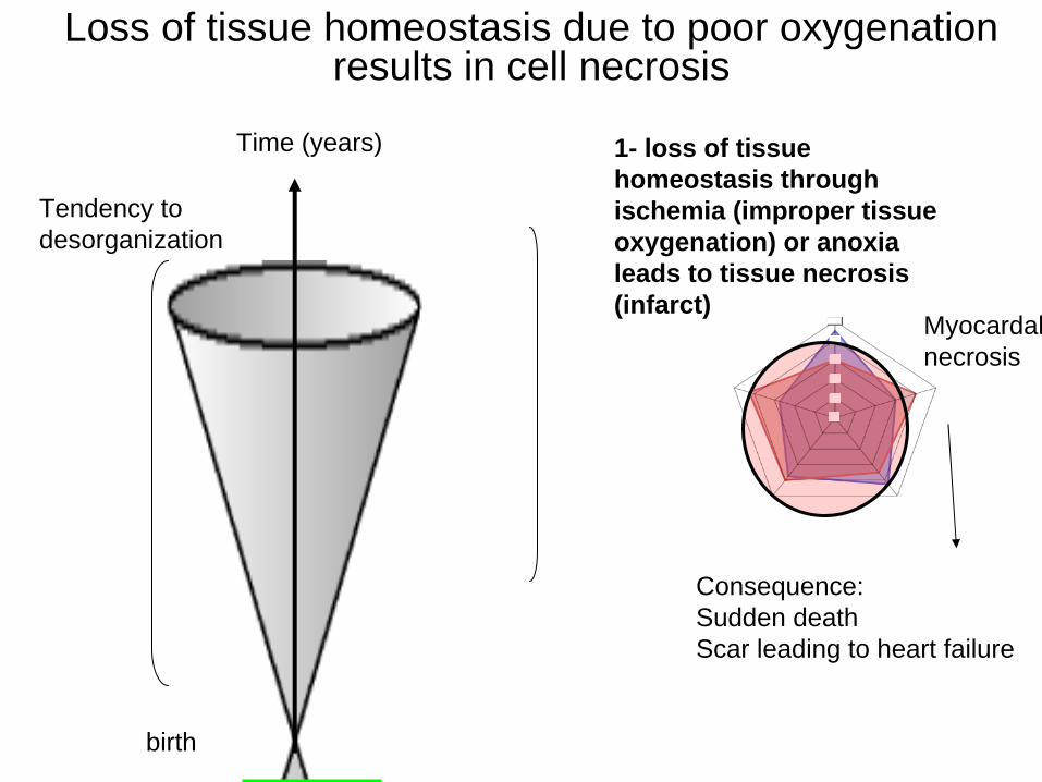
Loss of tissue homeostasis due to poor oxygenation results in cell necrosis
Time (years)
birth
Tendency to desorganization
1- loss of tissue homeostasis through ischemia (improper tissue oxygenation) or anoxia leads to tissue necrosis (infarct)
Myocardalnecrosis
Consequence:Sudden deathScar leading to heart failure
Chapter 2: the
pioneers…
Let’s go back to 1802…
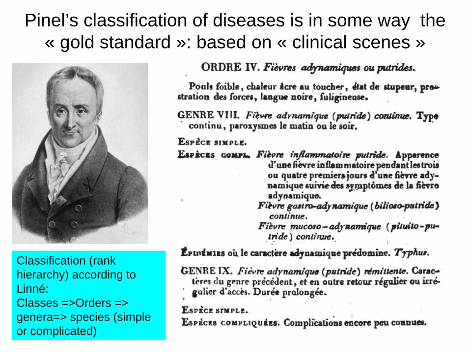
Pinel’s classification of diseases is in some way the « gold standard »: based on « clinical scenes »
Classification (rank hierarchy) according to Linné:Classes =>Orders => genera=> species (simple or complicated)
The limitations of existing classifications of diseases (1)
• Very complicated classifications (see Pinel)
• Diseases often named from the symptoms (a narrative by the patient) : how to detect the liars? ( many people want to escape their military duties…) (Corvisart)
The limitations of the existing classifications of diseases (2)
• terms that are used to name diseases are too vague
• case in point: « phtisis »: literally « consumption » has several meanings:– is the last phase in the course of progressive diseases
(e.g. cancer) associated with dramatic weight loss, anorexia, dramatic weakness : phtisis is then the evolutive convergence of several diseaes
– for others, should be restricted to pulmonary diseases causing weight loss, fever, hemoptysia…
– finally there are young patients dying of phtisis without internal lesions (called phtisis nervosa) (now referred to as anorexia nervosa)
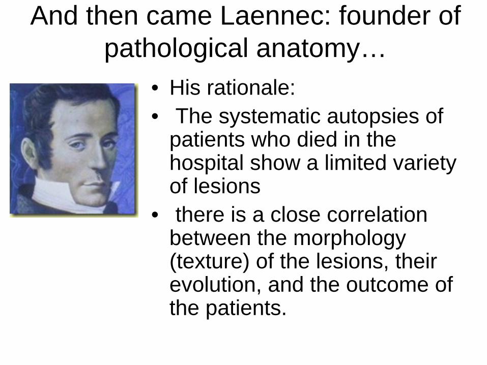
And then came Laennec: founder of pathological anatomy…
• His rationale:• The systematic autopsies of
patients who died in the hospital show a limited variety of lesions
• there is a close correlation between the morphology (texture) of the lesions, their evolution, and the outcome of the patients.
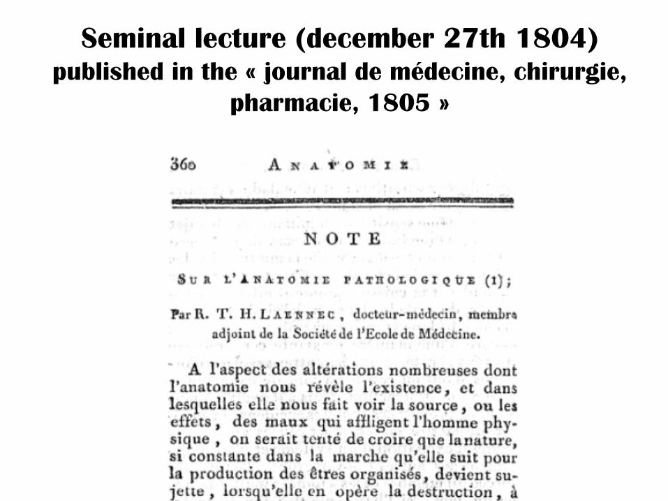
Seminal
lecture (december
27th 1804) published
in the «
journal de médecine, chirurgie,
pharmacie, 1805
»
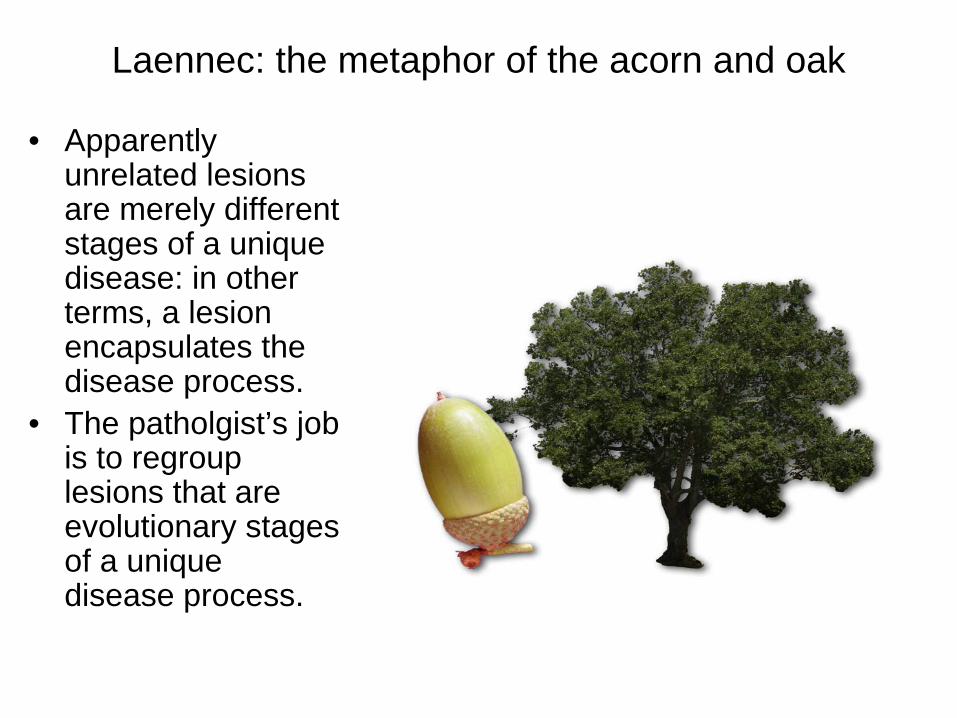
Laennec: the metaphor of the acorn and oak
• Apparently unrelated lesions are merely different stages of a unique disease: in other terms, a lesion encapsulates the disease process.
• The patholgist’s job is to regroup lesions that are evolutionary stages of a unique disease process.
Laennec introduces a morpho- chronological classification of lesions
• classification based on the assumption that the « texture » of a lesion recapitulates the disease process• Introducing a «morpho-chronological » denomination of lesions ex. « the tubercle » encompasses several lesional patterns. Pulmonary Tubercle = yellowish non translucid substance (crude state) becoming soft and friable on softening. May lead to a cavity upon evacuation or to infiltrates.
=> Phtisis becomes « tuberculosis »*Important notice: this nomenclature omits any causal element because too speculative
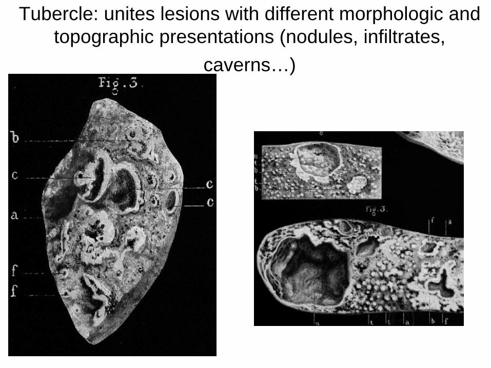
Tubercle: unites lesions with different morphologic and topographic presentations (nodules, infiltrates,
caverns…)
Definition of pathological anatomy by Laennec
« Science whose aim is to gain the knowledge of the visible alterations that the disease state produces in the human body. The opening of corpses is the way to get this knowledge. However in clinical practice, the pathological anatomy should be connected with the observation of symptoms or the alterations of functions asociated with alteration of organs » (In Panckoucke 1812)
Laennec’s legacy
• The morphology of a given lesion is predictive of its evolution and of the clinical outcome.
• The disease name should be based only on the representative anatomo- pathological lesions
• example: the term « phtisis » becomes only anatomical and will be replaced by « tuberculosis »
Main opponent to Laennec: Broussais (examen des doctrines médicales et
des systèmes de nosologie)
• A nomenclature of diseases based on pathological anatomy is irrelevant to the medicine as the name of the disease can be obtained only by the autopsy…
• No therapeutic relevance of this nomenclature
• No causal relationship
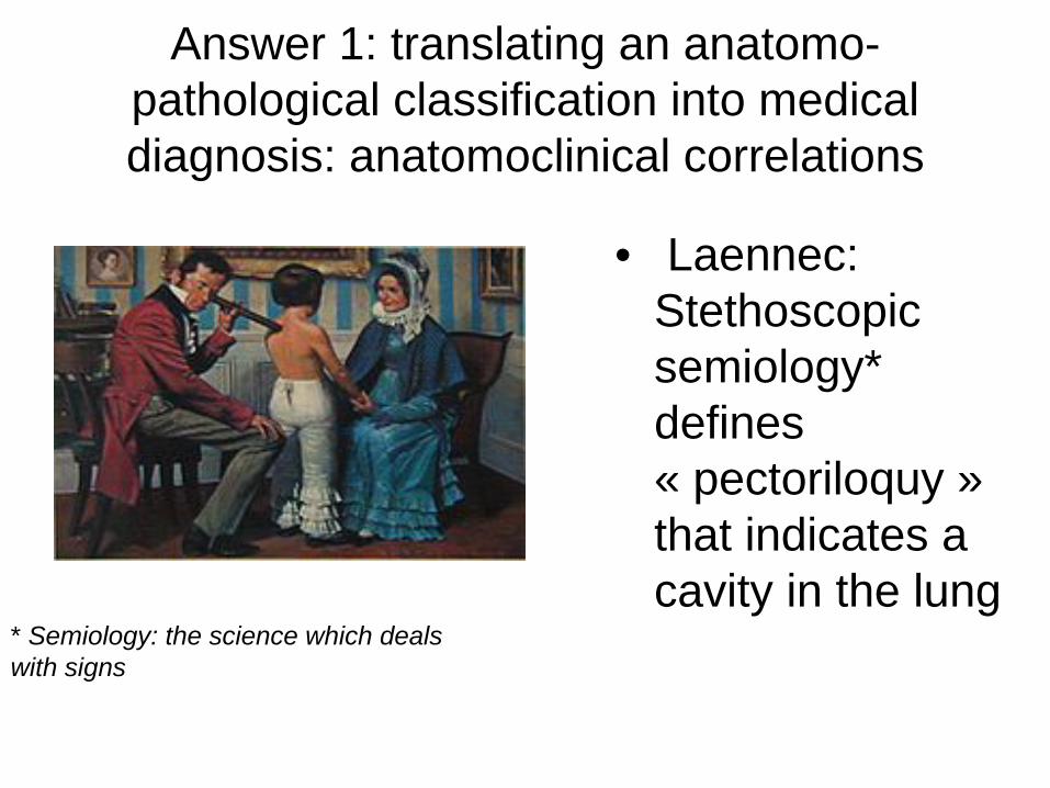
Answer 1: translating an anatomo- pathological classification into medical diagnosis: anatomoclinical correlations
• Laennec: Stethoscopic semiology* defines « pectoriloquy » that indicates a cavity in the lung
* Semiology: the science which deals with signs
The renowned french school of medicine, led mainly by anatomists,
refuses the microscope…..• Do I need a microscope to tell the
difference between an apple and a pear?…. ?
Chapter 3 : early times of microscopic
studies
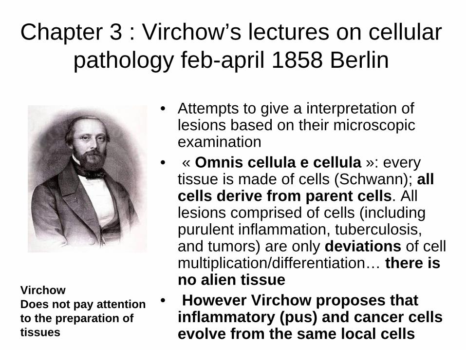
Chapter 3 : Virchow’s lectures on cellular pathology feb-april 1858 Berlin
• Attempts to give a interpretation of lesions based on their microscopic examination
• « Omnis cellula e cellula »: every tissue is made of cells (Schwann); all cells derive from parent cells. All lesions comprised of cells (including purulent inflammation, tuberculosis, and tumors) are only deviations of cell multiplication/differentiation… there is no alien tissue
• However Virchow proposes that inflammatory (pus) and cancer cells evolve from the same local cells
VirchowDoes not pay attention to the preparation of tissues
Tissue processing is a prerequisite for Microscopic examination of tissues
• Pathological diagnosis based on gross examination does not need a specific preparation/processing of tissues
• Microscopic examination of tissues requires a preparation/processing i.e. 1) tissue fixation (formalin), 2) tissue hardening (paraffin), 3) thin sectioning, 4) staining of tissue sections
• This pre-analytical phase is a prerequisite for the elaboration of a « microscopic semiology » needed for a microscopic diagnosis.
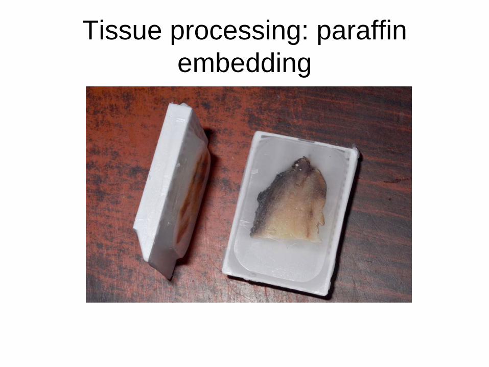
Tissue processing: paraffin embedding
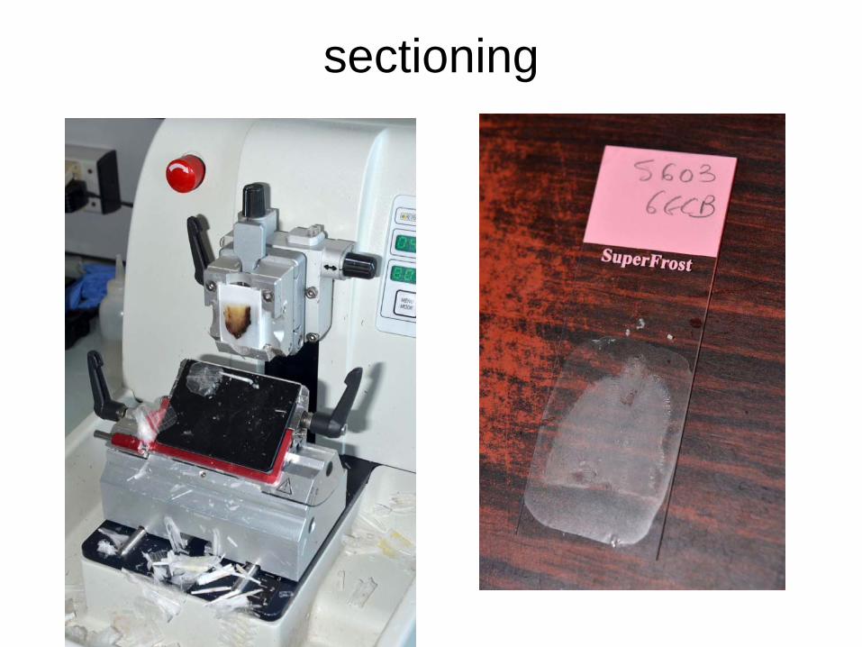
sectioning
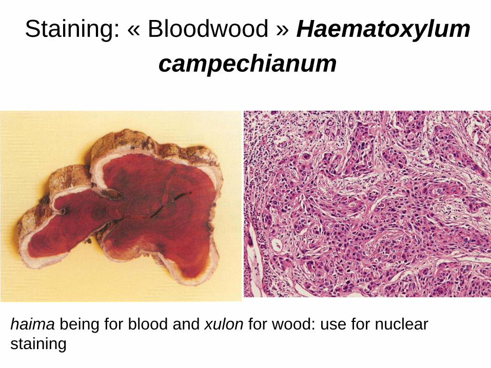
Staining: « Bloodwood » Haematoxylum campechianum
haima being for blood and xulon for wood: use for nuclear staining
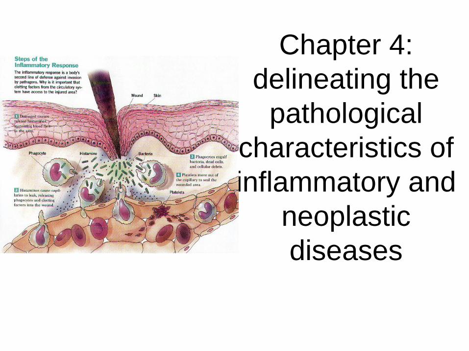
Chapter 4: delineating the
pathological characteristics of inflammatory and
neoplastic diseases
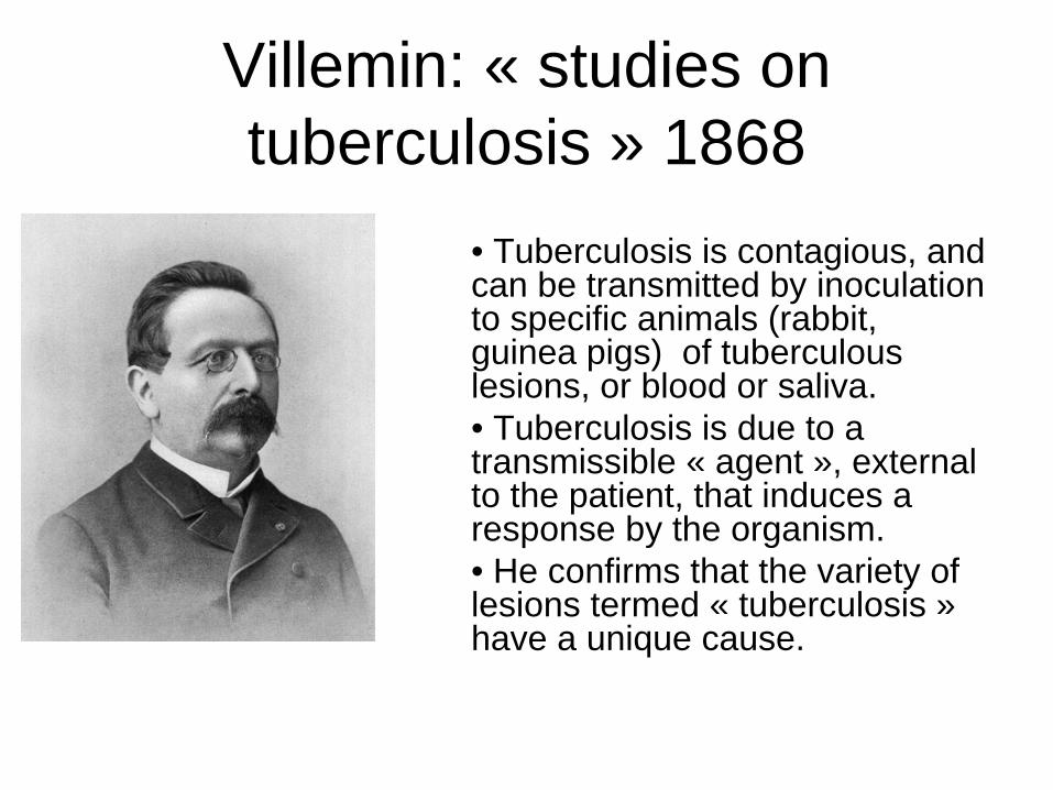
Villemin: « studies on tuberculosis » 1868
• Tuberculosis is contagious, and can be transmitted by inoculation to specific animals (rabbit, guinea pigs) of tuberculous lesions, or blood or saliva.• Tuberculosis is due to a transmissible « agent », external to the patient, that induces a response by the organism.• He confirms that the variety of lesions termed « tuberculosis » have a unique cause.
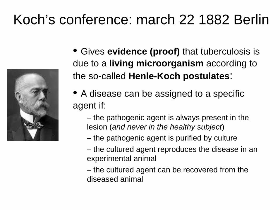
Koch’s conference: march 22 1882 Berlin
• Gives evidence (proof) that tuberculosis is due to a living microorganism according to the so-called Henle-Koch postulates:• A disease can be assigned to a specific agent if:
– the pathogenic agent is always present in the lesion (and never in the healthy subject)– the pathogenic agent is purified by culture– the cultured agent reproduces the disease in an experimental animal– the cultured agent can be recovered from the diseased animal
Koch’s postulates
• establish the principle of subordination of a lesion to a « specific » agent• question the validity of a classification of diseases based on the morphology of the lesions…• However an organism can harbor a pathogenic agent without being ill (healthy carrier)
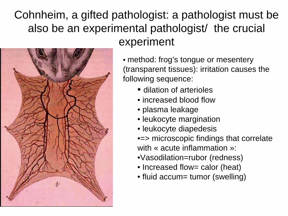
Cohnheim, a gifted pathologist: a pathologist must be also be an experimental pathologist/ the crucial
experiment• method: frog’s tongue or mesentery (transparent tissues): irritation causes the following sequence:
• dilation of arterioles• increased blood flow• plasma leakage• leukocyte margination• leukocyte diapedesis•=> microscopic findings that correlate with « acute inflammation »:•Vasodilation=rubor (redness)• Increased flow= calor (heat)• fluid accum= tumor (swelling)
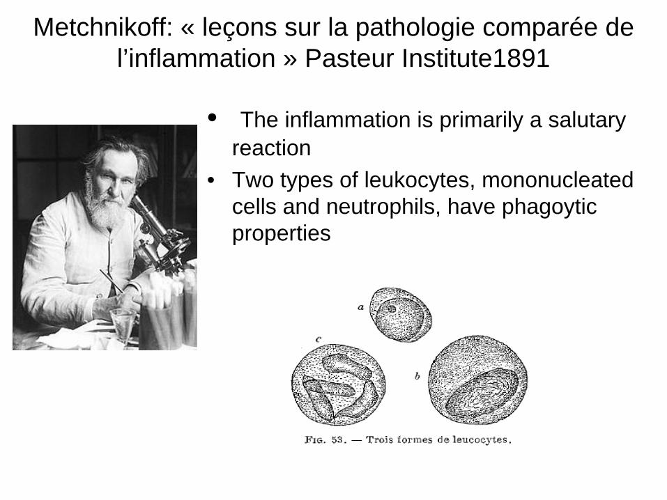
Metchnikoff: « leçons sur la pathologie comparée de l’inflammation » Pasteur Institute1891
• The inflammation is primarily a salutary reaction
• Two types of leukocytes, mononucleated cells and neutrophils, have phagoytic properties
Metchnikoff: « leçons sur la pathologie comparée de l’inflammation » Pasteur Institute 1891
• Acute Inflammation: « In an acute inflammation, there is a vasular dilatation, an activation of vascular endothelium and a exsudate with diapedesis, i.e. three steps that result in the accumulation of phagocytes in the in the injured area»
• Chronic Inflammation: ex. tuberculosis: « the tubercle is only made of phagocytic cells (derived from mononuclear cells)». It does not result from the multiplication of cells, but only from the recruitement of cells….. The so-called epithelioid cells (comprised of mononucleated cells) fuse in order to form giant cells…..
Chapter 4 : conclusion (1)
• New concepts drawn from observation and experimentation:
• Inflammatory lesions: lesions associated with the local recruitment of blood-derived cells - causative agents are external, transported by the blood: it is the dissemination of the agent that causes the extension of the disease
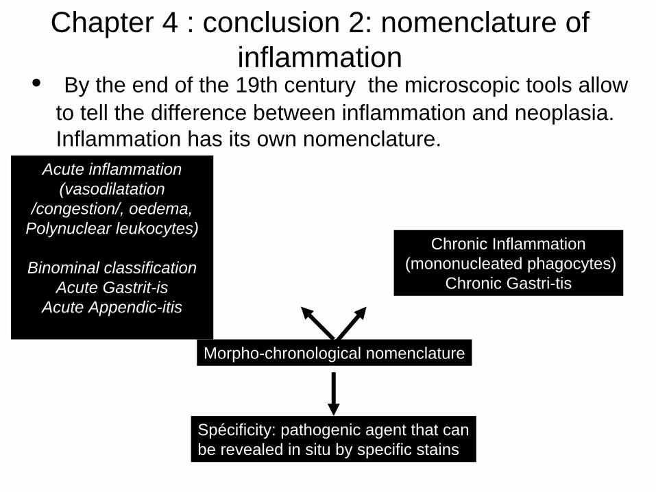
Chapter 4 : conclusion 2: nomenclature of inflammation
• By the end of the 19th century the microscopic tools allow to tell the difference between inflammation and neoplasia. Inflammation has its own nomenclature.
Acute inflammation(vasodilatation
/congestion/, oedema, Polynuclear leukocytes)
Binominal classificationAcute Gastrit-is
Acute Appendic-itisAcu
Chronic Inflammation(mononucleated phagocytes)
Chronic Gastri-tis
Morpho-chronological nomenclature
Spécificity: pathogenic agent that can be revealed in situ by specific stains
Chapter 4 : conclusion (3)
• New concepts drawn from observation and experimentation:
• neoplastic lesions: lesions associated with the local multiplication of resident cells - causative agents are (probably) internal to the cells. The metastasis of a malignant neoplasm is due to the dissemination of malignant cells.
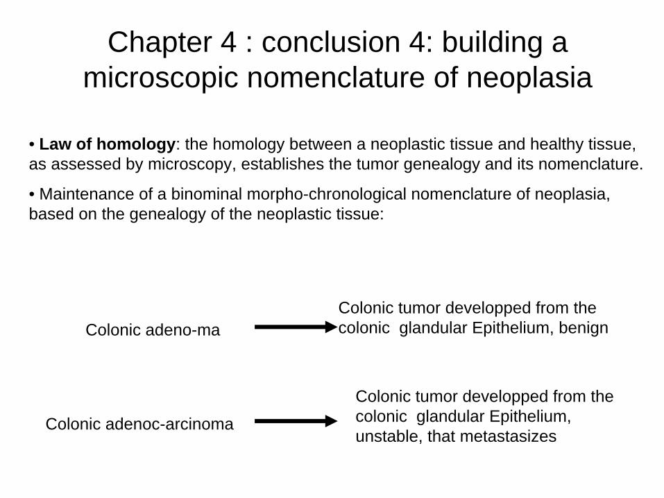
Chapter 4 : conclusion 4: building a microscopic nomenclature of neoplasia
• Law of homology: the homology between a neoplastic tissue and healthy tissue, as assessed by microscopy, establishes the tumor genealogy and its nomenclature.
• Maintenance of a binominal morpho-chronological nomenclature of neoplasia, based on the genealogy of the neoplastic tissue:
Colonic adeno-maColonic tumor developped from the colonic glandular Epithelium, benign
Colonic adenoc-arcinoma
Colonic tumor developped from the colonic glandular Epithelium, unstable, that metastasizes
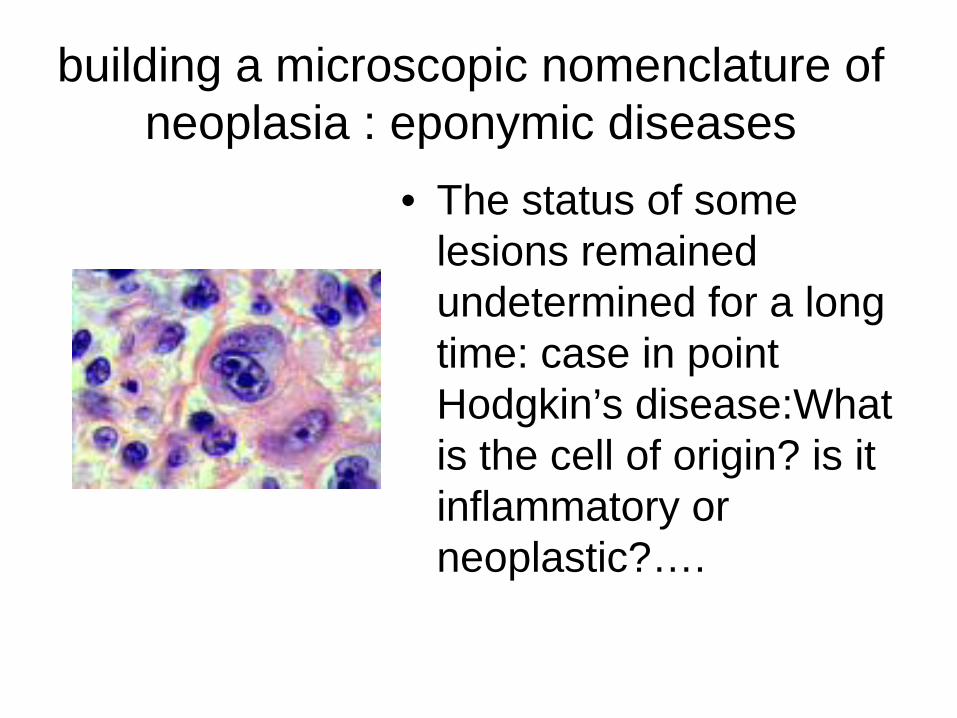
building a microscopic nomenclature of neoplasia : eponymic diseases
• The status of some lesions remained undetermined for a long time: case in point Hodgkin’s disease:What is the cell of origin? is it inflammatory or neoplastic?….
Chapter 5: the morphological
nomenclature of diseases becomes
therapeutically relevant
Halsted
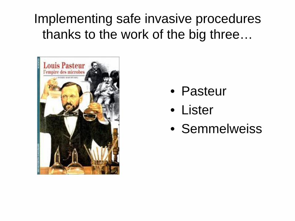
Implementing safe invasive procedures thanks to the work of the big three…
• Pasteur• Lister• Semmelweiss
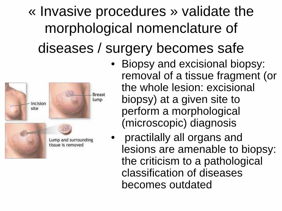
« Invasive procedures » validate the morphological nomenclature of
diseases / surgery becomes safe• Biopsy and excisional biopsy:
removal of a tissue fragment (or the whole lesion: excisional biopsy) at a given site to perform a morphological (microscopic) diagnosis
• practilally all organs and lesions are amenable to biopsy: the criticism to a pathological classification of diseases becomes outdated
Pathological diagnosis dictates the therapeutic strategy in oncology
• Most malignant epithelial tumors are primarily treated by oncologic surgery
• Lymphomas (malignant tumors of the lymphoid tissue) are sensitive to chemotherapy and are not eligible for surgery
• choriocarcinomas (highly malignant tumors of the placenta) are sensitive to chemotherapy
• seminoma (malignant tumor of the testis or ovary) is highly radiosensitive
Chapter 6: complementary
techniques : protein and DNA
imaging
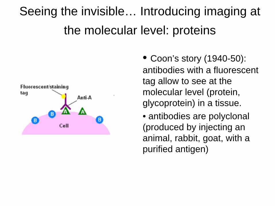
Seeing the invisible… Introducing imaging at the molecular level: proteins
• Coon’s story (1940-50): antibodies with a fluorescent tag allow to see at the molecular level (protein, glycoprotein) in a tissue.• antibodies are polyclonal (produced by injecting an animal, rabbit, goat, with a purified antigen)
Seeing the invisible… Introducing the molecular imaging of proteins
• Monoclonal antibodies: Köhler and Milstein– production of unlimited amounts of antibodies specific for a pre-
determined antigen– Allows cell immunophenotyping. Heterogeneity of the
nomenclature of antibodies and redundance of specificity have led to a denomination of antigens based on a standard classification: Clusters of Differentiation (CDs)
Example CALLA antigen (Common Acute Lymphoblastic Leukemia Antigen) recognized by the J5 monoclonal was designated CD10 Now known as a type II transmembrane protein found on pre-B cells, germinal-center B cells, some neutrophils, kidney cells, T-cell precursors, and epithelial cells that acts as a zinc metalloprotease cleaving peptide bonds on the amino side of hydrophobic amino acids; expressed in acute lymphocytic leukemia and follicular-center-cell lymphomas.
Seeing the invisible… introducing molecular pathology via in situ hybridization: reading the DNA
codeOncogenesis (cancerogenesis) proceeds partly via an accumulation of genomic errors or repeats (amplification) of some normal genes (called proto-oncogenes) that become oncogenic (called oncogenes)….
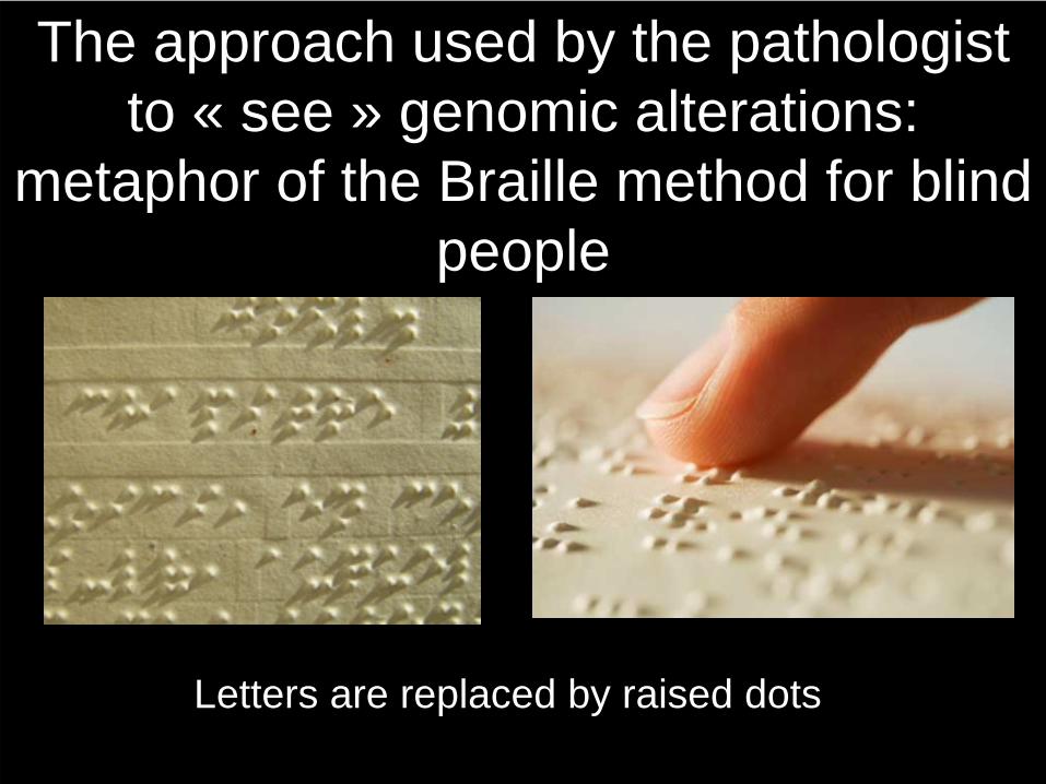
The approach used by the pathologist to « see » genomic alterations:
metaphor of the Braille method for blind people
Letters are replaced by raised dots
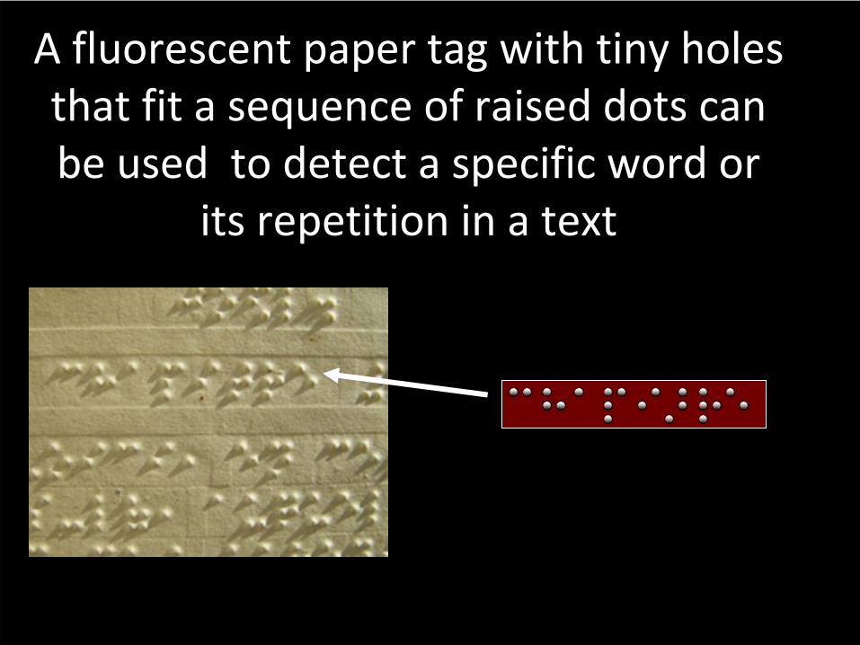
A fluorescent paper
tag with
tiny
holes that
fit a sequence
of raised
dots can
be
used
to detect
a specific
word
or its
repetition
in a text
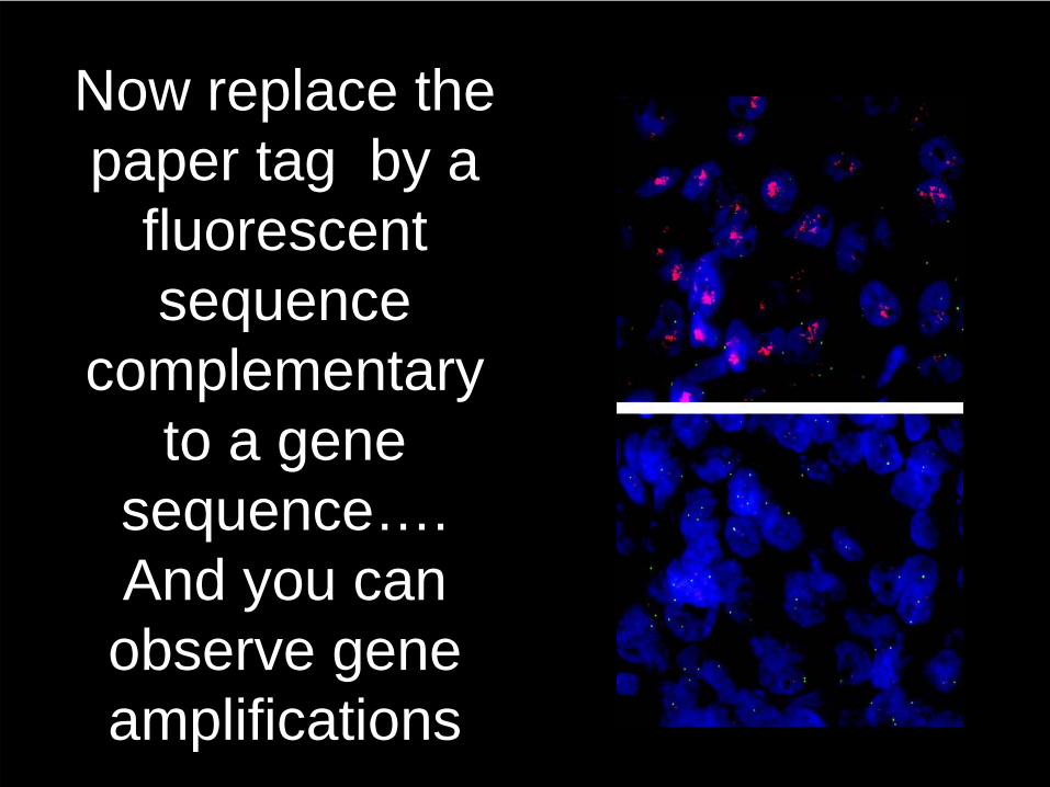
Now replace the paper tag by a
fluorescent sequence
complementary to a gene
sequence…. And you can observe gene amplifications
The role of the pathologist: top- down tissue analysis:
• Depending on the tissue available for examination, the pathologist uses 3 levels of analysis (semiology):– gross examination: form, texture, weigh of sample
(tumor, organ, biopsy etc…) dimensions– Microscopic examination: tissue architecture, type
of cells (inflammatory cells, tumor cells…)– Complementary methods:
• Immunohistochemistry: tumor immunophenotype• In situ hybridizaton: deciphering the abnormalities
of the DNA code (gene amplification, translocations…)
Chapter 6 conclusion: The role of the pathologist
• Laennec’s legacy: – the diseases get their names from the
morphology of the underlying lesions– the morphological diagnosis dictates
the therapeutic strategy– Anatomic pathology is the gold
standard for the diagnosis and treatment of neoplastic diseases
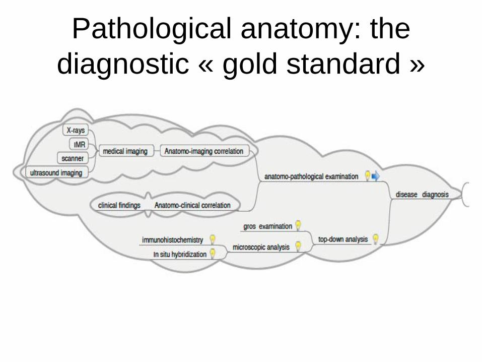
Pathological anatomy: the diagnostic « gold standard »