Embed Size (px)
Citation preview

Back to BasicsBack to BasicsNephrologyNephrology
20132013
Major issues in Nephrology, Electrolytes, Acid-base disturbances

CKD

K/DOQI Classification of Chronic K/DOQI Classification of Chronic Kidney DiseaseKidney Disease
StageStage GFR GFR ((≥≥3mo)3mo) Description Description
(ml/min/1.73m(ml/min/1.73m22))
1 1 90 90 Damage with normal Damage with normal GFRGFR
22 60-90 60-90 Mild Mild GFR GFR
33 30-59 30-59 Moderate Moderate GFR GFR
44 15-29 15-29 Severely Severely GFR GFR
5 5 <15 <15 Kidney FailureKidney Failure

In this K/DOQI staging, “kidney damage” means:
• Persistent proteinuriaPersistent proteinuria
• Persistent glomerular hematuriaPersistent glomerular hematuria
• Structural abnormality:Structural abnormality:– such as PCKD, reflux nephropathysuch as PCKD, reflux nephropathy
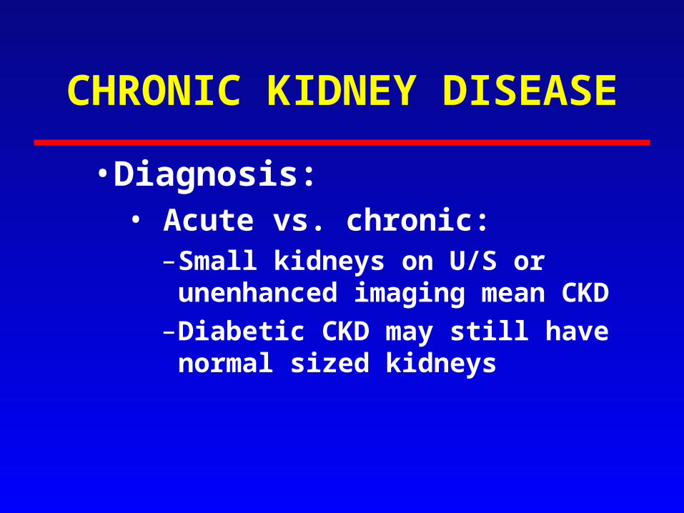
CHRONIC KIDNEY DISEASE
• Diagnosis: • Acute vs. chronic:
–Small kidneys on U/S or unenhanced imaging mean CKD
–Diabetic CKD may still have normal sized kidneys

CHRONIC KIDNEY DISEASE
• Common causes of CKD:• Diabetic nephropathy
• Vascular disease
• GN
• PKD

CHRONIC KIDNEY DISEASE
• Causes of CKD:• Best to divide as proteinuric or
non-proteinuric CKD
• Proteinuric is much more likely to have deterioration in GFR and higher cardiovascular morbidity and mortality

CHRONIC KIDNEY DISEASE
• Treatment• Delay progression:
• Treat underlying disease i.e. good glucose control for DM
• BP control to 140/90, (the current target); 130/80 for diabetics
• ACEI or ARB has extra benefit for proteinuric CKD
• Lower protein diet…maybe

CHRONIC KIDNEY DISEASE
• Treatment of the consequences of decreased GFR:– PO4:
• decrease dietary intake• PO4 binders such as CaCO3
– Hypocalcemia:• CaCO3, 1,25 OH D3

CHRONIC KIDNEY DISEASE
• Treatment of the consequences of decreased GFR:– Anemia:
• Erythropoetin current target Hb 95-105

CHRONIC KIDNEY DISEASE
• Uremic Complications:
Major:– Pericarditis– Encephalopathy– Platelet dysfunction

ARF

Question 1
Urine values compatible with pre-renal failure:
A)Osm < 300 mosm/LB)RBC castsC)Na+ < 20 mmol/LD)Fex Na+ > 2%
Click here in slide show mode

ARF
• Pre renal and ATN most common causes (quoted at 70% of cases of ARF)
• DDx:– Pre Renal– Intra Renal– Post Renal

U Na U Osm Fe Na
• Pre-Renal
• ATN
Urine: Pre-Renal vs. RenalAssessment of Function
Fe Na =U/P NaU/P Cr X 100
> 500 < 1%
> 40 < 350 > 2%
< 20
• Pigmented granular casts found in up to 70% of cases of ATN

Fe Urea
• Pre-Renal
• ATN
Urine: Pre-Renal vs. RenalAssessment of Function
Fe Urea = U/P UrU/P Cr X 100
> 55
< 35
• FeUrea might be useful to Dx pre renal ARF in those who received diuretics…but not all studies support its use.

ARF
• Investigations:– Pre Renal: Urine tests as noted and
responds to volume– Intra-Renal: look for GN, interstitial
nephritis as well as ATN– Post Renal: Imaging showing bilateral
hydronephrosis is highly specific for obstruction causing ARF

• If cannot control these by other means:HyperkalemiaPulmonary edemaAcidosisUremia
• (GFR < 10-15% for CRF)
Dialysis: Who Needs It?

• Hemodialysis is also used for intoxications with:– ASA– Li– Alcohols: i.e. methanol, ethylene glycol– Sometimes theophylline
Dialysis: Who Needs It?

Na+

Hyponatremia
• Pseudo: – If total osmolality is high: hyperglycemia/
mannitol– If total osmolality is normal, could be due to
very high serum lipoprotein or protein

Hyponatremia
• Volume status:– Hypovolemic: high ADH despite low
plasma osmolality – High total volume: CHF/ cirrhosis have
decreased effective circulating volume and high ADH despite low plasma osmolality

Hyponatremia
• Volume status:– If volume status appears normal:
• If urine osmolality is low: normal response to too much water intake…”psychogenic polydipsia”
• If urine osmolality is high: inappropriate ADH

Hyponatremia
• Treatment:– Hypovolemic:
• Replace volume
– Decreased effective volume:• Improve cardiac output if possible• Water restrict
– SIADH:• Water restrict

Hyponatremia
• Treatment:– Rate of correction of Na:
• Not more than 10 mmol in first 24 h and not more than 18 mmol over first 48 h of treatment
• Or Central Pontine Myelinosis may occur

Potassium

Hyperkalemia
• Real or Not:– Hemolysis of sample– Very high WBC, PLT– Prolonged tourniquet time

Hyperkalemia
• Shift of K from cells:– Insulin lack– High plasma osmolality– Acidosis– Beta blockers in massive doses

Hyperkalemia
• Increased total body K:– Decreased GFR plus:
• High diet K• KCl supplements• ACEI/ARB• K sparing diuretics
– Decreased Tubular K secretion

TTKG?• Requirements:
– Urine osmolality > 300– Urine Na+ > 25– Reasonable GFR
• TTKG =
[urine K[urine K++ (urine osmol/serum osmol)] (urine osmol/serum osmol)]
serum Kserum K++
<7, esp < 5 = hypoaldosteronism<7, esp < 5 = hypoaldosteronism
U/P K+/U/P Osm

Hyperkalemia
• Treatment– IV Ca– Temporarily shift K into cells:
• Insulin and glucose• Beta 2 agonists (not as reliable as insulin)• HCO3 if acidosis present
– Remove K

GFR

ASSESSMENT OF GFR:
0
200
400
600
800
1000
30 60 90 120
GFR
Cre
at

(140-age) x Kg x1.2 Creat
(x .85 for women)
ASSESSMENT OF GFR:
• Cockroft-Gault Cockroft-Gault estimated Creatinine estimated Creatinine clearanceclearance
UCr x V PCr
Need a Steady State for these to be valid
Creatinine clearance formula:

• Labs now calculate this for anyone who has a serum creatinine checked
• Use serum creatinine, age, sex
• Labs now calculate this for anyone who has a serum creatinine checked
• Use serum creatinine, age, sex
MDRD eGFRMDRD eGFR

GFR, in mL/min per 1.73 m2 =(170 x (PCr [mg/dL])exp[-0.999]) x (Age exp[-0.176]) x ((Surea [mg/dL])exp[-0.170]) x ((Albumin [g/dL])exp[+0.318])
where SUrea is the serum urea nitrogen concentration; and exp isthe exponential. The value obtained must be multiplied by 0.762 if the patient is female or by 1.180 if the patient is black.
Simplified:GFR, in mL/min per 1.73 m2 =186.3 x ((serum creatinine) exp[-1.154]) x (Age exp[-0.203])x (0.742 if female) x (1.21 if African American)
Simplified:GFR, in mL/min per 1.73 m2 =186.3 x ((serum creatinine) exp[-1.154]) x (Age exp[-0.203])x (0.742 if female) x (1.21 if African American)
MDRD eGFRMDRD eGFR
Do NOT memorize this formula

Limitations of GFR estimates:Limitations of GFR estimates: Not reliable for:Not reliable for:
• extremes of weight or different body extremes of weight or different body composition such as post composition such as post amputation, paraplegiaamputation, paraplegia
• acute changes in GFRacute changes in GFR
• use in pregnancy use in pregnancy
• eGFR greater than 60ml/min/1.73meGFR greater than 60ml/min/1.73m22

Proteinuria

Proteinuria
• Albumin vs. other protein– Dipstick tests albumin

PROTEINURIA
• Quantitative:
– 24 hour collection24 hour collection
– ACR: random albumin to creatinine ACR: random albumin to creatinine ratioratio
– PCR: random protein to creatinine PCR: random protein to creatinine ratioratio

PROTEINURIA
• Microalbuminuria: less than dipstick albumin
• Can use albumin to creatinine ratio on random urine sample… best done with morning urine sample

Random Random UrineUrine
24h 24h UrineUrine
Random Random UrineUrine
24h Urine24h Urine
ACRACR
(g/mol)(g/mol)
AlbuminAlbumin
(mg/24h)(mg/24h)
PCRPCR
(g/mol)(g/mol)
ProteinProtein
(mg/24h)(mg/24h)
NormalNormal MM
FF
<2.0<2.0
<2.8<2.8
<30<30 <20<20 <200<200
Micro-Micro-albuminuriaalbuminuria
MM
FF
2.0-302.0-30
2.8-302.8-30
30-30030-300
Macro-Macro-albuminuriaalbuminuria
>30>30 >300>300

Question 2
The definition of nephrotic syndrome includes::A)HypolipidemiaB)LipiduriaC)24 hr protein ≥2gD)hypertension

Nephrotic Syndrome
• Definition:– > 3 g proteinuria per day– Edema– Hypoalbuminemia– Hyperlipidemia and lipiduria are also
usually present

Nephrotic Syndrome
• Causes:– Secondary: DM, lupus– Primary:
• Minimal change disease• FSGS• Membranous nephropathy

Nephrotic Syndrome
• Complications:– Edema– Hyperlipidemia– Thrombosis…with membranous GN and
very low serum albumin

Nephrotic Syndrome
• Treatment:– Treat cause if possible– Treat edema, lipids– Try to decrease proteinuria

Hematuria

Hematuria
• Significance: ≥3 RBC's per hpf• DDx: Is it glomerular or not?• Glomerular:
– RBC casts– Dysmorphic RBCs in urine– Coinciding albuminuria may
indicate glomerular disease

Hematuria
• Other investigation:– Imaging of kidneys– Serum creatinine– Age over 40-50 rule out urologic
bleeding, i.e. urine cytology and referral for cystoscopy

Hematuria
• For glomerular hematuria without proteinuria DDx includes:– IgA nephropathy
– Thin GBM disease
– Hereditary nephritis

Ca++, PO4, Mg++

Ca++ and PO4--
Decreased GFR and increased PO4
Decreased Ca
1 OH of 25-OHD3
Increased PTH
Renal osteodystrophy

Magnesium
• Hypomagnesemia:– GI loss/lack of dietary Mg– Renal loss:
• Diuretics• Toxins esp cisplatin
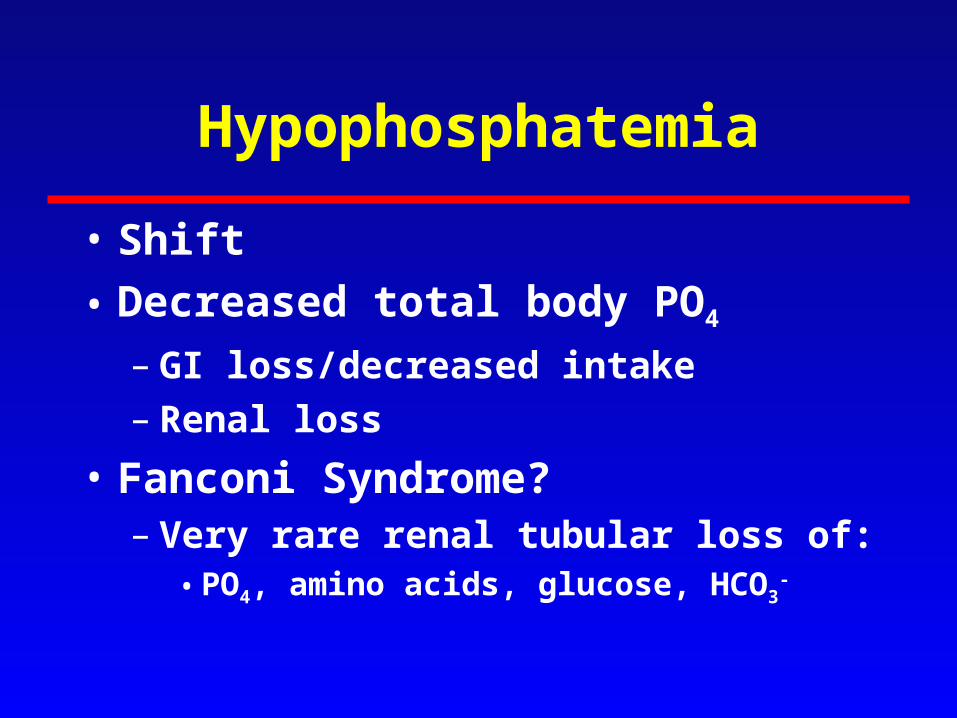
Hypophosphatemia
• Shift
• Decreased total body PO4
– GI loss/decreased intake– Renal loss
• Fanconi Syndrome?– Very rare renal tubular loss of:
• PO4, amino acids, glucose, HCO3-

Question 3
Most likely cause of Na 140 Cl 110 HCO3 10:A)RTAB)serum albumin 20C)resp alkalosisD)ketoacidosis

Acid-Base
• Approach to:– Resp or metabolic
– Compensated or not
– If metabolic: anion gap or not
– Anion gap = Na - (Cl + HCO3)

Acid-Base
• “MUDPILES”:– Methanol– Uremia– Diabetic/alcoholic
ketosis
– Paraldehyde– Isopropyl alcohol– Lactic acid– Ethylene glycol– Salicylate
Increased anion Gap acidosis:

Acid-Base
Metabolic acidosis with normal serum anion gap can be due to:
1) GI losses of HCO3
2) Renal tubular acidosis

Renal Tubular Acidosis
Hopefully will not need this.Normal renal response to acidosis is to increase
ammoniagenesis and more NH4 will be found in the urine
For those with close to normal GFR, the “urine anion gap” is a way to estimate urinary NH4
Urine anion gap = urine (Na+ + K+ – Cl-)
If it is positive there is decreased NH4+ production
and likely a renal component to the acidosis


AKI