Embed Size (px)
Citation preview

Page 1/19
Endochin-like quinolone-300 and ELQ-316 inhibitBabesia bovis, B. bigemina, B. caballi and Theileriaequi Marta G. Silva
Washington State UniversityReginaldo G. Bastos
washington state universityJ. Stone Doggett
Oregon Health & Science University - West CampusMichael K. Riscoe
Oregon Health & Science University - West CampusSovitj Pou
VA Portland Health CenterRolf Winter
VA Portland Health Care SystemRozalia A. Dodean
VA Portland Health Care SystemAaron Nilsen
Oregon Health & Science UniversityCarlos E. Suarez ( [email protected] )
"USDA-ARS Paci�c West Area"
Research
Keywords: Bovine babesiosis, equine piroplamsosis, Babesia bovis, Babesia bigemina, Babesia caballi,Theileria equi, endochin-like quinolones, ELQ-300, ELQ-316
Posted Date: November 11th, 2020
DOI: https://doi.org/10.21203/rs.3.rs-58018/v4
License: This work is licensed under a Creative Commons Attribution 4.0 International License. Read Full License

Page 2/19
Version of Record: A version of this preprint was published at Parasites & Vectors on December 3rd, 2020.See the published version at https://doi.org/10.1186/s13071-020-04487-3.

Page 3/19
AbstractBackground: The most common apicomplexan parasites causing bovine babesiosis are Babesia bovisand B. bigemina, while B. caballi and Theileria equi are responsible for equine piroplasmosis. Treatmentand control of these diseases are usually achieved using potentially toxic chemotherapeutics, such asimidocarb diproprionate, but drug-resistant parasites are emerging, and alternative effective and saferdrugs are needed. Endochin-like quinolones (ELQ)-300 and ELQ-316 proved safe and e�cacious againstrelated apicomplexans, such as Plasmodium spp., and ELQ-316 was also effective against B. microti,without showing toxicity in mammals.
Methods: Inhibitory effects of ELQ-300 and ELQ-316 were assessed on the growth of cultured B. bovis, B.bigemina, B. caballi and T. equi. Percentage of parasitized erythrocytes was measured by �ow cytometry.Effect of the ELQ drugs on the viability of horse and bovine peripheral blood mononuclear cells (PBMC)was assessed by monitoring cell metabolic activity using a colorimetric assay.
Results: We calculated IC50 ranging from 0.04 to 0.37 nM for ELQ-300, and from 0.002 to 0.1 nM for ELQ-316 at 72 h among all cultured parasites tested. None of the parasites tested were able to replicate incultures in the presence of the ELQ-300 and ELQ-316 at IC100, which range from 1.3 to 5.7 nM for ELQ-300and from 1.0 to 6.0 nM for ELQ-316 at 72 h. Neither ELQ-300 nor ELQ-316 altered the viability of equineand bovine PBMC at their IC100 in in vitro testing.
Conclusions: ELQ-300 and ELQ-316 showed signi�cant inhibitory activity on the main parasitesresponsible for bovine babesiosis and equine piroplasmosis at doses that are tolerable to host cells.These ELQ drugs may be viable candidates for developing alternative protocols for the treatment ofbovine babesiosis and equine piroplasmosis.
BackgroundTick-borne diseases caused by apicomplexan hemoparasites, such as Babesia and Theileria, imposeserious economic impact on the cattle and horse industries worldwide [1, 2]. Babesiosis and theileriosisshare similar acute disease signs, including anemia, loss of weight, anorexia and fever [3]. Usually,Babesia and Theileria are not eliminated in surviving animals and cause lifelong persistent infections.Important shared features among Babesia and Theileria species include a sexual reproductive cycle intheir Ixodes arthropod hosts and asexual reproduction in the red blood cells (RBC) of their vertebratehosts, a process that results in severe, potentially fatal hemolytic anemia [4-6].
Theileria equi and Babesia caballi are the etiological agents of equine piroplasmosis (EP), a disease thataffects horses, mules, donkeys, and zebras worldwide [7]. EP imposes severe and costly restrictions intransportation of high-performance horses between endemic and non-endemic areas to participate inequestrian sporting events [3, 6]. No vaccines are currently available against T. equi and B. caballi, andconsiderable resources have been spent to develop drugs to treat animals against the harmful effects ofacute EP and to prevent the loss of performance in chronically infected, high-value horses. Despite these

Page 4/19
efforts, horses that survive acute infection, especially when caused by T. equi, become persistentlyinfected, asymptomatic carriers, a condition that can be associated with the resurgence of outbreaks ofEP worldwide [8].
B. bovis and B. bigemina are two main causative agents of bovine babesiosis (BB), an acute andpersistent economically important disease of cattle that typically cause high mortality [1]. While B.bigemina is usually associated with relatively milder acute hemolytic disease, B. bovis is implicated in amore severe presentation of the acute phase of the disease, characterized by cytoadhesion of parasite-infected RBC in the brain capillaries, which resembles cerebral malaria and often leads to death [1].
Prevention and control of EP and BB have been typically achieved by controlling tick vector populations,the use of live attenuated vaccines in the case of BB, and chemotherapy. The live attenuated vaccinesavailable to prevent acute BB, which are in use only in a limited number of countries, are onlyrecommended for less than 1-year old animals and present several additional constraints, including therisk of reversion to virulence. Furthermore, cattle vaccinated with live attenuated vaccines may alsobecome persistently infected with the parasites, and can serve as a reservoir for tick acquisition andtransmission [9]. In addition, live vaccines can cause severe disease to immunocompromised and oldercattle, which may be more susceptible to the attenuated vaccine strains [9]. Given these scenarios, someanimals vaccinated with live Babesia vaccines also need to be treated with anti-babesial drugs to preventthe development of acute disease caused by virulent escapes within the population of parasites in theattenuated vaccine strains. Currently, babesicidal drugs are the only option available for preventing losesdue to babesiosis in adult vaccine-susceptible animals that need to be transported from non-endemic toBabesia endemic areas. Altogether, these aspects highlight the importance of having reliable babesicidaldrugs to control the spread of outbreaks and prevent development of acute disease in herds vaccinatedwith live attenuated Babesia vaccines.
Chemotherapy treatments based on diminazene aceturate and imidocarb dipropionate are the mosteffective and �rst-choice methods to manage animals with acute BB and EP [10, 11]. However, thee�cacy of these drugs is highly variable and treated animals need to be monitored closely for adverseeffects, especially when high doses are used for attempting clearance of the parasites, which is a usualoccurrence for valuable horses affected by EP [12]. In addition to toxic side effects, and although speci�cresistance to imidocarb by Babesia and Theileria parasites was not yet documented, the potential for thedevelopment of drug-resistance by Babesia parasites to other drugs such as amicarbalide isethionate hasbeen previously recorded [13]. Consequently, there is the need to search for new effective and less toxicalternative chemotherapeutics against BB and EP.
Endochin-like quinolone (ELQ) are potent selective inhibitors of the mitochondrial cytochrome bc1
complex, as demonstrated in Plasmodium falciparum, the causative agent of the most severe form ofhuman malaria [14-17]. ELQ compounds have shown to be highly effective against different species andmultiple stages of Plasmodium [18, 19]. Importantly, ELQ-300 and ELQ-316 have been selected as pre-clinical anti-malaria candidates, considering their reasonable oral bioavailability at e�cacious doses,

Page 5/19
long half-life, and metabolic stability [18, 19]. A recent study also demonstrated the e�cacy of ELQprodrugs combined with atovaquone to treat experimental babesiosis caused by B. microti in theimmunode�cient mouse model [20]. Data from this study showed that the combined therapy of ELQ andatovaquone resulted in complete clearance of the parasite with no disease recrudescence even more than100 days after discontinuation of the treatment [20]. Based on these conclusive parasite inhibitoryresults, we evaluated the effect of ELQ-300 and ELQ-316 on the in vitro growth of B. bovis, B. bigemina, B.caballi, and T. equi. Strong inhibition of the development of all these parasites, coupled with the lack oftoxic effects on host cells, suggests that these two compounds are promising candidates for futuredevelopment of novel alternative therapies to control BB and EP.
MethodsSynthesis of ELQ-300 and ELQ-316
ELQ-300 and ELQ-316 were synthesized as previously described [19, 21] (Fig. S1). Both compounds werekindly provided by the Department of Molecular Microbiology and Immunology, Oregon Health andScience University, Portland, OR, USA. Purity of both ELQ derivatives was assessed at >99% by protonNMR and gas chromatography – mass spectrometry. ELQ-300 and ELQ-316 were diluted in 100%dimethyl sulfoxide (DMSO) to prepare stock solutions. Stock solutions were kept at RT until use. Workingsolutions were freshly prepared in parasite culture medium before adding to the parasite cultures.
Cultures of B. bovis, B. bigemina, B. caballi and T. equi
B. caballi Puerto Rico strain [18], B. bovis Texas strain [19], B. bigemina Puerto Rico strain [20] andTheileria equi Florida strain [21] were grown in long-term microaerophilous stationary-phase cultures andincubated at 37°C in an atmosphere of 5% CO2, 5% O2 and 90% N2, as previously described [22-25]. B.bovis and B. bigemina were grown in 96-well plate using 180 µl per well of complete HL-1 culture media(pH 7.2; 2.38 g/L HEPES, 5 mL/L L-glutamine, 60 U/ml of penicillin G, 60 μg/ml of streptomycin, and 0.15μg/ml of amphotericin B (Sigma) supplemented with 40% bovine serum. Cultures contained asuspension of 10% and 5% packed cell volume of bovine erythrocytes for B. bovis and B. bigemina,respectively. B. caballi and T. equi were cultured under similar conditions, but culture media weresupplemented with 10% or 20% horse serum, respectively. In addition, B. caballi and T. equi culturescontained a suspension of 10% packed cell volume equine erythrocytes.
Parasite growth inhibition assay
Growth inhibition assays using ELQ-300 or ELQ-316 were performed on cultured B. bovis, B. bigemina, B.caballi and T. equi with a starting percentage of parasitized erythrocytes (PPE) of 0.2. Parasites weregrown as described above, using culture media containing different concentrations of ELQ-300 or ELQ-316 from 0.05 to 50 nM diluted in DMSO. Parasite cultures in the presence of DMSO (0.5 μl) and in theabsence of the ELQ compounds were used as a positive control for parasite growth. Extra wellscontaining uninfected bovine or equine RBC were prepared and used as negative controls for the �ow

Page 6/19
cytometric analysis. Fresh culture medium (150 µl/well) containing respective drug concentration wasreplaced daily to parasite cultures. These experiments were carried out in triplicate for each testedconcentration and controls, over a period of 72 h. PPE was monitored daily by �ow cytometry, aspreviously described [22, 23]. Fifty percent inhibitory concentration (IC50) values were calculated for ELQ-300 and ELQ-316 at 24, 48, and 72 h by extrapolation in which there is a 50% reduction of the PPE in thewells containing the ELQs compared with the positive control wells using nonlinear regression (GraphPadPrism version 8.0.2 for Windows, San Diego, California, USA). Similarly, 100% inhibitory concentration(IC100) values were also calculated at 72 h.
Flow cytometric analysis for detection of parasite growth in cultures
PPE of parasite cultures was determined by �ow cytometry, as previously described [26, 27]. Brie�y, 5 µlof cultures were collected from the bottom of the wells and centrifuged at 450 xg for 1 min at 4°C.Supernatant was discarded, and cell pellet was washed twice with 150 μl of phosphate buffer saline(PBS) pH 7.2. Then, cell pellet was suspended in 200 μl of 25 μg/μl hydroethidine (HE) (Invitrogen),incubated in 5% CO2 incubator at 37 °C for 20 min in the dark, and washed twice with 200 μl of PBS toremove the excess of HE. After that, the supernatant was discarded, and the cell pellet was suspended in200 μl of fresh PBS. Then, suspended cells were analyzed by �ow cytometry using a Guava® easyCyte�ow cytometer (Luminex) at a ratio of 800-1,000 cells/µl with 20,000 events collected. Results wereanalyzed by FCS Express v6 (De Novo Software). Normal, uninfected horse and cattle RBC were used asa negative control for the �ow cytometric analysis.
Effect of ELQ-300 and ELQ-316 IC100 on parasite growth
In vitro growth inhibition assays were performed over a period of 8 days using starting PPE at 0.2% and2%. Parasites were grown in the presence of calculated ELQ-300 or ELQ-316 IC100 values. Culturesgrowing in medium in the absence of ELQs and non-infected RBC maintained in medium only were usedas positive and negative controls, respectively. Culture medium containing respective compoundconcentrations was replaced daily, 150 µl medium per well, for a period of 72 h. Then, parasites werecultivated in media only and split every 48 h for a period of 8 days. PPE was evaluated at 24, 48, and 72h, and 8 days of culture by �ow cytometry.
Cytotoxicity assay
Cytotoxicity of ELQ-300 and ELQ-316 in ex vivo peripheral blood mononuclear cells (PBMC) of bovine andequine were examined by exposing cells to the compounds at their calculated IC50 and IC100. For thebovine PBMCs experiment, ELQ-300 IC50 and IC100 were 0.56 nM and 4.3 nM, respectively. And for theELQ-316 IC50 and IC100 were 0.07 nM and 3.92 nM, respectively. For the equine PBMCs experiment, ELQ-300 IC50 and IC100 were 0.23 nM and 5.94 nM, respectively. And for the ELQ-316 IC50 and IC100 were 0.11nM and 6.18 nM, respectively. Viability of bovine and horse PBMC was evaluated by monitoring cellmetabolic activity using a colorimetric assay. Brie�y, peripheral blood was collected from healthy cattle

Page 7/19
and horses via jugular venipuncture into Vacutainer® tubes containing ACD (Becton Dickinson) andPBMC were isolated using Histopaque® (Sigma) per standard protocol. Cells were then plated at 2x10⁴
cells/well in 96-well plates in complete Dulbecco’s modi�ed essential medium cDMEM (10% fetal bovineserum, 24 mM of HEPES, 2 mM of L-glutamine, 100 IU/ml penicillin, and 100 ug/ml streptomycin) andincubated with the ELQ compounds. The Cell Proliferation WST-1 reagent (Roche) was added to the cellcultures following the manufacture’s protocol at 24, 48, and 72 h after exposure to the ELQ compounds.Absorbance at 440 nm was measured using an ELISA plate reader at 4 h after adding WST-1 to the cells.Cells in cDMEM in the absence of the ELQ compounds and cells exposed to DMSO only (1/400 dilution,which corresponds to the highest volume used on the diluted ELQs) were used as negative controls.PBMC exposed to concanavalin (Con) A diluted in cDMEM (5 μg/ml) (Sigma) and Draxxin® [22] wereused as a positive control.
Statistical analysis
Growth of parasites in culture was analyzed using one-way ANOVA (GraphPad Prism version 8.0.2 forWindows, San Diego, California, USA). Values of P <0.05 were considered statistically signi�cantconcerning the effect of the ELQs on the parasite growth. Signi�cant differences in PBMC viability weremeasured by Student’s t test, and P values <0.05 were considered signi�cant.
Ethical Statement
The in vitro cultures of B. caballi, T. equi, B. bovis and B. bigemina require erythrocyte and serum equineand cattle donors. The protocols used for the bleeding of the horse and cattle donors for were approvedby the institutional IACUC of the University of Idaho (Project title: Bovine and Equine Bleeding, UI IACUC #2020-42).
Results And DiscussionELQ-300 and ELQ-316 inhibit the growth of B. bovis, B. bigemina, B. caballi, and T. equi
The effect of ELQ-300 and ELQ-316 on parasite growth, with a starting PPE of 0.2%, was evaluated usingseven different concentrations of each compound, ranging from 0.05 to 50 nM. Both tested drugssigni�cantly inhibited (P <0.05) the growth of B. bovis, B. bigemina, B. caballi, and T. equi (Fig. 1A-D andFig. 2A-D). In addition, the inhibitory effect of ELQ-300 and ELQ-316 was found to be dose-dependent forall four parasites tested. Calculated IC50 and IC100 values of ELQ-300 and ELQ-316 for each parasite areshown in Table 1. Overall, comparisons of the IC50 values among all parasites tested indicate increasedsusceptibility to ELQ-316 than to ELQ-300. The ELQ-316 IC50 varied from 0.002 to 0.1 nM, while in theELQ-300 compound it varied from 0.04 to 0.37 nM, as measured at 72 h of culture (Table 1).
Interestingly, our calculated values of IC50 for ELQ-300 and ELQ-316 are in the same range or lower thanvalues estimated for other related apicomplexans in previous studies. ELQ-316 IC50 values of 7.97, 0.66and 0.35 nM were established for Besnoitia besnoiti and Toxoplasma gondii tachyzoites, respectively [28,

Page 8/19
29]. In addition, a previous study demonstrated ELQ-300 IC50 values of 15.4 and 23.1 nM for P. knowlseiand P. falciparum, respectively [15]. Besides the acceptable IC50 inhibitory values found for ELQ-300, ourstudy showed even lower ELQ-316 IC50 values for B. bovis, B. bigemina, B. caballi, and T. equi, suggestingthat these parasites are also highly susceptible to these two drugs. In addition, the IC50 values obtainedfor ELQ-300 and ELQ-316 are lower than the values shown with anti-babesial drugs in recently publishedstudies, but in the same IC50 range of imidocarb dipropionate for the B. bovis and B. bigemina (Table S1).
Consistently, ELQ-300 and ELQ-316 completely abrogated the growth of all four parasites when tested attheir respective IC100. The calculated IC100 values ranged from 1.3 to 5.7 nM for ELQ-300, and from 1.0 to6.0 nM for ELQ-316 (Table 1). Overall, B. bigemina, displayed the lowest IC100 value out of the fourparasites tested, and appears to be the most susceptible parasite to ELQ-300. On the other hand andbased on the IC100 values (Table 1), T. equi appears to be more susceptible to ELQ-316 than the other fourparasites tested in this study. Taking the IC50 and IC100 data together, ELQ-300 and ELQ-316 are able toe�ciently inhibit the in vitro growth of B. bovis, B. bigemina, B. caballi, and T. equi blood stages. Notably,while the calculated IC100 of T. equi is unexpectedly high (500 times higher than the IC50) (Table 1), wecannot rule out, however, the possibility that the actual concentration of the drug in the culture well wasaffected by poor solubility in the culture media.
Growth inhibitory effect of ELQ-300 and ELQ-316 is independent of initial parasitemia
We then tested whether the e�ciency of the compounds is dependent on the parasite initial parasitemia,by comparing the effects of ELQ-300 and ELQ-316, at their respective IC100, on the four parasites growingin in vitro cultures with starting PPEs of 0.2% and 2%. Neither B. bovis,B. caballi nor T. equi were able togrow in in vitro cultures in the presence of the IC100 ELQ-300, regardless of their initial PPE (P <0.05) (Fig.3A, C, and D). Nonetheless, the addition of ELQ-300 to B. bigemina cultures with an initial PPE of 2% didnot result in a rapid decrease of parasitemia (Fig. 3B), in contrast to what was found when the initial PPEwas 0.2% (Fig. 3B).
Based on these results, a parasite rescue experiment was performed where the parasites were grown inculture in the presence of ELQ-300 for 3 days, then cultures were split 1:10, and maintained in media freeof the drug for 5 additional days. Parasite growth was not detected (P <0.05) by the end of this period oftime for B. bovis and T. equi, but that was not the case for B. bigemina and B. caballi (Fig. 3B and C).These results suggest the absence of pre-existing ELQ-300-resistant parasite subpopulations in the B.bovis and T. equi strains with the ability to survive the initial drug-inhibitory treatment among the parasitestrains tested. Collectively, these results are consistent with the relatively increased tolerance of B.bigemina and B. caballi to ELQ-300, compared to the other two parasites tested, as shown in Fig. 1B andC.
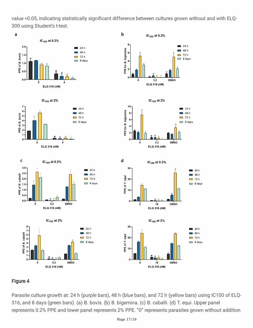
Interestingly, none of the four species of parasites tested in this study was able to grow in the presence ofthe ELQ-316 IC100 concentration regardless of their initial PPE at 72 h (P <0.05) (Fig. 4A-D). The same

Page 9/19
lack of parasite growth was observed after 8 days in the parasite rescue experiment, except for B. caballi(Fig. 4A-D), independent of the starting PPE. A possible interpretation of these results is that the B. caballistrain used in this study may contain a mix of subpopulations of parasites, each one with distinctdegrees of tolerance for ELQ-316. In contrast, the B. bovis, B. bigemina, and T. equi strains used in theseexperiments appear to be composed of subpopulations that are highly susceptible to ELQ-316. It wasbeyond the scope of this study to investigate the mechanism involved in the susceptibility for the ELQdrugs. However, one may speculate that such susceptibility can be due to variations/mutations in thecytochrome bc1 target sequence that affect the ELQ binding, differential uptake or elimination of thedrugs, or a combination of these factors [29-31]. It was recently shown that genetic alterations in the Qi
binding site of cytochrome bc1 complex (Cytb) of B. microti is associated with resistance to ELQ-316,which suggests that this cytochrome gene is as a potential target for the ELQ drugs [16]. Based on theseobservations, we performed alignment analysis of the Cytb genes of B. bovis, B. bigemina, B. caballi, andT. equi together with the B. microti Cytb. Our results indicated full conservation of the two canonical Qo
and Qi binding sites of Cytb in all sequences analyzed and a high level of amino acid identity, whichranged from 47.2 to 49.6 % in comparison to B. microti (Fig. 2) (Table S2). Overall, the results presentedhere set the rationale for further studies to alter and/or knock down the Cytb gene in these parasites andevaluate its potential effect on the susceptibility or resistance to the ELQ drugs.
ELQ-300 and ELQ-316 do not affect viability of equine and bovine PBMC
Cytotoxic assays were performed to assess whether ELQ-300 and ELQ-316 affect the viability of equineand bovine PBMC, which we used as surrogates of nucleated vertebrate host cells. The cytotoxic assayswere performed using the IC100 doses of ELQ-300 and ELQ-316 in in vitro cultures. For the bovine PBMCexperiment, ELQ-300 IC100 of 4.3 nM and ELQ-316 of 3.92 nM were used, respectively, whereas for theequine PBMC experiment, ELQ-300 IC100 of 5.94 nM and ELQ-316 IC100 of 6.18 nM were used,respectively. Viability of PBMC was similar regardless of the presence or absence of parasite lethal dosesof ELQ-300 or ELQ-316, strongly suggesting that cell viability was not compromised by any of these twodrugs under the experimental conditions used in the assays (Fig. 5A and B). In addition, signi�cantincrease (P <0.05) in cell proliferation was observed in bovine and horse PBMC exposed to ConA for 24 hand 48 h, respectively (Fig. 5A and B), indicating adequate sensitivity for the WST-1 proliferation assayused in this study. Taken together, results of cell viability revealed that ELQ-300 and ELQ-316, at theirrespective IC100, lack signi�cant toxic effect on in vitro cultivated bovine and horse PBMC. Although thedata presented here strongly suggest that these two drugs are appropriate candidates for the treatment ofBB and EP, it needs to be pointed out that we did not assess their effect of in vivo, and that our evidenceon the effectivity and safety of ELQ-300 and ELQ-316 was obtained from testing the effect of the drugson parasites growing in culture. In addition, investigation of the mechanism of action of ELQ-300 andELQ-316 in the parasites studied herein was also beyond the scope of this study, and it needs furtherexamination.
Conclusions

Page 10/19
Overall, results presented here demonstrate that both drugs tested in this study, ELQ-300 and ELQ-316, aree�cient in inhibiting the growth of in vitro cultured B. bovis, B. bigemina, B. caballi and T. equi.Importantly, IC100 doses of the ELQ drugs did not signi�cantly affect the viability of in vitro cultured cattleand horse PBMC. Collectively, �ndings of this study strongly suggest that ELQ-300 and ELQ-316 can bepotentially effective and safe candidates for the development of novel therapies to control BB and EP.However, it will be important to con�rm their mechanisms of action, and the drugs’s potential to select forresistant strains. Further studies in vivo using horses and bovines are needed to evaluate the e�cacy ofELQ-300 and ELQ-316 against acute and chronic BB and EP.
DeclarationsEthics declarations
Ethics approval and consent to participate
Not applicable.
Consent for publication
Not applicable.
Availability of data and material
The datasets supporting the conclusions of this article are included within the article and its additional�les.
Competing interests
The authors declare that they have no competing interests.
Funding
This work was funded by the United States Department of Agriculture-Agriculture Research ServiceCurrent Research Information System Project No. 2090-32000-039-00D, and the InternationalDevelopment Research Centre (IDRC) (Canada) project 108525-001. Dr. Riscoe’s laboratory receives�nancial support from the United States Department of Veterans Affairs, Veterans Health Administration,O�ce of Research and Development Program Award number i01 BX003312. MKR. is a recipient of a VAResearch Career Scientist Award (14S-RCS001). Research reported in this publication was also supportedby the US National Institutes of Health under award number AI100569 (MKR) and by the U.S. Departmentof Defense Peer Reviewed Medical Research Program (Log # PR181134 (MKR), and the National Instituteof Allergy and Infectious Diseases under award number R01AI141412. This work was also funded byCareer Development Award BX002440 and VA Merit Review Award BX004522 to JSD from U.S.Department of Veterans Affairs Biomedical Laboratory Research and Development.

Page 11/19
Author’s contribution
MGS, RGB and CES conceived and designed the study. MGS and RGB performed the inhibitory assaysand microscopy experiments, analyzed the data and drafted the manuscript. MGS and RGB performedthe �ow cytometry assay and cytotoxic assay. JSD, MKR, SP, RW, and AN manufactured and providedendochin-like quinolones, and gave technical guidance regarding their use. MGS, RGB, CES, JSD, andMKR wrote the manuscript. All authors read and approved the �nal manuscript.
Acknowledgements
The authors would like to acknowledge Paul Lacy for his help with the in vitroB. bovis, B. bigemina and B.caballi cultures and cultures supplies, Johanatan Hyunki and Ji Heon Lee for checking PPE on smears,and Jacob Laughery for providing uninfected bovine erythrocytes and serum, Lowell Kappemeyer forproviding the initial in vitroB. caballi and T. equi cultures and the uninfected equine erythrocytes, andMegan Blauert for animal handling and blood draw.
AbbreviationsCon A: concanavalin A
DMSO: dimethyl sulfoxide
ELQ: endochin-like quinolones
HE: Hydroethidine
IC: inhibitory concentration
nRBC: non-infected erythrocytes
PBMC: peripheral blood mononuclear cells
PBS: phosphate buffer saline
PPE: percentage of parasitized erythrocytes
References1. Uilenberg G. International collaborative research: Signi�cance of tick-borne hemoparasitic diseases
to world animal health. Vet Parasitol. 1995;57:19-41.
2. Wise LN, Kappmeyer LS, Silva MG, White SN, Grause JF, Knowles DP. Veri�cation of post-chemotherapeutic clearance of Theileria equi through concordance of nested PCR and immunoblot.Ticks Tick Borne Dis. 2018;9:135-40.

Page 12/19
3. Wise LN, Pelzel-McCluskey AM, Mealey RH, Knowles DP. Equine piroplasmosis. Vet Clin N Am-Equine.2014;30:677-93.
4. Florin-Christensen M, Suarez CE, Rodriguez AE, Flores DA, Schnittger L. Vaccines against bovinebabesiosis: where we are now and possible roads ahead. Parasitol. 2014;1-30.
5. Schnittger L, Rodriguez AE, Florin-Christensen M, Morrison DA. Babesia: A world emerging. Infection,genetics and evolution: Journal of Molecular Epidemiology and Evolutionary Genetics in InfectiousDiseases. 2012;12:1788-09.
�. Onyiche TE, Suganuma K, Igarashi I, Yokoyama N, Xuan X, Thekisoe O. A review on equinepiroplasmosis: Epidemiology, vector ecology, risk factors, host immunity, diagnosis and control. Int JEnviron Res Public Health. 2019;16:1736.
7. Kappmeyer LS, Thiagarajan M, Herndon DR, Ramsay JD, Caler E, Djikeng A, Gillespie JJ, et al.Comparative genomic analysis and phylogenetic position of Theileria equi. BMC Genomics.2012;13:603.
�. Siddra A. Hines JB, Mealey RH, Call DR, Graça T. Exposure to ambient air causes degradation anddecreased in vitro potency of buparvaquone and parvaquone. Vet Parasitol X. 2020;3:1000023.
9. Suarez CE, Noh S. Emerging perspectives in the research of bovine babesiosis and anaplasmosis.Vet Parasitol. 2011;180:109-25.
10. Kuttler KL, Johnson LW. Chemoprophylactic activity of imidocarb, diminazene and oxytetracyclineagainst Babesia bovis and B. bigemina. Vet Parasitol. 1986;2:107-18.
11. Yamasaki M, Watanabe N, Idaka N, Yamamori T, Otsuguro KI, Uchida N, et al. Intracellular diminazeneaceturate content and adenosine incorporation in diminazene aceturate-resistant Babesia gibsoniisolate in vitro. Exp Parasitol. 2017;183:92-8.
12. Schwint ON, Ueti MW, Palmer GH, Kappmeyer LS, Hines MT, Cordes RT, et al. Imidocarb dipropionateclears persistent Babesia caballi infection with elimination of transmission potential. AntimicrobAgents Chemother. 2009;53:4327-32.
13. Yeruham I, Pipano E, Davidson M. A �eld strain of Babesia bovis apparently resistant to amicarbalideisethionate. Trop Anim Health Prod. 1985;17:29-30.
14. Miley GP, Pou S, Winter R, Nilsen A, Li Y, Kelly JX, et al. ELQ-300 prodrugs for enhanced delivery andsingle-dose cure of malaria. Antimicrob Agents Chemother. 2015;59:5555-60.
15. van Schalkwyk DA, Riscoe MK, Pou S, Winter RW, Nilsen A, Duffey M, et al. Novel Endochin-likequinolones exhibit potent in vitro activity against Plasmodium knowlesi but do not synergize withproguanil. Antimicrob Agents Chemother. 2020;64:e02549-10.
1�. Winter R, Kelly JX, Smilkstein MJ, Hinrichs D, Koop DR, Riscoe MK. Optimization of endochin-likequinolones for antimalarial activity. Exp Parasitol. 2011;127:545-51.
17. Winter RW, Kelly JX, Smilkstein MJ, Dodean R, Hinrichs D, Riscoe MK. Antimalarial quinolones:synthesis, potency, and mechanistic studies. Exp Parasitol. 2008;118:487-97.

Page 13/19
1�. Frueh L, Li Y, Mather MW, Li Q, Pou S, Nilsen A, et al. Alkoxycarbonate ester prodrugs of preclinicaldrug candidate ELQ-300 for prophylaxis and treatment of malaria. ACS Infect Dis. 2017;3:728-35.
19. Nilsen A, LaCrue AN, White KL, Forquer IP, Cross RM, Marfurt J, et al. Quinolone-3-diarylethers: a newclass of antimalarial drug. Sci Transl Med. 2013;5:177ra137.
20. Lawres LA, Garg A, Kumar V, Bruzual I, Forquer IP, Renard I, et al. Radical cure of experimentalbabesiosis in immunode�cient mice using a combination of an endochin-like quinolone andatovaquone. J Exp Med. 2016;213:1307-18.
21. Doggett JS, Nilsen A, Forquer I, Wegmann KW, Jones-Brando L, Yolken RH, et al. Endochin-likequinolones are highly e�cacious against acute and latent experimental toxoplasmosis. Proc NatlAcad Sci USA. 2012; 109:15936-41.
22. Avarzed A, Igarashi I, Kanemaru T, Hirumi K, Omata Y, Saito A, et al. Improved in vitro cultivation ofBabesia caballi. J Vet Med Sci. 1997;59:479-81.
23. Levy MG, Ristic M. Babesia bovis: continuous cultivation in a microaerophilous stationary phaseculture. Science. 1980;207:1218-20.
24. Vega CA, Buening GM, Green TJ, Carson CA. In vitro cultivation of Babesia bigemina. Am J Vet Res.1985;46:416-20.
25. Zweygarth E, Just MC, de Waal DT. Continuous in vitro cultivation of erythrocytic stages of Babesiaequi. Parasitol Res. 1995;81:355-8.
2�. Silva MG, Villarino NF, Knowles DP, Suarez CE. Assessment of Draxxin((R)) (tulathromycin) as aninhibitor of in vitro growth of Babesia bovis, Babesia bigemina and Theileria equi. Int J ParasitolDrugs and Drug Resistance. 2018;8:265-70.
27. Wyatt CR, Goff W, Davis WC. A �ow cytometric method for assessing viability of intraerythrocytichemoparasites. J Immunol Methods. 1991;140:23-30.
2�. Eberhard N, Balmer V, Muller J, Muller N, Winter R, Pou S, et al. Activities of endochin-Like quinolonesagainst in vitro cultured Besnoitia besnoiti tachyzoites. Front Vet Sci. 2020;7:96. doi:10.3389/fvets.2020.00096.
29. Alday PH, Bruzual I, Nilsen A, Pou S, Winter R, Ben Mamoun C, et al. Genetic evidence for cytochromeb Qi site inhibition by 4(1H)-quinolone-3-diarylethers and antimycin in Toxoplasma gondii.Antimicrob Agents Chemother. 2017; 61:e01866-16.
30. McConnell EV, Bruzual I, Pou S, Winter R, Dodean RA, Smilkstein MJ, et al. Targeted structure-activityanalysis of endochin-like quinolones reveals potent Qi and Qo site inhibitors of Toxoplasma gondiiand Plasmodium falciparum cytochrome bc1 and identi�es ELQ-400 as a remarkably effectivecompound against acute experimental toxoplasmosis. ACS Infect Dis. 2018; 4:1574-84.
31. Silva MG, Domingos A, Esteves MA, Cruz ME, Suarez CE. Evaluation of the growth-inhibitory effect oftri�uralin analogues on in vitro cultured Babesia bovis parasites. Int J Parasitol Drugs and drugResistance. 2013;3:59-68.
Table

Page 14/19
Table 1 IC50 and IC100 of ELQ-300 and ELQ-316 calculated for B. bovis, B. bigemina, B.
caballi and T. equi at 72 h of culture. Results are presented as mean and standard deviation
based on triplicates for each experiment.
ELQ 300 ELQ 316
Species IC50 (nM) IC100 (nM) IC50 (nM) IC100 (nM)
B. bovis 0.09 ± 0.002 4.2 ± 0.10 0.07 3.8 ± 0.12
B. bigemina 0.37 ± 0.19 1.3 ± 0.05 0.05 3.0 ± 0.15
B. caballi 0.19 ± 0.04 5.7 ± 0.24 0.1 ± 0.006 6.0 ± 0.18
T. equi 0.04 ± 0.003 3.36 ± 0.83 0.002 1.0 ± 1.55
Figures
Figure 1

Page 15/19
Parasite culture growth at 24 h (purple bars), 48 h (blue bars), and 72 h (yellow bars) without and afteraddition of different concentrations of ELQ-300. (a) B. bovis (b) B. bigemina (c) B. caballi. (d) T. equi. “0”represents parasites in the absence of ELQ-300. Assays were carried out in triplicate and the error barsindicate standard error deviation for each ELQ-300 concentration tested. (*) Represents P-value <0.05,indicating statistically signi�cant difference between cultures grown without and with ELQ-300, usingStudent’s t-test.
Figure 2
Parasite culture growth at 24 h (purple bars), 48 h (blue bars), and 72 h (yellow bars) without and afteraddition of different concentrations of ELQ-316. (a) B. bovis. (b) B. bigemina. (c) B. caballi. (d) T. equi. “0”represents parasites in the absence of ELQ-316. Assays were carried out in triplicate and the error barsindicate standard error deviation for each ELQ-316 concentration tested. (*) Represents P-value <0.05,indicating statistically signi�cant difference between cultures grown without and with ELQ-316, usingStudent’s t-test.

Page 16/19
Figure 3
Parasite culture growth at 24 h (purple bars), 48 h (blue bars), and 72 h (yellow bars) using IC100 of ELQ-300, and 8 days (green bars). (a) B. bovis. (b) B. bigemina. (c) B. caballi. (d) T. equi. Upper panelrepresents 0.2% PPE, lower panel 2% PPE. “0” represents parasites grown without addition of ELQ-300.Assays were carried out in triplicate and the error bars indicate standard error deviation. (*) Represents P-
Page 17/19
value <0.05, indicating statistically signi�cant difference between cultures grown without and with ELQ-300 using Student’s t-test.
Figure 4
Parasite culture growth at: 24 h (purple bars), 48 h (blue bars), and 72 h (yellow bars) using IC100 of ELQ-316, and 8 days (green bars). (a) B. bovis. (b) B. bigemina. (c) B. caballi. (d) T. equi. Upper panelrepresents 0.2% PPE and lower panel represents 2% PPE. “0” represents parasites grown without addition

Page 18/19
of ELQ-316. Assays were carried out in triplicate and the error bars indicate standard error deviation. (*)Represents P-value <0.05, indicating statistically signi�cant difference between cultures grown withoutand with ELQ-300 using Student’s t-test.
Figure 5
Percentage of cell viability over a period of 72 h after incubation with IC50 and IC100 concentration ofELQs 300 and 316. “Cells+Med” represents PBMC cultivated without addition of the ELQ compounds.Cells in DMSO and Draxxin® were used as a negative control. ConA was used as a positive control for
Page 19/19
cell proliferation. (a) Bovine PBMC. (b) Horse PBMC. (*) Represents P-value <0.05 indicating statisticallysigni�cant differences compared to PBMC cultivated in medium only by using Student’s t-test. Bovineand horse PBMC were assayed in triplicate and the error bars indicate standard error deviation.
Supplementary Files
This is a list of supplementary �les associated with this preprint. Click to download.
Fig.S1.tiff
FigS2.tiff
graphicalabstract.pptx
SilvaandBastosetalELQSupplTables.docx