Embed Size (px)
Citation preview
Pediatr Blood Cancer 2007;49:812–816
B-Type Natriuretic Peptide as a Marker for Cardiac Dysfunctionin Anthracycline-Treated Children
Sanjeev Aggarwal, MD, Michael D. Pettersen, MD, Kanta Bhambhani, MD,Joellyn Gurczynski, Ronald Thomas, PhD, and Thomas L’Ecuyer, MD*
INTRODUCTION
Anthracyclines (AC) are highly effective chemotherapeutic
agents, whose full clinical potential is limited by cardiac toxicity
(ACT) [1–4]. ACT is progressive, may become manifested many
years after the completion of treatment and occurs at variable
threshold doses [5–8]. The evolution of AC-induced subclinical
cardiac dysfunction is not clearly understood and other than vigilant
monitoring, there are no explicit guidelines for its management
[6,9–11]. Therefore, each AC recipient requires monitoring for
cardiotoxicity for a long period of time, the duration of which is not
known.
Monitoring for ACT is most simply accomplished by echocar-
diographic measures, including fractional shortening (FS) and
ejection fraction (EF) [12]. However, these measures are dependent
on ventricular loading conditions and lack sensitivity for detecting
ACT [13,14]. FS alone is reduced in 28% of AC-treated patients,
whereas 57% have an abnormal left ventricular afterload, as
measured by end systolic wall stress (ESWS) or contractility
measured by the stress velocity index (SVI) [3]. These more
sophisticated parameters are technically difficult to obtain in
children. An objective, easily obtained biomarker of ventricular
function would greatly add to the clinical care of AC-treated
patients.
Plasma B-type natriuretic peptide (BNP) is a cardiac hormone
secreted from the ventricles, in response to ventricular volume and
pressure overload [15]. Levels increase in proportion to the severity
of congestive heart failure (CHF) in adults and children [16–18].
In the pediatric age group, values of BNP of 7� 5.9 and
10.1� 8.6 pg�ml�1 have been reported in healthy boys and girls,
respectively [19]. Comparable BNP levels are noted in neonates
after closure of patent ductus arteriosus and children with
cardiomyopathy, without clinical signs of heart failure [20,21].
The role of BNP in screening for late onset ACT remains to be
elucidated. One previous study found plasma BNP to be signi-
ficantly elevated in patients with a reduction of FS or EF [22].
The rationale for our study was that plasma BNP may be elevated
in AC-treated patients with late cardiac dysfunction. Our primary
objective was to evaluate plasma BNP as a screening test for
detecting late onset AC-induced cardiac dysfunction, in com-
parison to detailed sensitive indices, not ordinarily available from a
clinical echo.
Since the mid 1990s, cumulative AC doses have been
empirically limited to below 550 mg�m�2 due to the high incidence
of clinically important cardiotoxicity observed above this dose [23].
More recently, a further dose reduction to below 250 mg�m�2 has
been suggested to reduce the incidence of ACT [9]. Limited
information is available about the impact of progressive AC dose
curtailment on the incidence of late cardiac dysfunction. Our
secondary aim, therefore, was to define the prevalence of cardiac
dysfunction using load-independent echocardiographic measures in
a cohort of patients treated with low cumulative AC doses.
MATERIALS AND METHODS
This prospective non-interventional study was conducted at
Children’s Hospital of Michigan, after approval by the Human
Investigation Committee. Patients were enrolled between October
2003 and June 2004 after obtaining written informed consent from
parents or guardians.
Selection Criteria
Patients who had completed AC chemotherapy at least 1 year
prior to enrollment were included. Patients with congenital heart
Background. Anthracyclines (AC) are useful antineoplasticagents, whose utility is limited by progressive cardiotoxicity. Ourpurpose was to evaluate plasma B-type natriuretic peptide (BNP), asa screening test for detecting late cardiac dysfunction in AC-treatedchildren and to determine the prevalence of late cardiac dysfunctionat low cumulative AC doses. Materials and Methods. This was aprospective study in which patients who had completed AC therapyat least 1 year earlier, underwent a detailed echocardiogram and asimultaneous BNP level. Cardiac dysfunction was defined as any oneof the following: shortening fraction (FS) <29%, rate correctedvelocity of circumferential fiber shortening (VCFc)<0.9 c�sec�1, endsystolic wall stress (ESWS) >60 g�cm�2, abnormal VCFc: ESWSratio or decreased mitral inflow velocity (E/A) ratios, compared toage-specific norms. Results. The cohort (n¼63) included 37 males
with a median age of 13.1 years (range, 6.5–26.5 years). Cardiacdysfunction was found in 26 (41%) patients and in 40% ofpatients who received cumulative doses <150 mg�m�2. ESWS wasthe most common abnormality. Mean BNP levels in the subset withabnormal function were significantly higher than the normal group(23.4� 25.3 vs. 14.2� 8.9 pg�ml�1, P¼0.02). Conclusions. PlasmaBNP was significantly elevated in AC-treated patients with latecardiac dysfunction, although there was considerable overlap oflevels between groups with and without cardiac dysfunction. BNPmay need further evaluation as a serial index of cardiac function inthis population. Cardiac dysfunction was observed in a significantproportion of patients, even at low cumulative AC doses. PediatrBlood Cancer 2007;49:812–816. � 2006 Wiley-Liss, Inc.
Key words: Anthracyclines-induced cardiotoxicity; BNP
� 2006 Wiley-Liss, Inc.DOI 10.1002/pbc.21100
——————Department of Pediatrics, Wayne State University, Children’s Hospital
of Michigan, Michigan
*Correspondence to: Thomas L’Ecuyer, MD, Division of Cardiology,
Children’s Hospital of Michigan, 3901 Beaubien Blvd, Detroit, MI
48201. E-mail: [email protected]
Received 30 May 2006; Accepted 23 October 2006
disease, cardiomyopathy prior to initiation of chemotherapy, renal
failure (serum creatinine more than twice the age-specific norms),
patients who received mediastinal radiation, were pregnant or
critically ill, were excluded. Each patient underwent an echocardio-
gram and simultaneous blood draw for BNP assay for research
purposes.
Clinical Data
Clinical data obtained for each subject included demographics,
clinical diagnosis, the cumulative dose of AC received, time elapsed
since the completion of AC therapy, and cardiac symptoms.
Echocardiography
Echocardiograms were performed using a Phillips Sonos
5500 ultrasound machine. Each patient underwent M mode, 2D
and Doppler echocardiogram with simultaneous recording of
carotid pulse tracing, electrocardiogram, phonocardiogram, and
blood pressure. Echo parameters were selected to measure (a) left
ventricular systolic function (shortening fraction FS, ejection
fraction EF and rate corrected mean velocity of circumferential
fiber shortening VCFc), (b) afterload (end systolic wall stress
ESWS), (c) contractility (Stress velocity index SVI), and (d) left
ventricular diastolic function (peak E and A velocities and their
ratio). FS and EF were calculated as described [24,25]. Shortening
fraction below 29% and EF less than 64% were considered abnormal
[12,25]. VCFc was obtained by the method described by Colan et al.
[3,26] and considered abnormal below 0.90 c�sec�1. ESWS was
calculated by the method described by Grossman et al. and
considered abnormal above 60 g�cm�2 [3,27]. The SVI or the
relationship between VCFc and ESWS were measured by the
method described by Colan et al. [26]. A VCFc value two standard
deviations below normal in relation to ESWS was considered to
represent abnormal contractility [26,28]. E/A ratio was obtained by
pulse Doppler at the mitral valve inflow and compared to established
normal values [29]. All echocardiograms were read by a single
cardiologist (MP) who was blinded to patient data and results of the
BNP assay.
Abnormal cardiac function was defined by any one of the
following criteria: FS <29%; VCFc <0.90 c�sec�1; ESWS
>60 g�cm�2; abnormal VCFc: ESWS ratio; or decreased E/A ratio,
based on age-specific normal values. EF was not included in our
preset criteria of cardiac dysfunction due to inherent fallacies in its
echocardiographic measurement in the pediatric age group [25].
Plasma BNP Assay
Blood samples (3 ml) were collected in K-EDTA tubes within
3 hours of the echocardiogram and were centrifuged immediately at
3,500 rpm for 10 min. Platelet-free plasma was stored at �208C.
BNP assay was performed using the Triage BNP kit (Biosite
Diagnostic, San Diego, California).
Statistical Analysis
Statistical analyses were performed using SPSS (version 12)
software. Sample size was predetermined using a two-tailed test
with a significance (alpha) set at 0.05 to achieve a power of 81%,
based on the assumption that the BNP would be higher in patients
with abnormal heart function on echocardiogram by 20 pg�dl�1
(corresponding to means� SD of 9� 14.8 and 29� 31.2, respec-
tively, based on a prior study) [22]. The number of patients required
to achieve a power of 80% was 23 in each group. Since 50–60%
of pediatric patients who receive AC have abnormal contractility
and/or afterload, we enrolled 63 patients with an expectation of two
nearly equal subsets—those with and without cardiac dysfunction
[3]. Data were expressed as mean� SD for continuous variables and
as a number (percentage) for categorical variables. The two groups
were compared for continuous variables using the Student t-test for
independent samples [30]. Box and whisker plots of BNP levels
were used to show the distribution in the subgroups based on cardiac
function. Statistical significance was set asP< 0.05. All hypotheses
were two tailed.
RESULTS
Subject Characteristics
Eighty patients were identified from the echo database in the
cardiology clinic. Two could not be traced and 15 patients refused
consent. Sixty-three patients were enrolled in the study including 37
(59%) males and 26 (41%) females. The median age at enrollment
was 13.1 years (range, 6.5–26.5) and the median interval since
completion of AC treatment was 3.8 years (range, 1.1–17.5). The
clinical diagnoses included acute lymphocytic leukemia in 29
(46%), Wilms tumor in 12 (19%), osteosarcoma in 12 (19%), and
lymphoma in 10 patients (16%). Five patients were on cardiac
medications (ACE inhibitors and digoxin) for ventricular dysfunc-
tion, of whom three were symptomatic with CHF, while two had
asymptomatic abnormal LV function. None of our patients had a
history of acute cardiac failure immediately following an AC dose.
The median cumulative dose of AC received was 165 mg�m�2
(range, 45–520; mean 160 mg�m�2). The cumulative AC dose
received was less than 150 mg�m�2 in 29 (46%) patients, between
150 and 300 mg�m�2 in 20 (31.7%), between 300 and 450 mg/m2 in
13 (20.6%) patients, and 520 mg�m�2 in 1 (1.5%). No patient
received more than 550 mg�m�2.
Echocardiographic Data
Twenty-six (41%) patients had at least one cardiac abnormality
on echocardiogram. The most frequently detected abnormality
was ESWS (range, 61–86 g�cm�2; median 66 g�cm�2; mean
68.7 g�cm�2) in 20 patients (31.7%). The nine (14%) patients who
had abnormal shortening fractions (range, 23–28; median 26%;
mean 26%) also had low EF (range 54–63, mean 60). Seven out of
63 (11.1%) had abnormal VCFc (range 0.76–0.9; median
0.84 c�sec�1; mean 0.84 c�sec�1) and 6 patients (9.5%) had an
abnormal stress velocity index. All three patients with clinical CHF
had low FS; two had four abnormal parameters (FS, ESWS, VCFc,
and SVI). Diastolic function (peak E/A velocities and their ratio)
was normal in all patients, with the E/A ratios ranging from 1.33 to
3.05, with a mean (SD) of 2.06� 0.43 (median 2.03). Baseline
characteristics of the groups with normal and abnormal cardiac
function are shown (Table I). The two groups were similar in the
mean cumulative dose of AC received (204.7� 100.6 mg�m�2
abnormal cardiac function vs. 223.1� 127.7 mg�m�2 normal
cardiac function, P¼ 0.54) and interval post-AC treatment
Pediatr Blood Cancer DOI 10.1002/pbc
BNP and Anthracycline-Induced Cardiac Dysfunction 813

(5.6� 4.4 years abnormal cardiac function and 5.1� 3.7 years
normal cardiac function, P¼ 0.64).
Table II shows the number of patients with each cardiac
abnormality as a function of the cumulative dose received. Age at
treatment, interval of follow-up, and cumulative dose were not risk
factors for cardiac dysfunction on multivariate analysis. At all doses,
ESWS was the most frequently detected abnormality, present in 9 of
12 (75%) at cumulative AC doses below 150 mg�m�2, 7 of 10 (70%)
between 150 and 300 mg�m�2, and all 4 (100%) patients receiving
300–450 mg�m�2. Twelve out of 29 patients (40%) who had
received cumulative doses below 150 mg�m�2 had evidence of
cardiac dysfunction, including 4 patients who had received doses as
low as 90 mg�m�2. The patient who received a cumulative dose
of 520 mg�m�2 had normal cardiac function after 12.4 years of
follow-up.
Plasma BNP
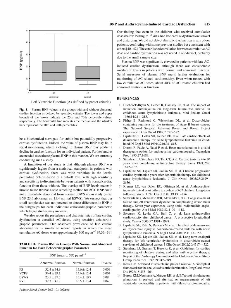
The median BNP level for the cohort was 12.2 pg�dl�1 (range,
5–117 pg�dl�1). Mean (�SD) plasma BNP levels were signi-
ficantly higher in the presence of abnormal cardiac function
(23.4� 25.3 pg�dl�1, n¼ 26 vs. 14.2� 8.9 pg�dl�1, n¼ 37,
P¼ 0.02; Fig. 1). Plasma BNP levels were higher when FS was
low (32.4� 34.9 (n¼ 9) vs. 15.6� 12.4 (n¼ 54), P< 0.008). When
all four systolic parameters were abnormal, plasma BNP was signi-
ficantly higher than when one to three parameters were abnormal
(69.2� 67.1 (n¼ 2) vs. 19.6� 17.3 (n¼ 24), P< 0.005). Plasma
BNP levels in the five subjects on cardiac medications were
significantly higher than the rest of the cohort (49.8� 40.2 vs.
15.2� 11.9 pg�ml�1, P< 0.001). When the five patients on
medications were excluded, BNP levels remained significantly
higher when cardiac function was abnormal than when function was
normal (19.4� 17.6 vs. 14.1� 8.9, P¼ 0.02). Table III depicts the
mean plasma BNP levels in the subgroups with normal and
abnormal function for each echocardiographic parameter. Plasma
BNP was significantly higher in patients with abnormal FS, VCFc,
and SVI, but not ESWS.
DISCUSSION
Among AC-treated patients, 41% had an abnormality of cardiac
function at a mean follow-up interval of 5.2 years, the majority of
whom were asymptomatic. Nine (14%) patients had abnormal FS
and EF, the commonly used clinical parameters. The most sensitive
echo parameter for diagnosis of subclinical ACT was ESWS,
representing 80% of cardiac abnormalities, while contractility by
SVI was abnormal in only 24% of patients. Patients receiving doses
as low as 90 mg�m�2 had cardiac dysfunction. Plasma BNP was
significantly higher in patients with cardiac dysfunction and when
multiple echo parameters were abnormal. Plasma BNP was
significantly elevated in the presence of abnormal contractility,
although the elevation of BNP with increased ESWS did not reach
statistical significance. These findings assume importance, given the
growing numbers of AC treated patients and that AC use is mainly
limited by cardiotoxicity [5–7,31].
Our finding of the association of an elevated BNP with late AC-
induced cardiac dysfunction is similar to studies in adults, where a
significant increase in plasma BNP has been noted in AC-treated
patients with clinical or subclinical heart failure [32–34]. Previous
data in children are limited. While one previous study reported
elevation of plasma BNP (29� 31.2 vs. 9� 14.8 pg�ml�1) in
patients with left ventricular dysfunction, compared to patients with
normal function, another reported higher BNP levels in the AC-
treated group as a whole, compared to untreated controls
(10.5� 10.2 vs. 4.09� 2.2 pg�ml�1) [22,35]. Both studies enrolled
patients soon after completion of AC therapy when LV dysfunction
could represent acute toxicity, which is not predictive of late ACT.
Neither study excluded patients who had received chest radiation,
which can worsen ACT. Moreover, VCFc, ESWS, and SVI were
not utilized.
To our knowledge, ours is the largest study to evaluate plasma
BNP for the detection of late ACTusing sensitive echocardiographic
indices. We believe our finding of a mild but significant elevation in
plasma BNP with cardiac dysfunction is of clinical interest.
Although largely asymptomatic, an increase in plasma BNP may
Pediatr Blood Cancer DOI 10.1002/pbc
TABLE I. Clinical and Demographic Characteristics
Normal
cardiac function
Abnormal
cardiac function P-value
Male gender 26/37 (70%) 11/26 (42%) 0.12
Median age at time of diagnosis of cancer (years) 7.1 5.7 0.74
Median age at time of enrollment (years) 12.1 14.3 0.96
Median interval Post-AC-treatment (years) 3.8 3.6 0.64
Median cumulative anthracycline dose (mg�m�2) 165 180 0.54
TABLE II. Effect of Cumulative AC Dose on Echocardiographic Parameters of Cardiac Function
Dose/(n) Any abnormality Abnormal FS Abnormal EF Abnormal ESWS Abnormal VCFc Abnormal SVI
<150 mg�m�2 (29) 12 3 3 9 3 2
150–300 mg�m�2 (20) 10 5 5 7 3 3
300–450 mg�m�2 (13) 4 1 1 4 1 1
>450 mg�m�2 (1) 0 0 0 0 0 0
26 9 9 20 7 6
814 Aggarwal et al.
be a biochemical surrogate for subtle but potentially progressive
cardiac dysfunction. Indeed, the value of plasma BNP may lie in
serial monitoring, where a change in plasma BNP may predict a
decline in cardiac function for an individual patient. Further studies
are needed to evaluate plasma BNP in this manner. We are currently
conducting such a study.
A limitation of our study is that although plasma BNP was
significantly higher from a statistical standpoint in patients with
cardiac dysfunction, there was wide variation in the levels,
precluding determination of a cut-off level with high sensitivity
and specificity to discriminate between patients with normal cardiac
function from those without. The overlap of BNP levels makes it
unwise to use BNP as a sole screening method for ACT. BNP could
not differentiate abnormal from normal ESWS in our study (mean
BNP 23.5 abnormal vs. 15.4 normal ESWS). We suspect that our
small sample size was not powered to detect differences in BNP in
the subgroups for each individual echocardiographic parameter,
which larger studies may uncover.
We also report the prevalence and characteristics of late cardiac
dysfunction at curtailed AC doses, using sensitive echocardio-
graphic parameters. Our observed prevalence of late cardiac
abnormalities is similar to recent reports in which the mean
cumulative AC doses were approximately 300 mg�m�2 [9,36–39].
Our finding that even in the children who received cumulative
doses below 150 mg�m�2, 40% had late cardiac dysfunction is novel
and disturbing. We did not detect diastolic dysfunction in any of our
patients, conflicting with some previous studies but consistent with
others [40–42]. The established correlation between cumulative AC
dose and cardiac dysfunction was not noted in our dataset, probably
due to the small sample size.
Plasma BNP was significantly elevated in patients with late AC-
induced cardiac dysfunction, although there was considerable
overlap of levels in patients with normal and abnormal function.
Serial measures of plasma BNP merit further evaluation for
monitoring of AC-related cardiotoxicity. Even when treated with
low cumulative AC doses, about 40% of AC-treated children had
abnormal ventricular function.
REFERENCES
1. Hitchcock-Bryan S, Gelber R, Cassady JR, et al. The impact of
induction anthracycline on long-term failure-free survival in
childhood acute lymphoblastic leukemia. Med Pediatr Oncol
1986;14:211–215.
2. Fisher B, Redmond C, Wickerham DL, et al. Doxorubicin-
containing regimens for the treatment of stage II breast cancer:
The National Surgical Adjuvant Breast and Bowel Project
experience. J Clin Oncol 1989;7:572–582.
3. Lipshultz SE, Colan SD, Gelber RD, et al. Late cardiac effects of
doxorubicin therapy for acute lymphoblastic leukemia in child-
hood. N Engl J Med 1991;324:808–815.
4. Dorent R, Pavie A, Nataf P, et al. Heart transplantation is a valid
therapeutic option for anthracycline cardiomyopathy. Transplant
Proc 1995;27:1683.
5. Steinherz LJ, Steinherz PG, Tan CT, et al. Cardiac toxicity 4 to 20
years after completing anthracycline therapy. Jama 1991;266:
1672–1677.
6. Lipshultz SE, Lipsitz SR, Sallan SE, et al. Chronic progressive
cardiac dysfunction years after doxorubicin therapy for childhood
acute lymphoblastic leukemia. J Clin Oncol 2005;23:2629–
2636.
7. Kremer LC, van Dalen EC, Offringa M, et al. Anthracycline-
induced clinical heart failure in a cohort of 607 children: Long-term
follow-up study. J Clin Oncol 2001;19:191–196.
8. Schwartz RG, McKenzie WB, Alexander J, et al. Congestive heart
failure and left ventricular dysfunction complicating doxorubicin
therapy. Seven-year experience using serial radionuclide angio-
cardiography. Am J Med 1987;82:1109–1118.
9. Sorensen K, Levitt GA, Bull C, et al. Late anthracycline
cardiotoxicity after childhood cancer: A prospective longitudinal
study. Cancer 2003;97:1991–1998.
10. Lipshultz SE, Rifai N, Dalton VM, et al. The effect of dexrazoxane
on myocardial injury in doxorubicin-treated children with acute
lymphoblastic leukemia. N Engl J Med 2004;351:145–153.
11. Lipshultz SE, Lipsitz SR, Sallan SE, et al. Long-term enalapril
therapy for left ventricular dysfunction in doxorubicin-treated
survivors of childhood cancer. J Clin Oncol 2002;20:4517–4522.
12. Steinherz LJ, Graham T, Hurwitz R, et al. Guidelines for cardiac
monitoring of children during and after anthracycline therapy:
Report of the Cardiology Committee of the Childrens Cancer Study
Group. Pediatrics 1992;89:942–949.
13. Ross J, Jr. Afterload mismatch and preload reserve: A conceptual
framework for the analysis of ventricular function. Prog Cardiovasc
Dis 1976;18:255–264.
14. Borow KM, Neumann A, Marcus RH, et al. Effects of simultaneous
alterations in preload and afterload on measurements of left
ventricular contractility in patients with dilated cardiomyopathy:
Pediatr Blood Cancer DOI 10.1002/pbc
Fig. 1. Plasma BNP values in the groups with and without abnormal
cardiac function as defined by specified criteria. The lower and upper
bounds of the boxes indicate the 25th and 75th percentile values,
respectively. The horizontal line indicates the median and the whisker
bars represent the 10th and 90th percentiles.
TABLE III. Plasma BNP in Groups With Normal and AbnormalFunction for Each Echocardiographic Parameter
BNP (mean� SD) pg�ml�1
P-valueAbnormal function Normal function
FS 32.4� 34.9 15.6� 12.4 0.009
VCFS 36.4� 39.1 15.6� 12.4 0.004
ESWS 23.5� 27.5 15.4� 10.7 0.09
SVI 32.3� 41.7 16.5� 13.4 0.04
BNP and Anthracycline-Induced Cardiac Dysfunction 815
Comparisons of ejection phase, isovolumetric and end-systolic
force-velocity indexes. J Am Coll Cardiol 1992;20:787–795.
15. Maeda K, Tsutamoto T, Wada A, et al. Plasma brain natriuretic
peptide as a biochemical marker of high left ventricular end-
diastolic pressure in patients with symptomatic left ventricular
dysfunction. Am Heart J 1998;135:825–832.
16. Maisel AS. B-type natriuretic peptide (BNP) levels: Diagnostic and
therapeutic potential. Rev Cardiovasc Med 2001;2:S13–18.
17. McCullough PA, Omland T, Maisel AS. B-type natriuretic
peptides: A diagnostic breakthrough for clinicians. Rev Cardiovasc
Med 2003;4:72–80.
18. Kunii Y, Kamada M, Ohtsuki S, et al. Plasma brain natriuretic
peptide and the evaluation of volume overload in infants and
children with congenital heart disease. Acta Med Okayama 2003;
57:191–197.
19. Koch A, Singer H. Normal values of B type natriuretic peptide in
infants, children, and adolescents. Heart 2003;89:875–878.
20. Sanjeev S, Pettersen M, Lua J, et al. Role of plasma B-type
natriuretic peptide in screening for hemodynamically significant
patent ductus arteriosus in preterm neonates. J Perinatol 2005;
25:709–713.
21. Westerlind A, Wahlander H, Lindstedt G, et al. Clinical signs of
heart failure are associated with increased levels of natriuretic
peptide types B and A in children with congenital heart defects or
cardiomyopathy. Acta Paediatr 2004;93:340–345.
22. Hayakawa H, Komada Y, Hirayama M, et al. Plasma levels of
natriuretic peptides in relation to doxorubicin-induced cardiotoxi-
city and cardiac function in children with cancer. Med Pediatr
Oncol 2001;37:4–9.
23. Gharib MI, Burnett AK. Chemotherapy-induced cardiotoxicity:
Current practice and prospects of prophylaxis. Eur J Heart Fail
2002;4:235–242.
24. Gutgesell HP, Paquet M, Duff DF, et al. Evaluation of left
ventricular size and function by echocardiography. Results in
normal children. Circulation 1977;56:457–462.
25. Park MK. Noninvasive Techniques. In: Park MK, editor. Pediatric
Cardiology for Practitioners. 4th edition. St. Louis: Mosby; 2002.
pp 67–82.
26. Colan SD, Borow KM, Neumann A. Left ventricular end-systolic
wall stress-velocity of fiber shortening relation: A load-indepen-
dent index of myocardial contractility. J Am Coll Cardiol 1984;4:
715–724.
27. Grossman W, Jones D, McLaurin LP. Wall stress and patterns of
hypertrophy in the human left ventricle. J Clin Invest 1975;56:56–
64.
28. Colan SD, Parness IA, Spevak PJ, et al. Developmental modulation
of myocardial mechanics: Age- and growth-related alterations in
afterload and contractility. J Am Coll Cardiol 1992;19:619–629.
29. O’Leary PW, Durongpisitkul K, Cordes TM, et al. Diastolic
ventricular function in children: A Doppler echocardiographic
study establishing normal values and predictors of increased
ventricular end-diastolic pressure. Mayo Clin Proc 1998;73:616–
628.
30. Yusuf S, W J, Probstfield J, Tyroler HA. Comparing Groups II.
Multiple Testing Problem. In: Lang TA, Michells S, editors. How to
report statistics in medicine annotated guideines for authors,
editors, and reviewers. Philadelphia: BMJ Publishing Group; 1997.
pp 81–92.
31. Von Hoff DD, Layard MW, Basa P, et al. Risk factors for
doxorubicin-induced congestive heart failure. Ann Intern Med
1979;91:710–717.
32. Suzuki T, Hayashi D, Yamazaki T, et al. Elevated B-type natriuretic
peptide levels after anthracycline administration. Am Heart J 1998;
136:362–363.
33. Okumura H, Iuchi K, Yoshida T, et al. Brain natriuretic peptide is a
predictor of anthracycline-induced cardiotoxicity. Acta Haematol
2000;104:158–163.
34. Nousiainen T, Vanninen E, Jantunen E, et al. Natriuretic peptides
during the development of doxorubicin-induced left ventricular
diastolic dysfunction. J Intern Med 2002;251:228–234.
35. Pinarli FG, Oguz A, Tunaoglu FS, et al. Late cardiac evaluation of
children with solid tumors after anthracycline chemotherapy.
Pediatr Blood Cancer 2005;44:370–377.
36. Lipshultz SE, Lipsitz SR, Mone SM, et al. Female sex and drug
dose as risk factors for late cardiotoxic effects of doxorubicin
therapy for childhood cancer. N Engl J Med 1995;332:1738–
1743.
37. Sorensen K, Levitt G, Sebag-Montefiore D, et al. Cardiac function
in Wilms’ tumor survivors. J Clin Oncol 1995;13:1546–1556.
38. Sorensen K, Levitt G, Bull C, et al. Anthracycline dose in childhood
acute lymphoblastic leukemia: Issues of early survival versus late
cardiotoxicity. J Clin Oncol 1997;15:61–68.
39. Pihkala J, Saarinen UM, Lundstrom U, et al. Myocardial function in
children and adolescents after therapy with anthracyclines and
chest irradiation. Eur J Cancer 1996;32A:97–103.
40. Marchandise B, Schroeder E, Bosly A, et al. Early detection of
doxorubicin cardiotoxicity: Interest of Doppler echocardiographic
analysis of left ventricular filling dynamics. Am Heart J 1989;
118:92–98.
41. Stoddard MF, Seeger J, Liddell NE, et al. Prolongation of
isovolumetric relaxation time as assessed by Doppler echocardio-
graphy predicts doxorubicin-induced systolic dysfunction in
humans. J Am Coll Cardiol 1992;20:62–69.
42. Schmitt K, Tulzer G, Merl M, et al. Early detection of doxorubicin
and daunorubicin cardiotoxicity by echocardiography: Diastolic
versus systolic parameters. Eur J Pediatr 1995;154:201–204.
Pediatr Blood Cancer DOI 10.1002/pbc
816 Aggarwal et al.