Embed Size (px)
Citation preview

The Rockefeller University Press, 0021-9525/99/03/823/15 $2.00The Journal of Cell Biology, Volume 144, Number 5, March 8, 1999 823–837http://www.jcb.org 823
The
b
4 Integrin Interactor p27
BBP/eIF6
Is an Essential Nuclear Matrix Protein Involved in 60S Ribosomal Subunit Assembly
Francesca Sanvito,*
‡
Simonetta Piatti,
§
Antonello Villa,*
i
Mario Bossi,*
i
Giovanna Lucchini,
§
Pier Carlo Marchisio,*
¶
and Stefano Biffo*
*DIBIT, Department of Biological and Technological Research, and
‡
Department of Pathology, San Raffaele Scientific Institute, 20132 Milano, Italy;
§
Dipartimento di Genetica e di Biologia dei Microrganismi, University of Milano, 20133 Milano, Italy;
i
Department of Pharmacology, University of Milano and CNR Center, 20129 Milano, Italy; and
¶
Department of Biomedical Sciences and Human Oncology, University of Torino School of Medicine, 10126 Torino, Italy
Abstract.
p27
BBP/eIF6
is an evolutionarily conserved protein that was originally identified as p27
BBP
, an inter-actor of the cytoplasmic domain of integrin
b
4 and, in-dependently, as the putative translation initiation factor eIF6. To establish the in vivo function of p27
BBP/eIF6
, its topographical distribution was investigated in mamma-lian cells and the effects of disrupting the correspond-
ing gene was studied in the budding yeast,
Saccharomy-ces cerevisiae.
In epithelial cells containing
b
4 integrin, p27
BBP/eIF6
is present in the cytoplasm and enriched at hemidesmosomes with a pattern similar to that of
b
4 in-tegrin. Surprisingly, in the absence and in the presence of the
b
4 integrin subunit, p27
BBP/eIF6
is in the nucleolus and associated with the nuclear matrix. Deletion of the
IIH S. cerevisiae
gene, encoding the yeast p27
BBP/eIF6
homologue, is lethal, and depletion of the correspond-ing gene product is associated with a dramatic decrease of the level of free ribosomal 60S subunit. Furthermore, human p27
BBP/eIF6
can rescue the lethal effect of the
iih
D
yeast mutation. The data obtained in vivo suggest an evolutionarily conserved function of p27
BBP/eIF6
in ri-bosome biogenesis or assembly rather than in transla-tion. A further function related to the
b
4 integrin sub-unit may have evolved specifically in higher eukaryotic cells.
Key words: epithelial cells • yeast • nucleolus • hemidesmosomes • intermediate filaments
F. Sanvito and S. Piatti contributed equally to this work.Address correspondence to Stefano Biffo, Lab. Istologia Molecolare
DIBIT, San Raffaele Scientific Institute, V. Olgettina 58, 20132 Milano,Italy. Tel.: 39-22-643-4857. Fax: 39-22-643-4855. E-mail: [email protected]
I
NTEGRINS
are heterodimeric receptors formed by theassembly of one
a
and one
b
transmembrane subunit.16
a
and 8
b
subunits heterodimerize to produce
.
20receptors that are differentially expressed in a wide varietyof tissues. Most integrins bind components of the extracel-lular matrix and can organize the cytoskeleton throughtheir cytoplasmic domain (for review see Hynes, 1992).Moreover, integrins are involved in the control of cellgrowth, apoptosis, and differentiation through the recruit-ing of several signal transduction and adaptor molecules(for review see Clark and Brugge, 1995; Giancotti, 1997;Howe et al., 1998). The steadily increasing role of integrinsin the organization of cellular metabolic processes has re-cently encompassed the process of protein synthesis. In-deed, clustering of some integrin subunits at points of focaladhesion results in the relocation of mRNA and ribo-somes to focal adhesion contacts (Chicurel et al., 1998).Although the molecular mechanisms of this process are
unknown, a picture in which most biochemical processesare spatially regulated, with integrins playing a major role,has emerged.
The integrin subunit
b
4 associates with
a
6 to form amultivalent laminin receptor present at high levels in mostepithelia. Its ligand engagement correlates with PI3 kinaseactivation (Shaw et al., 1997) and recruitment of the shcand grb2 adaptors (Mainiero et al., 1995). In addition, insquamous and transitional epithelia,
b
4 is required for theformation of hemidesmosomes, specialized structures pro-viding firm mechanical links between basal lamina and theintermediate filament cytoskeleton (for review see Gian-cotti, 1996). Loss of function of
b
4 both in human andmice results in hemidesmosome disruption, blistering, andperinatal death (Vidal et al., 1995; Dowling et al., 1996;van der Neut et al., 1996).
Mutations in the functional cytodomain of
b
4 result
in an inability to translocate into hemidesmosomes andbe targeted to the intermediate filament cytoskeleton(Spinardi et al., 1993; Niessen et al., 1997) accompanied bylethal forms of the blistering disease junctional
epidermol-ysis bullosa
associated with
pyloric atresia
(Vidal et al.,1995). These and other data strongly indicate that the
Dow
nloaded from http://rupress.org/jcb/article-pdf/144/5/823/1283692/9809068.pdf by guest on 15 D
ecember 2021

The Journal of Cell Biology, Volume 144, 1999 824
cytodomain of
b
4 exerts its function through the inter-action with cytoplasmic molecules led us to search forprotein interactors of the
b
4 cytodomain. Through an ex-tensive yeast two hybrid analysis, a previously unknownpeptide named p27
BBP
(BBP for beta4 binding protein)
1
that binds the
b
4 cytodomain was discovered. p27
BBP
di-rectly binds, in vitro and in vivo, a 300–amino acid longstretch of
b
4 integrin cytodomain, a region required fortargeting
b
4 to the hemidesmosomes and to the intermedi-ate filament cytoskeleton as determined by genetic stud-ies. In addition, p27
BBP
was found to be present at highlevels in the submembrane region of epithelial cells con-taining
b
4. Finally, the biochemical association of p27
BBP
with keratin intermediate filaments, suggested that p27
BBP
might be the molecular link between
b
4 and the cytoskele-ton (Biffo et al., 1997). The precise ultrastructural localiza-tion of p27
BBP
, in in vivo hemidesmosomes was not yet de-fined.
The finding that p27
BBP
homologues exist both in yeastand
Drosophila
, in which
b
4 integrin homologues are ab-sent (Biffo et al., 1997) suggested that p27
BBP
might alsohave a
b
4-independent function. Consistently, the cloningof a human cDNA encoding a protein named eIF6 (identi-cal to p27
BBP
) was almost concomitantly obtained by Si et al.(1997). The biological assay used to clone eIF6 was basedon its in vitro ability to inhibit the association between the40S and the 60S ribosomal subunits, and was not related tointegrin function. On the basis of its in vitro determinedproperties, it was suggested that eIF6 might act as a trans-lation initiation factor. The cloning and sequencing of eIF6unequivocally indicate that eIF6 and p27
BBP
are the sameprotein (Biffo et al., 1997; Si et al., 1997). Much more re-cently, eIF6/p27
BBP
has also been identified by anothergroup as a gene induced in mast cells by allergic reaction(Cho et al., 1998). To acknowledge the independent iden-tification of p27
BBP
and eIF6, the protein will be denotedas p27
BBP/eIF6
throughout this work.Both studies left a set of unresolved questions: (a)
Which is the precise cellular localization of p27
BBP/eIF6
andis it modulated by the presence of
b
4 integrin?; (b) Is theassociation of p27
BBP/eIF6
with the intermediate filamentcytoskeleton a unique feature of cells that contain
b
4?;(c) Is p27
BBP/eIF6
present in hemidesmosomes?; and (d)Which is the general, evolutionarily conserved, function ofp27
BBP/eIF6
? To address these questions, we used inte-grated approaches. First, the fine localization of p27
BBP/eIF6
was studied in cell lines, either containing
b
4 integrin ornot. Our studies show that p27
BBP/eIF6
is a nuclear matrix-associated protein present in the nucleolus of all cells ana-lyzed and enriched at the basal membrane of
b
4 express-ing epithelial cells. Second, the function of p27
BBP/eIF6
wasaddressed in
Saccharomyces cerevisiae
by constructing andcharacterizing a null mutant. The yeast p27
BBP/eIF6
homo-logue is essential for cell viability and its depletion resultsin an abnormal ribosomal profile, with a dramatic reduc-tion of the levels of free 60S ribosomal subunits. Taken to-gether these data indicate that the conserved role ofp27
BBP/eIF6
is linked to 60S ribosome subunit metabolism,
and that this process may be linked to the nuclear matrix.In higher organisms, novel functions of p27
BBP/eIF6
mayhave appeared that link this molecule to epithelial adhe-sion.
Materials and Methods
Antibodies and Cell Lines
The rabbit polyclonal antiserum against the COOH-terminal peptide ofp27
BBP/eIF6
(NH
2
-CTIATSMRDSLIDSLT-COOH) was tested for its spec-ificity by Western blotting and immunoprecipitation both on the recombi-nant protein and on cellular lysates (Biffo et al., 1997). Integrin
b
4 was de-tected with the rat mAb3E1 (10
m
g/ml; Chemicon International, Inc.), orwith the mouse mAb AA3 (Kajiji et al., 1989) at 10
m
g/ml (gift of VitoQuaranta, Scripps Research Institute, La Jolla, CA). The human autoanti-bodies against fibrillarin (Ochs and Smetana, 1991) were a generous giftof Robert Ochs (Scripps Research Institute) and were diluted 1:300. Cy-tokeratins were detected either with mouse monoclonal anticytokeratin8:18, IgG2a (Diagnostika) at 1:200, or with mouse monoclonal anticy-tokeratin 7/17 IgG1, according to the manufacturer’s protocol (C46;Euro-Diagnostica). Secondary antibodies were rhodamine- and fluores-cein-tagged swine anti–rabbit IgGs (1:50; DAKO Corp.), rhodamine-tagged goat anti–human IgGs (10
m
g/ml; Chemicon International, Inc.),rhodamine-tagged goat anti–mouse IgGs (7.5
m
g/ml; Molecular ProbesEurope) and fluorescein-tagged goat anti–mouse IgGs (1:50; AntibodiesInc.). In control experiments, primary antibodies were replaced by preim-mune sera or irrelevant mAbs. In addition, the p27
BBP/eIF6
antiserum waspreadsorbed with the peptide used for its generation (1
m
M, overnight,4
8
C), or with the bacterially produced human recombinant full length pro-tein (at 10
m
g/ml, 2 h at 4
8
C) purified by ion exchange chromatography.The cell lines and primary cells used in this study, as well as the condi-
tions for their propagation, are described in the American Type CultureCollection cell line catalogue or in the references between parentheses.They are as follows: mouse NIH/3T3 fibroblasts, human A431 epidermoidcarcinoma, human HeLa epitheloid carcinoma, human pancreatic carci-noma FG2 (Kajiji et al., 1989), human Jurkat T cells, transformed humankeratinocytes HaCat (Boukamp et al., 1988), human insulinoma cellsRin2A (Rouiller et al., 1990), and human neuroblastoma SK-N-MC. The804G rat epithelial cell line clone A was a gift of F. Giancotti (MemorialSloan-Kettering Cancer Center, New York) and has been described inSpinardi et al. (1993).
Mouse resting splenocytes, human fibroblasts from the umbilical cord,and
Xenopus
oocytes were gifts of A. Cabibbo, E. Bianchi, and E.Pannese (all at DIBIT, Milano, Italy) and were obtained by standard pro-cedures.
Actinomycin Treatment
Cells were treated with actinomycin D (Boehringer Mannheim GmbH) atthe final concentration of 5
m
g/ml for 1, 4, and 12 h, washed, and fixed asdescribed. In some experiments cells were allowed to recover after treat-ment by switching them to their normal medium.
Electron Microscopy on Human Amnion
Human fresh amniotic membrane (obtained immediately upon deliveryfrom the Department of Obstetrics, San Raffaele Hospital, Milano, Italy)was dissected and pieces of tissue were fixed with 4% paraformaldehydeand 0.25% glutaraldehyde in 125 mM sodium phosphate buffer, pH 7.4,for 45 min at 4
8
C. The samples were infiltrated with polyvinylpyrrolidoneand frozen in a 3:1 (vol/vol) mixture of propane and isopentane cooledwith liquid nitrogen. Ultrathin cryosections (50–100 nm thick) were ob-tained using an Ultracut ultramicrotome equipped with a Reichert FC4cryosectioning apparatus. The cryosections were processed as described inVilla et al. (1993) using the rabbit anti-p27
BBP/eIF6
antiserum and themouse monoclonal anticytokeratin 7/17 IgG1. Cryosections were exam-ined in an electron microscope (Hitachi H-7000).
Western Blot Analysis
All samples were denatured before loading in Laemmli buffer (Laemmli,1970) and run on denaturing 12% SDS–acrylamide gel, transferred to Im-mobilon P membranes (Millipore Corp.), and blotted with the rabbit
1.
Abbreviations used in this paper:
BBP, beta4 binding protein; ECL,chemiluminescence detection system; eIF, eukaryotic (translation) initia-tion factor; HA, hemagglutinin; IIH, integrin interacting homologue.
Dow
nloaded from http://rupress.org/jcb/article-pdf/144/5/823/1283692/9809068.pdf by guest on 15 D
ecember 2021

Sanvito et al.
p27
BBP/eIF6
Function
825
p27
BBP/eIF6
antiserum at 1:1,000 dilution as previously described (Biffo et al.,1997). In the control of the fractionation experiment, a mouse monoclonalanticytokeratin 8/18 IgG2a at 1:200 was used. Detection was always per-formed by the commercially available chemiluminescence detection sys-tem (ECL) technique (Nycomed Amersham).
Extraction of Nuclear Matrix and Ribosomal Proteins
Intermediate filaments/nuclear matrix filaments fractions were preparedexactly according to He et al. (1990). Briefly, all the soluble proteins, thenonintermediate filament cytoskeleton, DNA associated proteins, andproteins loosely associated with the nuclear matrix proteins were removedby sequential washes in buffers (Triton X-100, 250 mM ammonium sul-phate, DNase I, and 2 M NaCl). At the end of this procedure, a cytoplas-mic and nuclear intermediate filament network containing keratins,lamins, and intermediate filament-associated proteins was left. The effi-ciency of the extraction was routinely controlled by DNA staining, or byimmunostaining for keratins.
Preparation of ribosomes was performed through established proce-dures and exactly as described in Madjar (1994). 804G clone A cell linemonolayer was washed and scraped with cold PBS. The pellet was resus-pended in cold buffer A (0.25 M sucrose, 25 mM KCl, 5 mM MgCl
2
, 50 mMTris-HCl, pH 7.4), stirred slowly with a vortex, while adding NP-40 to a fi-nal concentration of 0.7%, and kept on ice for 10 min. The suspension wascentrifuged at 750
g
for 10 min at 4
8
C and the resulting pellet containingnuclei and insoluble proteins was resuspended in Laemmli buffer for bio-chemical analysis. The supernatant was centrifuged at 12,500
g
, 10 min at4
8
C and the resulting pellet containing mitochondria was resuspended inLaemmli buffer. The low-speed supernatant was added to 0.32 vol ofbuffer C (0.25 M sucrose, 2 M KCl, 5 mM MgCl
2
, 50 mM Tris-HCl, pH7.4), layered on top of a 1 M sucrose cushion, and ultracentrifuged(TL100; Beckman Instruments, Inc.) at 245,000
g
, 2 h at 4
8
C. The high-speed pellet containing ribosomal proteins was resuspended in Laemmlibuffer. The high-speed supernatant was precipitated with cold 10% TCA,for 45 min on ice, centrifuged at 14,000 rpm at 4
8
C, and resuspended inLaemmli buffer for biochemical analysis.
Indirect Immunofluorescenceand Immunocytochemistry
Immunofluorescence was performed as previously reported (Marchisio et al.,1991). In brief, the following was performed: cell monolayers were fixed in3% paraformaldehyde in PBS, pH 7.6, containing 2% sucrose for 10 minat room temperature; permeabilized in Hepes–Triton X-100 buffer for 5min at 4
8
C (20 mM Hepes, 300 mM sucrose, 50 mM NaCl, 3 mM MgCl
2
,0.5% Triton X-100, pH 7.4); and blocked with 5% BSA in PBS for 30 minat room temperature. Next, the cells were incubated in primary antibodies(diluted in 5% BSA in PBS) for 2 h at room temperature, washed in 0.2%BSA in PBS, and treated with secondary antibodies diluted in PBS. Stain-ing for F-actin was performed with 200 nM fluorescein-labeled phalloidin(Sigma Chemical Co.) for 20 min at 37
8
C in the dark and with 2
m
g/mlDNA counterstaining (Hoechst 33342; Sigma Chemical Co.). Oncemounted in Mowiol 4-88 (Hoechst AG), coverslips were analyzed with aconfocal microscope (MRC-1024; Bio-Rad Laboratories) equipped with akrypton/argon laser. To reduce bleed through, double-label confocal im-ages (XY and XZ sections) were acquired sequentially. Micrographs weretaken using either a Focus Imagecorder Plus (Focus Graphics Inc.) onKodak film or a Professional color Point II dye sublimation printer(Seiko). Stained cells were observed in parallel with a Zeiss Axiophot mi-croscope equipped for epifluorescence and a 63
3
planapochromatic lens;pictures were taken on Kodak T-MAX 400 films exposed at 1000 ISO anddeveloped at 1600 ISO in T-MAX developer for 10 min at 20
8
C.In some experiments, p27
BBP/eIF6
was revealed by immunoperoxidaselabeling using the avidin–biotin amplification method (ABC Kit Vec-tastain; Vector Labs Inc.). Briefly, after incubation with the primary anti-serum cell monolayers were washed and treated with a goat anti–rabbit bi-otin-conjugated antibody for 30 min at room temperature, followed by thepreformed avidin–biotin complex (ABC). The staining was revealed byhorseradish peroxidase and 3,3
9
-diaminobenzidine as chromogen (Bio-Genex Labs).
Electron Microscopy on Extracted Cells
FG2 cells were extracted to reveal the nuclear matrix as described aboveand fixed with fresh 3.7% paraformaldehyde in digestion buffer for 30 min
at 4
8
C. They were washed once in digestion buffer, once in TBS-1 (10 mMTris-HCl, pH 7.7, 150 mM NaCl, 3 mM KCl, 1.5 mM MgCl
2
, 0.05% Tween20, 0.1% BSA, 0.2% glycine), and blocked in 5% BSA in TBS-1 for 30min at room temperature. The cells were incubated in rabbit p27
BBP/eIF6
antiserum, 1:200 in 5% BSA in TBS-1 rocking overnight at 4
8
C. After thisincubation, they were sequentially washed in TBS-1, blocked with 5%BSA in TBS-1, 10 min at room temperature, and incubated with 5-nmgold bead-conjugated goat anti–rabbit antibody (Jackson ImmunoRe-search Laboratories, Inc.), 1:40 in TBS-2 (20 mM Tris-HCl, pH 8.2, 140mM NaCl, 0.1% BSA) rocking for 1 h at room temperature. Cells werewashed in TBS-1, postfixed in 2.5% glutaraldehyde in 0.1 M cacodylatebuffer, pH 7.4, and centrifuged at low speed (600 g for 5 min). The cell pel-lets were included in diethylene glycol distearate as described by Nicker-son et al. (1994). Resinless sections were examined in a Hitachi H-7000electron microscope.
Database SearchesHomology searches were performed with the Blast programs availablethrough http://www.ncbi.nlm.nih.gov, or by the alerting system of EMBL(http://www.bork.embl-heidelberg.de/Alerting). The accession number ofthe sequences retrieved are: Homo sapiens, Y11435; S. cerevisiae, Z49919;Caenorhabditis elegans, Z99709; Arabidopsis thaliana, AC003000; Metha-nococcus jannaschii, U67463; S. acidocaldarius, P38619; Methanobacte-rium thermoautotrophicum, AE000920; P. bomkoshii, AB009481; andArchaeoglobus fulgidus, AE000961. The alignment was created using theCLUSTAL W algorithm (Thompson et al., 1994). The phylogram of thealigned proteins were produced with the GROWTREE program in GCGwith the Jukes-Cantor distance matrix and neighbor-joining method.
Yeast Strains and MediaAll strains used are derivatives of W303 (MATa, ade2-1, trp1-1, leu2-3,112, his3-11,15, ura3, can1–100). Cells were grown in YEP medium (1%yeast extract, 2% bactopeptone, 50 mg/liter adenine) supplemented witheither 2% glucose (YEPD) or 2% raffinose and 1% galactose (YEPRG).Transformants carrying the kanMX4 cassette were selected on YEPDplates containing 400 mg/ml G418 (US Biological).
Plasmid Construction and Genetic Manipulations of Yeast StrainsStandard techniques were used for genetic crosses (Rose et al., 1990) andDNA manipulations (Sambrook et al., 1989). The yeast integrin interact-ing homologue (IIH1) gene was cloned by PCR using as a template thegenomic DNA of strain W303 and oligonucleotides oSP44 (59CAG-AATAGTCGGAGAAGCGGAC39) and oSP45 (59GTAAGGTGCA-AGATCAGACAAAG 39). To construct a IIH1 chromosomal deletion(iih1::kanMX4), the heterologous kanMX4 cassette was amplified by PCRusing plasmid pFA6a-kanMX4 (Wach et al., 1994) as a template andoligonucleotides oSP43 (59CGCATACAACTGTAAACAGACTTGA-GGAAGGAGGGGAATCCCCTCAGGAGATCGATGAATTCGAG-CTC-G39) and oSP46 (59GCCTCATCCCTCGTTCTTATAGTATAA-TTACAAGAAGCAATACGACAGCGTACGCTGCAGGTCGAC39)as primers. The amplification product contained the kanMX4 cassetteflanked by IIH1 sequences (underlined in the oligonucleotide sequences)and was used to transform the diploid strain W303. G418-resistant trans-formants were shown by PCR analysis to be heterozygous for the replace-ment of most of the IIH1 chromosomal ORF with the kanMX4 cassette.By sporulation and tetrad analysis of one of these transformants (ySP478),iih1::kanMX4 segregants were shown to be inviable, since all tetrads con-tained only two viable spores that were always G418 sensitive.
To construct the GAL–IIH1 fusion (pSP43), the IIH1 XbaI/SspI frag-ment was cloned in XbaI (SalI) of a Yiplac211-derived plasmid (Gietz andSugino, 1988) that carried the BamHI–EcoRI GAL1-10 promoter frag-ment (c2139). To construct the GAL-Hsp27BBP/eIF6 fusion (pSP40), a PCRfragment containing a hemagglutinin (HA)-tagged version of Hsp27BBP/eIF6
was amplified using as a template the pSG5-p27 expression plasmid inwhich the entire open reading frame of human p27BBP/eIF6 gene was clonedin frame with a 10–amino acid HA tag in the NH2 terminus (the originalvector is described in Green et al., 1988, the HA-modified vector was agift of V. Zappavigna, DIBIT). The oligonucleotides YSTAG (59CGG-AATTCAACAATAATGTACCCATACGAGCTTCCA39) and YAS-TAG2 (59CGGAATTCCTAGGTGAGGCTGTCAATGAGGGA39),where the sequences of the human gene and of the HA tag are underlined
Dow
nloaded from http://rupress.org/jcb/article-pdf/144/5/823/1283692/9809068.pdf by guest on 15 D
ecember 2021

The Journal of Cell Biology, Volume 144, 1999 826
were used as primers. The obtained PCR product was cut with EcoRI andcloned in the EcoRI site of c2139. Both GAL-IIH1 and GAL-Hsp27BBP/eIF6
fusions were integrated at the ura3 locus of ySP478 by cutting pSP43 andpSP40 with ApaI before transformation. As a result, strains ySP650(GAL-IIH1 single copy), ySP653 (GAL-Hsp27BBP/eIF6 single copy), andySP652 (GAL-Hsp27BBP/eIF6 multiple copies), respectively were gener-ated. The copy number of the integrated plasmids was checked by South-ern analysis. Strain ySP661 (MATa, iih1::kanMX4, and ura3::URA3::GAL-IIH1) was obtained after sporulation and tetrad dissection ofySP650, whereas ySP664 (MATa, iih1::kanMX4, and ura3::URA3::GAL-Hsp27BBP/eIF6) was obtained after sporulation and tetrad dissection ofySP653.
Polysome and Western Blot Analysis ofRibosomal FractionsPolyribosome preparation and polysome analysis were done exactly ac-cording to Foiani et al. (1991). Briefly, cell cultures of W303 (wt), ySP661(iih1D, GAL-IIH1), and ySP664 (iih1D, GAL-HSp27BBP/eIF6) were grownin YEPRG medium and shifted to YEPD at time 0 to repress the GALpromoter. Yeast extracts were prepared from 300 ml of cell culture atOD 5 0.5–1 (Foiani et al., 1991), layered on a 7–47% sucrose gradient in50 mM Tris-acetate, pH 7.0, 50 mM NH4Cl, 12 mM MgCl2, and 1 mMdithiothreitol and centrifuged at 48C in a SW41 Beckman rotor for 2 h at39,000 rpm. Gradient analysis was performed with a gradient collectorwith continuous monitoring at A254.
For protein analysis, the collected fractions were precipitated withTCA to a final concentration of 10% and left on ice for 30 min. Fractionswere centrifuged at 15,000 g, for 15 min at 48C, and resuspended inLaemmli buffer. Equal amounts of extracts were run on denaturing 12%acrylamide gels and blotted as described above.
Results
p27BBP/eIF6 Is Present in all Cell Lines, atEarly Developmental Stages and Is Associated withthe Cytoskeleton
It was previously observed that p27BBP/eIF6 mRNA washighly expressed during mouse embryonic developmentand that highly conserved homologues were present inthe unicellular organism S. cerevisiae (Biffo et al., 1997).These data suggested that p27BBP/eIF6 might have a generalrole in cellular processes that is not limited to epithelialcells expressing b4 integrin only. To test this hypothesis,the expression of p27BBP/eIF6 was first measured by West-ern blot analysis with a polyclonal antiserum directedagainst the COOH terminus of p27BBP/eIF6 on total proteinlysates from immortalized cell lines of various origin (seeMaterials and Methods for original references). So far,p27BBP/eIF6 has been detected in all cell lines analyzed. Fig.1 (left) shows the levels of p27BBP/eIF6 in the immortalizedcell lines NIH/3T3 (nontransformed mouse fibroblasts),Jurkat (human T cells), SK-N-MC (human neuroblas-toma), A431, HeLa, HaCaT, and FG2 (transformed hu-man epithelial cell lines), and Rin2A (human insulinoma).Constitutive expression of p27BBP/eIF6 was also detected intwo out of two primary cultures tested, respectively, hu-man primary fibroblasts, and mouse resting splenocytes.
It was previously observed that p27BBP/eIF6 mRNA wasabundant during embryonic development and declined inthe adult, where it was mainly retained in epithelial tissues(Biffo et al., 1997). To test the hypothesis that p27BBP/eIF6
protein may be present already at early phases of develop-ment, its onset was studied in embryos. The protein wasfound to be expressed from the earliest developmentalstage and later on. Fig. 1 (right) shows p27BBP/eIF6 in the
Xenopus egg, between fertilization and the beginning ofsegmentation. Comparable results were obtained in mice.As previously observed for p27BBP/eIF6 mRNA, in theadult, high levels of p27BBP/eIF6 protein were retainedmostly in epithelial tissues, and testis (not shown).
In epithelial cells containing b4 integrin, z50% ofp27BBP/eIF6 was associated with the intermediate filamentcytoskeleton (Biffo et al., 1997). The association of p27BBP/eIF6
with the cytoskeleton was analyzed in cell lines not con-taining b4 integrin. For this purpose, cells were first ex-tracted with detergent containing buffers (Materials andMethods) and the various fractions were analyzed byWestern blot. Part of p27BBP/eIF6 was always found in thecytoskeletal fraction. However, the extent of the associa-tion varied according to the cell line (not shown). Fig. 1(right) shows the results of the fractionation experimentsin Xenopus eggs, where at least half of p27BBP/eIF6 wasfound to be resistant to detergent extraction and associ-ated with the cytoskeleton.
Nucleolar Localization of p27BBP/eIF6
The topographical distribution of p27BBP/eIF6 was studiedin detail by immunofluorescence, immunocytochemistry,and electron microscopy. To summarize our findings, asshown in Figs. 2–4, p27BBP/eIF6 was present in the nucleus,with a clear nucleolar pattern. The nucleolar staining ofp27BBP/eIF6 was present in all the organisms analyzed so far(from worms to humans) and in all cell lines. In addition,in some cell lines containing b4 integrin, p27BBP/eIF6 was
Figure 1. p27BBP/eIF6 is present in all cell lines, and in a cytoskele-tal associated pool. (Left) 30 mg of total protein extracts from es-tablished cell lines and primary cultures were run on 12% acryl-amide gels, transferred on immobilon-P membranes and blottedwith the p27BBP/eIF6 antiserum, followed by ECL detection. Thearrow points at the p27BBP/eIF6 band. The cell lines were mouseNIH/3T3 fibroblasts, human A431 epidermoid carcinoma, hu-man HeLa epitheloid carcinoma, human pancreatic carcinomaFG2, human Jurkat T cells, transformed human keratinocytesHaCaT, human insulinoma cells Rin2A, and human neuroblas-toma SK-N-MC. Primary cultures were mouse resting spleno-cytes and human fibroblasts. (Right) p27BBP/eIF6 in Xenopus oo-cytes extracted either in Laemmli buffer (total), or according tothe nuclear matrix/intermediate filaments procedure of He et al.(1990, see Materials and Methods). The lane marked soluble con-tains cytosolic proteins extracted with an isotonic Triton X-100buffer. The lane marked insoluble contains nuclear matrix/inter-mediate filament-associated proteins left after sequential extrac-tion with ammonium sulphate, nuclease, and high salt.
Dow
nloaded from http://rupress.org/jcb/article-pdf/144/5/823/1283692/9809068.pdf by guest on 15 D
ecember 2021
Sanvito et al. p27BBP/eIF6 Function 827
clearly evident in the cytoplasm (see Fig. 5). All the immu-noreactivity described is specific, since both the nuclearand the cytoplasmic staining could be routinely abolishedby preincubating the antiserum with either the peptideused for immunization or with the recombinant protein(Fig. 2, A and B). In addition, a similar nucleolus-enrichedstaining pattern could be seen on NIH/3T3 fibroblaststransfected with a HA-tagged version of p27BBP/eIF6, fol-
lowed by immunofluorescence with a mouse anti-HAmAb (not shown).
The nuclear staining of p27BBP/eIF6 and its dynamic fea-tures will be described using the FG2 cell line as a model.In the interphase nucleus, p27BBP/eIF6 was clearly concen-trated in the nucleolus (Fig. 2, A and C). This pattern wassimilar to the one obtained with an antiserum recognizingthe nucleolar protein, fibrillarin (Fig. 2 E). The nucleolar
Figure 2. p27BBP/eIF6 is alwayspresent in the nucleolus. Theexperiments were performedon the FG2 cell line. Identicalresults are obtained with allcell lines. FG2 cells werestained with the specificp27BBP/eIF6 antiserum, re-vealed by immunoperoxi-dase (A and B) or immuno-fluorescence (C and D) andwith a human antiserumrecognizing the nucleolarmarker fibrillarin (nucleoan-tiserum; E and F) followedby immunofluorescence. Astrong p27BBP/eIF6 nuclearstaining, as well as a weakercytoplasmic staining are visi-ble in A. The staining wascompletely eliminated bypre-adsorption of the antise-rum with the pure recombi-nant p27BBP/eIF6 protein. (B)The immunofluorescencepattern of p27BBP/eIF6 in nor-mal untreated cells is shownwhere one to three intenselylabeled dots are present innuclei (C). By comparison,the pattern of the nucleolarantigen fibrillarin is shown(E); the large labeled cell inthe middle is in earlyprophase. The effects oftreatment with actinomycinD for 4 h on the pattern ofboth p27BBP/eIF6 (D) andfibrillarin (F) are shown. Thistreatment results in the lossof the nucleolus: note thatboth p27BBP/eIF6 and fibril-larin largely redistributewithin the nucleus. Bar(A–F), 5 mm. Immunoelec-tron microscopy localizationof p27BBP/eIF6 within the nu-cleolus of epithelial cells(G). Cells were sequentiallytreated with p27BBP/eIF6 anti-serum followed by 5-nmgold-conjugated secondaryantibodies. Note the highnumber of gold particles scat-
tered throughout the nucleolus (n). Bar, 0.1 mm. Proteins from the 804G cell line were separated in the following fractions: soluble, nu-clear (also containing highly insoluble intermediate filament-associated proteins), mitochondrial, and ribosomal as described in the Ma-terials and Methods.
Dow
nloaded from http://rupress.org/jcb/article-pdf/144/5/823/1283692/9809068.pdf by guest on 15 D
ecember 2021

The Journal of Cell Biology, Volume 144, 1999 828
colocalization was supported by double immunofluores-cence studies with fibrillarin and p27BBP/eIF6 (not shown).
To establish whether p27BBP/eIF6 was dynamically associ-ated with the nucleolus, epithelial cells were treated withlow doses of actinomycin D and p27BBP/eIF6 localizationwas analyzed after 1, 4, and 12 h. This treatment causedthe collapse of the nucleolus and the redistribution of nu-cleolar-associated proteins (Schofer et al., 1996). In actino-mycin D–treated cells, both p27BBP/eIF6 (Fig. 2 D) and thenucleolar antigen, fibrillarin (Fig. 2 F), reversibly weak-ened their association with the nucleolus and becamemostly diffuse in the cell’s nucleus. Importantly, no effecton p27BBP/eIF6 localization was seen when cells weretreated with the protein synthesis inhibitors, cyclohexi-mide and puromycin (not shown). Nucleolar localiza-tion of p27BBP/eIF6 was confirmed by immunoelectron mi-croscopy, using the anti-p27BBP/eIF6 antiserum, followedby 5-nm gold-labeled secondary antibodies. 5-nm goldbeads were strongly concentrated within the nucleolus(Fig. 2 G).
Next, we tested whether p27BBP/eIF6 was stably associ-ated with ribosomal proteins in the cytoplasm of the 804G-clone A epithelial cell line. For this purpose, ribosomesand ribosomal proteins were separated from all of thefollowing: mitochondria, nuclear matrix/intermediate fil-aments, and soluble proteins. Afterwards, the differentfractions were tested for the presence of p27BBP/eIF6 byWestern blot analysis. As shown in Fig. 2 H, most of theprotein was present in the nuclear matrix/intermediate fil-ament cytoskeleton fraction. A faint band was associatedwith the ribosomal fraction.
p27BBP/eIF6 Redistributes during Mitosis
The strong nucleolar-associated pattern of p27BBP/eIF6 wasvisible in all cell lines during interphase, as well as in vari-ous normal and neoplastic tissues (Sanvito, F., manuscriptin preparation). Therefore, it was expected that during mi-tosis, when the nucleolus disappears, the protein would beredistributed. Indeed, during cell division p27BBP/eIF6 dra-
Figure 3. p27BBP/eIF6 be-comes associated with con-densed chromosomes duringmitosis. FG2 cells werestained using the rabbit anti–p27BBP/eIF6 antiserum. The ar-rowheads point to cells atspecific phases of mitosis:prophase (B), metaphase(D), anaphase (F), and telo-phase (H) as detected withHoechst dye. In earlyprophase (A), the nuclearmembrane dissolves andp27BBP/eIF6 appears diffuse.During metaphase p27BBP/eIF6
staining concentrates in thecondensed chromosomes, atthe metaphasic plate (C);then in anaphase it associateswith the segregating chroma-tids (E). In G, with the for-mation of the membranes ofthe two nuclei, p27BBP/eIF6 ap-pears again diffuse in andaround the nucleus, beforeconcentrating again in thenucleolus (all surroundingcells). Bar, 5 mm.
Dow
nloaded from http://rupress.org/jcb/article-pdf/144/5/823/1283692/9809068.pdf by guest on 15 D
ecember 2021

Sanvito et al. p27BBP/eIF6 Function 829
matically changed its topographical pattern. At prophase,the immunoreactivity tended to become more dispersed atfirst. Later, it became associated with the periphery ofcondensed chromosomes (Fig. 3, A and B). At metaphase,p27BBP/eIF6 was enriched in the central mass of chromatinformed by the condensed chromosomes of the metaphasicplate (Fig. 3, C and D) and this pattern was even morenoted at anaphase (Fig. 3, E and F). With the onset of telo-phase and the reappearance of the nucleolar organization,p27BBP/eIF6 first scattered and then regained its associationwith the nucleolus (Fig. 3, G and H). The redistribution ofp27BBP/eIF6 during the mitotic phases was not associatedwith its proteolytic degradation. Furthermore, no obviousphysical association of p27BBP/eIF6 with tubulin was ob-served (not shown). A similar redistribution was observedfor some nucleolar antigens, chromosome passengers,which redistribute around chromosomes during mitosis(Earnshaw and Bernat, 1991), as well as for some nuclearmatrix-associated antigens, whose immunoreactivity be-come more dispersed during mitosis (Nickerson et al.,1992).
p27BBP/eIF6 Is Associated with the Nuclear Matrix
To investigate whether the nucleolar p27BBP/eIF6 was asso-ciated with the nuclear matrix, FG2 cells were extractedwith a sequential treatment by means of detergents, DNase,RNase, and high salts (He et al., 1990), and then analyzedby immunofluorescence and electron microscopy. Thistreatment removed .90% of the proteins, and 95% of theDNA. In addition, the treatment uncovered a nuclear ma-trix consisting of a nuclear lamina connected to the cyto-plasmic intermediate filaments and of an internal mesh-work of polymorphic fibers connecting the lamina tomasses within the nucleus. In conditions that lead to thecomplete loss of DNA (Fig. 4 D), the p27BBP/eIF6 staining,associated with the nucleolus was clearly retained (Fig. 4,C–E). Also, the nuclear staining of p27BBP/eIF6 was unaf-fected after digestion of residual RNA with RNase (notshown).
To establish whether the residual staining of p27BBP/eIF6
was present in specific structures, extracted cells were ex-amined by immunoelectron microscopy. By this analysis,immunoreactivity of p27BBP/eIF6 was always found to be as-sociated with the residual thick filaments of the nuclearmatrix (Fig. 4 F). Taken together these data show thatin the nucleolus and in the nucleus a relevant part ofp27BBP/eIF6 is tightly associated with the nuclear matrix.
Topographical Relationships of p27BBP/eIF6 and b4 at Hemidesmosomes in Epithelial Cells
In epithelial cells containing the b4 integrin, the pattern ofimmunoreactivity of p27BBP/eIF6 was slightly different andis briefly described using the epithelial cell line 804G cloneA. This cell line contains human b4 integrin, clustered inrosettes of hemidesmosomes (Spinardi et al., 1993). As aresult, when stained with antibodies against b4, these cellsexhibit a typical Swiss cheeselike pattern in which intenseb4 staining surrounds cytoplasmic areas devoid of integrin(Fig. 5, A and D). Confocal laser scanning microscopyanalysis in the horizontal section (x, y) of p27BBP/eIF6 im-munolocalization in these cells clearly showed a cytoplas-
mic staining partially superimposable to the one for b4integrin (Fig. 5, A and D, b4; B and C, p27BBP/eIF6; E, b4–p27BBP/eIF6). Most importantly, in the vertical (x, z) andhorizontal (x, y) sections, both b4 and p27BBP/eIF6 stainingswere excluded from the small circular areas forming theholes of the Swiss cheeselike pattern (Fig. 5, A9 and D; B9and C). However, staining with the labeled actin-bindingdrug, phalloidin, showed that these holes contained othercytoskeletal components such as actin and actin-bindingproteins (data not shown; Spinardi, L., manuscript in prep-aration). These data suggest that in epithelial cells thatrequire b4 to form hemidesmosomes, p27BBP/eIF6 can bespecifically recruited in the intermediate filament’s cyto-skeleton converging on these adhesion structures.
To extend these observations, the presence of p27BBP/eIF6
was analyzed by immunoelectron microscopy on cryosec-tions of human amnion, a tissue that contains hemidesmo-somes clustered at the basal cell surface. Consistent withthe pattern observed in the 804G clone A cells, p27BBP/eIF6
was detected at the level of inner plaque of the hemides-mosome, where it seemed associated with a thin filamentnetwork (Behzad, 1995) running between the intermediatefilaments and the hemidesmosomal dense plaque (5-nmgold beads; Fig. 5, F and H, arrowheads). A specific immu-nolabeling was also noticed in the cytoplasm associatedwith filamentous structures (e.g., the area indicated by thearrow in Fig. 5, G and H), and also at the inner face of des-mosomes (Fig. 5 I). In agreement with the association withthe intermediate filament cytoskeleton, p27BBP/eIF6 immu-nolocalization was resistant to high salt extraction (notshown). However, the p27BBP/eIF6 positive structures (5-nmgold beads) were within intermediate filament bundles, asshown by a double staining with antikeratin antibodies(15-nm gold beads; Fig. 5, G and H, arrows).
p27BBP/eIF6 Is Essential for Yeast Cell Viability
To gain more insights into p27BBP/eIF6 function, several ap-proaches were taken, but our efforts to manipulate the lev-els of p27BBP/eIF6 in mammalian cell lines were not success-ful. Briefly, the expression of p27BBP/eIF6 antisense mRNAin NIH/3T3 cells led only to a small decrease of proteinlevels and established clones could not be derived (San-vito, F., unpublished observations). Furthermore, tran-sient expression of several mutated constructs in COS cellsled in some cases to accumulation of p27BBP/eIF6 either inthe nucleus or in the cytoplasm, and was toxic to the cells(Sanvito, F., unpublished observations). These observa-tions, together with the nucleolar localization and the factthat the protein is conserved from yeast to humans (Biffoet al., 1997; Si et al., 1997), might suggest a conserved func-tion for this protein, which should be independent of b4integrin (S. cerevisiae does not have b4 homologues). Thepossibility that p27BBP/eIF6 has an ancestral function is fur-ther supported by the finding that putative genes encodingpeptides homologous to human p27BBP/eIF6 are present inthe genome of different Archibacteria and are also foundin plants (Fig. 6).
The analysis of the conserved amino acid sequencesdoes not provide any insight into p27BBP/eIF6 function.However, the fact that S. cerevisiae contains a p27BBP/eIF6
homologue, 80% identical to the human protein, allowed
Dow
nloaded from http://rupress.org/jcb/article-pdf/144/5/823/1283692/9809068.pdf by guest on 15 D
ecember 2021

The Journal of Cell Biology, Volume 144, 1999 830
Figure 4. p27BBP/eIF6 is associated with the nuclear matrix. FG2 cells were stained for p27BBP/eIF6 (A, C, and E) and counterstained withHoechst dye for nuclear DNA (B and D) before (A and B) or after (C, D, and E) extraction of all soluble proteins to reveal the nuclearmatrix/intermediate filament cytoskeleton, according to the procedure of He et al. (1990). B (before) and D (after) show the completeloss of nuclear DNA provided by this treatment. In contrast, A (before) and C (after) show that a conspicuous part of p27 BBP/eIF6 is re-tained in the nucleolus. E shows a p27BBP/eIF6 immunoperoxidase staining of cells, after extraction. Note that although the strongest cy-toskeleton associated staining is in the nucleolus (large black dots), a clear residual staining is also visible in the nucleus. (F) To revealthe intimate association of p27BBP/eIF6 with the nuclear matrix, immunoelectron microscopy was performed on resinless sections of ex-tracted cells, exposing the polymorphous nuclear matrix filaments (as in Nickerson et al., 1992). Cells were first treated with p27 BBP/eIF6
antibodies, followed by gold-conjugated antibodies. The electron micrograph shows a nucleus surrounded by the nuclear lamina (L),which anchored the intermediate filaments of the cytoskeleton (IF). The fibers of the nuclear matrix are strongly decorated with 5-nmgold particles indicating the location of p27BBP/eIF6. Intense gold labeling was seen throughout the nuclear matrix filaments. Bars: (A–E)4 mm; (F) 0.15 mm.
Dow
nloaded from http://rupress.org/jcb/article-pdf/144/5/823/1283692/9809068.pdf by guest on 15 D
ecember 2021

Sanvito et al. p27BBP/eIF6 Function 831
us to analyze the functional role of the protein in the yeastmodel. The yeast protein is encoded by a single copy gene,which we called IIH1. We disrupted one chromosomalcopy of the IIH1 gene in a diploid strain (see Materialsand Methods), followed by sporulation of the obtainedIIH1/iih1D heterozygous strain. Tetrad dissection and
analysis showed that all tetrads contained only two viablespores (Fig. 7), none of which carried the disruptionmarker KanMX4, indicating that deletion of IIH1 was le-thal. Spores carrying the iih1D allele were able to germi-nate, but arrested cell division either in the first or the sec-ond cell cycle.
Figure 5. In epithelial cellsp27BBP/eIF6 distribution is sim-ilar to the one of b4 integrinand is present in hemidesmo-somes and desmosomes.(A–E) The 804G clone Acell line that expresses hu-man b4 integrin and formshemidesmosomes in vitrowas double-labeled for b4 in-tegrin (A, A9, and D) andp27BBP/eIF6 (B, B9, and C),and the results were analyzedin the confocal microscope(E). In horizontal sections (x,y), b4 integrin staining isconcentrated in the Swisscheese pattern formed byhemidesmosome rosettes (Aand D). The labeling gener-ated by the p27BBP/eIF6 antise-rum yields a pattern similarand superimposable to thatof b4 (B and C). In verticalsections (x, z) part of p27BBP/
eIF6 is observed at the basalsurface of epithelial cells,where b4 is localized. Notethat both b4 and p27BBP/eIF6
stainings are excluded fromthe small circular areas char-acterizing the Swiss cheesepattern (A9 and B9). Bar, 10mm. (F–I) Electron micros-copy studies were performedon ultracryosections of hu-man amnion immunolabeledwith p27BBP/eIF6 antiserum (Fand I) or double-immunola-beled with both anticytoker-atins (15-nm gold beads) andp27BBP/eIF6 antiserum (5-nmgold beads) (G and H). Thebasal cytoplasm of amnionepithelial cells contains alarge number of hemidesmo-somes, especially in the distalportions of basal cell footprocesses. As shown in F andH, p27BBP/eIF6 is localized inthe innermost plaque ofhemidesmosomes (arrow-heads) in a region composedof a discrete network of thinfilaments between the dense
plaque and the intermediate filaments. p27BBP/eIF6 immunolabeling is also detected throughout the cytoplasm associated with thin fila-ments, which are adjacent to intermediate filaments, stained with anticytokeratin (G and H, arrows). p27BBP/eIF6 is also present in thecytoskeletal filament network that converges upon desmosomes (I). Bars, 0.125 mm (F); 0.1 mm (G and H); and 0.08 mm (I).
Dow
nloaded from http://rupress.org/jcb/article-pdf/144/5/823/1283692/9809068.pdf by guest on 15 D
ecember 2021

The Journal of Cell Biology, Volume 144, 1999 832
We asked whether the human protein could rescue thelethality caused by deletion of the IIH1 gene. For this pur-pose, we constructed fusion genes where the yeast or thehuman p27BBP/eIF6 coding sequences were expressed undercontrol of the yeast galactose inducible GAL1-10 pro-moter. These fusions were integrated in either single ormultiple copies at the yeast URA3 locus of IIH1/iih1D het-
erozygous diploid strains. Subsequently, these integratedfusions underwent induced sporulation to analyze viabilityof their meiotic segregants under galactose-induced condi-tions. As shown in Fig. 7, most tetrads derived from any ofthese diploid strains contained either three or four viablespores, as expected if expression of human p27BBP/eIF6
(Hsp27BBP/eIF6) was able to rescue the lethality caused bythe iih1D allele. These data indicate that human and yeast
Figure 7. Deletion of the yeast IIH gene results in cell lethality,which can be rescued by expression of its human counterpart.Diploid yeast strains ySP478, ySP650, ySP653 and ySP652, withthe indicated genotypes (see text and Materials and Methods)were allowed to sporulate, followed by tetrad dissection on galac-tose containing medium, and incubation at 288C. The four sporesfrom each tetrad are aligned vertically.
Figure 8. p27BBP/eIF6 depletion causes accumulation of G1 cells.Cell cultures of strains W303 (WT) and ySP664 (iih1D GAL-Hsp27BBP/eIF6), logarithmically growing in galactose-containingmedium, were transferred to glucose-containing medium at time0. Samples for FACS® analysis were taken at the indicated times.
Figure 6. p27BBP/eIF6 homo-logues are present in Archi-bacteria. (Left) alignment ofthe p27BBP/eIF6 protein fromvarious species (human,Y11435; S. cerevisiae, Z49919;C. elegans, Z99709; A.thaliana, AC003000; theArchibacteria M. jannaschii,U67463; S. acidocaldarius,P38619; M. thermoautotrophi-cum, AE000920; P. bomko-shii, AB009481; and A. fulgi-dus, AE000961) obtainedwith the CLUSTAL W algo-rithm (for details see Materi-als and Methods). Identicalamino acids are boxed, andhomology regions are shad-owed. (Right) Phylogenetictree of the p27BBP/eIF6 proteinobtained with the GROW-TREE program.
Dow
nloaded from http://rupress.org/jcb/article-pdf/144/5/823/1283692/9809068.pdf by guest on 15 D
ecember 2021

Sanvito et al. p27BBP/eIF6 Function 833
p27BBP/eIF6 share a common function. However, expressionof human p27BBP/eIF6 seems to complement the defect lessefficiently than its yeast counterpart; as indicated by theslower growth of the clones derived from spores express-ing a single copy of the human gene and the iih1D allele(Fig. 7). This might be due to inefficient translation of thehuman mRNA gene in yeast (CAI-S.c. 5 0.076); consis-tently with this hypothesis, the slow growth phenotype wassubstantially abolished when multiple copies of the GAL-Hsp27BBP/eIF6 fusion were integrated at the ura3 locus (Fig. 7).
Depletion of p27BBP/eIF6 Causes Accumulation ofG1 Cells
To study the function of p27BBP/eIF6 in yeast cells, we char-acterized the phenotype caused by its depletion. For thispurpose, wild-type and iih1D cells, carrying either theGAL-IIH1 or the GAL-Hsp27BBP/eIF6 fusion and logarith-mically growing in galactose, were transferred to glucose-containing medium, to switch off the GAL promoter. Theswitch to a glucose-containing medium resulted in the pro-gressive loss of the p27BBP/eIF6 protein (not shown). Sincethe shut-off of the GAL-Hsp27BBP/eIF6 fusion caused amuch quicker arrest of cell division than that of the GAL-IIH1 fusion, we used the GAL-Hsp27BBP/eIF6 fusion-expressing strain for all the described depletion experi-ments. As shown by the FACS® profiles in Fig. 8, yeastcells depleted of p27BBP/eIF6, progressively stopped grow-ing and accumulated as G1 cells with 1C DNA content.This phenotype is consistent with a role of p27BBP/eIF6 inprotein synthesis since yeast cells need to grow in cell massand reach a critical size before they can enter the S phase.
p27BBP/eIF6 Depletion Correlates with the Loss of Free 60S Ribosomal Subunit
The arrest of p27BBP/eIF6 depleted cells in G1, the fact thatp27BBP/eIF6 has been independently identified as a putativetranslation initiation factor (Si et al., 1997) and our obser-vation that p27BBP/eIF6 is detected in the nucleolus of allcell lines, suggested that this protein might be involved inprotein synthesis and/or ribosome assembly. To under-stand the relevance of p27BBP/eIF6 in one of these processesin yeast, the polysome profiles of wild-type– and p27BBP/eIF6-depleted cells were analyzed. For this purpose, wild-typeand iihD strains carrying the GAL-Hsp27BBP/eIF6 weregrown in galactose-containing medium, and then shifted toglucose-containing medium to switch off the GAL pro-moter. As a control, a strain where the iih1D allele lethal-ity was rescued by the GAL-IIH1 fusion was also used.
As shown in Fig. 9, the ribosomal profiles of wt andiih1D GAL-IIH1 strains were very similar at time 0,whereas iih1D GAL-Hsp27BBP/eIF6 cells, consistently withtheir slow growth phenotype, already showed a markeddecrease in the amount of both the 60S subunit and thepolysome fraction at the same time point. In contrast, thelevels of the free 40S subunit seemed unaffected or slightlyincreased. This phenotype was even more dramatic 6 h af-ter shifting to the glucose-containing medium of iih1DGAL-Hsp27BBP/eIF6 cells (Fig. 9). Furthermore, an accu-mulation of half-mer polysomes (i.e., 80S 1 60S) was de-tectable under these conditions. These data suggest that
Figure 9. p27BBP/eIF6 depletion results in a reduced level of free60S ribosomal subunit. Cell cultures of strains W303 (WT) andySP664 (iih1D GAL-Hsp27BBP/eIF6), logarithmically growing ingalactose-containing medium, were transferred to glucose-con-taining medium at time 0. Samples for polyribosome preparationand polysome analysis were taken at time 0 and 6 h after shiftedto glucose-containing medium. Polysome profiles after the su-crose gradient centrifugation of yeast extracts (see Materials andMethods) are shown in the top, and FACS® profiles of the corre-sponding cell cultures are shown in the bottom. As a control, thesame procedure was applied to strain ySP661 (iih1D GAL-IIH1),whose polysome profile (left) and FACS® profile (right) at time 0are shown in the bottom.
Dow
nloaded from http://rupress.org/jcb/article-pdf/144/5/823/1283692/9809068.pdf by guest on 15 D
ecember 2021
The Journal of Cell Biology, Volume 144, 1999 834
p27BBP/eIF6 might have a primary function in the correct as-sembly of the 60S ribosomal subunit in yeast.
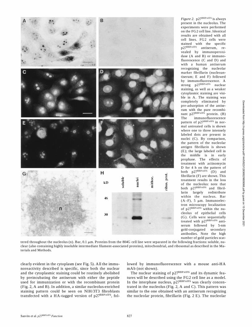
Cofractionation of human p27BBP/eIF6 in yeast cells wasanalyzed in parallel. For this end, fractions from the ribo-somal gradients were precipitated with TCA and analyzedby Western blot using antibodies against the human pro-tein. As shown in Fig. 10, p27BBP/eIF6 was detected in the80S and in the free 60S fractions, but absent from poly-somes.
Discussionp27BBP/eIF6 was simultaneously identified by two laborato-ries using two different approaches. It was isolated in ourlaboratory as a cytoplasmic interactor of the b4 integrinsubunit, and we have shown that it can specifically bindthe cytodomain of b4 in vitro (Biffo et al., 1997). However,the discovery of p27BBP/eIF6 homologues in organisms thatdo not contain b4 indicated that this protein might have afunction independent of b4. Along this line, p27BBP/eIF6
was independently identified by Si et al. (1997) as a puta-tive translation initiation factor, able to inhibit the associa-tion between the 60S and the 40S ribosomal subunits.
In this study, we have shown that although in epithelialcells p27BBP/eIF6 is coherent with b4 at hemidesmosomes,its association with the cytoskeleton is not a unique fea-ture of epithelial cells. Indeed the protein is in the nuclearmatrix of all growing cells. Consistently with its conservednucleolar expression pattern and sequence, p27BBP/eIF6 isnecessary for growth in yeast cells where its loss correlateswith a reduced level of the free 60S ribosomal subunit. Thein vivo findings were unexpected because they were con-sistent with a role of p27BBP/eIF6 in ribosomal biogenesisrather than in mRNA translation. In addition, the associa-tion of p27BBP/eIF6 with the nuclear matrix suggested thatthis process was linked to the nuclear cytoskeleton. Theability of the human protein to complement yeast muta-tion further suggested a conserved function for p27BBP/eIF6.
An Evolutionarily Conserved Function for p27BBP/eIF6 in 60S Metabolism
Database analysis indicates that p27BBP/eIF6 is a very an-cient, evolutionarily conserved protein. It is striking tonote that the homology is not restricted to a particular do-main of the protein, and that even the length of the proteinis constant among different species (246 amino acids in C.elegans; 245 in humans, fly, yeast, and A. thaliana; and215–222 in different Archibacteria). These data suggestthat p27BBP/eIF6 may have a critical and conserved function.Indeed, we have shown that the deletion of the S. cerevi-siae IIH1 gene, encoding the p27BBP/eIF6 homologue, is le-thal to yeast cells, and that the human protein can comple-ment the yeast-null mutation. Some lines of evidencesuggest that also in mammalian cells, p27BBP/eIF6 may berequired for growth because of the following: (a) the in-ability to produce stable p27BBP/eIF6 mRNA antisense ex-pressing mammalian cells (not shown); (b) the ubiquitousp27BBP/eIF6 expression in all immortalized cell lines so faranalyzed; (c) and the presence of a single p27BBP/eIF6 genein the human genome (Sanvito et al., 1998). The generationof p27BBP/eIF6-null mice will help to understand whetherp27BBP/eIF6 is also necessary for growth in higher verte-brates. Unfortunately, extensive sequence analysis did notyield significant clues to understand p27BBP/eIF6 function.
To gain some insights into this problem, we used twocomplementary approaches: the depletion of p27BBP/eIF6
in the genetically manipulable yeast model, and the studyof its topographical localization and biochemical proper-ties in mammalian cell lines and tissues. Yeast cells de-pleted of p27BBP/eIF6 are progressively arrested in G1, aphenotype consistent with a defect in either protein syn-thesis or ribosomal biogenesis. This fact, and the localiza-tion of p27BBP/eIF6 in nucleoli prompted us to analyze theeffect of its depletion on the polysome profile. These ex-periments provide useful information about how p27BBP/eIF6,based on its in vitro ribosomal anti-association activity,could be a translation initiation factor (Si et al., 1997).Polysome profiles of p27BBP/eIF6-depleted yeast cells showeda dramatic reduction in the peak of free 60S subunits andthe appearance of half-mer polysomes. Similar polysomeprofiles have been observed for mutants defective in ribo-somal proteins of the 60S ribosomal subunit (Moritz et al.,1991; Deshmukh et al., 1993; Vilardell and Warner, 1997),or for components involved in pre-rRNA processing and60S ribosomal subunit assembly (Ripmaster et al., 1992;Sun and Woolford, 1994; Hong et al., 1997; Weaver et al.,1997; Zanchin et al., 1997; Kressler et al., 1998). Thus, theprimary function of p27BBP/eIF6 in yeast is likely related tothe 60S ribosomal subunit metabolism.
Polysome profiles of yeast cells, defective in translationinitiation factor proteins, are generally characterized bythe reduction of the rate of polysomes accompanied by thegradual accumulation of both the free 40S and 60S sub-units. Therefore, the polysome profile of p27BBP/eIF6-depletedyeast cells does not support its primary function as a trans-lation initiation factor. However, on the basis of the invitro data of Si et al. (1997), and in view of the presence ofp27BBP/eIF6 also in the cytoplasm of some human cells, thepossibility that this protein might have a function also as acytosolic initiation factor cannot be ruled out, as such ac-
Figure 10. p27BBP/eIF6 cosedi-ments with 60S and 80S ribo-somal subunits. Fractions col-lected after the sucrosegradient centrifugation (top)of extracts from strain ySP-664 (iih1D GAL-Hsp27BBP/eIF6),logarithmically growing ingalactose-containing me-dium, were precipitated byTCA, and equal amounts ofprotein extracts were run on12% acrylamide gels, trans-ferred on immobilon-P mem-branes and probed with theanti–p27BBP/eIF6 antiserum,followed by the ECL detec-
tion method (bottom). Note that p27BBP/eIF6 (arrow) is highly en-riched in the free 60S and 80S fractions, but clearly absent fromthe polysome fraction. The high molecular weight band in lane 1is a nonspecific product recognized by secondary antibodies.
Dow
nloaded from http://rupress.org/jcb/article-pdf/144/5/823/1283692/9809068.pdf by guest on 15 D
ecember 2021

Sanvito et al. p27BBP/eIF6 Function 835
tivity could be masked by the predominant defect in 60Smetabolism.
The polysome profile does not enlighten the precise roleplayed by p27BBP/eIF6 in 60S metabolism. The protein maybe necessary for ribosome assembly/transport, or may actas a structural ribosomal protein. On the basis of the avail-able data, this last possibility is less likely to be true. Infact, the amount of p27BBP/eIF6 sedimenting with the ribo-somal fraction in several cell lines represents only a minorfraction of the total p27BBP/eIF6 content. Furthermore, nop27BBP/eIF6 was detected in the polysome fraction.
p27BBP/eIF6 accumulates in the nucleolus of all the ana-lyzed cell lines, where its pattern follows nucleolar evolu-tion (redistribution at mitosis, when the nucleolar organiz-ing region disappears, and redistribution after actinomycinD treatment). Since the nucleolus is the site where riboso-mal subunits are assembled, it seems plausible to speculatethat p27BBP/eIF6 might be involved in 60S ribosomal biogen-esis. The process of ribosome biogenesis is complex andinvolves several factors (for review see Woolford andWarner, 1991; Eichler and Craig, 1994) including proteinswith diverse functions such as RNA helicases, transcrip-tion factors, and nucleases. Further studies will address theprecise role that p27BBP/eIF6 might play in this process.
Finally, it is possible that p27BBP/eIF6 may be involved inthe transport of the 60S subunit from the nucleus to the cy-toplasm. To date, very little is known about this process(for review see Shaw and Jordan, 1995), and only a few nu-cleolar proteins have been found to shuttle between thenucleolus and the cytoplasm. In this context, three ob-servations are particularly intriguing: (a) the presence ofp27BBP/eIF6 both in a soluble pool and in a cytoskeletalbound compartment; (b) the existence of trace amounts ofsoluble cytoplasmic p27BBP/eIF6 in all cells; and (c) the abil-ity of p27BBP/eIF6 to bind also the mature 60S subunit (Si et al.,1997).
It is also worth noting that the nucleolar localization ofp27BBP/eIF6 is observed in the absence of a consensus nu-clear localization signal. Therefore, either p27BBP/eIF6 car-ries an unknown sequence for nuclear targeting or it is tar-geted into the nucleus by binding an additional factor inthe cytoplasm. The second hypothesis is supported by thefact that even in its most soluble form, p27BBP/eIF6 parti-tions in gel filtration as a high molecular weight complex(unpublished observation). The molecular dissection ofthis high molecular weight complex may shed light on themechanism by which p27BBP/eIF6 is transported to the nu-cleus.
p27BBP/eIF6 in the Nuclear Matrix/Intermediate Filaments Fraction
Our study shows that a relevant fraction of p27BBP/eIF6 ishighly insoluble in vivo and is associated both with the nu-clear matrix and with the intermediate filament pool. Inthe cytoplasm, electron microscopy studies have detectedp27BBP/eIF6 on thin cytoplasmic filaments of unknown com-position that are spatially separated from the classicalkeratin intermediate filaments, and converge both uponhemidesmosomes and desmosomes. To our knowledge,beside keratins, only another intermediate filament asso-ciated protein, IFAP300, has been described both in
hemidesmosomes and desmosomes (Skalli et al., 1994). Inthis context, it is interesting to note that a recent thoroughelectron microscopy analysis of human hemidesmosomeshas shown the presence of a novel filamentous structure inthe proximity of the inner plaque of the hemidesmosome(Behzad et al., 1995).
Nuclear matrix consists of both thick polymorphous fila-ments and of thin filaments known as core filaments (Heet al., 1990). In the nucleus, p27BBP/eIF6 is associated withpolymorphous thick filaments, and is absent from the corefilaments. This observation is fully consistent with the no-tion that core filaments may be formed by nuclear RNA,and that p27BBP/eIF6 distribution is resistant to RNase di-gestion (He et al., 1990). The localization of p27BBP/eIF6 inthe nuclear matrix is of extreme interest in the context ofribosome biogenesis. Our data provide an intriguing linkbetween the nuclear cytoskeleton and the process of ribo-some assembly.
In recent years growing evidence has indicated thatmost nuclear and cytoplasmic processes including tran-scription, DNA replication, and protein synthesis are spa-tially organized in association with the cytoskeleton. Thecombined roles of p27BBP/eIF6 protein in 60S assembly, itsassociation with the cytoskeleton, and its ability to bind b4integrin (Biffo et al., 1997) and the mature 60S ribosomesubunit (Si et al., 1997) belong to an integrated view of cellregulation that encompasses structure as well as biochemi-cal processes (Chicurel et al., 1998).
p27BBP/eIF6 and b4 Integrin
We have previously shown that p27BBP/eIF6 binds specifi-cally to the cytodomain of b4 integrin in vitro and in yeast(Biffo et al., 1997). Our previous data, and specifically theassociation of p27BBP/eIF6 with keratin intermediate fila-ments, strongly suggested that this interaction could occuralso in vivo and be necessary for targeting b4 to hemides-mosomes and intermediate filaments. Since intermediatefilament-associated proteins can be solubilized only uponSDS treatment, rendering the maintenance of biochemicalinteractions impossible, an association between b4 andp27BBP/eIF6 in tissues could not be proved. We now providetwo further elements suggesting that p27BBP/eIF6 is func-tionally associated to the b4 integrin in vivo: (a) its pecu-liar Swiss cheese distribution is superimposable to that ofb4 in cells that form hemidesmosomes; and (b) the pres-ence of the protein, in vivo, in hemidesmosomes of the hu-man amnion. Further experiments are needed to clarifythe functional significance of b4–p27BBP/eIF6 interaction,and specifically whether p27BBP/eIF6 may direct b4 to hemi-desmosomes. Alternatively, as it has been recently sug-gested, on the basis of in vitro evidence and yeast two-hybridassays, the crucial step in targeting b4 to hemidesmosomesis the interaction with the large intermediate filament-associated protein, HD-1 (Niessen et al., 1997; Rezniczecket al., 1998). If this is the case also in vivo, then the role ofp27BBP/eIF6 binding to b4 may be related to a nonstructuralfunction of b4 integrin, similar to that shown in the case ofthe recruitment of shc and grb2 (Mainiero et al., 1995) orof PI3 kinase (Shaw et al., 1997).
In the absence of further evidence, we may reasonablysuggest that p27BBP/eIF6 has an evolutionarily conserved
Dow
nloaded from http://rupress.org/jcb/article-pdf/144/5/823/1283692/9809068.pdf by guest on 15 D
ecember 2021

The Journal of Cell Biology, Volume 144, 1999 836
function linked to 60S ribosome biogenesis, and one ac-quired during evolution in epithelial cells containing b4 in-tegrin. At least one precedent of a protein with a dualfunction acquired during evolution, i.e., b-catenin/arma-dillo, has already been reported. This remarkable proteincan be found both at sites of cell–cell adhesion in connec-tion to cadherins and in the nucleus where it can signal inconjunction with LEF-1 (for review see Willert and Nusse,1998).
We thank R. Ochs for the kind gift of human antibodies directed againstfibrillarin, F. Giancotti for permission to use clone A of the 804G cell line,and J. Nickerson for advice on nuclear matrix preparation. We are in-debted to N. Offenhaeuser for useful suggestion and criticism throughoutthis work, to A. Hinnebusch and M. Foiani for useful suggestions, G.Serini for having pushed us to perform some experiments, E. Bianchi forher criticism, and E. Rizzo for the preparation and purification of the re-combinant p27BBP protein used in the preadsorption experiments and theexperiments with actinomycin D.
The financial support of Telethon-Italy (grant 762 and E.712) is grate-fully acknowledged. The work was supported also by Associazione Ital-iana per la Ricerca sul Cancro, Giovanni Armenise-Harvard Foundation,and MURST to P.C. Marchisio, and by CNR Target Project on Biotech-nology Grant CT.97.01180.PF49(F).
Received for publication 15 September 1998 and in revised form 14 Janu-ary 1999.
References
Behzad, F., C.J. Jones, S. Ball, T. Alvares, and J.D. Aplin. 1995. Studies ofhemidesmosomes in human amnion: the use of a detergent extraction proto-col for compositional and ultrastructural analysis and preparation of ahemidesmosome-enriched fraction from tissue. Acta Anat. 152:170–184.
Biffo, S., F. Sanvito, S. Costa, L. Preve, R. Pignatelli, L. Spinardi, and P.C.Marchisio. 1997. Isolation of a novel beta4 integrin-binding protein(p27(BBP)) highly expressed in epithelial cells. J. Biol. Chem. 272:30314–30321.
Boukamp, P., R.T. Petrussevska, D. Breitkreutz, J. Hornung, A. Markham, andN.E. Fusenig. 1988. Normal keratinization in a spontaneously immortalizedaneuploid human keratinocyte cell line. J. Cell Biol. 106:761–771.
Chicurel, M.E., R.H. Singer, C.J. Meyer, and D.E. Ingber. 1998a. Integrin bind-ing and mechanical tension induce movement of mRNA and ribosomes tofocal adhesions. Nature. 392:730–733.
Chicurel, M.E., C.S. Chen, and D.E. Ingber. 1998b. Cellular control lies in thebalance of forces. Curr. Opin. Cell Biol. 10:232–239.
Cho, S.H., J.J. Cho, I.I. Kim, H. Vliagoftis, D.D. Metcalfe, and C.K. Oh. 1998.Identification and characterization of the inducible murine mast cell gene,imc-415. Biochem. Biophys. Res. Commun. 9:123–127.
Clark, E.A., and J.S. Brugge. 1995. Integrins and signal transduction pathways:the road taken. Science. 268:233–239.
Deshmukh, M., Y.F. Tsay, A.G. Paulovich, and J.L. Woolford, Jr. 1993. Yeastribosomal protein L1 is required for the stability of newly synthesized 5SrRNA and the assembly of 60S ribosomal subunits. Mol. Cell. Biol. 13:2835–2845.
Dowling, J., Q.C. Qu, and E. Fuchs. 1996. Beta4 integrin is required forhemidesmosome formation, cell adhesion, and cell survival. J. Cell Biol. 134:559–572.
Earnshaw, W.C., and R.L. Bernat. 1991. Chromosomal passengers: toward anintegrated view of mitosis. Chromosoma. 100:139–146.
Eichler, D.C., and N. Craig. 1994. Processing of eukaryotic ribosomal RNA.Prog. Nuc. Acid Res. Mol. Biol. 49:197–239.
Foiani, M., A.M. Cigan, C.J. Paddon, S. Harashima, and A.G. Hinnebusch.1991. GCD2, a translational repressor of the GCN4 gene, has a general func-tion in the initiation of protein synthesis in Saccharomyces cerevisiae. Mol.Cell Biol. 11:3203–3216.
Giancotti, F.G. 1996. Signal transduction by the alpha 6 beta 4 integrin: chartingthe path between laminin binding and nuclear events. J. Cell Sci. 109:1165–1172.
Giancotti, F.G. 1997. Integrin signaling: specificity and control of cell survivaland cell cycle progression. Curr. Opin. Cell Biol. 9:691–700.
Gietz, R.D., and A. Sugino. 1988. New yeast–Escherichia coli shuttle vectorsconstructed with in vitro mutagenized yeast genes lacking six-base pair re-striction sites. Gene. 74:527–534.
Green, S., I. Issemann, and E. Sheer. 1988. A versatile in vivo and in vitro eu-karyotic expression vector for protein engineering. Nucleic Acids Res. 16:369.
He, D.C., J.A. Nickerson, and S. Penman. 1990. Core filaments of the nuclear
matrix. J. Cell Biol. 110:569–580.Hong, B., J.S. Brockenbrough, P. Wu, and J.P. Aris. 1997. Nop2p is required for
pre-rRNA processing and 60S ribosome subunit synthesis in yeast. Mol.Cell. Biol. 17:378–388.
Howe, A., A.E. Aplin, S.K. Alahari, and R.L. Juliano. 1998. Integrin signalingand cell growth control. Curr. Opin. Cell Biol. 10:220–231.
Hynes, R.O. 1992. Integrins: versatility, modulation, and signaling in cell adhe-sion. Cell. 69:11–25.
Kajiji, S., R.N. Tamura, and V. Quaranta. 1989. A novel integrin (alpha E beta4) from human epithelial cells suggests a fourth family of integrin adhesionreceptors. EMBO (Eur. Mol. Biol. Organ.) J. 8:673–680.
Kressler, D., J. de la Cruz, M. Rojo, and P. Linder. 1998. Dbp6p is an essentialputative ATP-dependent RNA helicase required for 60S-ribosomal-subunitassembly in Saccharomyces cerevisiae. Mol. Cell. Biol. 18:1855–1865.
Laemmli, U.K. 1970. Cleavage of structural proteins during the assembly of thehead of bacteriophage T4. Nature. 227:680–685.
Madjar, J.J. 1994. Preparation of ribosomes and ribosomal proteins from cul-tured cells. In Cell Biology: A Laboratory Handbook. Academic Press Inc.,Orlando, FL. 657–661.
Mainiero, F., A. Pepe, K.K. Wary, L. Spinardi, M. Mohammadi, J. Schlessinger,and F.G. Giancotti. 1995. Signal transduction by the alpha 6 beta 4 integrin:distinct beta 4 subunit sites mediate recruitment of Shc/Grb2 and associationwith the cytoskeleton of hemidesmosomes. EMBO (Eur. Mol. Biol. Organ.)J. 14:4470–4481.
Marchisio, P.C., S. Bondanza, O. Cremona, R. Cancedda, and M. De Luca.1991. Polarized expression of integrin receptors (a6b4, a2b1, a3b1, andavb5) and their relationship with the cytoskeleton and basement membranematrix in cultured human keratinocytes. J. Cell Biol. 112:761–773.
Nickerson, J.A., G. Krockmalnic, K.M. Wan, C.D. Turner, and S. Penman.1992. A normally masked nuclear matrix antigen that appears at mitosis oncytoskeleton filaments adjoining chromosomes, centrioles, and midbodies. J.Cell Biol. 116:977–987.
Nickerson, J.A., G. Krockmalnic, and S. Penman. 1994. Isolation and visualiza-tion of the nuclear matrix, the nonchromatin structure of the nucleus. In CellBiology: A Laboratory Handbook. Academic Press Inc., Orlando, FL. 622–627.
Niessen, C.M., E.H. Hulsman, L.C. Oomen, I. Kuikman, and A. Sonnenberg.1997. A minimal region on the integrin beta4 subunit that is critical to its lo-calization in hemidesmosomes regulates the distribution of HD1/plectin inCOS-7 cells. J. Cell Sci. 110:1705–1716.
Ochs, R.L., and K. Smetana. 1991. Detection of fibrillarin in nucleolar rem-nants and the nucleolar matrix. Exp. Cell Res. 197:183–190.
Rezniczek, G.A., J.M. de Pereda, S. Reipert, and G. Wiche. 1998. Linking inte-grin alpha6beta4-based cell adhesion to the intermediate filament cytoskele-ton: direct interaction between the beta4 subunit and plectin at multiple mo-lecular sites. J. Cell Biol. 141:209–225.
Ripmaster, T.L., G.P. Vaughn, and J.L. Woolford, Jr. 1992. DRS1 to DRS7,novel genes required for ribosome assembly and function in Saccharomycescerevisiae. Mol. Cell. Biol. 13:7901–7912.
Rose, M.D., F. Winston, and P. Hieter. 1990. Methods in yeast genetics. ColdSpring Harbor Laboratory, Cold Spring Harbor, NY. 192 pp.
Rouiller, D.G., V. Cirulli, and P.A. Halban. 1990. Differences in aggregationproperties and levels of the neural cell adhesion molecule (NCAM) betweenislet cell types. Exp. Cell Res. 191:305–312.
Sambrook, J., E.F. Fritsch, and T. Maniatis. 1989. Molecular Cloning. A Labo-ratory Manual. Cold Spring Harbor Laboratory, Cold Spring Harbor, NY.
Sanvito, F., G. Arrigo, O. Zuffardi, M. Agnelli, P.C. Marchisio, and S. Biffo.1998. Localization of p27 beta4 binding protein gene (ITGB4BP) to humanchromosome region 20q11.2. Genomics. 51:111–112.
Schofer, C., K. Weipoltshammer, M. Almeder, M. Muller, and F. Wachtler.1996. Redistribution of ribosomal DNA after blocking of transcription in-duced by actinomycin D. Chromosome Res. 4:384–391.
Shaw, L.M., I. Rabinovitz, H.H. Wang, A. Toker, and A.M. Mercurio. 1997.Activation of phosphoinositide 3-OH kinase by the alpha6beta4 integrinpromotes carcinoma invasion. Cell. 91:949–960.
Shaw, P.J., and E.G. Jordan. 1995. The nucleolus. Annu. Rev. Cell. Dev. Biol.11:93–121.
Si, K., J. Chaudhuri, J. Chevesich, and U. Maitra. 1997. Molecular cloning andfunctional expression of a human cDNA encoding translation initiation fac-tor 6. Proc. Natl. Acad. Sci. USA. 94:14285–14290.
Skalli, O., J.C. Jones, R. Gagescu, and R.D. Goldman. 1994. IFAP 300 is com-mon to desmosomes and hemidesmosomes and is a possible linker of inter-mediate filaments to these junctions. J. Cell Biol. 125:159–170.
Spinardi, L., Y.L. Ren, R. Sanders, and F.G. Giancotti. 1993. The beta 4 subunitcytoplasmic domain mediates the interaction of alpha 6-beta 4 integrin withthe cytoskeleton of hemidesmosomes. Mol. Biol. Cell. 4:871–884.
Sun, C., and J.L. Woolford, Jr. 1994. The yeast NOP4 gene product is an essen-tial nucleolar protein required for pre-rRNA processing and accumulationof 60S ribosomal subunits. EMBO (Eur. Mol. Biol. Organ.) J. 13:3217–3235.
Thompson, J.D., D.G. Higgins, and T.J. Gibson. 1994. CLUSTAL W: improv-ing the sensitivity of progressive multiple sequence alignment through se-quence weighting, position-specific gap penalties and weight matrix choice.Nucleic Acids Res. 22:4673–4680.
van der Neut, R., P. Krimpenfort, J. Calafat, C.M. Niessen, and A. Sonnenberg.1996. Epithelial detachment due to absence of hemidesmosomes in integrin
Dow
nloaded from http://rupress.org/jcb/article-pdf/144/5/823/1283692/9809068.pdf by guest on 15 D
ecember 2021
Sanvito et al. p27BBP/eIF6 Function 837
beta 4 null mice. Nat. Genet. 13:366–369.Vidal, F., D. Aberdam, C. Miquel, A.M. Christiano, L. Pulkkinen, J. Uitto, J.P.
Ortonne, and G. Meneguzzi. 1995. Integrin beta 4 mutations associated withjunctional epidermolysis bullosa with pyloric atresia. Nat. Genet. 10:229–234.
Vilardell, J., and J.R. Warner. 1997. Ribosomal protein L32 of Saccharomycescerevisiae influences both the splicing of its own transcript and the process-ing of rRNA. Mol. Cell. Biol. 174:1959–1965.
Villa, A., P. Podini, M.C. Panzeri, H.D. Soling, P. Volpe, and J. Meldolesi. 1993.The endoplasmic-sarcoplasmic reticulum of smooth muscle: immunocy-tochemistry of vas deferens fibers reveals specialized subcompartments dif-ferently equipped for the control of Ca21 homeostasis. J. Cell Biol. 121:1041–1051.
Wach, A., A. Brachat, R. Pohlmann, and P. Philippsen. 1994. New heterologousmodules for classical or PCR-based gene disruptions in Saccharomyces cer-evisiae. Yeast. 10:1793–1808.
Weaver, P.L., C. Sun, and T.H. Chang. 1997. Dbp3p, a putative RNA helicasein Saccharomyces cerevisiae, is required for efficient pre-rRNA processingpredominantly at site A3. Mol. Cell. Biol. 17:1354–1365.
Willert, K., and R. Nusse. 1998. Beta-catenin: a key mediator of Wnt signaling.Curr. Op. Genet. Dev. 8:95–102.
Zanchin, N.I., P. Roberts, A. De Silva, F. Sherman, and D.S. Goldfarb. 1997.Saccharomyces cerevisiae Nip7p is required for efficient 60S ribosome sub-unit biogenesis. Mol. Cell. Biol. 17:5001–5015.
Dow
nloaded from http://rupress.org/jcb/article-pdf/144/5/823/1283692/9809068.pdf by guest on 15 D
ecember 2021

The Journal of Cell Biology, Volume 144, 1999 838
Dow
nloaded from http://rupress.org/jcb/article-pdf/144/5/823/1283692/9809068.pdf by guest on 15 D
ecember 2021