Embed Size (px)
Citation preview

Singapore Med J 2009; 50(3) : e114C a s e R e p o r t
Department of Pathology & Microbiology,Section of Haematology,The Aga Khan University Hospital,PO Box 3500,Stadium Road, Karachi 74800,Pakistan
Ali N, MBBSResident Moiz B, MBBS, MCPS, FCPSAssistant Professor and Consultant
Correspondence to:Dr Natasha AliTel: (92) 21 486 1548Fax: (92) 21 493 4294Email: [email protected]
Azurophilic inclusions in plasma cellsAli N, Moiz B
ABSTRACTWe report a 45-year-old man with complaints of chest pain and weight loss who was referred for a bone marrow/trephine procedure to the Aga Khan University Hospital. Bone marrow examination showed plasmacytosis of 95% with plasma cells containing coarse Auer rod-like azurophilic inclusions, which failed to stain positively with Sudan Black B or periodic acid Schiff stain. These inclusions have rarely been previously reported as they are more significant morphologically rather than having a prognostic value.
Keywords : azurophilic granules, multiple myeloma, plasma cells
Singapore Med J 2009; 50(3): e114-e115
INTRODUCTIONSeveral cases of Auer rod-like inclusions in cells of multiple myeloma have been published previously.(1) Other associations include prolymphocytic leukaemia and hypogammaglobulinaemia.(2) On special stain profile, these granules are negative for Sudan Black B stain and show occasional positivity for periodic acid Schiff (PAS) stain. We describe a patient with multiple myeloma showing azurophilic granules in plasma cells.
CASE REPORTA 45-year-old man was referred to the clinical laboratory of The Aga Khan University Hospital, Pakistan, for bone marrow examination due to persistent chest pain and weight loss for the last four months. His past history was insignificant except for a blood transfusion two weeks ago. He had no previous history of hospitalisation. A provisional diagnosis of secondary deposits was made clinically and bone marrow examination was performed for proper evaluation. His complete blood count showed a haemoglobin level of 6.5 g/dL, haematocrit 20.6, mean corpuscular volume 78.5 fL, mean corpuscular haemoglobin 25 pg, white blood cells 6.8 × 10⁹/L and platelets 237 × 109/L. A peripheral smear revealed a dimorphic blood picture, with target cells and gross rouleaux formation. White blood cells and platelets showed normal morphology (Fig. 1). Bone marrow aspirate was hypercellular, with diffuse infiltration of plasma cells comprising 95% of the total nucleated cell population. The abnormal plasma cells ranged from 20 µm to 30 µm in size with an oval
nucleus, fine chromatin and deep basophilic cytoplasm. Approximately 80% of these cells contained coarse azurophilic Auer rod-like inclusions/granules occupying the majority of the cytoplasmic space except the perinuclear area (Figs. 2 & 3). The remainder of these cells showed an eccentrically-placed nucleus, with vacuolated cytoplasm and granules occupying the periphery of the cytoplasm. These cells were negative for Sudan Black B
Fig. 1 Peripheral blood film shows gross rouleaux formation (Leishmann stain, × 40).
Fig. 2 Bone marrow aspirate shows plasma cells with coarse azurophilic granules (Leishmann stain, × 100).
Fig. 3 Bone marrow aspirate shows pleomorphic plasma cells with flaming cytoplasm and azurophilic granules (Leishmann stain, × 100).
Singapore Med J 2009; 50(3) : e115
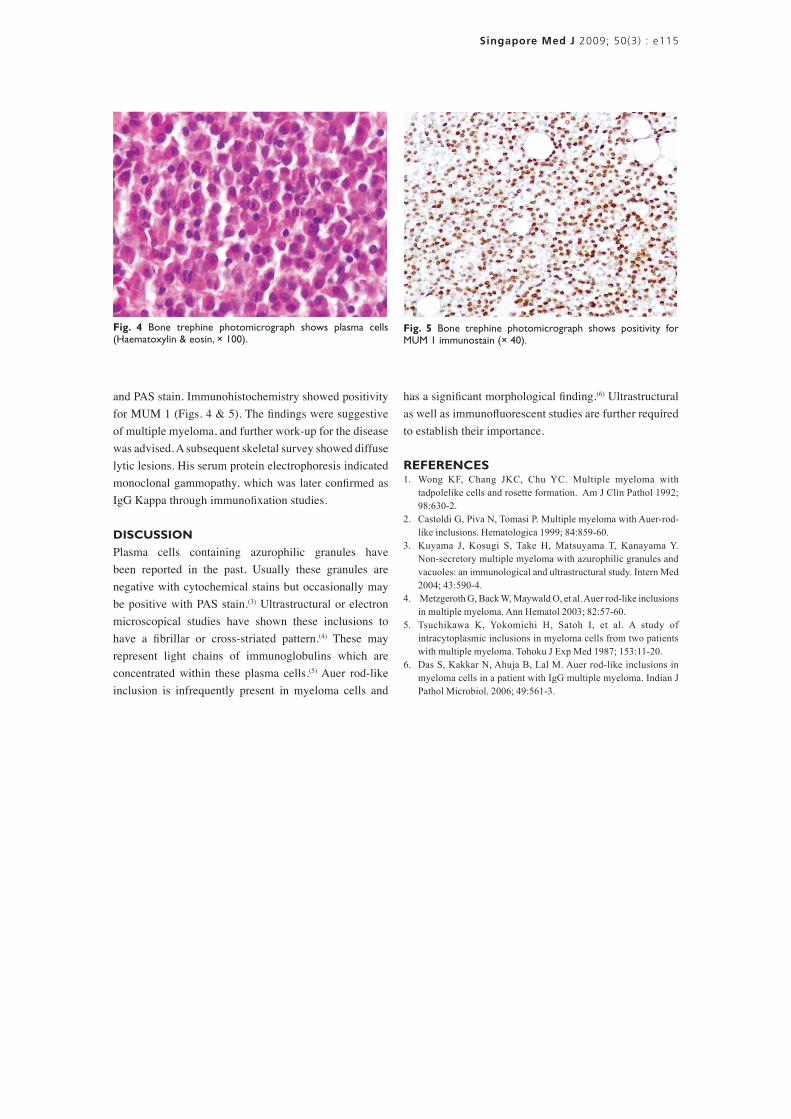
and PAS stain. Immunohistochemistry showed positivity for MUM 1 (Figs. 4 & 5). The findings were suggestive of multiple myeloma, and further work-up for the disease was advised. A subsequent skeletal survey showed diffuse lytic lesions. His serum protein electrophoresis indicated monoclonal gammopathy, which was later confirmed as IgG Kappa through immunofixation studies.
DISCUSSIONPlasma cells containing azurophilic granules have been reported in the past. Usually these granules are negative with cytochemical stains but occasionally may be positive with PAS stain.(3) Ultrastructural or electron microscopical studies have shown these inclusions to have a fibrillar or cross-striated pattern.(4) These may represent light chains of immunoglobulins which are concentrated within these plasma cells.(5) Auer rod-like inclusion is infrequently present in myeloma cells and
Fig. 5 Bone trephine photomicrograph shows positivity for MUM 1 immunostain (× 40).
Fig. 4 Bone trephine photomicrograph shows plasma cells (Haematoxylin & eosin, × 100).
has a significant morphological finding.(6) Ultrastructural as well as immunofluorescent studies are further required to establish their importance.
REFERENCES1. Wong KF, Chang JKC, Chu YC. Multiple myeloma with
tadpolelike cells and rosette formation. Am J Clin Pathol 1992; 98:630-2.
2. Castoldi G, Piva N, Tomasi P. Multiple myeloma with Auer-rod-like inclusions. Hematologica 1999; 84:859-60.
3. Kuyama J, Kosugi S, Take H, Matsuyama T, Kanayama Y. Non-secretory multiple myeloma with azurophilic granules and vacuoles: an immunological and ultrastructural study. Intern Med 2004; 43:590-4.
4. Metzgeroth G, Back W, Maywald O, et al. Auer rod-like inclusions in multiple myeloma. Ann Hematol 2003; 82:57-60.
5. Tsuchikawa K, Yokomichi H, Satoh I, et al. A study of intracytoplasmic inclusions in myeloma cells from two patients with multiple myeloma. Tohoku J Exp Med 1987; 153:11-20.
6. Das S, Kakkar N, Ahuja B, Lal M. Auer rod-like inclusions in myeloma cells in a patient with IgG multiple myeloma. Indian J Pathol Microbiol. 2006; 49:561-3.


![Plasma cell neoplasmas [Read-Only]](https://img.pdfslide.us/doc/110x75/61c307aa2d33612dab6737b3/plasma-cell-neoplasmas-read-only.jpg)