Embed Size (px)
Citation preview
Clinically Tested to be the World’s Most Accurate Toric Marking Device
AXsys® Studay Data and Press Release Reference
Press Release Reference
46 CataraCt & refraCtive Surgery today july 2012
CataraCt and refraCtive Surgery innovative SurgiCal inStrumentS
I give my patients the option of having a toric IOL implanted during cataract surgery. As opposed to con-ventional IOLs, toric IOLs correct preexisting corneal astigmatism during cataract surgery. Achieving the best
result with a toric IOL requires properly marking the axis on which the toric IOL should be implanted, which can be determined with toric IOL web-based calculators. For every 1º that the axis is misaligned, a toric lens loses 3% of its corrective effect.1 I could not achieve an accurate marking with conventional toric IOL markers, because I was unable to confirm that I was holding the instrument perfectly horizontally. In addition, I found it difficult with bubble and pendulum markers to simultaneously focus on the marker and the patient’s eye. I therefore worked with ASICO LLC to design an electronic toric marker that uses my senses of sight and hearing to help me easily and accurately pinpoint the horizontal axis and stay focused on my patient.
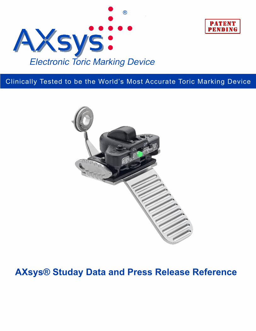
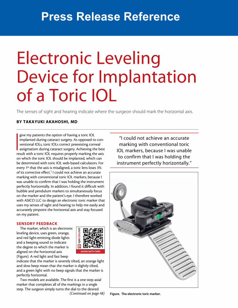
SENSORY FEEDBACKThe marker, which is an electronic
leveling device, uses green, orange, and red light-emitting diode lights and a beeping sound to indicate the degree to which the marker is aligned on the horizontal axis (Figure). A red light and fast beep indicate that the marker is severely tilted, an orange light and slow beep mean that the marker is slightly tilted, and a green light with no beep signals that the marker is perfectly horizontal.
Two models are available. The first is a one-step axial marker that completes all of the markings in a single step. The surgeon simply turns the dial to the desired
eyetube.net
eyetube.net/?v=hefet
electronic leveling device for implantation of a toric iolThe senses of sight and hearing indicate where the surgeon should mark the horizontal axis.
BY TAKAYuKi AKAhOShi, MD
Figure. The electronic toric marker.
“i could not achieve an accurate marking with conventional toric
iol markers, because i was unable to confirm that i was holding the
instrument perfectly horizontally.”
eyetube.net
(Continued on page 48)
Press Release Reference Press Release ReferenceCataraCt and refraCtive Surgery innovative SurgiCal inStrumentS
48 CataraCt & refraCtive Surgery today july 2012
axis, asks the patient to sit up and look straight, and marks the axis when the light indicator on the marker is green and the beeping stops.
The two-step electronic toric marker is for patients with narrow eyelids or deep-set eyes. This model precisely marks the 0º and 180º positions preoperatively, and the ophthalmologist uses these as points of reference to mark the desired axis during the surgery.
The sensitivity of the device can be adjusted to five settings, ranging from 0.2º to 1.0º. If they wish, surgeons can choose to completely turn off the beeping or pro-gram the instrument to beep only when the toric mark-er is perfectly horizontal.
DESiGN FEATuRESThe surgeon holds the flat handle with his or her
thumb and index finger. The electronic device is attached to the body of the marker by a magnet and can easily be removed for sterilization. The head on both designs may be used for all types of eyes, includ-ing those with small orbits.
TiPSWhen using the toric marker, I apply topical anesthe-
sia and wait until the secretion of tears subsides. I paint the blades of the toric marker with a marking pen, and set the desired axis on the dial. I ask the patient to sit upright while gazing straight ahead with both eyes open. I gently hold the patient’s eyelids with my fingers to prevent the patient from closing his or her eyes as the marker approaches. The surgeon should sit at the same eye level as the patient; this position is helpful for placing the marker on the center of the cornea. n
Takayuki Akahoshi, MD, is the director of ophthalmology at Mitsui Memorial Hospital in Tokyo. He acknowledged no financial inter-est in the products or companies mentioned herein. Dr. Akahoshi may be reached at +81 3 3862 9111; [email protected].
1. Visser N, Berendschot TT, Bauer NJ, et al. Accuracy of toric intraocular lens implantation in cataract and refractive surgery. J Cataract Refract Surg. 2011;37(8):1394-1402.
“the surgeon should sit at the same eye level as the patient;
this position is helpful for placing the marker on the center
of the cornea.”
(Continued from page 46)
40
EUROTIMES | Volume 17 | Issue 10
Global surveyBausch + Lomb has released the findings of the company’s first-ever Barometer of Global Eye Health, a global survey of more than 11,000 consumers in 11 countries.
“Among the survey’s findings are that people would rather lose 10 years of their life (67 per cent) than lose their eyesight. And they would be more willing to take a 50 per cent pay cut than lose 50 per cent of their vision,” said a Bausch + Lomb spokesman. “More than 70 per cent of Americans are not getting annual eye exams,” he said, “and 44 per cent believe that they do not need an eye test unless there is a problem.”
The global survey was developed in co-operation with eye care professionals from around the world and further validated by an additional 147 professionals from 26 different countries.n www.bausch.com.
ad-EUR-1-2 hoch-1202v2-pva RZ.indd 1 29.02.12 13:43
Feature
INdusTRy NEWsRecent developments in the vision care industry
Precision Laser SystemOptiMedica Corp has announced expanded international market adoption of its Catalys Precision Laser System with new installations at the premier Shinagawa LASIK Center in Tokyo and Santa Maria Eye Clinic in Sakaide, Kagawa, Japan.
“The Catalys Precision Laser System allows surgeons to perform custom cataract procedures with unparalleled precision, exceptional patient comfort and a markedly streamlined workflow. Dr Minoru Tomita and Dr Kunihiro Nagahara, of the respective centres, have joined a rapidly growing installed base of Catalys customers that include more than 20 centres in seven countries,” said a company spokeswoman. n www.optimedica.com
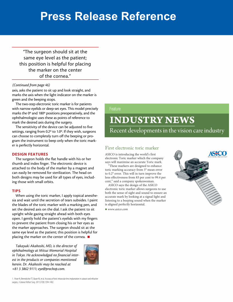
First electronic toric markerASICO is introducing the world’s first electronic Toric marker which the company says will maximise an accurate Toric mark.
“These markers are designed to enhance toric marking accuracy from 5° mean error to 0.2° error. This will in turn improve the lens effectiveness from 83 per cent to 99.4 per cent,” said a company spokeswoman.
ASICO says the design of the ASICO electronic toric marker allows surgeons to use both the sense of sight and sound to ensure an accurate mark by looking at a signal light and listening to a beeping sound when the marker is aligned perfectly horizontal.n www.asico.com
Single-use instrumentsAfter the launch of disposable instruments for cataract surgery then keratoplasty, Moria has announced the extension of its ONE® range with three single-use instruments for LASIK and FemtoLASIK surgery.
These are a Kratz wire speculum (reference #17227 – pictured), a Sinskey manipulating hook (reference #17223) and a manipulating hook (reference #17230).
“The Moria ONE® range of single-use instruments is ideally suited for LASIK surgery,” said a spokesman for the company. These instruments are sterile, individually packaged in sealed peel-apart pouches, in a box of 10. Each reference can be ordered separately,” he said. “The instruments are convenient and provide an exceptional value and they also eliminate the time and expense of re-processing soiled instruments,” said the spokesman.n www.moria-surgical.com
Press Release Reference
1/29/13 www.ophthalmologymanagement.com/articleviewer.aspx?articleID=107873
1/6www.ophthalmologymanagement.com/articleviewer.aspx?articleID=107873
Enhancing Outcomes.Advancing Your Practice. SEARCH
Article Feedback & Ideas
CLASSIFIEDS
Pre-ownedequipment, practicesfor sale, openpositions, helpfulpracticemanagementresources and more!
Click here to viewthe latest classifiedsfrom OphthalmologyManagement.
0 1
Article Date: 1/1/2013 Print Friendly Page
How to Manage Post-op ResidualAstigmatism
Strategies to optimize toric IOL outcomes and manageresidual astigmatism after surgery.
BY Jessica Shonfeld, MD, Mujtaba A. Qazi, MD, Jay S. Pepose, MD,PHD
In cataract patients with regular corneal astigmatism, toric IOLs offer
the potential for excellent uncorrected postoperative visual acuity.
However, the surgeonmust carefully consider a number of steps,
both preoperatively and intraoperatively, to avoid postoperative
residual astigmatism. Despite careful application of advanced tools
and techniques, postoperative refractive astigmatism can be
unexpectedly high — approximately 10% of patients with more than
1.00 D.1,2 Fortunately, a number of postoperative solutions can
address this situation.
Measuring Astigmatic Magnitude and Axis
One of the most critical elements of preoperative planning is the
measure of the magnitude and orientation of astigmatism. To
minimize measurement errors, one must use multiple sources; for
example, by comparing manual keratometry values to automated
keratometry and topography.
Videokeratography provides the greatest details of the anterior and
posterior cornea and regional corneal pachymetry, and helps assess
for corneal irregularity. The IOLMaster (Carl-Zeiss Meditec)
extrapolates astigmatic axis from six data points spaced 60° apart.3
This can be helpful if the steep meridian is at 0 or 60, but if the axis is
different, such as at 90° or 145°, you may find inaccuracies in
measurement. The Lenstar LS900 (Haag-Streit) surveys two rings of
16 data points spaced 22.5° apart.
Most studies comparing multiple diagnostic modalities have reported
close concordance, so readings should not vary by more than 0.50 D
and 10° between instruments. This is important because a toric IOL
can lose 30%of its astigmatic effect if it is misaligned 10°.4 It
becomes more important in the higher-powered toric IOLs; 30% of
the astigmatic effect of the AcrySof SN6AT9 (Alcon Surgical) is almost
1.50 D.
Select the Appropriate Patient
When selecting an ideal toric IOL candidate, keep in mind the multiple
patient characteristics that may foretell a poor outcome. Patients
with dry eye will often manifest discrepancies between keratometry
and topography measurements. Poorly managed dry eye disease
preoperatively can lead to selecting the wrong axis. Patients with
irregular corneal astigmatism — those with anterior basement
E-NEWSLETTER SUBSCRIPTION
AMD Update is a monthly e-newsletterdedicated to bringing to theophthalmologist the latest and mostuseful clinical information and literaturereviews on the management of AMD ina quick, easy to review format
Subscribe Now
Current Issue Archive Supplements Digital Supplements & Videos Ophthalmic ASC Helpful Resources Calendar of Events Contact Us
1/29/13 www.ophthalmologymanagement.com/articleviewer.aspx?articleID=107873
2/6www.ophthalmologymanagement.com/articleviewer.aspx?articleID=107873
membrane dystrophy or advanced keratoconus — may not be ideal
candidates due to their variable refractive and corneal astigmatism.
When obtaining biometry measurements, we find it helpful to
approach the toric IOL patient much like a preoperative refractive
patient. Avoid taking measurements if the patient has not
discontinued soft contact lens wear for at least two weeks and gas
permeable contact lens wear for four weeks before the evaluation.
Repeating these measurements a few weeks apart helps to assure
that the cornea has reached stability from the impact of contact lens
warpage, particularly in patients with an extended history of rigid
contact lens use. Additionally, we must pay attention to proper head
positioning when obtaining keratometry and topography.
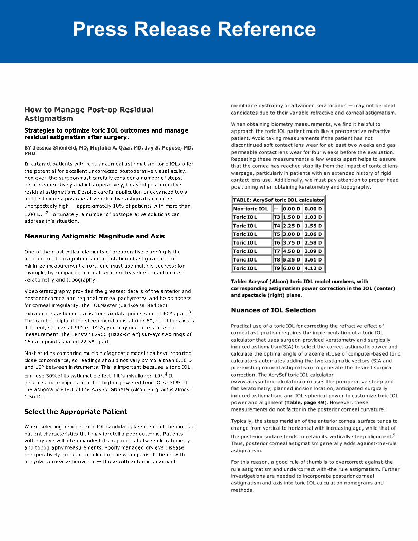
TABLE: AcrySof toric IOL calculator
Non-toric IOL -- 0.00 D 0.00 D
Toric IOL T3 1.50 D 1.03 D
Toric IOL T4 2.25 D 1.55 D
Toric IOL T5 3.00 D 2.06 D
Toric IOL T6 3.75 D 2.58 D
Toric IOL T7 4.50 D 3.09 D
Toric IOL T8 5.25 D 3.61 D
Toric IOL T9 6.00 D 4.12 D
Table: Acrysof (Alcon) toric IOL model numbers, with
corresponding astigmatism power correction in the IOL (center)
and spectacle (right) plane.
Nuances of IOL Selection
Practical use of a toric IOL for correcting the refractive effect of
corneal astigmatism requires the implementation of a toric IOL
calculator that uses surgeon-provided keratometry and surgically
induced astigmatism(SIA) to select the correct astigmatic power and
calculate the optimal angle of placement.Use of computer-based toric
calculators automates adding the two astigmatic vectors (SIA and
pre-existing corneal astigmatism) to generate the desired surgical
correction. The AcrySof toric IOL calculator
(www.acrysoftoriccalculator.com) uses the preoperative steep and
flat keratometry, planned incision location, anticipated surgically
induced astigmatism, and IOL spherical power to customize toric IOL
power and alignment (Table, page 49). However, these
measurements do not factor in the posterior corneal curvature.
Typically, the steep meridian of the anterior corneal surface tends to
change from vertical to horizontal with increasing age, while that of
the posterior surface tends to retain its vertically steep alignment.5
Thus, posterior corneal astigmatism generally adds against-the-rule
astigmatism.
For this reason, a good rule of thumb is to overcorrect against-the
rule astigmatism and undercorrect with-the rule astigmatism. Further
investigations are needed to incorporate posterior corneal
astigmatism and axis into toric IOL calculation nomograms and
methods.
Press Release Reference Press Release Reference
1/29/13 www.ophthalmologymanagement.com/articleviewer.aspx?articleID=107873
3/6www.ophthalmologymanagement.com/articleviewer.aspx?articleID=107873
Figure 1. Electronic One-step Toric Reference Marker (ASICO).
SIA: A Little Can Mean a Lot
Knowing one’s surgically induced astigmatism (SIA) is critical. SIA can
affect both the magnitude and direction of the principal astigmatic
meridians of the cornea and have a greater impact in cases of smaller
amounts of preoperative cylinder. Studies have reported 0.20 D to
1.20 D of SIA depending upon incision size and location.4
The SIA Calculator, an online tool developed by Warren Hill,MD,
(www.SIA-calculator.com) can help you customize SIA for clear, near-
clear and scleral incisions. You can then calculate the optimal incision
angle to minimize the postoperative residual astigmatism, sometimes
rendering a change in surgical approach from temporal to superior, or
vice versa.
Surgical Tips and Tricks
Once in the operating room, correctly placing the toric IOL can be just
as challenging as the preoperative planning. These perioperative
steps can help avoid problems postoperatively.
► Mark, remark and reinforce. Mark the cornea twice: once
preoperatively with a reference marker in the upright position to
compensate for cyclotorsion; and during surgery with a toric axis
marker. Markers with either a bubble-level or digital guidance (Figure
1) can facilitate reliable preoperative marking. Reinforcing the
reference marks with a sterile marking pen can help prevent fading
during surgery. Circumlinear rhexis formation, thorough cortical
cleanup, and anterior and posterior capsular polishing are
prerequisites for proper toric IOL centration and in-the-bag stability.
The astigmatic axis of the toric IOL should be aligned with the steep
corneal meridian. Leaving the lens 20° shy of the target axis can
make the final adjustments more precise. Careful removal of
viscoelastic from the bag and behind the IOL can prevent later IOL
rotation.
► Remove all viscoelastic behind the IOL. As with multifocal IOLs,
the ideal location of the toric IOL is centered on the visual axis. This
leaves the IOL slightly nasally positioned in the bag to attain proper
centration and usually requires haptic placement to be oblique or
vertical. Haptics oriented horizontally can cause the lens to shift
temporally after surgery. In long myopic eyes, the IOL will have a
greater tendency to rotate. In these cases, removing all the
viscoelastic behind the IOL is critical to minimize postoperative
rotation.
► Do not over-inflate the bag. By the same token, over-inflation of
the bag can also facilitate postoperative IOL rotation. A longer-
shelved corneal incision can help maintain a stable chamber
intraoperatively and postoperatively. Some surgeons have elected to
implant a capsular tension ring in highly myopic eyes to further
prevent postoperative rotation.
1/29/13 www.ophthalmologymanagement.com/articleviewer.aspx?articleID=107873
4/6www.ophthalmologymanagement.com/articleviewer.aspx?articleID=107873
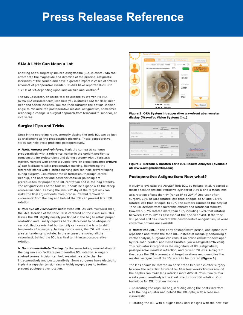
► Use intraoperative aberrometry. Intraoperative aberrometry,
with instruments such as Wavetec’s ORA system and other surgical
microscope mounted autorefractors, can give an on-demand
pseudophakic refraction (Figure 2) to further guide the toric lens
alignment. One study showed that using aberrometry to
intraoperatively measure and enhance the effects of limbal relaxing
incisions (LRIs) resulted in almost a sixfold reduction in the odds ratio
for subsequent excimer laser enhancement.6
Figure 2. ORA System intraoperative wavefront aberrometer
display (WaveTec Vision Systems Inc.).
Figure 3. Berdahl & Hardten Toric IOL Results Analyzer (available
at: www.astigmatismfix.com).
Postoperative Astigmatism: Now what?
A study to evaluate the AcrySof Toric IOL, by Holland et al, reported a
mean absolute residual refractive cylinder of 0.59 D and a mean lens
axis rotation of less than 4° (range 0° to 20°).2 One year after
surgery, 78% of IOLs rotated less than or equal to 5° and 93.4%
rotated less than or equal to 10°. The authors concluded the AcrySof
Toric IOL demonstrated favorable efficacy and rotational stability.
However, 6.7% rotated more than 10°, including 1.2% that rotated
between 15° to 20° as assessed at the one-year visit. If the toric
IOL patient still has unacceptable postoperative astigmatism, several
corrective options are available.
► Rotate the IOL. In the early postoperative period, one option is to
reposition and rotate the toric IOL. Instead of manually performing a
vector analysis, surgeons can consult an online calculator developed
by Drs. John Berdahl and David Hardten (www.astigmatismfix.com).
This calculator incorporates the magnitude of IOL astigmatism,
postoperative manifest refraction, and current IOL axis. A diagram
illustrates the IOL’s current and target locations and quantifies the
residual astigmatism if the IOL were to be rotated (Figure 3).
The lens should be rotated no earlier than two weeks after surgery
to allow the refraction to stabilize. After four weeks fibrosis around
the haptics can make lens rotation more difficult. Thus, two to four
weeks postoperatively is the ideal time for toric IOL rotation. Our
technique for IOL rotation involves:
• Re-inflating the capsular bag, including along the haptic interface
with the bag equator and behind the IOL optic, with a cohesive
viscoelastic.
• Rotating the IOL with a Kuglen hook until it aligns with the new axis

Press Release Reference1/29/13 www.ophthalmologymanagement.com/articleviewer.aspx?articleID=107873
5/6www.ophthalmologymanagement.com/articleviewer.aspx?articleID=107873
of orientation.
• Using bimanual I/A via small paracentesis incisions to remove the
residual viscoelastic.
• Reconfirming correct alignment of the toric IOL with axis markings at
the end of the case.
► Limbal relaxing incisions. LRIs are safe, easy and relatively
reliable for managing residual astigmatism, especially if the
postoperative spherical equivalent is close to plano. Several LRI
nomograms have been developed, primarily for techniques employing
either guarded or adjustable diamond blades. We currently utilize Dr.
Louis D. Nichamin’s nomogram,7 which adjusts arc length as a
function of refractive astigmatism and patient age with a fixed 600-
μm incision depth. The Accurate LRI Calculator (MicroMedical Devices)
is pachymetry software that can calculate the depth and incision arc
based on patient age, corneal rigidity and IOP. Use an adjustable
diamond blade with micrometer depth markings with this method.
AMO’s LRI calculator is a vector-analysis based online tool that allows
surgeons to enter keratometry or topography data and receive an
individualized diagram showing where to place the LRI incisions. A
growing number of studies have shown promising results with
femtosecond laser-assisted astigmatism correction and offer options
for further customization, including intrastromal keratotomy.8
► Refractive surgery. Excimer laser procedures can help neutralize
residual postoperative refractive cylinder and sphere, as well as
higher-order aberrations. We prefer waiting for the refraction to
stabilize after YAG capsulotomy before performing excimer
photoablation. This minimizes variables related to capsular fibrosis
and contraction.
We do not modify our LASIK nomogram for pseudophakes, but do
make an adjustment for patient age. Make sure to review the
centroid pattern for wavefront- guided ablations to rule out the
presence of any artifact fromthe IOL or capsulotomy edge. Tracking
can be affected if the post-cataract pupil is irregular or iris
transillumination defects are present. As with toric IOL procedures,
horizontal and vertical alignment marks are placed to rule out
significant cyclotorsion during the LASIK procedure.
► Glasses or contacts. With refractive cataract surgery, patients
have a goal of spectacle independence, so they can be resistant to
this last option. However, in a randomized multicenter trial of the
AcrySof toric IOL (n=256 eyes),2 almost 40% of the toric IOL group
had some degree of spectacle dependence six months after surgery.
In this regard, it is important that surgeons stress to patients during
preoperative counseling that they might need corrective eyewear
after surgery or need to undergo an additional surgical procedure.
Achieving Patient Satisfaction
Toric IOL implantation has shown itself to be an effective treatment
option in patients with corneal astigmatism, with good rotational
stability and expanding ranges of cylindrical power correction. Recent
and future advances in IOL design, wound and rhexis construction,
intraoperative aberrometry, and postoperative light adjustment of
the IOL optic offer further opportunities to reduce residual refractive
and astigmatic error. By consistently following recommended
guidelines regarding patient selection and intraoperative techniques,
the goal of satisfied patients is highly attainable.
Nevertheless, we must educate candidates for toric IOLs about the
possibility that they may need additional refractive procedures to
improve their uncorrected visual acuity, and that part-time spectacle
wear may augment their vision postoperatively. OM
References
1. Ahmed IK, Rocha G, Slomovic AR, et al. Visual function and the
patient experience after bilateral implantation of toric intraocular
1/29/13 www.ophthalmologymanagement.com/articleviewer.aspx?articleID=107873
5/6www.ophthalmologymanagement.com/articleviewer.aspx?articleID=107873
of orientation.
• Using bimanual I/A via small paracentesis incisions to remove the
residual viscoelastic.
• Reconfirming correct alignment of the toric IOL with axis markings at
the end of the case.
► Limbal relaxing incisions. LRIs are safe, easy and relatively
reliable for managing residual astigmatism, especially if the
postoperative spherical equivalent is close to plano. Several LRI
nomograms have been developed, primarily for techniques employing
either guarded or adjustable diamond blades. We currently utilize Dr.
Louis D. Nichamin’s nomogram,7 which adjusts arc length as a
function of refractive astigmatism and patient age with a fixed 600-
μm incision depth. The Accurate LRI Calculator (MicroMedical Devices)
is pachymetry software that can calculate the depth and incision arc
based on patient age, corneal rigidity and IOP. Use an adjustable
diamond blade with micrometer depth markings with this method.
AMO’s LRI calculator is a vector-analysis based online tool that allows
surgeons to enter keratometry or topography data and receive an
individualized diagram showing where to place the LRI incisions. A
growing number of studies have shown promising results with
femtosecond laser-assisted astigmatism correction and offer options
for further customization, including intrastromal keratotomy.8
► Refractive surgery. Excimer laser procedures can help neutralize
residual postoperative refractive cylinder and sphere, as well as
higher-order aberrations. We prefer waiting for the refraction to
stabilize after YAG capsulotomy before performing excimer
photoablation. This minimizes variables related to capsular fibrosis
and contraction.
We do not modify our LASIK nomogram for pseudophakes, but do
make an adjustment for patient age. Make sure to review the
centroid pattern for wavefront- guided ablations to rule out the
presence of any artifact fromthe IOL or capsulotomy edge. Tracking
can be affected if the post-cataract pupil is irregular or iris
transillumination defects are present. As with toric IOL procedures,
horizontal and vertical alignment marks are placed to rule out
significant cyclotorsion during the LASIK procedure.
► Glasses or contacts. With refractive cataract surgery, patients
have a goal of spectacle independence, so they can be resistant to
this last option. However, in a randomized multicenter trial of the
AcrySof toric IOL (n=256 eyes),2 almost 40% of the toric IOL group
had some degree of spectacle dependence six months after surgery.
In this regard, it is important that surgeons stress to patients during
preoperative counseling that they might need corrective eyewear
after surgery or need to undergo an additional surgical procedure.
Achieving Patient Satisfaction
Toric IOL implantation has shown itself to be an effective treatment
option in patients with corneal astigmatism, with good rotational
stability and expanding ranges of cylindrical power correction. Recent
and future advances in IOL design, wound and rhexis construction,
intraoperative aberrometry, and postoperative light adjustment of
the IOL optic offer further opportunities to reduce residual refractive
and astigmatic error. By consistently following recommended
guidelines regarding patient selection and intraoperative techniques,
the goal of satisfied patients is highly attainable.
Nevertheless, we must educate candidates for toric IOLs about the
possibility that they may need additional refractive procedures to
improve their uncorrected visual acuity, and that part-time spectacle
wear may augment their vision postoperatively. OM
References
1. Ahmed IK, Rocha G, Slomovic AR, et al. Visual function and the
patient experience after bilateral implantation of toric intraocular1/29/13 www.ophthalmologymanagement.com/articleviewer.aspx?articleID=107873
6/6www.ophthalmologymanagement.com/articleviewer.aspx?articleID=107873
0 1
lenses. J Cataract Refract Surg. 2010; 36:609-616.
2. Holland E, Lane S, Horn JD, Ernest P, Arless R, Miller KM. The
AcrySof toric intraocular lens in subjects with cataracts and corneal
astigmatism: a randomized, subjectmasked, parallel-group, 1-year
study. Ophthalmology. 2010; 117:2104-2111
3. IOLMaster: A Practical Operational Guide. Dublin, DA: Carl Zeiss
Meditec. Section. 4-14.
4. Nienke Visser, Noël J.C. Bauer and Rudy M.M.A. Nuijts (2012). Toric
Intraocular Lenses in Cataract Surgery. Goggin M. Astigmatism -
Optics, Physiology and Management. Rijeka, Croatia: InTech; 2012:
267-291.
5. Weikert MP, Koch DD, Wang L. Contribution of posterior corneal
astigmatism to the total corneal astigmatism. Paper presented at
ASCRS, Chicago, April 21, 2012.
6. Packer M. Effect of intraoperative aberrometry on the rate of
postoperative enhancement: retrospective study. J Cataract Refract
Surg. 2010; 36:747-755
7. Nichamin LD. Astigmatism control. Ophthalmol Clin N Am.
2006;19:485-493
8. Duna Raoof-Daneshvar and Shahzad I. Mian (2012). Femtosecond
Laser-Assisted Astigmatism Correction. Goggin M. Astigmatism -
Optics, Physiology and Management. Rijeka, Croatia: InTech; 2012:
193-209.
Mujtaba Qazi, MD is Director of Clinical Studies at Pepose
Vision Institute in St. Louis. He is a consultant at Bausch &
Lomb.
Jessica Schonfeld, MD is a Cornea and Refractive Surgery
Clinical Fellow at Pepose Vision Institute.
Jay Pepose, MD, PhD, heads the Pepose Vision Institute
in St. Louis.
, Volume: , Issue: , page(s):
Prev Page Table of Contents Archives
Now more than ever before, ophthalmologists are required to think as an MD and a CEO.The right balance of clinical and practice management skills is critical for a practice toflourish. Each month only one publication delivers the essential strategies needed tonavigate and grow today’s ophthalmology practice. Led by Chief Medical Editor LarryPatterson, MD, Ophthalmology Management provides all the tools ophthalmologists needto succeed, bringing them the latest practice management pearls, clinical advancementsand medical economics they need to help their practices grow.
Visit PentaVision's Other Publications
About Publication | This Month | New Ophthalmologist | Calendar of Events | Supplements
Archive | Magazine Subscription | Subscription to eNewsletter | Privacy Policy | Contact Us
Press Release Reference Studay Data
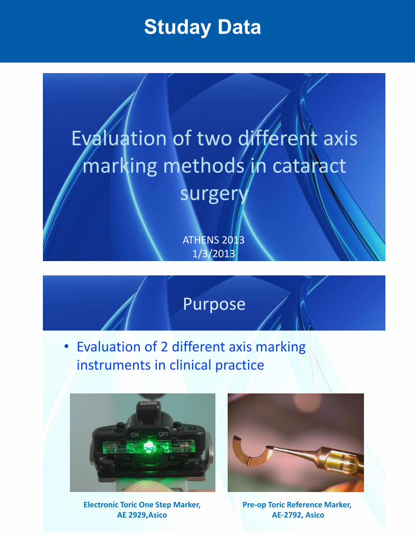
Evaluation of two different axis marking methods in cataract
surgery
ATHENS 20131/3/2013
Purpose
• Evaluation of 2 different axis marking instruments in clinical practice
Electronic Toric One Step Marker, AE 2929,Asico
Pre-op Toric Reference Marker,AE-2792, Asico
Studay Data
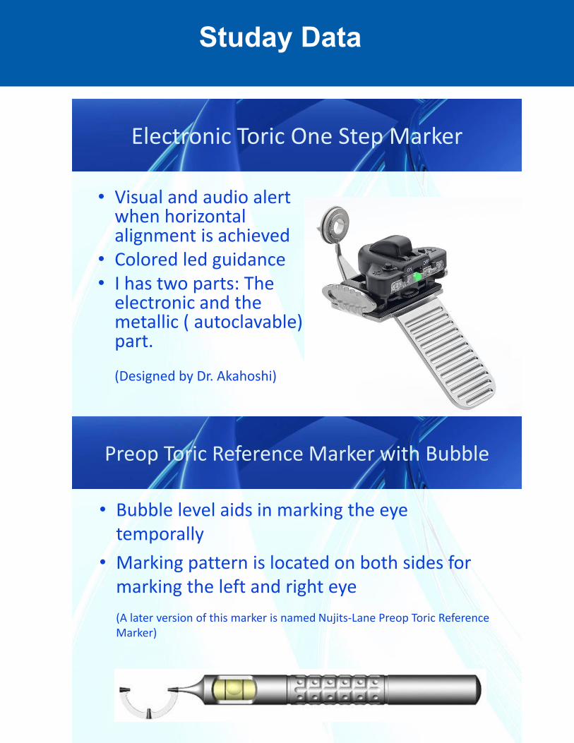
Electronic Toric One Step Marker
• Visual and audio alertwhen horizontal alignment is achieved
• Colored led guidance • I has two parts: The
electronic and the metallic ( autoclavable) part.
(Designed by Dr. Akahoshi)
Preop Toric Reference Marker with Bubble
• Bubble level aids in marking the eye temporally
• Marking pattern is located on both sides for marking the left and right eye(A later version of this marker is named Nujits-Lane Preop Toric Reference Marker)
Studay Data
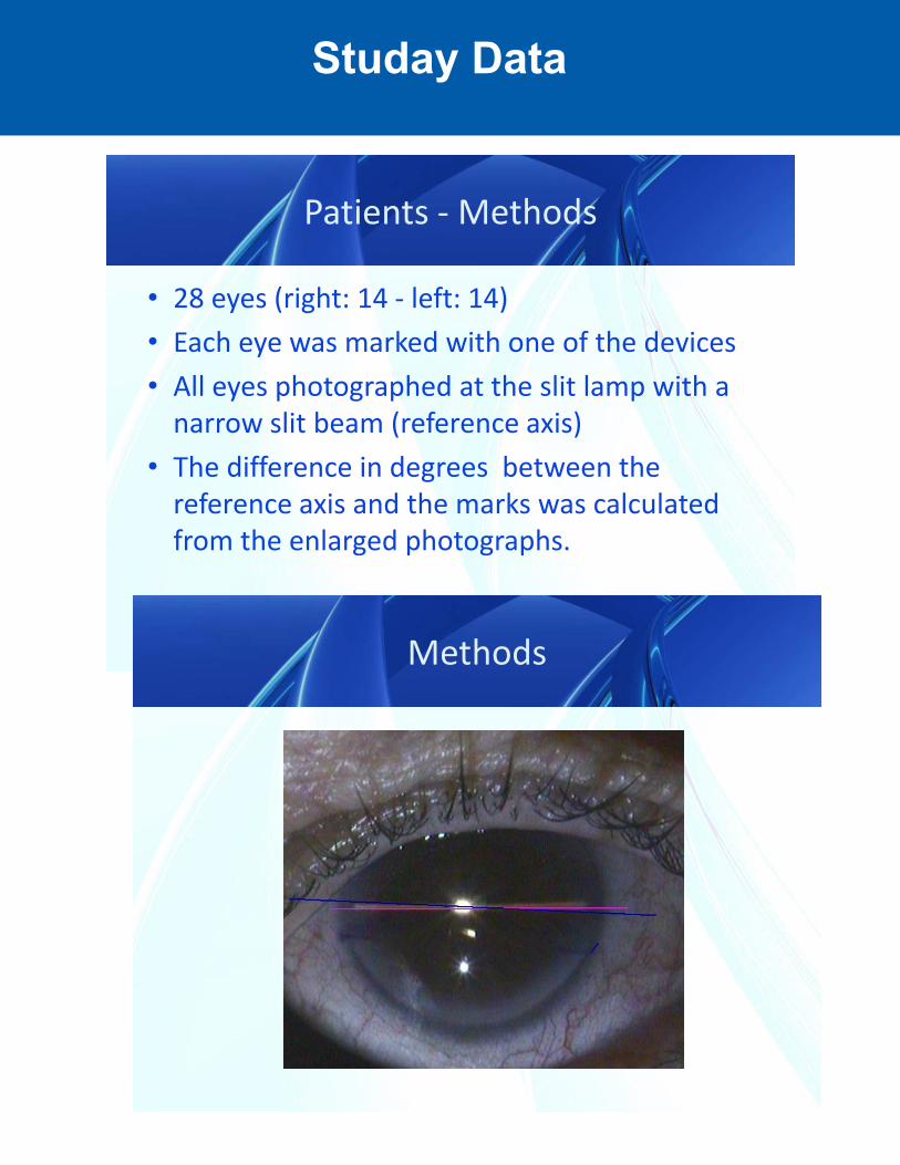
Patients - Methods
• 28 eyes (right: 14 - left: 14)• Each eye was marked with one of the devices• All eyes photographed at the slit lamp with a
narrow slit beam (reference axis)• The difference in degrees between the
reference axis and the marks was calculated from the enlarged photographs.
Methods
Studay Data
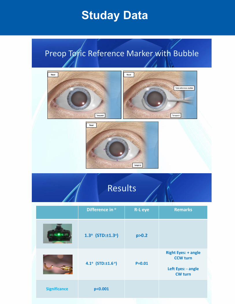
Preop Toric Reference Marker with Bubble
Results
Difference in o R-L eye Remarks
1.3o (STD:±1.3o) p>0.2
4.1o (STD:±1.6 o) P=0.01
Right Eyes: + angle CCW turn
Left Eyes: - angle CW turn
Significance p<0.001
Studay Data
Discussion
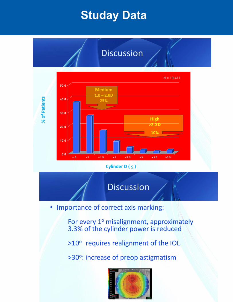
0.0
10.0
20.0
30.0
40.0
50.0
<.5 <1 <1.5 <2 <2.5 <3 <3.5 >3.5
Medium1.0 – 2.0D
25%
High >2.0 D
10%
Cylinder D ( < )
% o
f Pat
ient
s
N = 10,411
Discussion
• Importance of correct axis marking:
For every 1o misalignment, approximately 3.3% of the cylinder power is reduced
>10o requires realignment of the IOL
>30o: increase of preop astigmatism
Studay Data
Discussion
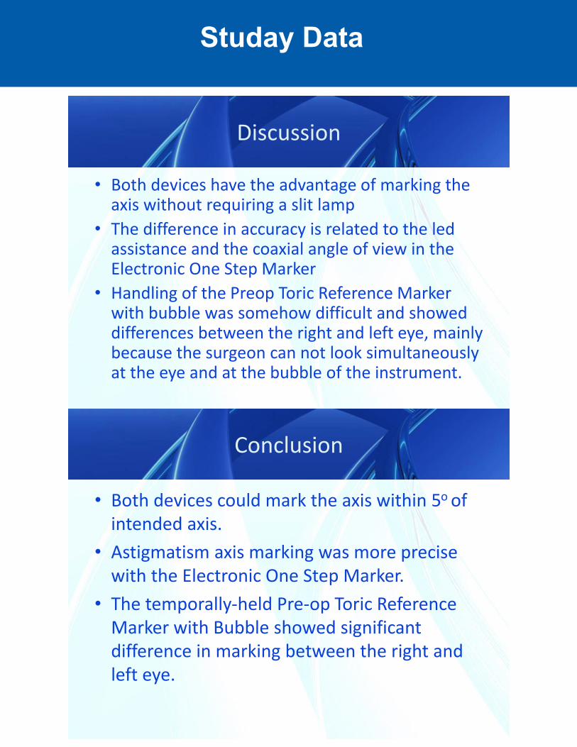
• Both devices have the advantage of marking the axis without requiring a slit lamp
• The difference in accuracy is related to the led assistance and the coaxial angle of view in the Electronic One Step Marker
• Handling of the Preop Toric Reference Marker with bubble was somehow difficult and showed differences between the right and left eye, mainly because the surgeon can not look simultaneously at the eye and at the bubble of the instrument.
Conclusion
• Both devices could mark the axis within 5o of intended axis.
• Astigmatism axis marking was more precise with the Electronic One Step Marker.
• The temporally-held Pre-op Toric Reference Marker with Bubble showed significant difference in marking between the right and left eye.