Embed Size (px)
Citation preview

193
Axenic cultivation and characterization of Leishmaniamexicana amastigote-like forms
P. A. BATES1, C. D. ROBERTSON1, L. TETLEY2 and G. H. COOMBS1
1 Laboratory for Biochemical Parasitology, Department of Zoology, University of Glasgow, Glasgow G12 8QQ2 Electron Microscopy Centre, Department of Zoology, University of Glasgow, Glasgow G12 8QQ
(Received I November 1991; revised 26 February 1992; accepted 2 March 1992)
SUMMARY
A new method is described which has made possible the long-term axenic cultivation of Leishmania mexicana amastigote-like forms in Schneider's Drosophila medium supplemented with 20 °0 (v/v) foetal calf serum. Unlike previous methods,it utilizes direct culture of parasites obtained from the lesions of infected animals rather than adaptation of promastigotesin vitro. Ultrastructural (possession of megasomes), biochemical (cysteine proteinase activity and gelatin SDS—PAGEbanding pattern) and infectivity (in vivo) data are presented which show the close similarity of the cultured forms to lesionamastigotes. The axenically cultured forms grew optimally at a temperature of 32-33 °C, providing further evidence fortheir amastigote nature. It was found that adjustment of the pH of the growth medium to 5-4 was required in order toretain the amastigote morphology of the cultured parasites. This supports the notion that leishmanial amastigotes areacidophiles.
Key words: Leishmania mexicana, axenic culture, amastigotes, megasomes, cysteine proteinase, infectivity.
INTRODUCTION
Leishmania parasites exist as amastigotes within thephagolysosomes of macrophages in their mammalianhosts (Alexander & Vickerman, 1975; Chang &Dwyer, 1976). The mechanisms which amastigotesemploy to survive in this apparently inhospitableenvironment are only partly understood, due in partto the difficulty of conducting biochemical studiesdirectly on parasites within their host cells. Isolationof amastigotes from infected animals (Childs,McRoberts & Foster, 1976; Brazil, 1978; Infanteet al. 1980; Hart, Vickerman & Coombs, 1981a;Saraiva et al. 1983; Meade et al. 1984; Glaser et al.1990) or from macrophage cultures infected in vitro(Chang & Dwyer, 1978; Berens & Marr, 1979;Berman, Dwyer & Wyler, 1979; Chang, 1980 a;Looker et al. 1986; Martinez, Looker & Marr, 1988)is often labour-intensive and/or provides insufficientmaterial for biochemical studies. Also such methodsleave doubts concerning the purity of the amastigotepreparation and the presence in it of adsorbed hostcomponents. The alternative, axenic in vitro cul-tivation of amastigote-like forms, has proved rela-tively difficult to achieve when compared withpromastigote culture, but has been reported forL. pifanoi, L. panamensis, L. braziliensis andL. donovani (Pan, 1984; Eperon & McMahon-Pratt,1989a; Doyle et al. 1991). These difficulties havedetermined that the biochemical characterization ofLeishmania has centred on cultured extracellularpromastigotes as a model system, and investigations
on amastigotes have lagged somewhat behind. Inthis paper we present a new method for the axeniccultivation of Leishmania mexicana in a form thatfrom morphological, biochemical and biological dataappears similar to lesion amastigotes. Evidence isalso presented that, compared with promastigotes,L. mexicana amastigotes require an elevated tem-perature and a relatively acidic environment forgrowth. The use of this system should facilitatestudies on the biology of L. mexicana amastigotes.
MATERIALS AND METHODS
In vitro culture
The following protocol was developed (see Resultssection) and used routinely for the axenic culture ofL. mexicana amastigotes. Infections of L. mexicana(MNYC/BZ/62/M379) were maintained in femaleCBA mice as previously described (Hart et al.1981 a). Mice with medium-sized unruptured lesions(10—15 mm diameter) were selected as a source ofamastigotes, so as to minimize the danger ofcontamination with unwanted organisms. Mice werekilled by terminal anaesthesia and rinsed with 70%(v/v) ethanol to sterilize the skin. The lesions wereexcised aseptically in a laminar flow hood and allsubsequent manipulations performed under sterileconditions. A single lesion was able to providesufficient amastigotes on each occasion that cultureswere established.
Parasitology (1992), 105, 193-202 Printed in Great Britain
P. A. Bates and others 194
The lesion was gently homogenized in 10 ml ofcomplete growth medium (see below) using a sterile10 ml syringe plunger to push the tissue through afine wire mesh in a Petri dish. The resulting crudehomogenate usually contained 108-109 freeamastigotes/ml and was centrifuged at 1000^ for5 min at ambient temperature (22 °C). The pelletcontaining large clumps of amastigotes, erythrocytesand other host cells was discarded, and the super-natant medium containing 107—108 amastigotes/mlwas retained and further centrifuged at 2000 g for5 min at 22 °C. The resulting supernatant mediumcontaining host cell debris was discarded and theamastigote pellet resuspended in 10 ml of freshgrowth medium. Cultures were initiated by sub-inoculation of the amastigote preparation into 10 mlof fresh growth medium, to a final density of5 x 10°/ml, in 25 cm2 tissue-culture flasks. Cultureswere maintained stationary in the sealed flasks,on their side at 32-33 °C with an initial gas phase ofair, and subpassaged every 5-6 days at the latelog/early stationary phase of growth (see Resultssection). This was achieved by passing culturedamastigotes through a sterile 26-gauge needle tobreak up any large clumps of cells, and initiating newcultures at 1—5 x 10° cells/ml as desired.
Growth medium consisted of Schneider's Droso-phila Medium (Gibco Ltd, Paisley, Scotland) supple-mented with 20 °0 (v/v) foetal calf serum (Gibco)and 25 fig gentamicin sulphate/ml. A variety of pHconditions were tested initially (see Results section),and these experiments established that for routineculturing the optimal initial pH was 54 + 0 1 ,obtained using 1 M HC1.
Promastigotes were obtained by transformation oflesion amastigotes and cultured in HOMEM supple-mented with 10 °0 foetal calf serum and 25 figgentamicin sulphate/ml as previously described(Hart, Vickerman & Coombs, 19816; Mallinson &Coombs, 1989).
Cell counting was performed using ImprovedNeubauer haemocytometers under phase-contrastmicroscopy. Amastigotes isolated from lesions andthose cultured in vitro had a tendency to clumptogether. Such clumps were disrupted for thepurposes of counting by passing the cells 3 timesthrough a 26-gauge needle.
Ultrastructure
Transmission electron microscopy of axenic amasti-gotes was performed as previously described forlesion amastigotes (Coombs et al. 1986).
Proteinases
Enzyme assays and gelatin SDS-PAGE were per-formed as previously described (Robertson &Coombs, 1990).
Infectivity
The infectivity of lesion amastigotes, axenic amasti-gotes and promastigotes was determined essentiallyas described by Alexander (1988). Lesion amasti-gotes were used immediately after isolation. Axenicamastigotes were taken from day 5-6 cultures andpromastigotes from day 7 cultures, i.e. at stationaryphase when the numbers of metacyclic promastigotesare maximal (Mallinson & Coombs, 1986, 1989). Toobviate any possible complicating effects of thelength of in vitro culture on the infectivity ofparasites, matched cultures of amastigotes andpromastigotes which had been cultured in vitro foridentical periods and within 7 subpassages from aninfected animal were used to infect mice. For eachform, 5 x 10⁶ parasites were inoculated subcu-taneously into the shaven rump of each 5- to 6-week-old female CBA mouse used. After 4 weeks andthereafter at 2-weekly intervals until 20 weeks, micewere shaved, examined and the diameter of lesionsdetermined using a Mitutoyo micrometer. Infec-tivity was calculated as the mean lesion diameter,including all animals in a group and treating thosewithout lesions as possessing a lesion of 0-0 diameter.We do not assume that the data are normallydistributed, and therefore a non-parametric test, theWilcoxon—Mann—Whitney, was used to determinethe statistical significance of any observed differencesin lesion diameter between groups of mice.
RESULTS
In vitro culture
Initial experiments were conducted to optimizeconditions for axenic cultivation of amastigotes.Three factors were varied: medium composition,temperature and pH. As a result of these experiments,described fully below, a standard growth mediumcomprising Schneider's Drosophila medium supple-mented with 20 °0 (v/v) foetal calf serum and with apH of 5-4 was developed. Establishment of culturesused a partially purified preparation of amastigotes(see Materials and Methods section), which con-tained low numbers of erythrocytes and other hostcells. However, their presence did not affect theviability of the amastigotes as assessed by theirability to grow in vitro. Such contaminants weredegraded and undetectable within 24 h of culture.
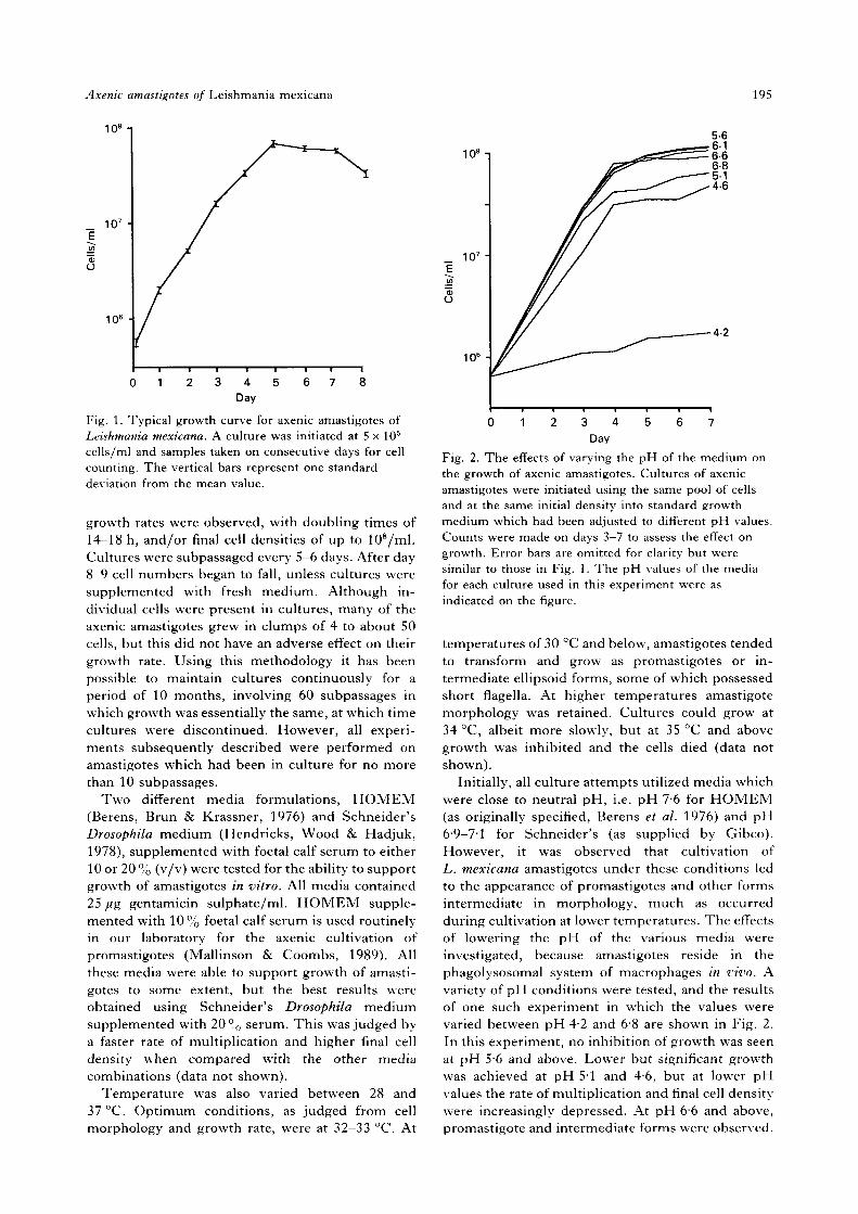
A typical growth curve for axenic amastigotes ofL. mexicana cultured in standard growth medium isshown in Fig. 1. In common with other micro-organisms cultured in vitro, a logarithmic growthphase was observed, followed by a period of slowergrowth, a stationary phase and eventually cell deathleading to a decrease in density. Typically, no lagphase was observed. Generally, doubling timesvaried between 18 and 22 h and the final cell densitywas in the range 4 to 6 x 10⁷/ml. Occasionally, faster
Axenic amastigotes of Leishmania mexicana
108
195
107
O
106
0 1 2 3 4 5 6 7 8Day
Fig. 1. Typical growth curve for axenic amastigotes ofLeishmania mexicana. A culture was initiated at 5 x 10⁵
cells/ml and samples taken on consecutive days for cellcounting. The vertical bars represent one standarddeviation from the mean value.
growth rates were observed, with doubling times of14-18 h, and/or final cell densities of up to 108/ml.Cultures were subpassaged every 5-6 days. After day8-9 cell numbers began to fall, unless cultures weresupplemented with fresh medium. Although in-dividual cells were present in cultures, many of theaxenic amastigotes grew in clumps of 4 to about 50cells, but this did not have an adverse effect on theirgrowth rate. Using this methodology it has beenpossible to maintain cultures continuously for aperiod of 10 months, involving 60 subpassages inwhich growth was essentially the same, at which timecultures were discontinued. However, all experi-ments subsequently described were performed onamastigotes which had been in culture for no morethan 10 subpassages.
Two different media formulations, HOMEM(Berens, Brun & Krassner, 1976) and Schneider'sDrosophila medium (Hendricks, Wood & Hadjuk,1978), supplemented with foetal calf serum to either10 or 20 % (v/v) were tested for the ability to supportgrowth of amastigotes in vitro. All media contained25 fig gentamicin sulphate/ml. HOMEM supple-mented with 10% foetal calf serum is used routinelyin our laboratory for the axenic cultivation ofpromastigotes (Mallinson & Coombs, 1989). Allthese media were able to support growth of amasti-gotes to some extent, but the best results wereobtained using Schneider's Drosophila mediumsupplemented with 20 °o serum. This was judged bya faster rate of multiplication and higher final celldensity when compared with the other mediacombinations (data not shown).
Temperature was also varied between 28 and37 °C. Optimum conditions, as judged from cellmorphology and growth rate, were at 32-33 °C. At
10 s - i
107
CD
o
106
0 1 I 3 4 5 6 7Day
Fig. 2. The effects of varying the pH of the medium onthe growth of axenic amastigotes. Cultures of axenicamastigotes were initiated using the same pool of cellsand at the same initial density into standard growthmedium which had been adjusted to different pH values.Counts were made on days 3—7 to assess the effect ongrowth. Error bars are omitted for clarity but weresimilar to those in Fig. 1. The pH values of the mediafor each culture used in this experiment were asindicated on the figure.
temperatures of 30 °C and below, amastigotes tendedto transform and grow as promastigotes or in-termediate ellipsoid forms, some of which possessedshort flagella. At higher temperatures amastigotemorphology was retained. Cultures could grow at34 °C, albeit more slowly, but at 35 °C and abovegrowth was inhibited and the cells died (data notshown).
Initially, all culture attempts utilized media whichwere close to neutral pH, i.e. pH 76 for HOMEM(as originally specified, Berens et al. 1976) and pH6-9—7'1 for Schneider's (as supplied by Gibco).However, it was observed that cultivation ofL. mexicana amastigotes under these conditions ledto the appearance of promastigotes and other formsintermediate in morphology, much as occurredduring cultivation at lower temperatures. The effectsof lowering the pH of the various media wereinvestigated, because amastigotes reside in thephagolysosomal system of macrophages in vivo. Avariety of pH conditions were tested, and the resultsof one such experiment in which the values werevaried between pH 42 and 68 are shown in Fig. 2.In this experiment, no inhibition of growth was seenat pH 5-6 and above. Lower but significant growthwas achieved at pH 51 and 4-6, but at lower pHvalues the rate of multiplication and final cell densitywere increasingly depressed. At pH 66 and above,promastigote and intermediate forms were observed.
P. A. Bates and others 196
; £^PI ^
• r >.?~'"*!,
" • ' * • . » ' -i- r *
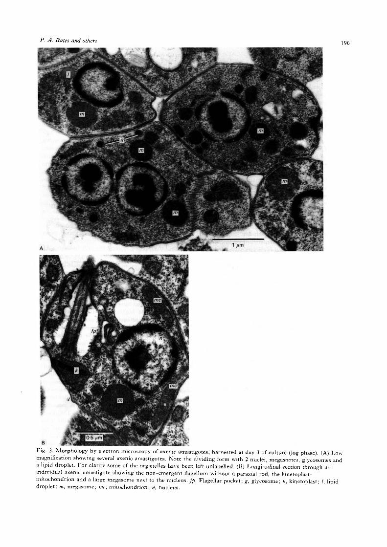
Fig. 3. Morphology by electron microscopy of axenic amastigotes, harvested at day 3 of culture (log phase). (A) Lowmagnification showing several axenic amastigotes. Note the dividing form with 2 nuclei, megasomes, glycosomes anda lipid droplet. For clarity some of the organelles have been left unlabelled. (B) Longitudinal section through anindividual axenic amastigote showing the non-emergent flagellum without a paraxial rod, the kinetoplast-mitochondrion and a large megasome next to the nucleus./£, Flagellar pocket; g, glycosome; k, kinetoplast; /, lipiddroplet; m, megasome; me, mitochondrion; n, nucleus.
Axenic amastigotes of Leishmania mexicana 197
M,(kDa)
58
36
24
20
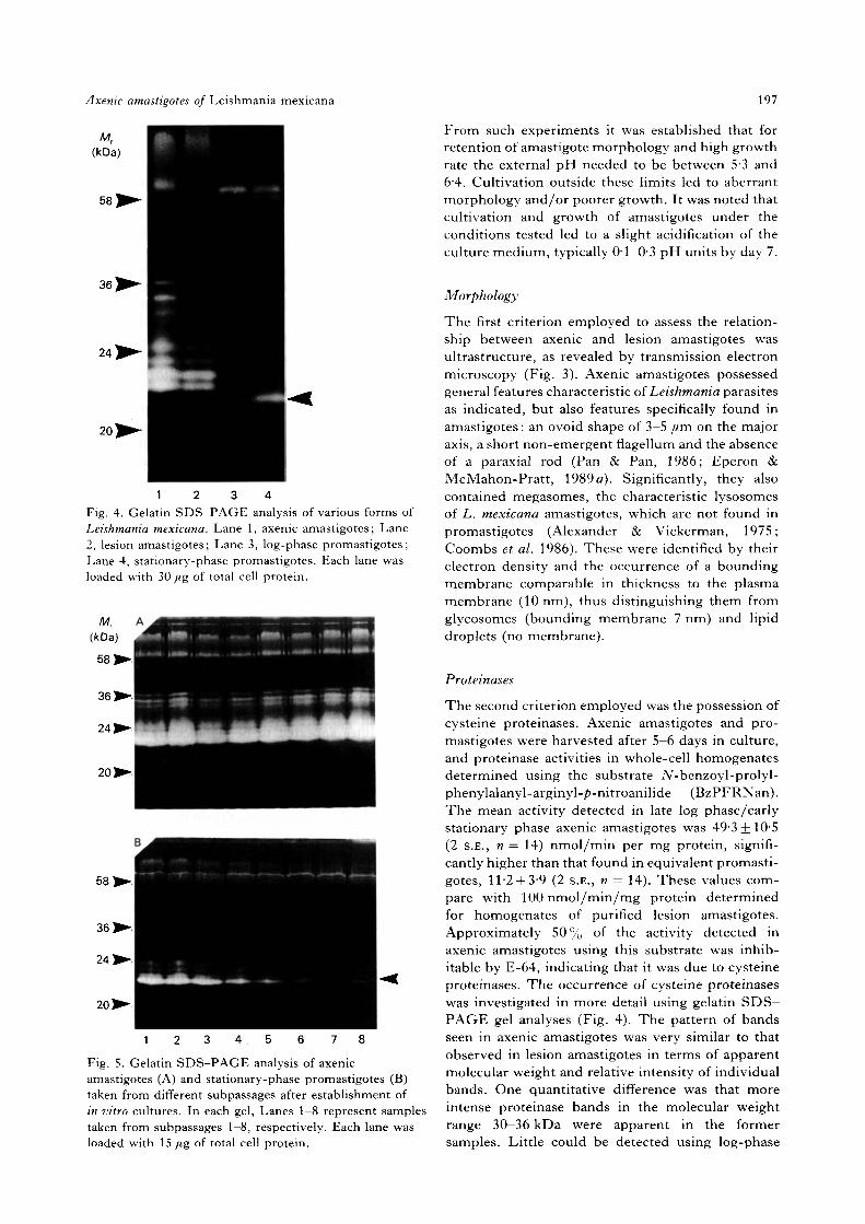
1 2 3 4Fig. 4. Gelatin SDS-PAGE analysis of various forms ofLeishmania mexicana. Lane 1, axenic amastigotes; Lane2, lesion amastigotes; Lane 3, log-phase promastigotes;Lane 4, stationary-phase promastigotes. Each lane wasloaded with 30 fig of total cell protein.
24
20
58;
36]
24]
20 3
Fig. 5. Gelatin SDS-PAGE analysis of axenicamastigotes (A) and stationary-phase promastigotes (B)taken from different subpassages after establishment ofin vitro cultures. In each gel, Lanes 1—8 represent samplestaken from subpassages 1—8, respectively. Each lane wasloaded with 15 fig of total cell protein.
From such experiments it was established that forretention of amastigote morphology and high growthrate the external pH needed to be between 53 and6-4. Cultivation outside these limits led to aberrantmorphology and/or poorer growth. It was noted thatcultivation and growth of amastigotes under theconditions tested led to a slight acidification of theculture medium, typically 0-1-0-3 pH units by day 7.
Morphology
The first criterion employed to assess the relation-ship between axenic and lesion amastigotes wasultrastructure, as revealed by transmission electronmicroscopy (Fig. 3). Axenic amastigotes possessedgeneral features characteristic of Leishmania parasitesas indicated, but also features specifically found inamastigotes: an ovoid shape of 3-5 fim on the majoraxis, a short non-emergent flagellum and the absenceof a paraxial rod (Pan & Pan, 1986; Eperon &McMahon-Pratt, 1989 a). Significantly, they alsocontained megasomes, the characteristic lysosomesof L. mexicana amastigotes, which are not found inpromastigotes (Alexander & Vickerman, 1975;Coombs et al. 1986). These were identified by theirelectron density and the occurrence of a boundingmembrane comparable in thickness to the plasmamembrane (10 nm), thus distinguishing them fromglycosomes (bounding membrane 7 nm) and lipiddroplets (no membrane).
Proteinases
The second criterion employed was the possession ofcysteine proteinases. Axenic amastigotes and pro-mastigotes were harvested after 5-6 days in culture,and proteinase activities in whole-cell homogenatesdetermined using the substrate iV-benzoyl-prolyl-phenylalanyl-arginyl-/>-nitroanilide (BzPFRNan).The mean activity detected in late log phase/earlystationary phase axenic amastigotes was 49-3 + 10-5(2 S.E., n = 14) nmol/min per mg protein, signifi-cantly higher than that found in equivalent promasti-gotes, 11-2 + 3-9 (2 S.E., n = 14). These values com-pare with 100 nmol/min/mg protein determinedfor homogenates of purified lesion amastigotes.Approximately 50 % of the activity detected inaxenic amastigotes using this substrate was inhib-itable by E-64, indicating that it was due to cysteineproteinases. The occurrence of cysteine proteinaseswas investigated in more detail using gelatin SDS—PAGE gel analyses (Fig. 4). The pattern of bandsseen in axenic amastigotes was very similar to thatobserved in lesion amastigotes in terms of apparentmolecular weight and relative intensity of individualbands. One quantitative difference was that moreintense proteinase bands in the molecular weightrange 30—36 kDa were apparent in the formersamples. Little could be detected using log-phase
P. A. Bates and others 198
M,(kDa)
58
36,
24
Fig. 6. Gelatin SDS-PAGE analysis of proteinasesduring the growth cycle of Leishmania mexicana axenicamastigotes. A culture was initiated at 5x10° cells/mlwith axenic amastigotes (Lane 1) and samples removedon consecutive days of in vitro culture (Lanes 2—9). Eachlane was loaded with IS fig of total cell protein.
12-,
08 10 12
WeeksFig. 7. A comparison of the infectivity of lesionamastigotes (A—A), axenic amastigotes ( • — • ) andstationary-phase promastigotes (O—O) in groups of 24,30 and 30 female CBA mice, respectively. The barsrepresent two standard errors from the mean lesiondiameter.
promastigotes, but enzymes were detected insamples of stationary-phase promastigotes. A lowermolecular weight cysteine proteinase characteristi-cally found in the latter (arrowed, Fig. 4) wasapparently absent from either form of amastigote.
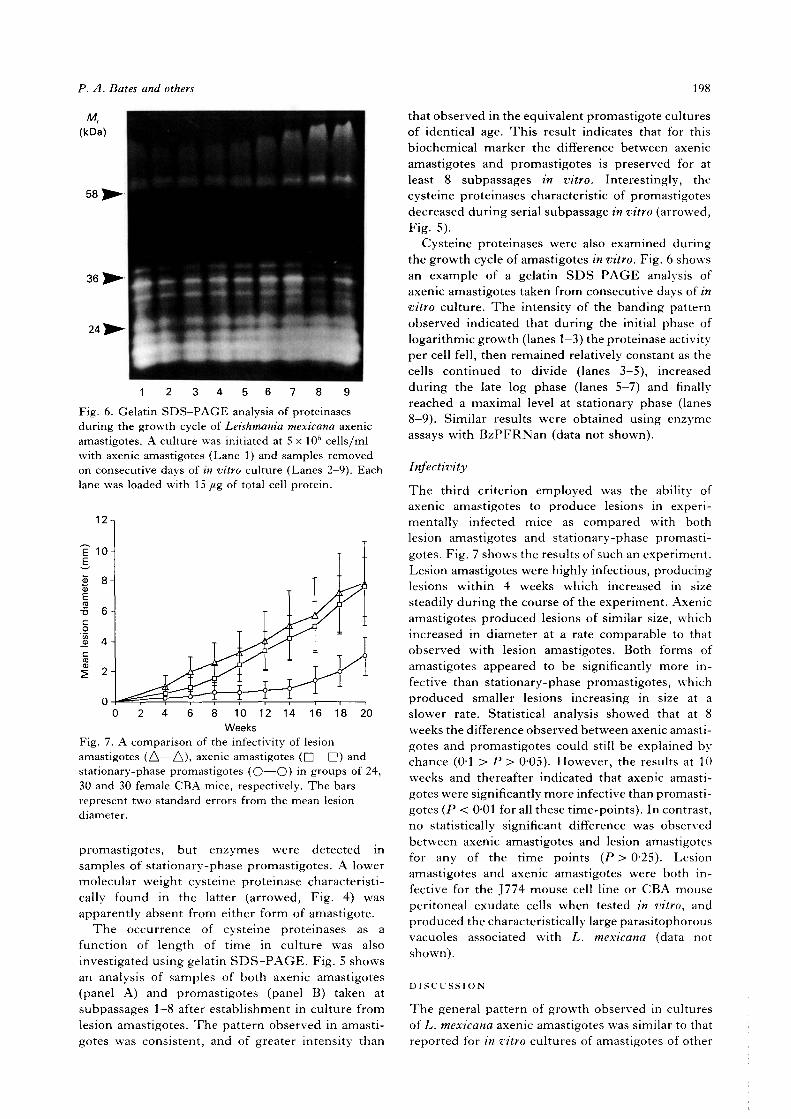
The occurrence of cysteine proteinases as afunction of length of time in culture was alsoinvestigated using gelatin SDS—PAGE. Fig. 5 showsan analysis of samples of both axenic amastigotes(panel A) and promastigotes (panel B) taken atsubpassages 1-8 after establishment in culture fromlesion amastigotes. The pattern observed in amasti-gotes was consistent, and of greater intensity than
that observed in the equivalent promastigote culturesof identical age. This result indicates that for thisbiochemical marker the difference between axenicamastigotes and promastigotes is preserved for atleast 8 subpassages in vitro. Interestingly, thecysteine proteinases characteristic of promastigotesdecreased during serial subpassage in vitro (arrowed,Fig. 5).
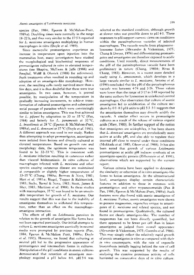
Cysteine proteinases were also examined duringthe growth cycle of amastigotes in vitro. Fig. 6 showsan example of a gelatin SDS—PAGE analysis ofaxenic amastigotes taken from consecutive days of invitro culture. The intensity of the banding patternobserved indicated that during the initial phase oflogarithmic growth (lanes 1-3) the proteinase activityper cell fell, then remained relatively constant as thecells continued to divide (lanes 3-5), increasedduring the late log phase (lanes 5-7) and finallyreached a maximal level at stationary phase (lanes8-9). Similar results were obtained using enzymeassays with BzPFRNan (data not shown).
Infectivity
The third criterion employed was the ability ofaxenic amastigotes to produce lesions in experi-mentally infected mice as compared with bothlesion amastigotes and stationary-phase promasti-gotes. Fig. 7 shows the results of such an experiment.Lesion amastigotes were highly infectious, producinglesions within 4 weeks which increased in sizesteadily during the course of the experiment. Axenicamastigotes produced lesions of similar size, whichincreased in diameter at a rate comparable to thatobserved with lesion amastigotes. Both forms ofamastigotes appeared to be significantly more in-fective than stationary-phase promastigotes, whichproduced smaller lesions increasing in size at aslower rate. Statistical analysis showed that at 8weeks the difference observed between axenic amasti-gotes and promastigotes could still be explained bychance (0-1 > P > 0-05). However, the results at 10weeks and thereafter indicated that axenic amasti-gotes were significantly more infective than promasti-gotes (P < 0-01 for all these time-points). In contrast,no statistically significant difference was observedbetween axenic amastigotes and lesion amastigotesfor any of the time points (P > 025). Lesionamastigotes and axenic amastigotes were both in-fective for the J774 mouse cell line or CBA mouseperitoneal exudate cells when tested in vitro, andproduced the characteristically large parasitophorousvacuoles associated with L. mexicana (data notshown).
DISCUSSION
The general pattern of growth observed in culturesof L. mexicana axenic amastigotes was similar to thatreported for in vitro cultures of amastigotes of other
Axenic amastigotes of Leishmania mexicana
species (Pan, 1984; Eperon & McMahon-Pratt,1989a). Doubling times were normally in the range18-22 h, and thus very similar to the 17-5 h reportedfor L. mexicana amastigotes multiplying in humanmacrophages in vitro (Doyle et al. 1989).
Since metacyclic promastigotes experience anincrease in temperature upon injection into themammalian host, many investigators have studiedthe morphological and biochemical responses ofpromastigotes cultured in vitro to elevated temper-atures (see Shapira, McEwen & Jaffe (1988) andSmejkal, Wolff & Olenick (1988) for references).Such treatments often resulted in rounding up andadoption of an amastigote-like morphology. How-ever, the resulting cells rarely survived more than afew days, and it is thus doubtful that these were trueamastigotes. In two cases, however, it provedpossible, by manipulation of the temperature ingradually increasing increments, to achieve trans-formation of cultured promastigotes and subsequentserial passage of parasites which were convincinglysimilar to lesion amastigotes. This was first achievedfor L. pifanoi by adaptation to 33 or 35 °C (Pan,1984) and latterly for L. panamensis at 32 °C,L. braziliensis at 28 °C (Eperon & McMahon-Pratt,1989a), and L. donovani at 37 °C (Doyle et al. 1991).A different approach was used in our study. Ratherthan attempting to adapt promastigotes, amastigoteswere isolated from lesions and directly cultured atelevated temperatures. Based on growth rate andmorphology data, the optimum temperature wasfound to be 32-33 °C. This is consistent withL. mexicana infection resulting in cutaneous ratherthan visceral leishmaniasis. In vitro cultures ofmacrophages infected with L. mexicana and otherspecies causing cutaneous disease are maintainedat comparable or slightly higher temperatures of33-35 °C (Chang, 1980a; Berman & Neva, 1981;Hart et al. 1981a; Biegel, Topper & Rabinovitch,1983; Sacks, Barral & Neva, 1983; Scott, James &Sher, 1985; Martinez et al. 1988). In these studieswith macrophages, 37 °C was found to be an unsuit-able temperature for growth of L. mexicana. Ourresults suggest that this was due to the inability ofamastigotes themselves to withstand this tempera-ture, rather than an effect on the macrophage-amastigote interaction.
The effects of pH on Leishmania parasites inrelation to the growth of amastigote-like forms havenot been reported previously. Our initial attempts toculture L. mexicana amastigotes axenically in neutralmedia were prompted by previous reports (Pan,1984; Eperon & McMahon Pratt, 1989 a; Doyleet al. 1991). However, we found that culturing atneutral pH led to the progressive appearance ofpromastigotes and intermediate forms in cultures.Manipulation of the pH conditions in axenic culturesdemonstrated that retention of amastigote mor-phology required a pH below 6-4. pH 5-4 was
199
selected as the standard condition, although growthat slower rates was possible down to pH 4-5. Theseresponses to pH support current views on conditionswithin the parasitophorous vacuoles of infectedmacrophages. The vacuole results from phagosome—lysosome fusion (Alexander & Vickerman, 1975;Chang & Dwyer, 1976) and differentiating promasti-gotes and amastigotes are therefore exposed to acidicconditions. Until recently, direct measurements ofthe pH of the parasitophorous vacuole have beenpreliminary in nature (Chang, 19806; Rivas &Chang, 1983). However, in a recent more detailedstudy using L. amazonensis, which develops in alarge vacuole similar to L. mexicana, Antoine et al.(1990) concluded that the pH of the parasitophorousvacuole was between 4-74 and 526. These valueswere lower than the range of 5-17 to 548 reported bythe same authors for lysosomes in uninfected controlmacrophages. Our observation that culture of axenicamastigotes led to acidification of the culture me-dium by 0-1-0-3 pH units to pH 5-3-5-1 suggests thatthe amastigotes themselves may directly acidify thevacuole. A similar effect occurs in promastigotecultures as a result of the release of various organic
acids (Marr, 1980). In further support of the notionthat amastigotes are acidophiles, it has been shownthat L. donovani amastigotes are metabolically moreactive at acidic pH, and also able to regulate theirinternal pH close to neutral under these conditions(Mukkada et al. 1985 ; Glaser et al. 1988). It has alsobeen noted that growth of various Leishmaniapromastigotes at acidic pH induces synthesis of anamastigote-specific protein (Zilberstein et al. 1991),observations which are supported by the currentreport.
Various criteria have been applied to determinethe similarity or otherwise of in vitro amastigote-likeforms to lesion amastigotes. At the ultrastructurallevel, amastigotes display certain characteristicfeatures in addition to those in common withpromastigotes and other trypanosomatids (Pan &Pan, 1986; Eperon & McMahon-Pratt, 1989 a). Suchfeatures were also found with axenic amastigotes ofL. mexicana. Futher, axenic amastigotes were shownto possess megasomes, organelles unique to amasti-gotes of L. mexicana and related species, and notfound in promastigotes. Qualitatively, the culturedforms are clearly amastigote-like. The number ofmegasomes has not been directly quantified, butthere appeared to be fewer per cell than in lesionamastigotes as judged from overall appearance(Alexander & Vickerman, 1975 ; Coombs et al. 1986).This may simply reflect the relatively rapid growthof axenic amastigotes in vitro as compared with theirin vivo counterparts, with the rate of organellebiosynthesis initially lagging behind the rate of cellgrowth. This was supported by the results ofanalysing the cysteine proteinase activity of cellsharvested on consecutive davs of in vitro culture.

P. A. Bates and others 200
These enzymes probably reside in the megasomes(Pupkis, Tetley & Coombs, 1986), and it was notedthat, after falling slightly during the early log phase,the banding pattern became more intense instationary-phase cells. Therefore, in this respectstationary-phase axenic amastigotes most closelyresembled the lesion amastigotes.
Various biochemical criteria have been used tocompare axenically cultured and lesion amastigotesof L. pifanoi (Pan, McMahon-Pratt & Honigberg,1984; Pan, 1986; Pan & McMahon-Pratt, 1988;Rainey et al. 1991), L. panamensis and L. braziliensis(Eperon & McMahon-Pratt, 19896). In our study weexamined L. mexicana axenic amastigotes forcysteine proteinases. Proteinases in general, andthese enzymes in particular, were found at thehighest specific activity in lesion amastigotes, atsomewhat lower levels in stationary-phase promasti-gotes and at very low levels in multiplicativepromastigotes (North & Coombs, 1981; Coombs,1982; Pupkis & Coombs, 1984; Pupkis et al. 1986;Lockwood et al. 1987). Using BzPFRNan, lesionamastigotes were found to possess approximately 10times and axenic amastigotes 5 times the level ofproteinase activity of stationary-phase promasti-gotes. This result is similar to those of previouscomparisons of lesion amastigotes and promastigotes(Coombs, 1982; Pupkis & Coombs, 1984; Pupkiset al. 1986). It also suggests that proteinase activity inaxenic amastigotes is slightly lower than in lesionamastigotes, in agreement with the apparently lowernumbers of megasomes (see above).
A more detailed analysis of cysteine proteinaseswas performed using gelatin SDS—PAGE, where aprominent group of bands in the apparent molecular-weight range 20-25 kDa have been demonstrated inL. mexicana (Lockwood et al. 1987; Robertson &Coombs, 1990; Coombs, Robertson & Mottram,1991). The pattern observed in lesion amastigotesand axenic amastigotes was qualitatively identical inthis region. However, there appeared to be moreintense banding in the molecular weight range30—36 kDa in samples from axenic amastigotes.Whether these have any relationship to the lowermolecular weight enzymes, precursors for example,is unknown. Differences in the apparent molecularweights of cysteine proteinases in promastigotes andamastigotes have been noted before (North &Coombs, 1981; Coombs et al. 1991) and here wedemonstrated that a low molecular weight enzymefound characteristically in stationary-phase promasti-gotes was undetectable in both axenic and lesionamastigotes.
The third criterion, and a key biological feature ofaxenic amastigotes, is that they should show in-fectivity in vivo similar to that of lesion amastigotes,which have been reported to be generally moreinfective than cultured promastigotes. This criterionhas been partly met for L. pifanoi axenic amastigote-
like forms, which were compared with amastigotesderived from in vitro-infected J774 macrophagecultures (Pan & Honigberg, 1985). These two formswere shown to be equally infective to the J774 mousecell line or to hamsters via footpad inoculation. Inthe current study we applied a more stringent test,comparing axenic amastigotes with those isolatedfrom lesions of infected animals for their ability tocause rump lesions in naive mice. The resultsindicated that L. mexicana axenic amastigotes weresimilar in infectivity to those isolated directly frominfected animals, and both of these were significantlymore infective than promastigotes.
In summary, we have reported for the first time asuccessful method for the direct axenic cultivation ofleishmanial amastigotes. For each of the criteria wehave employed, the data presented demonstrate thatthe cultured forms are indeed amastigote-like andnot rounded aflagellate promastigotes. Not surpris-ingly, some quantitative differences were observed incomparison with lesion amastigotes, and these arethe subject of further investigation. Similar dif-ferences have also been observed with other species(Rainey et al. 1991). Such differences may simplyarise from different growth rates, but could indicatethat some further refinement of culture conditionswould be beneficial. It should be borne in mind,however, that amastigotes in animals may themselvesnot all be identical, varying for instance with theirlocation in the lesion. Nevertheless, as long as thecaveats of any in vitro system are borne in mind, themethod described should provide an excellent modelenabling further biochemical studies of L. mexicanaamastigotes including certain types of work whichare otherwise impossible to conduct.
This work was supported by the Wellcome Trust (P. A.B.,G.H.C., C.D.R.), the E.C. (G.H.C., C.D.R.) and theCaledonian Research Foundation/Royal Society of Edin-burgh (P.A.B.).
REFERENCES
ALEXANDER, J. (1988). Sex differences and cross-immunity in DBA/2 mice infected with heishmaniamexicana and h. major. Parasitology 96, 297—302.
ALEXANDER, J. & VICKERMAN, K. (1975). Fusion of hostcell secondary lysosomes with the parasitophorousvacuoles of heishmania mexicana-infectedmacrophages. Journal of Protozoology 22, 502-8.
ANTOINE, J . - C , PRINA, E., JOUANNE, C. & BONGRAND, P.
(1990). Parasitophorous vacuoles of heishmaniaamazonensis-infected macrophages maintain an acidicpH. Infection and Immunity 58, 779—87.
BERENS, R. L. & MARR, J. J. (1979). Growth of heishmaniadonovani amastigotes in a continuous macrophage-likecell culture. Journal of Protozoology 26, 453-6.
BERENS, R. L., BRUN, R. & KRASSNER, S. M. ( 1 9 7 6 ) . A
simple monophasic medium for axenic culture ofhemoflagellates. Journal of Parasitology 62, 360-5.
Axenic amastigotes of Leishmania mexicana 201
BERMAN, J. D., DWYER, D. M. & WYLER, D. J. ( 1 9 7 9 ) .
Multiplication of Leishmania in human macrophagesin vitro. Infection and Immunity 26, 375-9.
BERMAN, J. D. & NEVA, F. A. (1981). Effects of temperatureon multiplication of Leishmania amastigotes withinhuman monocyte-derived macrophages in vitro.American Journal of Tropical Medicine and Hygiene30, 318-21.
BIEGEL, D., TOPPER, G. & RABINOVITCH, M. ( 1 9 8 3 ) .
Leishmania mexicana: temperature sensitivity ofisolated amastigotes and of amastigotes infectingmacrophages in culture. Experimental Parasitology 56,289-97.
BRAZIL, R. P. (1978). Isolation of the intracellular stagesof Leishmania mexicana amazonensis using cellulosecolumn. Annals of Tropical Medicine and Parasitology72, 579-80.
CHANG, K.-P. (1980a). Human cutaneous leishmania in amouse macrophage cell line: propagation and isolationof intracellular parasites. Science 209, 1240-2.
CHANG, K.-P. (19806). Endocytosis of Leishmania-infected macrophages. Fluorometry of pinocytic rate,lysosome-phagosome fusion and intralysosomal pH.In The Host Invader Interplay (ed. H. Van DenBossche), pp. 231-4. Amsterdam: Elsevier.
CHANG, K.-P. & DWYER, D. M. (1976). Multiplication of ahuman parasite (Leishmania donovani) inphagolysosomes of hamster macrophages in vitro.Science 193, 678-80.
CHANG, K.-P. & DWYER, D. M. (1978). Leishmania
donovani. Hamster macrophage interactions in vitro:cell entry, intracellular survival, and multiplication ofamastigotes. Journal of Experimental Medicine 147,515-30.
CHILDS, G. E., MCROBERTS, M. J. & FOSTER, K. A. ( 1 9 7 6 ) .
Partial purification of amastigotes from cutaneouslesions of American leishmaniasis. Journal ofParasitology 62, 676-9.
COOMBS, G. H. (1982). Proteinases of Leishmaniamexicana and other flagellate protozoa. Parasitology84, 149-55.
COOMBS, G. H., TETLEY, I.., MOSS, V. A. & VICKERMAN, K.
(1986). Three-dimensional structure of the leishmaniaamastigote as revealed by computer-aidedreconstruction from serial sections. Parasitology 92,13-23.
COOMBS, G. H., ROBERTSON, C. D. & MOTTRAM, J. C. ( 1 9 9 1 ) .
Cysteine proteinases of leishmanias. In BiochemicalProtozoology (ed. Coombs, G. H. & North, M. J.), pp.208—20. London: Taylor & Francis.
DOYLE, P. S., ENGEL, J. C, GAM, A. A. & DVORAK, J. A.
(1989). Leishmania mexicana mexicana: quantitativeanalysis of the intracellular cycle. Parasitology 99,311-16.
DOYLE, P. S., ENGEL, J. C , PIMENTA, P. F., DA SILVA, P. P. &
DWYER, D. M. (1991). Leishmania donovani: long-termculture of axenic amastigotes at 37 °C. ExperimentalParasitology 73, 326-34.
EPERON, s. & MCMAHON-PRATT, D. (1989a). Extracellularcultivation and morphological characterization ofamastigote-like forms of Leishmania panamensis andL. braziliensis. Journal of Protozoology 36, 502-10.
EPERON, s. & MCMAHON-PRATT, D. (19896). Extracellular
amastigote-like forms of Leishmania panamensis and L.braziliensis. II. Stage- and species-specific monoclonalantibodies. Journal of Protozoology 36, 510-18.
GLASER, T. A., BAATZ, J. E., KREISHMAN, G. P. & MUKKADA,
A. j . (1988). pH homeostasis in Leishmania donovaniamastigotes and promastigotes. Proceedings of theNational Academy of Sciences, USA 85, 7602-6.
GLASER, T. A., WELLS, S. J., SPITHILL, T. W., PETTITT, J. M.,
HUMPHRIS, D. c. & MUKKADA, A. j . (1990). Leishmaniamajor and L. donovani: a method for rapid purificationof amastigotes. Experimental Parasitology 71, 343-5.
HART, D. T., VICKERMAN, K. & COOMBS, G. H. ( 1 9 8 1 a ) . A
quick simple method for purifying Leishmaniamexicana amastigotes in large numbers. Parasitology82, 345-55.
HART, D. T., VICKERMAN, K. & COOMBS, G. H. ( 1 9 8 1 6 ) .
Transformation in vitro of Leishmania mexicanaamastigotes to promastigotes: nutritional requirementsand the effect of drugs. Parasitology 83, 529—41.
HENDRICKS, L. D., WOOD, D. E. & HAJDUK, M. E. ( 1 9 7 8 ) .
Haemoflagellates: commercially available liquid mediafor rapid cultivation. Parasitology 76, 309—16.
INFANTE, R. B., HERNANDEZ, A. G., RIGGIONE, F. &
DAWIDOWICZ, K. (1980). A new method for the partialpurification of leishmania amastigotes from cutaneouslesions. Parasitology 80, 105-12.
LOCKWOOD, B. C , NORTH, M. J., MALLINSON, D. J. &
COOMBS, G. H. (1987). Analysis of Leishmaniaproteinases reveals developmental changes in species-specific forms and a common 68-kDa activity. FEMSMicrobiology Letters 48, 345-50.
LOOKER, D. L., MARTINEZ, S., HORTON, J. M. & MARR, J. J.
(1986). Growth of Leishmania donovani amastigotes inthe continuous human macrophage cell line U937:studies of drug efficacy and metabolism. Journal ofInfectious Diseases 154, 323-7.
MALLINSON, D. J. & COOMBS, G. H. (1986). Molecularcharacterisation of the metacyclic forms of Leishmania.IRCS Medical Science 14, 557-8.
MALLINSON, D. j . & COOMBS, G. H. (1989). Biochemicalcharacteristics of the metacyclic forms of Leishmaniamajor and L. mexicana mexicana. Parasitology 98,7-15.
MARR, j . J. (1980). Carbohydrate metabolism inLeishmania. In Biochemistry and Physiology ofProtozoa, Vol. 3 (ed. M. Levandowsky and S. H.Hutner), pp. 313-40. London: Academic Press.
MARTINEZ, S., LOOKER, D. L. & MARR, J. J. (1988). A tissue
culture system for the growth of several species ofLeishmania: growth kinetics and drug sensitivities.American Journal of Tropical Medicine and Hygiene38, 304-7.
MEADE, J. C , GLASER, T. A., BONVENTRE, P. F. & MUKKADA,
A. j . (1984). Enzymes of carbohydrate metabolism inLeishmania donovani amastigotes. Journal ofProtozoology 31, 156-61.
MUKKADA, A. ] . , MEADE, J. C , GLASER, T. A. & BONVENTRE,
p. F. (1985). Enhanced metabolism of Leishmaniadonovani amastigotes at acid pH: an adaptation forintracellular growth. Science 229, 1099-101.
NORTH, M. ]. & COOMBS, G. H. (1981). Proteinases ofLeishmania mexicana amastigotes and promastigotes:analysis by gel electrophoresis. Molecular andBiochemical Parasitology 3, 293—300.
P. A. Bates and others 202
PAN, A. A. (1984). Leishmania mexicana: Serial cultivationof intracellular stages in a cell-free medium.Experimental Parasitology 58, 72—80.
PAN, A. A. & HONIGBERG, B. M. (1985). Leishmaniamexicana pifanoi: in vivo and in vitro interactionsbetween amastigotes and macrophages. Zeitschrift furParasitenkunde 71, 3-13.
PAN, A. A. & MCMAHON-PRATT, D. (1988). Monoclonalantibodies specific for the amastigote stage ofLeishmania pifanoi. I. Characterization of antigensassociated with stage- and species-specificdeterminants. Journal of Immunology 140, 2406-14.
PAN, A. A., MCMAHON-PRATT, D. & HONIGBERG, B. M.
(1984). Leishmania mexicana pifanoi: antigeniccharacterization of promastigote and amastigote stagesby solid-phase radioimmunoassay. Journal ofParasitology 70, 834-5.
PAN, A. A. & PAN, s. c. (1986). Leishmania mexicana:comparative fine structure of amastigotes andpromastigotes in vitro and in vivo. ExperimentalParasitology 62, 254-65.
PUPKIS, M. F. & COOMBS, G. H. (1984). Purification andcharacterization of proteolytic enzymes of Leishmaniamexicana mexicana amastigotes and promastigotes.Journal of General Microbiology 130, 2375-83.
PUPKIS, M. F., TETLEY, L. & COOMBS, G. H. ( 1 9 8 6 ) .
Leishmania mexicana: amastigote hydrolases inunusual lysosomes. Experimental Parasitology 62,29-39.
RAINEY, P. M., SPITHILL, T. W., MCMAHON-PRATT, D. &
PAN, A. A. (1991). Biochemical and molecularcharacterization of Leishmania pifanoi amastigotes incontinuous axenic culture. Molecular and BiochemicalParasitology 49, 111-18.
RIVAS, L. & CHANG, K.-P. (1983). Intraparasitophorous
vacuolar pH of Leishmania mexicana-infectedmacrophages. Biological Bulletin 165, 536-7.
ROBERTSON, C. D. & COOMBS, G. H. ( 1 9 9 0 ) .
Characterisation of three groups of cysteineproteinases in the amastigotes of Leishmania mexicanamexicana. Molecular and Biochemical Parasitology 42,269-76.
SACKS, D. L., BARRAL, A. & NEVA, F. A. ( 1 9 8 3 ) .
Thermosensitivity patterns of Old vs. New Worldcutaneous strains of Leishmania growing within mouseperitoneal macrophages in vitro. American Journal ofTropical Medicine and Hygiene 32, 300-4.
SARAIVA, E. M. B., PIMENTA, P. F. P . , PEREIRA, M. E. A. & DE
SOUZA, w. (1983). Isolation and purification ofamastigotes of Leishmania mexicana amazonensis by agradient of metrizamide. Journal of Parasitology 69,627-9.
SCOTT, P. A., JAMES, s. & SHER, A. (1985). The respiratoryburst is not required for killing of intracellular andextracellular parasites by a lymphokine-activatedmacrophage cell line. European Journal of Immunology,15, 553-8.
SHAPIRA, M., MCEWEN, J. G. & JAFFE, C. L. ( 1 9 8 8 ) .
Temperature effects on molecular processes whichlead to stage differentiation in Leishmania. EMBOJournal!, 2895-901.
SMEJKAL, R. M., WOLFF, R. & OLENICK, J. G. ( 1 9 8 8 ) .
Leishmania braziliensis panamensis: increasedinfectivity resulting from heat shock. ExperimentalParasitology 65, 1-9.
ZILBERSTEIN, D., BLUMENFELD, N., LIVEANU, V., GEPSTEIN,
A. & JAFFE, c. L. (1991). Growth at acidic pH inducesan amastigote stage-specific protein in Leishmaniapromastigotes. Molecular and Biochemical Parasitology45, 175-8.