Embed Size (px)
Citation preview
AD_________________
Award Number: W81XWH-12-1-0375 TITLE: A Label-Free and Chemical-Selective Microendoscope to Enhance Prostate Cancer Surgical
Outcomes
PRINCIPAL INVESTIGATOR: Stephen Wong CONTRACTING ORGANIZATION: Methodist Hospital Research Institute Houston, TX 77030 REPORT DATE: October 2014 TYPE OF REPORT: Annual PREPARED FOR: U.S. Army Medical Research and Materiel Command Fort Detrick, Maryland 21702-5012 DISTRIBUTION STATEMENT: Approved for Public Release; Distribution Unlimited The views, opinions and/or findings contained in this report are those of the author(s) and should not be construed as an official Department of the Army position, policy or decision unless so designated by other documentation.
REPORT DOCUMENTATION PAGE Form Approved
OMB No. 0704-0188 Public reporting burden for this collection of information is estimated to average 1 hour per response, including the time for reviewing instructions, searching existing data sources, gathering and maintaining the data needed, and completing and reviewing this collection of information. Send comments regarding this burden estimate or any other aspect of this collection of information, including suggestions for reducing this burden to Department of Defense, Washington Headquarters Services, Directorate for Information Operations and Reports (0704-0188), 1215 Jefferson Davis Highway, Suite 1204, Arlington, VA 22202-4302. Respondents should be aware that notwithstanding any other provision of law, no person shall be subject to any penalty for failing to comply with a collection of information if it does not display a currently valid OMB control number. PLEASE DO NOT RETURN YOUR FORM TO THE ABOVE ADDRESS. 1. REPORT DATE October 2014
2. REPORT TYPE Annual
3. DATES COVERED 30 Sep 2013 – 29 Sep 2014
4. TITLE AND SUBTITLE A Label-Free and Chemical-Selective Microendoscope to Enhance
5a. CONTRACT NUMBER
Prostate Cancer Surgical Outcomes
5b. GRANT NUMBER W81XWH-12-1-0375
5c. PROGRAM ELEMENT NUMBER
6. AUTHOR(S) Xiaoyun Xu, Xu Chen, Zhengfan Liu, Olen Rambow, Xi Wang, Stephen Wong
5d. PROJECT NUMBER
5e. TASK NUMBER
E-Mail: [email protected]
5f. WORK UNIT NUMBER 7. PERFORMING ORGANIZATION NAME(S) AND ADDRESS(ES)
AND ADDRESS(ES)
8. PERFORMING ORGANIZATION REPORT NUMBER
Houston Methodist Research Institute, Houston, TX 77030
9. SPONSORING / MONITORING AGENCY NAME(S) AND ADDRESS(ES) 10. SPONSOR/MONITOR’S ACRONYM(S) U.S. Army Medical Research and Materiel Command
Fort Detrick, Maryland 21702-5012 11. SPONSOR/MONITOR’S REPORT NUMBER(S) 12. DISTRIBUTION / AVAILABILITY STATEMENT Approved for Public Release; Distribution Unlimited 13. SUPPLEMENTARY NOTES
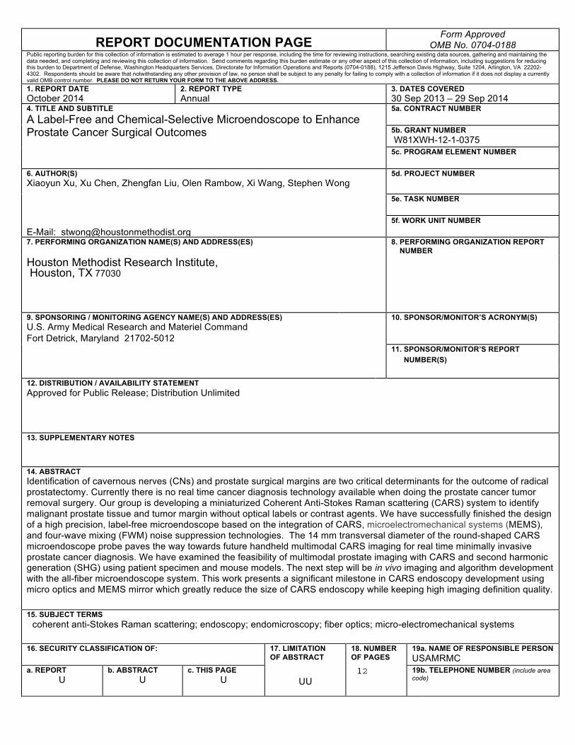
14. ABSTRACT Identification of cavernous nerves (CNs) and prostate surgical margins are two critical determinants for the outcome of radical prostatectomy. Currently there is no real time cancer diagnosis technology available when doing the prostate cancer tumor removal surgery. Our group is developing a miniaturized Coherent Anti-Stokes Raman scattering (CARS) system to identify malignant prostate tissue and tumor margin without optical labels or contrast agents. We have successfully finished the design of a high precision, label-free microendoscope based on the integration of CARS, microelectromechanical systems (MEMS), and four-wave mixing (FWM) noise suppression technologies. The 14 mm transversal diameter of the round-shaped CARS microendoscope probe paves the way towards future handheld multimodal CARS imaging for real time minimally invasive prostate cancer diagnosis. We have examined the feasibility of multimodal prostate imaging with CARS and second harmonic generation (SHG) using patient specimen and mouse models. The next step will be in vivo imaging and algorithm development with the all-fiber microendoscope system. This work presents a significant milestone in CARS endoscopy development using micro optics and MEMS mirror which greatly reduce the size of CARS endoscopy while keeping high imaging definition quality. 15. SUBJECT TERMS coherent anti-Stokes Raman scattering; endoscopy; endomicroscopy; fiber optics; micro-electromechanical systems
16. SECURITY CLASSIFICATION OF:
17. LIMITATION OF ABSTRACT
18. NUMBER OF PAGES
19a. NAME OF RESPONSIBLE PERSON USAMRMC
a. REPORT U
b. ABSTRACT U
c. THIS PAGE U
UU
12
19b. TELEPHONE NUMBER (include area code)
Table of Contents
Page
1. Introduction………………………………………………………………….………..…..…..4
2. Keywords…………………………………………………………………………………..…...4
3. Overall Project Summary ………………………………………………………………..…...4
4. Key Research Accomplishments………………………………………………….……..…..8
5. Conclusion ………………………………………………………………………………..…...9
6. Publications, Abstracts, and Presentations ………………………………………………9
7. Inventions, Patents, and Licenses……………………………………………………………9
8. Reportable Outcomes ………………………………………………………………………..9
9. Other Achievements …………………………………………………………………………..9
10. References ………………………………………………………………………….…...……9
11. Appendices ……………………………………………………………………………...……10
4
1. Introduction Identification of cavernous nerves (CNs) and prostate surgical margins are two critical determinants for the outcome of radical prostatectomy. Coherent Anti-Stokes Raman scattering (CARS) is a label-free imaging technique which can differentiate prostate cancer tissues and cavernous nerves (CNs) based on intrinsic macromolecular contrast without the use of any labeling agents. One major obstacle for intraoperative and surgical application of CARS is the bulky size of the original CARS system which was built on free space optics. To overcome this hurdle, we replaced the bulk optics with their fiber optic counterparts to build an all-fiber-based CARS microendoscope in order to be able to visualize prostate and cancer tissues during prostatectomy and evaluate CARS imaging for prostate cancer tissue differentiation.
2. Keywords Coherent anti-Stokes Raman scattering; non-linear optics; endomicroscopy; fiber optics; micro-electromechanical systems; prostate cancer; prostatectomy, in vivo cancer differentiation; optical diagnostics; medical imaging; image quantification; machine learning
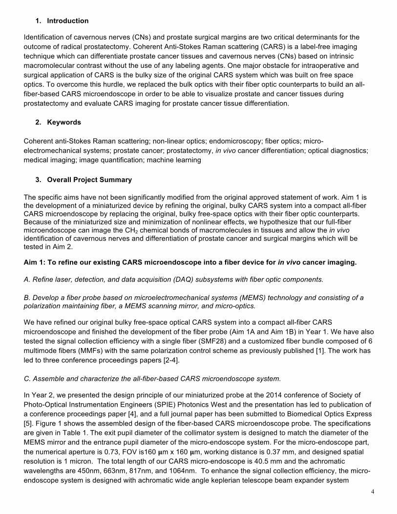
3. Overall Project Summary The specific aims have not been significantly modified from the original approved statement of work. Aim 1 is the development of a miniaturized device by refining the original, bulky CARS system into a compact all-fiber CARS microendoscope by replacing the original, bulky free-space optics with their fiber optic counterparts. Because of the miniaturized size and minimization of nonlinear effects, we hypothesize that our full-fiber microendoscope can image the CH2 chemical bonds of macromolecules in tissues and allow the in vivo identification of cavernous nerves and differentiation of prostate cancer and surgical margins which will be tested in Aim 2. Aim 1: To refine our existing CARS microendoscope into a fiber device for in vivo cancer imaging. A. Refine laser, detection, and data acquisition (DAQ) subsystems with fiber optic components. B. Develop a fiber probe based on microelectromechanical systems (MEMS) technology and consisting of a polarization maintaining fiber, a MEMS scanning mirror, and micro-optics. We have refined our original bulky free-space optical CARS system into a compact all-fiber CARS microendoscope and finished the development of the fiber probe (Aim 1A and Aim 1B) in Year 1. We have also tested the signal collection efficiency with a single fiber (SMF28) and a customized fiber bundle composed of 6 multimode fibers (MMFs) with the same polarization control scheme as previously published [1]. The work has led to three conference proceedings papers [2-4]. C. Assemble and characterize the all-fiber-based CARS microendoscope system. In Year 2, we presented the design principle of our miniaturized probe at the 2014 conference of Society of Photo-Optical Instrumentation Engineers (SPIE) Photonics West and the presentation has led to publication of a conference proceedings paper [4], and a full journal paper has been submitted to Biomedical Optics Express [5]. Figure 1 shows the assembled design of the fiber-based CARS microendoscope probe. The specifications are given in Table 1. The exit pupil diameter of the collimator system is designed to match the diameter of the MEMS mirror and the entrance pupil diameter of the micro-endoscope system. For the micro-endoscope part, the numerical aperture is 0.73, FOV is160 𝛍m x 160 𝛍m, working distance is 0.37 mm, and designed spatial resolution is 1 micron. The total length of our CARS micro-endoscope is 40.5 mm and the achromatic wavelengths are 450nm, 663nm, 817nm, and 1064nm. To enhance the signal collection efficiency, the micro-endoscope system is designed with achromatic wide angle keplerian telescope beam expander system
5
delivering the collimated light to maximum cover the back aperture of light focusing system in order to increase the NA of the micro-endoscope.
Figure 1. Assembled design of the fiber-based CARS microendoscope probe. Table 1. Basic system parameters of CARS microendoscope probe Basic System Descriptions Parameters
Excitation Wavelength range for delivery 817nm(pump), 1064nm(stokes)
Signal wavelength to be collected from tissue 663nm(CARS) 500nm(TPEF) 400nm(SHG)
Input aperture (diameter of exit pupil) Agree with the dimension of MEMS scanning mirror 1.12mm
Distance from the MEMS mirror to objective lens 4mm
NA(after immersion in water) 0.75 Actual resolution 0.78 micron or smaller
Field of view 160𝜇mx160𝜇m High spatial overlap on sample image side is achieved with 0.46 𝛍m resolution at the center field of view and 0.57 𝛍m at the marginal field of view, the ‘ideal’ resolution is half the goal to achieve for the as-built system. Misalignment in the microendoscope system would generate mismatch between the pump and Stokes beams and create difficulty for CARS signal generation. Tolerance budget is used to ensure that each component is manufactured adhering strictly to the specifications and integrated well into the CARS microendoscope probe
6
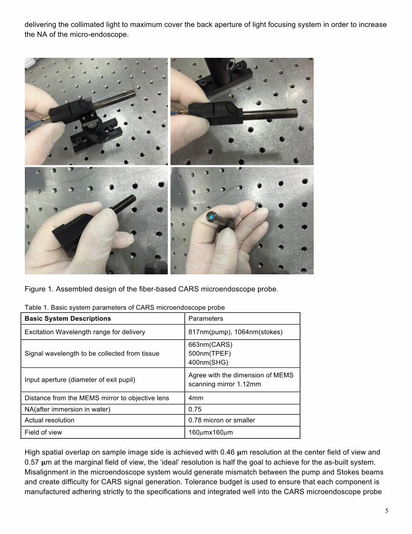
with the highest precision possible. Monte Carlo simulation of the CARS microendoscope probe was performed using the following parameters listed in Table 2. Table 2. Tolerance budget of CARS micro-endoscope probe.
Radius Airspace and glass thickness
Index tolerance V#
Surface Irregularity
Element wedge
Element tilt
Element decenter
0.1 mm 0.025mm 0.0005 0.8% 0.25 fringe 8 𝜇m TIR 0.001 radians 20 𝜇m
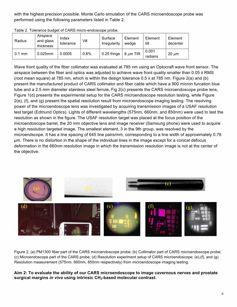
Wave front quality of the fiber collimator was evaluated at 785 nm using an Optocraft wave front sensor. The airspace between the fiber and optics was adjusted to achieve wave front quality smaller than 0.05 λ RMS (root mean square) at 785 nm, which is within the design tolerance 0.5 λ at 785 nm. Figure 2(a) and (b) present the manufactured product of CARS collimator and fiber cable which have a 900 micron furcation blue tube and a 2.5 mm diameter stainless steel ferrule, Fig 2(c) presents the CARS microendoscope probe lens, Figure 1(d) presents the experimental setup for the CARS microendoscope resolution testing, while Figure 2(e), (f), and (g) present the spatial resolution result from microendoscope imaging testing. The resolving power of the microendoscope lens was investigated by acquiring transmission images of a USAF resolution test target (Edmund Optics). Lights of different wavelengths (575nm, 660nm, and 850nm) were used to test the resolution as shown in the figure. The USAF resolution target was placed at the focus position of the microendoscope barrel, the 20 mm objective lens and image receiver (Samsung phone) were used to acquire a high resolution targeted image. The smallest element, 3 in the 9th group, was resolved by the microendscope. It has a line spacing of 645 line pairs/mm, corresponding to a line width of approximately 0.78 µm. There is no distortion in the shape of the individual lines in the image except for a conical defocus deformation in the 660nm resolution image in which the transmission resolution image is not at the center of the objective.
Figure 2. (a) PM1300 fiber part of the CARS microendoscope probe; (b) Collimator part of CARS microendoscope probe; (c) Microendoscope part of the CARS probe; (d) Resolution experiment setup of CARS microendoscope; (e),(f), and (g) Resolution measurement (575nm, 660nm, 850nm respectively) from microendoscope imaging testing. Aim 2: To evaluate the ability of our CARS microendoscope to image cavernous nerves and prostate surgical margins in vivo using intrinsic CH2-based molecular contrast.
(a) (c)
(b)
(g) (e) (f) (d)
7
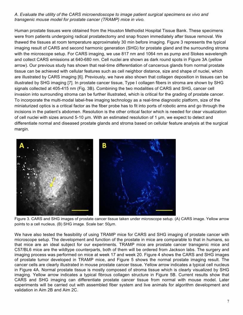
A. Evaluate the utility of the CARS microendoscope to image patient surgical specimens ex vivo and transgenic mouse model for prostate cancer (TRAMP) mice in vivo. Human prostate tissues were obtained from the Houston Methodist Hospital Tissue Bank. These specimens were from patients undergoing radical prostatectomy and snap frozen immediately after tissue removal. We thawed the tissues at room temperature approximately 30 min before imaging. Figure 3 represents the typical imaging result of CARS and second harmonic generation (SHG) for prostate gland and the surrounding stroma with the microscope setup. For CARS imaging, we use 817 nm and 1064 nm as pump and Stokes wavelength and collect CARS emissions at 640-680 nm. Cell nuclei are shown as dark round spots in Figure 3A (yellow arrow). Our previous study has shown that real-time differentiation of cancerous glands from normal prostate tissue can be achieved with cellular features such as cell neighbor distance, size and shape of nuclei, which are illustrated by CARS imaging [6]. Previously, we have also shown that collagen deposition in tissues can be illustrated by SHG imaging [7]. In prostate cancer tissue, Type I collagen fibers in stroma are shown by SHG signals collected at 405-415 nm (Fig. 3B). Combining the two modalities of CARS and SHG, cancer cell invasion into surrounding stroma can be further illustrated, which is critical for the grading of prostate cancer. To incorporate the multi-modal label-free imaging technology as a real-time diagnostic platform, size of the miniaturized optics is a critical factor as the fiber probe has to fit into ports of robotic arms and go through the incisions in the patient’s abdomen. Resolution is the other critical factor which is needed for clear visualization of cell nuclei with sizes around 5-10 µm. With an estimated resolution of 1 µm, we expect to detect and differentiate normal and diseased prostate glands and stroma based on cellular feature analysis at the surgical margin.
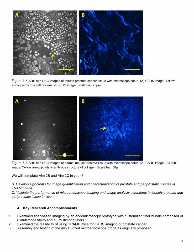
Figure 3. CARS and SHG images of prostate cancer tissue taken under microscope setup. (A) CARS image. Yellow arrow points to a cell nucleus. (B) SHG image. Scale bar: 50µm. We have also tested the feasibility of using TRAMP mice for CARS and SHG imaging of prostate cancer with microscope setup. The development and function of the prostate in mice are comparable to that in humans, so that mice are an ideal subject for our experiments. TRAMP mice are prostate cancer transgenic mice and C57/BL6 mice are the wildtype counterparts, both of them will be ordered from Jackson labs. The surgery and imaging process was performed on mice at week 17 and week 20. Figure 4 shows the CARS and SHG images of prostate tumor developed in TRAMP mice, and Figure 5 shows the normal prostate imaging result. The cancer cells are clearly illustrated in mouse prostate cancer tissue. Yellow arrow indicates a typical cell nucleus in Figure 4A. Normal prostate tissue is mostly composed of stroma tissue which is clearly visualized by SHG imaging. Yellow arrow indicates a typical fibrous collagen structure in Figure 5B. Current results show that CARS and SHG imaging can differentiate prostate cancer tissue from normal with mouse model. Later experiments will be carried out with assembled fiber system and live animals for algorithm development and validation in Aim 2B and Aim 2C.
8
Figure 4. CARS and SHG images of mouse prostate cancer tissue with microscope setup. (A) CARS image. Yellow arrow points to a cell nucleus. (B) SHG image. Scale bar: 25µm.
Figure 5. CARS and SHG images of normal mouse prostate tissue with microscope setup. (A) CARS image. (B) SHG image. Yellow arrow points to a fibrous structure of collagen. Scale bar: 50µm. We will complete Aim 2B and Aim 2C in year 3. B. Develop algorithms for image quantification and characterization of prostate and periprostatic tissues in TRAMP mice. C. Validate the performance of microendoscope imaging and image analysis algorithms to identify prostate and periprostatic tissue in vivo.
4. Key Research Accomplishments
1. Examined fiber-based imaging by an endomicroscopy prototype with customized fiber bundle composed of 6 multimode fibers and 18 multimode fibers
2. Examined the feasibility of using TRAMP mice for CARS imaging of prostate cancer 3. Assembly and testing of the miniaturized microendoscope probe as originally proposed
9
5. Conclusion We have successfully finished the design of a high precision, label-free microendoscope based on the integration of CARS, MEMS, and FWM noise suppression technologies. The high spatial resolution (0.78 𝜇m) and distortion-free images of CARS microendoscope are obtained from USAF target. Image testing results show our microendoscope probe meet the expected computational modeling design specification with good optical alignment, and at the same time, unwanted fiber-generated non-resonant FWM noise is eliminated using our polarization scheme applied in this probe optical system. 14 mm transversal diameter of the round-shaped CARS microendoscope probe paves the path towards a future handheld multimodal CARS imaging for real-time, minimally invasive prostate cancer diagnosis. We have examined the feasibility of multimodal prostate imaging with CARS and SHG using patient specimens and mouse models. The next step will be in vivo imaging and algorithm development with an all-fiber microendoscope system.
6. Publication, Abstracts, and Presentations
1) Zhengfan Liu, Zachary A. Satira, Xi Wang, Xiaoyun Xu, Xu Chen, Kelvin Wong, Shufen Chen, Jianguo Xin, Stephen T.C. Wong. Fiber bundle-based endomicroscopy prototype with two collection channels for simultaneous multimodal coherent anti-Stokes Raman scattering and second-harmonic generation imaging, Multiphoton Microscopy in the Biomedical Sciences XIV. Proceedings of the SPIE, 894814, 2014
2) Xu Chen, Xi Wang, Xiaoyun Xu, Jie Cheng, Zhengfan Liu, Sheng Weng, Michael J. Thrall, Alvin C. Goh, Daniel T. McCormick, Kelvin Wong, Stephen T.C. Wong. Miniaturized CARS Microendoscope Probe Design for Label-free Intraoperative Imaging, Multiphoton Microscopy in the Biomedical Sciences XIII. Proceedings of the SPIE, 89470Y, 2014
3) Xu Chen, Daniel T. McCormick, Kelvin Wong, Stephen T.C. Wong. CARS Microendoscope Probe Design for High Precision Label-free Intraoperative Imaging, submitted to Biomedical Optics Express
7. Inventions, Patents, and Licenses Nothing to report
8. Reportable Outcomes 1) A miniaturized CARS probe has been designed and is being fabricated for in vivo identification of
cavernous nerves and differentiation of prostate cancer and surgical margins.
2) The prototype of fiber bundle-based endomicroscopy has been tested with two collection channels for simultaneous multimodal coherent anti-Stokes Raman scattering and second-harmonic generation imaging. 9. Other Achievements
1) This DOD award supports three postdoctoral trainees (Xu Chen, Xiaoyun Xu, and Xi Wang)
2) This award also provides research opportunities for one masters student (Olen Rambow, graduated Jun
2014) and one PhD student (Zhengfan Liu)
10. References 1) Zhiyong Wang, Liang Gao, Pengfei Luo, Yaliang Yang, Ahmad A. Hammoudi, Kelvin K. Wong, and
Stephen T. C. Wong. Coherent anti-Stokes Raman scattering microscopy imaging with suppression of four-wave mixing in optical fibers. Optics Express, 19(9), 7960-7970 (2011)
10
2) Zhengfan Liu, Zhiyong Wang, Xi Wang, Xiaoyun Xu, Xu Chen, Jie Cheng, Xiaoyan Li, Shufen Chen, Jianguo Xin, Stephen T.C. Wong. Fiber bundle based probe with polarization for coherent anti-Stokes Raman scattering microendoscopy imaging, Multiphoton Microscopy in the Biomedical Sciences XIII. Proceedings of the SPIE, Oral Presentation, 85880F, 2013
3) Zhengfan Liu, Zachary A. Satira, Xi Wang, Xiaoyun Xu, Xu Chen, Kelvin Wong, Shufen Chen, Jianguo Xin, Stephen T.C. Wong. Fiber bundle-based endomicroscopy prototype with two collection channels for simultaneous multimodal coherent anti-Stokes Raman scattering and second-harmonic generation imaging, Multiphoton Microscopy in the Biomedical Sciences XIV. Proceedings of the SPIE, Oral Presentation, 894814, 2014
4) Xu Chen, Xi Wang, Xiaoyun Xu, Jie Cheng, Zhengfan Liu, Sheng Weng, Michael J. Thrall, Alvin C. Goh, Daniel T. McCormick, Kelvin Wong, Stephen T.C. Wong. Miniaturized CARS Microendoscope Probe Design for Label-free Intraoperative Imaging, Multiphoton Microscopy in the Biomedical Sciences XIII. Proceedings of the SPIE, Oral Presentation, 89470Y, 2014
5) Xu Chen, Daniel T. McCormick, Kelvin Wong, Stephen T.C. Wong. CARS Microendoscope Probe Design for High Precision Label-free Intraoperative Imaging, submitted to Biomedical Optics Express
6) Liang Gao, Haibo Zhou, Michael J. Thrall, Fuhai Li, Yaliang Yang, Zhiyong Wang, Pengfei Luo, Kelvin K. Wong, Ganesh S. Palapattu, Stephen T.C. Wong. Coherent anti-Stokes Raman scattering microscopy imaging with suppression of four-wave mixing in optical fibers, Biomedical Optics Express, 2(4), 915-926 (2011)
7) Xiaoyun Xu, Jie Cheng, Michael J. Thrall, Zhengfan Liu, Xi Wang, and Stephen T.C. Wong. Multimodal non-linear optical imaging for label-free differentiation of lung cancerous lesions from normal and desmoplastic tissues. Biomedical Optics Express 4, 2855-2868 (2013)
Appendices
Our work has led to publication of two conference proceedings papers in the past funding year.
11
12