Embed Size (px)
Citation preview
LETTERS TO THE EDITOR
ULTRASOUND VIEW OF A TRAUMATICTWO-LEVEL MEDIAN NERVE LESION
After falling and landing on his outstretched hand in amotorbike accident, a 40-year-old man experienced atraumatic distal fracture of the radius and ulna on theleft side; surgical treatment was successful.
Soon after the trauma, however, the patient com-plained of paresthesias in the first 3 fingers and palm ofthe left hand, severe impairment of hand movements,mild weakness of wrist flexion, and pain in his arm.Physical examination showed severe weakness of the ab-ductor pollicis brevis (APB) (Medical Research CouncilScale (MRC) 1/5] and the opponens pollicis (OP)(MRC 1/5), while the flexor digitorum profundus of thesecond and third digits (FDP), the flexor pollicis longus(FPL), and the flexor carpi radialis (FCR) were less weak(MRC 3/5). Although weakness of the proximal musclescould have been pain-related, the consistency of musclecontraction suggested neurogenic involvement. A sen-sory deficit in the median nerve territory (thenar areaand fingers) was observed. Electrophysiological studiesshowed severe involvement of the APB and the OP (de-nervation signs and deficit of recruitment); there wasonly reduced neurogenic recruitment in the median-in-nervated forearm muscles [flexor digitorum profundus(FDP) of the second and third digits and FCR]. Thesefindings, together with the clinical deficit, suggested aproximal median nerve lesion with greater involvementof distal median nerve branches.
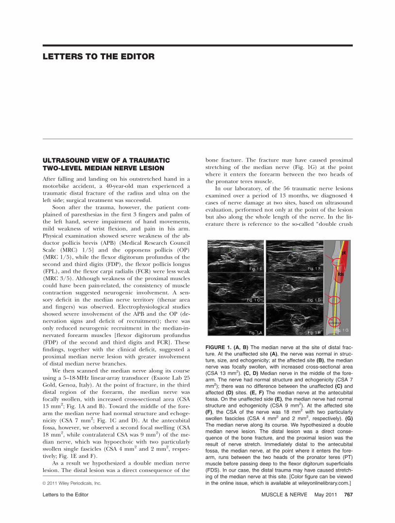
We then scanned the median nerve along its courseusing a 5–18-MHz linear-array transducer (Esaote Lab 25Gold, Genoa, Italy). At the point of fracture, in the thirddistal region of the forearm, the median nerve wasfocally swollen, with increased cross-sectional area (CSA13 mm2; Fig. 1A and B). Toward the middle of the fore-arm the median nerve had normal structure and echoge-nicity (CSA 7 mm2; Fig. 1C and D). At the antecubitalfossa, however, we observed a second focal swelling (CSA18 mm2, while contralateral CSA was 9 mm2) of the me-dian nerve, which was hypoechoic with two particularlyswollen single fascicles (CSA 4 mm2 and 2 mm2, respec-tively; Fig. 1E and F).
As a result we hypothesized a double median nervelesion. The distal lesion was a direct consequence of the
bone fracture. The fracture may have caused proximalstretching of the median nerve (Fig. 1G) at the pointwhere it enters the forearm between the two heads ofthe pronator teres muscle.
In our laboratory, of the 56 traumatic nerve lesionsexamined over a period of 13 months, we diagnosed 4cases of nerve damage at two sites, based on ultrasoundevaluation, performed not only at the point of the lesionbut also along the whole length of the nerve. In the lit-erature there is reference to the so-called ‘‘double crush
FIGURE 1. (A, B) The median nerve at the site of distal frac-
ture. At the unaffected site (A), the nerve was normal in struc-
ture, size, and echogenicity; at the affected site (B), the median
nerve was focally swollen, with increased cross-sectional area
(CSA 13 mm2). (C, D) Median nerve in the middle of the fore-
arm. The nerve had normal structure and echogenicity (CSA 7
mm2); there was no difference between the unaffected (C) and
affected (D) sites. (E, F) The median nerve at the antecubital
fossa. On the unaffected side (E), the median nerve had normal
structure and echogenicity (CSA 9 mm2). At the affected site
(F), the CSA of the nerve was 18 mm2 with two particularly
swollen fascicles (CSA 4 mm2 and 2 mm2, respectively). (G)
The median nerve along its course. We hypothesized a double
median nerve lesion. The distal lesion was a direct conse-
quence of the bone fracture, and the proximal lesion was the
result of nerve stretch. Immediately distal to the antecubital
fossa, the median nerve, at the point where it enters the fore-
arm, runs between the two heads of the pronator teres (PT)
muscle before passing deep to the flexor digitorum superficialis
(FDS). In our case, the distal trauma may have caused stretch-
ing of the median nerve at this site. [Color figure can be viewed
in the online issue, which is available at wileyonlinelibrary.com.]VC 2011 Wiley Periodicals, Inc.
Letters to the Editor MUSCLE & NERVE May 2011 767

syndrome’’ and ‘‘reversed double crush syndrome’’2 inrelation to nerve compression, but the clinical picture,underlying mechanisms, and timing of the double crushsyndrome are different from those hypothesized in a trau-matic ‘‘double-level’’ nerve lesion. We believe that injurydue to stretching could occur where the nerve is located atan anatomical–physiological angle or site of fixation.
In conclusion, ultrasound evaluation simplifies diag-nosis of this kind of lesion, which is difficult to detectwith electrophysiology alone. Treatment and rehabilita-tion are thus enhanced.
Giovanna Anna Liotta, MD1,2
Antonella Di Pasquale, MD2,3
Marta Lucchetta, MD2,4
Maria Antonia Alberti, MD5
Luca Padua, MD, PhD2,6
1Dipartimento di Neuroscienze, Scienze Psichiatriche edAnestesiologiche, Universit‘ di Messina, Messina, Italy
2Fondazione Don Carlo Gnocchi Onlus, Italy
3Dipartimento di Neuroscienze, Universita La Sapienza di Roma,Italy
4Dipartimento di Neuroscienze, Universit di Padova, Padova, Italy
5IDIBELL, Unitat de Neuromuscular, Neurology DepartmentHospital Universitari de Bellvitge, Barcelona, Spain
6Dipartimento di Neuroscienze, Universit Cattolica del SacroCuore, Roma, Italy
1. Liotta G, Granata G, Librante A, Di Pasquale A, Caliandro P, Marti-noli C, et al. Atypical double nerve lesion after humeral fracture:diagnosis by ultrasound. Muscle Nerve 2010;41:287–288.
2. Dahlin LB, Lunborg G. The neurone and its response to peripheralnerve compression. J Hand Surg [Br] 1990;1:5–10.
Published online 14 March 2011 in Wiley Online Library(wileyonlinelibrary.com). DOI 10.1002/mus.22011
---------------------------------------------------------
TALIPES EQUINOVARUS AS LEADINGSYMPTOM OF CONGENITAL MYOTONICDYSTROPHY TYPE 2
In their study, Renard and colleagues described a 2.5-year-old patient with congenital bilateral talipes equino-varus (CTEV).1 Both in the patient and his mother,CCTG expansions typical for myotonic dystrophy type 2(DM2) were found in the ZNF9 gene with 85 and 88repeats, respectively. Mother and patient were otherwiseclinically asymptomatic. The authors discussed a possibleassociation of CTEV with DM2.
In 2008, we reported the first patient with possiblecongenital DM2.2 In addition to reduced intrauterinemovements and postnatal moderate muscular hypotonia,the patient also had congenital pes equinovarus. Ourpatient is now 5 years old and shows mild speech andmotor retardation. Myotonic or other symptoms havenot been noticed so far.
Our initial observation is in line with the findings ofRenard and colleagues and suggests that a congenitalvariant of DM2 exists with CTEV as the leading early
symptom. Many different neuromuscular diseases havebeen linked to congenital CTEV. The clinical findings inthese two patients suggest that, in addition to myotonicdystrophy type 1,3 DM2 could also be considered as apossible underlying disorder in patients with congenitalCTEV, with or without other signs of an unclassifiedneuromuscular disease. Careful clinical and family analy-sis is recommended and, if appropriate, molecular test-ing could help to reach the right diagnosis.
Editor’s note. Renard and colleagues were asked ifthey wished to reply to this letter. Because it agreed withtheir initial report, they felt that no reply was necessary.
Bernd Kruse, MD1
Andreas Gal, MD2
1Department of Pediatric Epileptology, Fachkliniken Wangen,Germany
2Institute of Human Genetics, University Medical Centre,Hamburg–Eppendorf, Germany
1. Renard D, Rivier F, Dimeglio A, Labauge P. Congenital talipes equi-novarus associated with myotonic dystrophy type 2. Muscle Nerve2010;42:457.
2. Kruse B, Wohrle D, Steinbach P, Gal A. Does proximal myotonicmyopathy show anticipation? Hum Mutat 2008;29:E100–102.
3. Canavese F, Sussman M. Orthopaedic manifestations of congenitalmyotonic dystrophy during childhood and adolescence. J PediatrOrthop 2009;29:208–213.
Published online in Wiley Online Library(wileyonlinelibrary.com). DOI 10.1002/mus.22032
---------------------------------------------------------
AWAJI ISLAND MODIFIED CRITERIA FORALS—INCREASED SENSITIVITY WITHOUTCHANGE IN SPECIFICITY: ARE THEYREALLY TWO SIDES OF THE SAME COIN?
We read with interest the article by Chen and colleagueson the use of the Awaji criteria for the electrodiagnosis(EDX) of amyotrophic lateral sclerosis (ALS) and itsimpact on sensitivity of diagnosis.1 We agree that in manypatients with suspected ALS the presence of fasciculationpotentials, especially with widespread distribution on EDXstudies, might suggest the diagnosis of motor neuron dis-ease (MND). In our opinion, to substitute such findingsfor fibrillation potentials/positive sharp waves may proveto be premature. The strict Lambert criteria, which weresubsequently modified in the El Escorial criteria, makecertain such errors of oversensitivity could be avoided.The implications of their observations could be far reach-ing for both patients and physicians alike in clinical prac-tice, and hence we offer the following comments.
Since the publication of the original Awaji criteria in2008,2 new data have been published that show a statisti-cally non-significant occurrence of complex fasciculationpotentials in both benign fasciculation syndrome and inALS.3 These observations suggest that even in the rightclinical settings complex fasciculation potentials may notbe specific for ALS.
Based on the proposed electrodiagnostic criteria thedefinitive ALS category in the Chen et al. study increasedonly by 1 case (2 definite before to 3 definite after EDX
768 Letters to the Editor MUSCLE & NERVE May 2011
examination) and not surprisingly, the authors reportedan increase in the number of ALS cases classified asprobable (24–37 EDX cases).1 Further in their experi-ence 2 definite ALS cases evolved into 7 definite cases byclinical diagnosis alone. It would be of interest to knowthe breakdown of the diagnostic class these 5 patientsbelonged to in the initial Awaji criteria. In Chen et al.report an ALS specialist evaluated 50% of the patients inthe study group, resulting in a pretest probability bias to-ward correct diagnosis. This is probably different fromroutine clinical practice when neurologists are con-fronted with such cases. A larger cohort of patients withlong-term follow-up could provide the statistical evidenceto support such change in EDX practice.
Raghav Govindarajan, MD
Nestor Galvez-Jimenez, MD, MSc
Department of Neurology, Electromyography Laboratory,Cleveland Clinic, Weston, Florida, USA
1. Chen A, Weimer L, Brannagan TIII, Colin M, Andrews J, MitsumotoH, et al. Experience with the Awaji Island modifications to the ALSdiagnostic criteria. Muscle Nerve 2010;42:831–832.
2. de Carvalho M, Dengler R, Eisen A, England JD, Kaji R, Kimura J,et al. Electrodiagnostic criteria for diagnosis of ALS. Clin Neuro-physiol 2008;119:497–503.
3. Mills KR. Characteristics of fasciculations in amyotrophic lateralsclerosis and the benign fasciculation syndrome. Brain 2010;133:3458–3469.
Published online 14 March 2011 in Wiley Online Library(wileyonlinelibrary.com). DOI 10.1002/mus.22027
---------------------------------------------------------
REPLY
We appreciate the interest of Drs. Govindarajan andGalvez-Jimenez in our work as well as their comments.In response, we first emphasize that the Awaji modifica-tions apply to the electrodiagnostic criteria, which bythemselves are never sufficient for the diagnosis of ALS.Second, the Awaji guidelines use the combination of fas-ciculation potentials with chronic neurogenic changes(defined as motor unit potentials of increased durationor amplitude, and/or reduced recruitment) as electro-diagnostic evidence of lower motor neuron dysfunctionin the diagnosis of ALS. In our study, we did not use orpropose using fasciculation potentials alone to diagnosemotor neuron disease.
With regard to the characteristics of fasciculationpotentials seen in ALS and benign fasciculation syn-drome, we had no information on the waveform mor-phology or the firing frequency of the fasciculationpotentials recorded in our study; as a retrospective analy-sis we used tabulated EMG reports. We did not claim inour study that the fasciculation potentials seen in ALSare more complex or unstable, as proposed by theexpert consensus made in Awaji.
Regarding the initial Awaji diagnoses for the 5patients who showed clinical progression and who laterwere diagnosed with clinically definite ALS, 4 (cases 2, 8,61, and 69) were initially diagnosed with probable ALS,whereas 1 (case 66) was initially diagnosed with possible
ALS by either Airlie House or Awaji criteria (see the sup-plementary table accompanying the article). We did notdiscuss these results in our investigation, because (a) thesample size was small, and (b) it was not our objective todetermine whether the certainty of the diagnosis wascorrelated or predictive of clinical progression, althoughthat is certainly an important issue.
Since the publication of our study, several reportshave emerged suggesting that the use of the Awaji guide-lines can be useful in the early diagnosis of ALS.1–3 Weagree that a larger prospective study without selectionbias would be helpful in determining the utility of theAwaji guidelines in routine clinical practice with the aimtoward the earlier diagnosis of ALS.
Amy Chen, MD, PhD1
Louis Weimer, MD2
Thomas Brannagan III, MD2
Jinsy Andrews, MD3
Hiroshi Mitsumoto, MD2
Petra Kaufmann, MD, MSc2,4
1Department of Neurology, University of Rochester, Rochester,New York, USA
2Department of Neurology, Columbia University, New York, NewYork, USA
3Hospital for Special Care, University of Connecticut, New Britain,Connecticut, USA
4National Institute of Neurological Disorders and Stroke, NationalInstitutes of Health, Bethesda, Maryland, USA
1. Krarup C. Lower motor neuron involvement examined by quantita-tive electromyography in amyotrophic lateral sclerosis. Clin Neuro-physiol 2011;122:414–422.
2. Douglass CP, Kandler RH, Shaw PJ, McDermott CJ. An evaluation ofneurophysiological criteria used in the diagnosis of motor neurondisease. J Neurol Neurosurg Psychiatry 2010;81:646–649.
3. Boekestein WA, Kleine BU, Hageman G, Schelhaas HF, Zwarts MJ.Sensitivity and specificity of the ‘Awaji’ electrodiagnostic criteria foramyotrophic lateral sclerosis: retrospective comparison of the Awajiand revised El Escorial criteria for ALS. Amyotroph Lateral Scler2010;11:497–501.
Published online in Wiley Online Library(wileyonlinelibrary.com). DOI 10.1002/mus.22026
---------------------------------------------------------
CAN WE ACCURATELY MEASURE THEONSET LATENCY TO THE FIRST DORSALINTEROSSEOUS?
I have read the short report by Takahashi and Robinsonwith interest.1 First dorsal interosseous (FDI) and abductordigiti minimi (ADM) compound muscle action potentials(CMAP) from two healthy subjects were recorded usingfour bipolar montages (A–D) and four monopolar mon-tages (E–F). The investigators proposed that the initialdeflections of the belly–tendon ‘‘FDI’’ (montages C andD) and ‘‘ADM’’ (montage A) CMAPs might be affectednot by the activation of FDI and ADM themselves, but byvolume-conducted potentials from more proximal ulnar-in-nervated intrinsic muscles. These volume-conducted poten-tials should be incorporated in ‘‘belly–CMAP’’ (montages
Letters to the Editor MUSCLE & NERVE May 2011 769

A–D) but also in ‘‘tendon–CMAP’’ (montages F–H).From my point of view, the ‘‘tendon–CMAP’’ is not a
volume-conducted potential, but rather it is a far-fieldpotential. More precisely, it is a stationary wave originat-ing from myotendinous junctions from all ulnar-inner-vated muscles, including more proximal muscles thanthe FDI.2–5 Proof of this is illustrated in Figure 1. Whenthe distance between the stimulation site at the wristand the recording electrode E1 placed over digit 3 (E2contralateral) increases, latencies to potential peaks donot change, indicating that these potentials are not vol-ume-conducted. Thus, potentials called ‘‘belly–CMAP’’correspond to the summation of the FDI CMAP andmyotendinous far-field potentials, whereas potentialscalled ‘‘tendon–CMAP’’ are mainly related to myotendi-nous far-field potentials. Consequently, the initial posi-tive deflection in montage B (FDI index) indicates thatpositive far-field potentials, originating from myotendi-nous junctions, are steeper when recorded over theindex than over the FDI belly.
Francois Charles Wang, PhD
Department of Neurophysiology, CHU Liege, Liege, Belgium
1. Takahashi N, Robinson LR. Can we accurately measure the onset la-tency to the first dorsal interosseous? Muscle Nerve 2011;43:129–132.
2. Dumitru D, Jewett DL. Far-field potentials. Muscle Nerve 1993;16:237–254.
3. Dumitru D, King JC. Far-field potentials in muscle. Muscle Nerve1991;14:981–989.
4. Dumitru D, King JC. Far-field potentials in muscle: a quantitativeinvestigation. Arch Phys Med Rehabil 1992;73:270–274.
5. Dumitru D, King JC. Median/ulnar premotor potential identificationand localization. Muscle Nerve 1995;18:518–525.
Published online in Wiley Online Library(wileyonlinelibrary.com). DOI 10.1002/mus.22030
---------------------------------------------------------
REPLY
We appreciate the interest and the comments by Dr.Wang regarding our short report on the origin of theresponse recorded from over the first dorsal interosseous(FDI) muscle.
We believe that Dr. Wang’s letter and figure augmentthe information provided in our report. In our report, weused the term ‘‘volume-conducted potentials’’ to indicatethat potentials originated not from directly under the elec-trode, but from a distance. Specifically, we consider volumeconduction as ‘‘spread of current from a potential sourcethrough a conducting medium, such as body tissues.’’1
We do not disagree with Dr. Wang that these vol-ume-conducted potentials could be, at least in part, far-field potentials from myotendinous junctions of proxi-mal muscles, although we cannot be sure which musclescontribute and how much of the far-field potential isfrom the myotendinous junction vs. proximal musclebellies. Because the differences between ‘‘near-field’’ and‘‘far-field’’ are somewhat arbitrary,1 we did not use thatterm in our report.
Our objective in the short report was to demonstratethe significant influence of the tendon electrode on thebelly–tendon compound muscle action potential wave-forms recorded from FDI. Dr. Wang’s information pro-vides additional information on possible origin of thepotentials recorded at the tendon and reinforces theneed to carefully interpret responses recorded in thisregion of the hand.
Nobushige Takahashi, MD
Lawrence R. Robinson, MD
Department of Rehabilitation Medicine, University of WashingtonSchool of Medicine, Seattle, Washington
1. American Association of Electrodiagnostic Medicine glossary ofterms in electrodiagnostic medicine. Muscle Nerve 2001;24(suppl10):S1–50.
Published online in Wiley Online Library(wileyonlinelibrary.com). DOI 10.1002/mus.22031
---------------------------------------------------------
FIGURE 1. Three monopolar recordings (superimposed curves)
with increasing distance between the ulnar stimulation site at
the wrist and the active recording electrode E1 placed over digit
3 (E2 contralateral). Latencies to potential peaks do not
increase with increasing distance.
770 Letters to the Editor MUSCLE & NERVE May 2011