Embed Size (px)
Citation preview

BRANSON W. RITCHIE, DVM, PhD Assistant Professor, Avian and Zoologic Medicine Department of Small Animal Medicine College of Veterinary Medicine University of Georgia Athens, Georgia
GREG J. HARRISON, DVM Director, The Bird Hospital Lake Worth, Florida President, Harrison’s Bird Diets Omaha, Nebraska
LINDA R. HARRISON, BS President, Wingers Publishing, Inc. Former Editor, Journal of the Association of Avian Veterinarians Lake Worth, Florida
AVIAN MEDICINE: PRINCIPLES AND APPLICATION
Close window to return to IVIS

ytology is designed to be a rapid, inexpen-sive “in-house” diagnostic procedure, andthe use of cytodiagnosis should be easilywithin the realm of any veterinary clini-
cian. The basic cytodiagnosis of inflammation, tissuehyperplasia, malignant neoplasia and normal cellu-larity are easily differentiated from each other (seeFigures 10.10, 10.11). One who is well versed inmammalian cytodiagnosis should have little troublein the interpretation of avian samples. The goal is toachieve a quick presumptive or definitive diagnosisduring the patient’s initial visit to the veterinaryclinic in an effort to provide an immediate and spe-cific treatment plan. Cytology can then be used tomonitor the success of therapy by evaluating changesin microbial and cell populations within or on thehost. Cytology should be considered as a part of theminimum database in birds with discharges, massesor swellings. Cytologic evaluation of tissue imprintsand fluids collected during a postmortem examina-tion can be used to develop a presumptive diagnosisthat can guide disease management decisions withinthe flock until a definitive diagnosis is provided byculture, DNA probe or histopathology. Cytologicalsamples are of greatest value if they are collectedfresh and immediately processed for evaluation. Toobtain a cytologic sample and send it to an outsidelaboratory defeats the purpose and usefulness of cy-tology. By cytologically examining antemortem andpostmortem samples, the clinician can compare theinformation that is derived from cytology, radio-graphs, CBC, serum chemistries and histopathology.This will serve to improve understanding of thepathogenesis and cellular effects of a disease process.
C C H A P T E R
10CYTOLOGY
Terry W. Campbell
Published in IVIS with the permission of the editorClose window to return to IVIS www.ivis.org
Avian Medecine : Principles and Application, B.W. Ritchie, G.J Harrison and L.R. Harrison (Eds.)
Sample Collection
A variety of sample collection methods can be used toobtain samples for cytologic examination.4 Themethod of choice depends upon the location and na-ture of the material being sampled. Cytologic samplecollection methods can be divided into two broadcategories: aspiration and contact smears.
Sample Collection by Aspiration
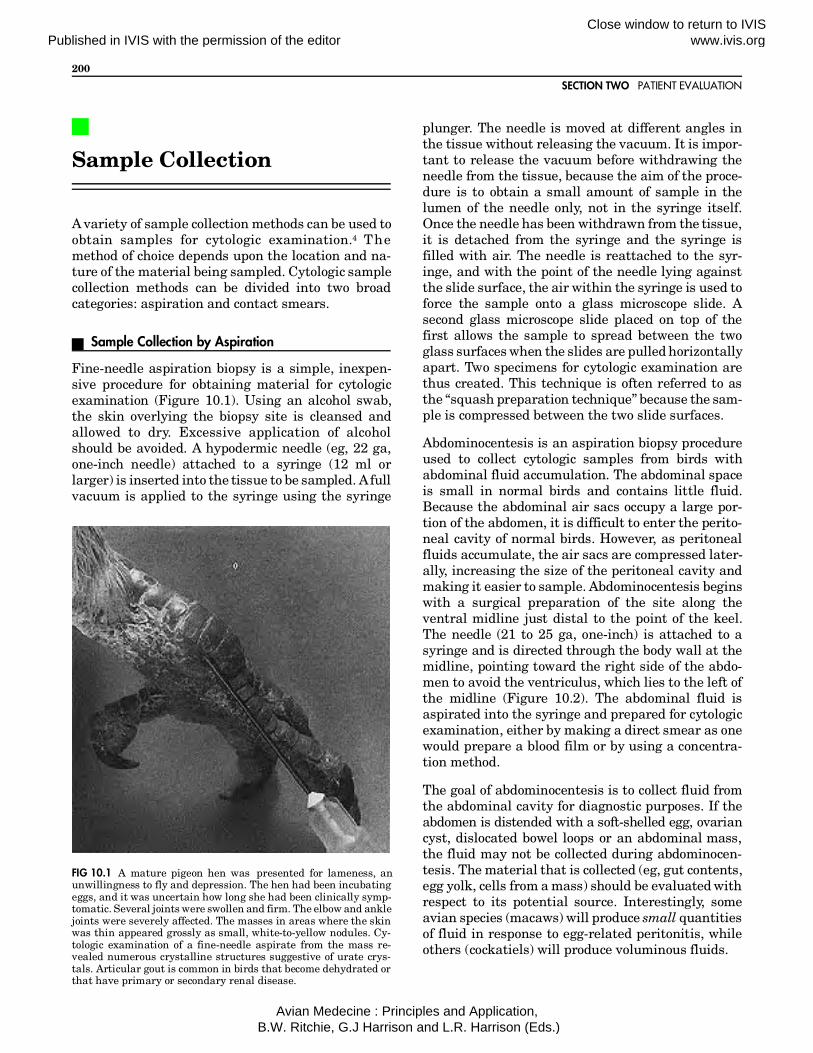
Fine-needle aspiration biopsy is a simple, inexpen-sive procedure for obtaining material for cytologicexamination (Figure 10.1). Using an alcohol swab,the skin overlying the biopsy site is cleansed andallowed to dry. Excessive application of alcoholshould be avoided. A hypodermic needle (eg, 22 ga,one-inch needle) attached to a syringe (12 ml orlarger) is inserted into the tissue to be sampled. A fullvacuum is applied to the syringe using the syringe
plunger. The needle is moved at different angles inthe tissue without releasing the vacuum. It is impor-tant to release the vacuum before withdrawing theneedle from the tissue, because the aim of the proce-dure is to obtain a small amount of sample in thelumen of the needle only, not in the syringe itself.Once the needle has been withdrawn from the tissue,it is detached from the syringe and the syringe isfilled with air. The needle is reattached to the syr-inge, and with the point of the needle lying againstthe slide surface, the air within the syringe is used toforce the sample onto a glass microscope slide. Asecond glass microscope slide placed on top of thefirst allows the sample to spread between the twoglass surfaces when the slides are pulled horizontallyapart. Two specimens for cytologic examination arethus created. This technique is often referred to asthe “squash preparation technique” because the sam-ple is compressed between the two slide surfaces.
Abdominocentesis is an aspiration biopsy procedureused to collect cytologic samples from birds withabdominal fluid accumulation. The abdominal spaceis small in normal birds and contains little fluid.Because the abdominal air sacs occupy a large por-tion of the abdomen, it is difficult to enter the perito-neal cavity of normal birds. However, as peritonealfluids accumulate, the air sacs are compressed later-ally, increasing the size of the peritoneal cavity andmaking it easier to sample. Abdominocentesis beginswith a surgical preparation of the site along theventral midline just distal to the point of the keel.The needle (21 to 25 ga, one-inch) is attached to asyringe and is directed through the body wall at themidline, pointing toward the right side of the abdo-men to avoid the ventriculus, which lies to the left ofthe midline (Figure 10.2). The abdominal fluid isaspirated into the syringe and prepared for cytologicexamination, either by making a direct smear as onewould prepare a blood film or by using a concentra-tion method.
The goal of abdominocentesis is to collect fluid fromthe abdominal cavity for diagnostic purposes. If theabdomen is distended with a soft-shelled egg, ovariancyst, dislocated bowel loops or an abdominal mass,the fluid may not be collected during abdominocen-tesis. The material that is collected (eg, gut contents,egg yolk, cells from a mass) should be evaluated withrespect to its potential source. Interestingly, someavian species (macaws) will produce small quantitiesof fluid in response to egg-related peritonitis, whileothers (cockatiels) will produce voluminous fluids.
FIG 10.1 A mature pigeon hen was presented for lameness, anunwillingness to fly and depression. The hen had been incubatingeggs, and it was uncertain how long she had been clinically symp-tomatic. Several joints were swollen and firm. The elbow and anklejoints were severely affected. The masses in areas where the skinwas thin appeared grossly as small, white-to-yellow nodules. Cy-tologic examination of a fine-needle aspirate from the mass re-vealed numerous crystalline structures suggestive of urate crys-tals. Articular gout is common in birds that become dehydrated orthat have primary or secondary renal disease.
SECTION TWO PATIENT EVALUATION
200
Published in IVIS with the permission of the editorClose window to return to IVIS www.ivis.org
Avian Medecine : Principles and Application, B.W. Ritchie, G.J Harrison and L.R. Harrison (Eds.)

Fluid samples having low cellularity require a con-centration procedure for easier examination of thecells. A variety of techniques can be used to concen-trate cells on microscope slides. A simple method is tomarginate the cells on a smear made by the conven-tional wedge technique used for making blood films.A drop of the fluid sample is placed on a microscopeslide and spread slowly using a spreader slide. Justprior to reaching the end of the smear, the spreaderslide is quickly backed slightly into the advancingsmear, just before lifting it from the surface of theslide containing the smear. This should produce aslide with the marginated cells concentrated at theend of the film.
Cells can be concentrated by centrifugation in a man-ner similar to that used in mammalian urinalysisprocedures. The fluid is placed in a plastic test tubeand centrifuged at 600 G (gravity) for ten minutes.Unlike urine sediments, cytologic sediments frompoorly cellular fluids do not have a visible button orpellet at the bottom of a spun tube. Therefore, theconcentrated cells are usually obtained by aspiratingthe fluid at the bottom of the tube into a pipette orsyringe. The sample is then placed onto a microscopeslide and a smear is made in the manner describedfor concentrating cells in a smear. Special cytocentri-fuge equipmenta is available for concentrating cellson microscope slides while absorbing the fluid ontofilter paper. This equipment is expensive and notpractical for the average veterinary laboratory.
Because centrifugation distorts the appearance ofthe cells, a cell concentration method that utilizesgravity provides a concentrated sample with normalappearing cells. A simple, inexpensive sedimentationdevice can be made for use in the veterinary labora-tory. This device consists of a base to support the slideand a clamping mechanism to hold the fluid columnonto the microscope slide (Figure 10.3). The columnthat holds the fluid is made from a one millimetertuberculin syringe barrel with the tip removed. Thebase of the syringe barrel allows for the syringe to beheld in place by a clamp (usually made of wood). Apiece of filter paper (eg, Whatman #2) is cut to thedimensions of the microscope slide and a standard 2mm paper hole punch is used to create a hole in thecenter of the filter paper. The filter paper is placed ontop of the slide, and the base of the tuberculin syringebarrel is placed on top of the filter paper with theopening of the syringe superimposed over the hole in
FIG 10.2 For abdominocentesis, the needle is attached to a syringeand is directed through the body wall at the midline, pointingtoward the right side of the abdomen. 1) Caudal edge of sternum2) liver 3) ventriculus and 4) intestines.
FIG 10.3 Centrifugation can distort the appearance of cells thatare intended for cytologic evaluation. A simple device that usesgravity to concentrate cells provides cytologic samples of betterquality than centrifugation (courtesy of Terry Campbell).
CHAPTER 10 CYTOLOGY
201
Published in IVIS with the permission of the editorClose window to return to IVIS www.ivis.org
Avian Medecine : Principles and Application, B.W. Ritchie, G.J Harrison and L.R. Harrison (Eds.)

the filter paper. The clamp is used to secure thecolumn to the slide. A small amount of fluid (eg, 0.2to 0.5 ml) is placed into the syringe column. Whenallowed to stand undisturbed, the fluid is drawn bygravity and absorbed into the filter paper. The cellsin the fluid fall onto the surface of the slide wherethey adhere. Once the fluid has drained from thecolumn, the apparatus is disassembled and the slideis allowed to air dry. After staining, the cells can befound concentrated in the two millimeter circle cre-ated by the filter paper and column.
Cytologic evaluation of the ingluvies (crop) can beperformed from samples obtained by aspiration. Thisis indicated in birds showing clinical signs of regur-gitation, vomiting, delayed emptying of the crop orother crop disorders. A crop aspirate is obtained byinserting a sterile plastic, metal or rubber feedingtube through the mouth and esophagus into theingluvies (see Figure 15.6). The tube should passfreely and not be forced into the crop. Passage of thetube is facilitated by extending the head and neck tostraighten the esophagus. The crop content is gently
aspirated into the tube using a syringe attached tothe free end. Excessive vacuum should be avoided toprevent damage to the crop mucosa. In cases wherematerial cannot be aspirated for examination, awash sample can be obtained by infusing a smallamount of sterile isotonic saline into the crop andaspirating the fluid back into the tube and syringe.
Aspiration of the infraorbital sinus of birds sufferingfrom sinusitis can provide diagnostic material forculture and cytologic examination. One technique ofsinus aspiration in psittacine birds samples the largesinus between the eye and the external nares (Figure10.4). With the head and body properly restrained, aneedle (eg, 22 ga one-inch) is passed through thefleshy skin at the commissure of the mouth. Theneedle is directed toward a point midway betweenthe eye and external nares, keeping parallel with theside of the head. The needle passes under the zygo-matic bone, which lies between the lower corner ofthe rhinotheca (upper beak) and the ear. Often thepassage of the needle is improved by keeping thebird’s mouth open with an oral speculum. Once the
FIG 10.4 Aspiration of the infraorbital diverticulum of the infraorbital sinus in psittacine birds can be performed by a) passing a needlethrough the fleshy skin at the commissure of the mouth and directing it toward a point midway between the eye and external nares, b)keeping parallel with the side of the head and passing under the zygomatic arch. 1) Zygomatic arch 2) mandible 3) oral cavity.
SECTION TWO PATIENT EVALUATION
202
Published in IVIS with the permission of the editorClose window to return to IVIS www.ivis.org
Avian Medecine : Principles and Application, B.W. Ritchie, G.J Harrison and L.R. Harrison (Eds.)
needle has entered the sinus, the sinus contents canbe aspirated. A caudally misdirected needle couldresult in penetration of the ocular orbit; however,more commonly, a misdirected needle results in pene-tration of the surrounding muscles, causing periph-eral blood contamination of the sample. It is impor-tant to note that in some species (eg, some passerinebirds), the sinuses may not communicate with eachother as they do in psittacine birds. Therefore, abilateral sinusitis may require bilateral aspirations.(Ed note: If a routine sinus flush does not produce anadequate sample, the anesthetized bird may be heldwith the head parallel to the floor and the affectedsinus down. The sinus is flushed from underneathwith the needle directed up; see Chapter 22).
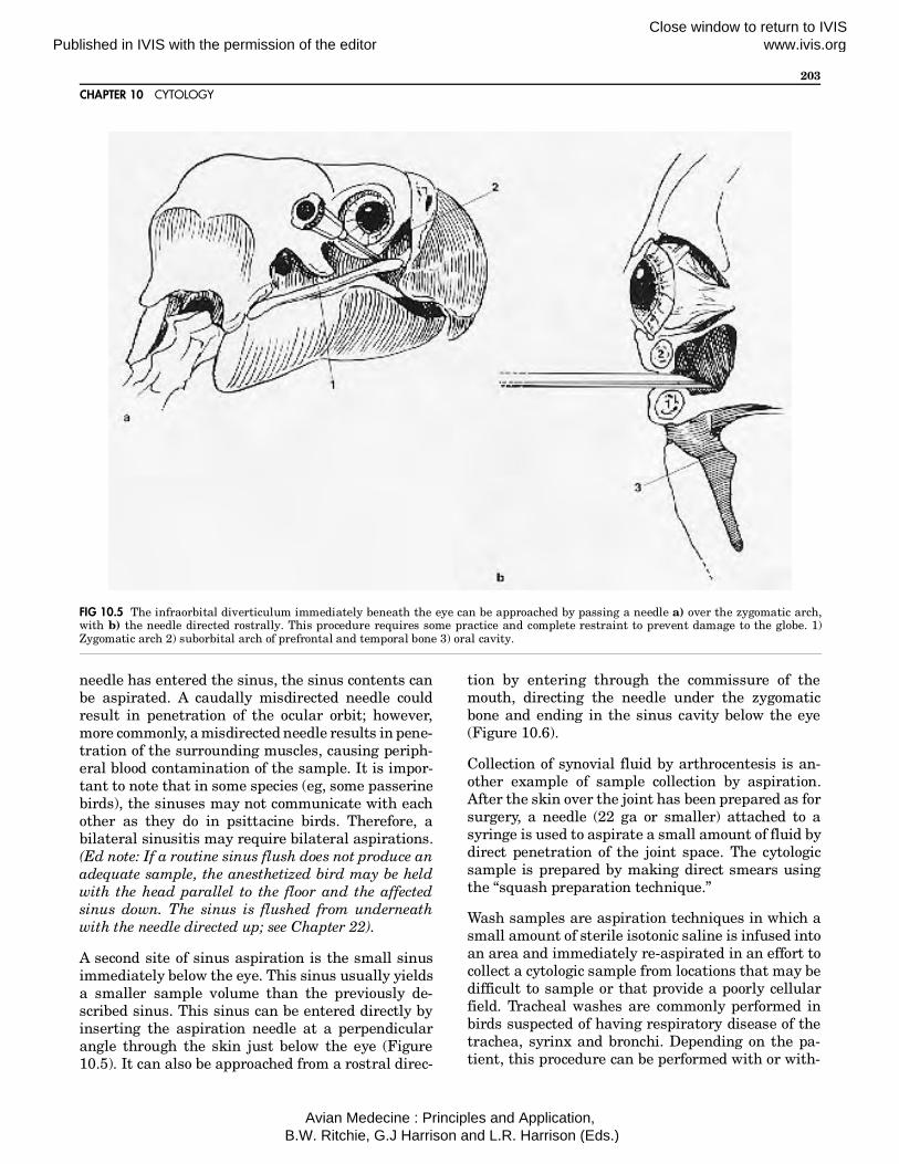
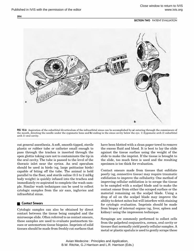
A second site of sinus aspiration is the small sinusimmediately below the eye. This sinus usually yieldsa smaller sample volume than the previously de-scribed sinus. This sinus can be entered directly byinserting the aspiration needle at a perpendicularangle through the skin just below the eye (Figure10.5). It can also be approached from a rostral direc-
tion by entering through the commissure of themouth, directing the needle under the zygomaticbone and ending in the sinus cavity below the eye(Figure 10.6).
Collection of synovial fluid by arthrocentesis is an-other example of sample collection by aspiration.After the skin over the joint has been prepared as forsurgery, a needle (22 ga or smaller) attached to asyringe is used to aspirate a small amount of fluid bydirect penetration of the joint space. The cytologicsample is prepared by making direct smears usingthe “squash preparation technique.”
Wash samples are aspiration techniques in which asmall amount of sterile isotonic saline is infused intoan area and immediately re-aspirated in an effort tocollect a cytologic sample from locations that may bedifficult to sample or that provide a poorly cellularfield. Tracheal washes are commonly performed inbirds suspected of having respiratory disease of thetrachea, syrinx and bronchi. Depending on the pa-tient, this procedure can be performed with or with-
FIG 10.5 The infraorbital diverticulum immediately beneath the eye can be approached by passing a needle a) over the zygomatic arch,with b) the needle directed rostrally. This procedure requires some practice and complete restraint to prevent damage to the globe. 1)Zygomatic arch 2) suborbital arch of prefrontal and temporal bone 3) oral cavity.
CHAPTER 10 CYTOLOGY
203
Published in IVIS with the permission of the editorClose window to return to IVIS www.ivis.org
Avian Medecine : Principles and Application, B.W. Ritchie, G.J Harrison and L.R. Harrison (Eds.)
out general anesthesia. A soft, smooth-tipped, sterileplastic or rubber tube or catheter small enough topass through the trachea is inserted through theopen glottis taking care not to contaminate the tip inthe oral cavity. The tube is passed to the level of thethoracic inlet near the syrinx. An oral speculumshould be used in birds (eg, large psittacine birds)capable of biting off the tube. The animal is heldparallel to the floor, and sterile saline (0.5 to 2 ml/kgbody weight) is quickly infused into the trachea andimmediately re-aspirated to complete the wash sam-ple. Similar wash techniques can be used to collectcytologic samples from the air sacs, ingluvies andinfraorbital sinus.
Contact Smears
Cytologic samples can also be obtained by directcontact between the tissue being sampled and themicroscope slide. Often referred to as contact smears,these samples are used to evaluate postmortem tis-sues or antemortem tissue biopsies. Imprints of solidtissues should be made from freshly cut surfaces that
have been blotted with a clean paper towel to removethe excess fluid and blood. It is best to lay the slideagainst the tissue surface using the weight of theslide to make the imprint. If the tissue is brought tothe slide, too much force is used and the resultingspecimen is too thick for evaluation.
Contact smears made from tissues that exfoliatepoorly (eg, connective tissue) may require traumaticexfoliation to improve the cellularity. One method ofimproving cellular exfoliation is to scrape the tissueto be sampled with a scalpel blade and to make thecontact smear from either the scraped surface or thematerial remaining on the scalpel blade. Using adrop of oil on the scalpel blade may improve theability to detect mites but will interfere with stainingfor cytologic evaluation. Imprints should be madefrom biopsy of internal organs (eg, liver, spleen andkidney) using the impression technique.
Scrapings are commonly performed to collect cellsfrom the palpebral conjunctiva, cornea, oral cavity ortissues that normally yield poorly cellular samples. Ametal or plastic spatula is used to gently scrape these
FIG 10.6 Aspiration of the suborbital diverticulum of the infraorbital sinus can be accomplished by a) entering through the commissure ofthe mouth, directing the needle under the zygomatic bone and b) ending in the sinus cavity below the eye. 1) Zygomatic arch 2) suborbitalarch 3) oral cavity.
SECTION TWO PATIENT EVALUATION
204
Published in IVIS with the permission of the editorClose window to return to IVIS www.ivis.org
Avian Medecine : Principles and Application, B.W. Ritchie, G.J Harrison and L.R. Harrison (Eds.)

tissues, and the exfoliated cells are transferred to amicroscope slide.
Cytologic samples can also be obtained using a sterileswab.b Once the sample has been collected, the swabis gently rolled across the surface of a clean micro-scope slide, using light pressure in order to avoid celldamage. The swab should be rolled in one directiononly and not rolled back and forth across the smearto prevent the creation of an excessively thick smear.Cytologic samples of internal tissues can be obtainedusing endoscopic equipment. Samples can be ob-tained either from the tip of the endoscope or byusing brushes or biopsy forceps. The sample is ap-plied directly to a microscope slide.
Evaluationof the Cytologic Sample
Tables 10.1 to 10.3 describe the use of stains mostcommonly available for cytology.
Classification of Cells and Cellular Responses
The cells observed in the cytologic sample can beclassified as either hemic, epithelial, mesenchymalor nervous tissue cells.13 Hemic cells are those cellsfound in the blood and the hematopoietic tissues (seeChapter 9). It is extremely important to recognize
TABLE 10.1 Cytologic Stains Commonly Used in Avian Practice
1. Romanowsky stains (Wright’s and Wright-Giemsa)These stains are commonly used for peripheral blood films and routinecytology. They require air-dried smears. Commercially prepared quickstains are available to simplify the staining procedure. These stainscan be used to prepare a permanently stained slide.
2. New Methylene Blue stainThis is a routine cytologic stain used as a wet preparation on driedsmears. It does not provide a permanent stain. It is useful in thedemonstration of fibrin, lipid droplets, fungal hyphae and other struc-tures that stain poorly with alcohol-based stains.
3. Acid-fast stainThis specific stain is used to demonstrate acid-fast positive organisms,such as Mycobacterium sp. Acid-fast positive organisms stain red,whereas other bacteria stain blue. This stain is not used to evaluatecells.
4. Gram’s stainThis is a microbiologic stain used primarily for the classification ofbacteria grown on culture media. Gram-positive organisms stain deepviolet, whereas gram-negative organisms stain red. Because of thenature of material on most cytologic preparations, it is difficult to
achieve uniformity of staining on the smear. This stain is not used toevaluate cells.
5. Macchiavello’s stainThis stain is used to identify chlamydia and mycoplasma inclusions.Chlamydia elementary bodies (0.2 - 0.3 µ) stain red, whereas the initialbodies (0.9-1.0 µ) stain blue. Mycoplasma colonies resemble chlamy-dia. Other particles in some smears may stain red and make theinterpretation of the smear difficult. This stain is not used to evaluatecells.
6. Gimenez stainThis stain is used to identify chlamydia inclusions which stain redagainst a blue-green cellular background. There is less interferencewith non-chlamydia particles staining red with this stain as comparedto Macchiavello’s stain. This stain is not used to evaluate cells.
7. Stamp stainThis stain is used to detect chlamydia and rickettsia, which appear assmall, bright red, “cocci” intracytoplasmic inclusions.
8. Giemsa stainThis stain is used to identify chlamydia and mycoplasma.
TABLE 10.2 Results of Cytologic Staining
STAIN USE RESULTSAcid-fast stain Mycobacterium. . . . . . . . red
Other bacteria . . . . . . . . blueLeukocytes . . . . . . . . . . . blueCellular debris . . . . . . . . blue
Giemsa stain Cell nuclei. . . . . . . . . . . . reddish purpleChlamydial elementary bodies . . . purple initial bodies . . . . . . . . blueMycoplasma. . . . . . . . . . pink or purple
Gram’s stain Gram-positive bacteria. . violetGram-negative bacteria . redEukaryatic cells (except yeast) . . . . . . redYeast . . . . . . . . . . . . . . . deep violet
ModifiedGimenez stain
Chlamydial elementary bodies . . . red initial bodies . . . . . . . . blueHeterophil granules . . . . redEosinophil granules . . . . redMycoplasma. . . . . . . . . . like chlamydia
New methyleneblue stain
Granulocytes . . . . . . . . . purple nuclei, pale blue cytoplasmErythrocytes . . . . . . . . . . purple nuclei, distinct cytoplasmic border, cytoplasm greenish blueHeterophil granules . . . . not stainedEosinophil granules . . . . not stainedFibrin . . . . . . . . . . . . . . . not stained
Stamp stain Chlamydia, rickettsia . . . bright redCocci tissue, other organisms . . . . . green
Sudan III stain Fat globules . . . . . . . . . . red-orangeCell nuclei. . . . . . . . . . . . blueCell cytoplasm . . . . . . . . green
Wright’s stain Blood cells(see hematology)
CHAPTER 10 CYTOLOGY
205
Published in IVIS with the permission of the editorClose window to return to IVIS www.ivis.org
Avian Medecine : Principles and Application, B.W. Ritchie, G.J Harrison and L.R. Harrison (Eds.)

hemic cells because these cells can be either impor-tant features of the cellular response or commoncontaminants of the cytologic sample.
Epithelial cells typically exfoliate easily and arefound in clusters or sheets.13 Epithelial cells vary inshape depending upon their origin. They can be oval,cuboidal, columnar or polygonal (eg, squamous epi-thelial cells). Epithelial cells typically have an abun-dant cytoplasm, small round-to-oval nuclei and dis-tinct cytoplasmic margins. Cells from secretoryepithelium may contain cytoplasmic granules orvacuoles.
Mesenchymal cells tend to exfoliate poorly and nor-mally occur as single cells. These cells vary in shapeand usually have indistinct cytoplasmic margins.The fibroblast is the most frequently encounteredcell of this group. Fibroblasts are typically spindle-shaped with small nuclei that usually follow theshape of the cell. The cytoplasm has indistinct mar-gins. Fibroblasts usually exfoliate as single cellsrather than in sheets or clusters.
Nervous tissue cells are rare in cytologic specimens.13
They may be seen as deeply basophilic, stellate cellswith cytoplasmic projections.
During the cytologic examination, an assessment ofthe cells is made by identifying the majority of thecell types, the morphology of the cells and characterof the noncellular background. The goal of cytology isto identify the cellular message and classify the cellresponse into one of the basic cytodiagnostic groups.These groups include inflammation, tissue hyper-plasia or benign neoplasia, malignant neoplasia andnormal cellularity.14
Inflammation
A cytodiagnosis of inflammation is made when anincreased number of inflammatory cells is detectedin the cytologic sample. The inflammatory cells ofbirds are heterophils, lymphocytes, plasma cells andmacrophages (Figure 10.7). Peripheral blood hetero-phils and lymphocytes have been described in thehematology chapter. It should be emphasized thatheterophils found in tissues and fluids other thanperipheral blood may not appear the same as thosefound in hemic tissue. Heterophils found in inflam-matory lesions often degranulate and may resemblemammalian neutrophils. Depending upon the mi-croenvironment, they may appear degenerate.Plasma cells are large, oval lymphocytes with an
abundant, deeply basophilic cytoplasm; an eccentric,mature nucleus; and a prominent perinuclear halo(Golgi). Macrophages are large cells with an abun-dant cytoplasm that may contain small granules,vacuoles or foreign material. Macrophages and theirnuclei vary in shape and can coalesce into multinu-cleated giant cells.
FIG 10.7 Cytology is an effective technique for differentiating be-tween masses caused by infectious agents and those caused byneoplasia. In this goose, several fine-needle aspirates from a softtissue mass associated with a humeral fracture revealed numerousdegenerating heterophils and macrophages containing phago-cytized bacteria suggestive of osteomyelitis. There were no pleo-morphic cells, abnormal nuclei or mitotic figures suggestive of aneoplasm. Because neoplastic cells were not demonstrated, theclient chose surgical removal of the humerus, which was unevent-ful. The presence of feather follicles (arrows) visible on the dis-placed antebrachium should not be confused with intralesional gasproduction.
SECTION TWO PATIENT EVALUATION
206
Published in IVIS with the permission of the editorClose window to return to IVIS www.ivis.org
Avian Medecine : Principles and Application, B.W. Ritchie, G.J Harrison and L.R. Harrison (Eds.)

Eosinophils may be included in the list of inflamma-tory cells; however, eosinophilic inflammation iseither extremely rare in birds or difficult to detectbased on routine cytologic methods. Heterophils andeosinophils may be difficult to differentiate in cy-tologic samples using the standard Romanowskystains. Eosinophils of domestic fowl stain peroxidase-positive and heterophils stain peroxidase-negativewith the benzidine or p-phenylenediamine meth-ods.9,11 A suspected eosinophilic inflammatory re-sponse may be confirmed by peroxidase staining;however, one must keep in mind that cytochemicalstaining may vary among avian species. Avian eosi-nophils may not behave in the same manner as mam-malian eosinophils.2,7,8,10 Because these cells weregiven the same name, there is an implied similarfunction, but the function of avian eosinophils iscurrently unknown.11
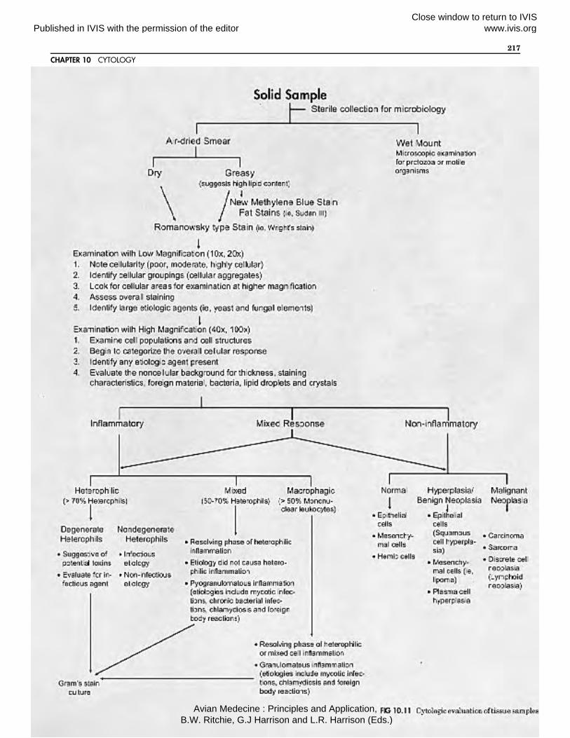
The inflammatory response is classified as eitherheterophilic, mixed-cell or macrophagic inflamma-tion based upon the types of inflammatory cells pre-sent.
Heterophilic inflammation is represented by a pre-dominance of heterophils (greater than 70 percent ofthe inflammatory cells) in the cellular response. Het-erophilic inflammation indicates an acute inflamma-tory response in birds.11 It is important to examinethe heterophils closely for signs of degeneration orphagocytized material.
Degenerate heterophils indicate a toxic microenvi-ronment, usually caused by microbial toxins. Degen-erative changes in heterophils include increased cy-toplasmic basophilia, vacuolation, degranulation andnuclear karyolysis. If bacterial phagocytosis can bedemonstrated, the cytodiagnosis of septic hetero-philic inflammation can be made. If only extracellu-lar bacteria are found, it cannot be determined thatthere is a bacterial etiology since the extracellularbacteria may represent either normal flora (depend-ing upon the location of the inflammation) or con-taminants of the sample.
Because macrophages migrate quickly (within a fewhours of onset) into inflammatory lesions, mixed-cellinflammation is the most commonly found inflamma-tory response in birds.11 Mixed-cell inflammation isrepresented by the presence of heterophils and mono-nuclear leukocytes (eg, macrophages, plasma cellsand lymphocytes). Heterophils represent at least 50percent of the inflammatory cells in mixed-cell in-flammatory responses. Mixed-cell inflammation usu-
ally represents an established, active inflammation.The heterophils in this type of inflammation areusually nondegenerate, suggesting a microenviron-ment free of microbial toxins even though there maybe a bacterial etiology.
Macrophagic inflammation is indicated by the pre-dominance of macrophages (greater than 50 percent)in the inflammatory response. This type of inflamma-tion does not necessarily imply chronicity, but may besuggestive of a number of etiologies (eg, intracellularpathogens). Macrophagic inflammation is common tocertain avian diseases. These include avian tubercu-losis, chlamydiosis, foreign body reaction, mycoticinfections and cutaneous xanthomatosis. Multinu-cleated giant cell formation is often associated withmacrophagic inflammation. Giant cells can appearwithin hours of the onset of some inflammatory re-sponses and, unlike in mammals, their presence doesnot imply chronic inflammation.1,5,12
Tissue Hyperplasia or Benign Neoplasia
Tissue hyperplasia resulting from cellular injury orchronic stimulation is difficult to differentiate frombenign neoplasia based upon cytology. Cells fromhyperplastic tissue appear mature and do not exhibitmuch pleomorphism. They may appear immature byexhibiting increased cytoplasmic basophilia owing tothe increased RNA activity within the cell.14 Prolifer-ating cells may also exhibit an increase in mitoticfigures; however, the nuclear features do not showimmaturity. Examples of tissue hyperplasia, fre-quently seen in birds, include the fibrous and epi-thelial cell proliferation adjacent to chronic inflam-matory lesions, thyroid hyperplasia (especially inbudgerigars) and squamous hyperplasia secondaryto hypovitaminosis A. A common benign neoplasm ofbirds is the lipoma, especially in budgerigars (seeColor 25).
Malignant Neoplasia
Cells obtained from malignant neoplasms show vary-ing degrees of pleomorphism. The severity of themalignancy increases with the greater degree of pleo-morphism. The appearance of the cell nucleus canprovide important clues to the detection of a malig-nant neoplasm.14 Increased nuclear size, which isreflected by an increased nucleus to cytoplasm (N:C)ratio, is suggestive of an abnormal cell. Nuclear an-isocytosis (variation in size) and pleomorphism (vari-able nuclear shapes) are features of malignant cells.Multinucleation can also be a feature of malignancy.
CHAPTER 10 CYTOLOGY
207
Published in IVIS with the permission of the editorClose window to return to IVIS www.ivis.org
Avian Medecine : Principles and Application, B.W. Ritchie, G.J Harrison and L.R. Harrison (Eds.)

The nuclear chromatin may also be abnormal inmalignant cells. Coarse, hyperchromatic chromatinis suggestive of neoplasia. Other nuclear features ofmalignant cells include abnormal nucleoli (very largeor multiple, such as greater than five), irregularnuclear margins, abnormal or increased mitotic fig-ures and abnormal lobation, especially in cells thatnormally do not have lobed nuclei.
Cytoplasmic features of malignant cells include in-creased basophilia, abnormal vacuolation or inclu-sions, decreased volume, variation in cell marginsand variability in the staining.14 Abnormal cytoplas-mic inclusions may include satellite nuclei (smallnuclear fragments) and phagocytized cells.
Once a decision has been made for the cytodiagnosisof malignant neoplasia, an attempt to classify theneoplasm should be made. The four basic classifica-tions of malignant neoplasms based upon cytologicfeatures include carcinomas, sarcomas, discrete-cellneoplasia and poorly differentiated neoplasia.14 Car-cinomas are malignancies of the epithelial cells;therefore, the abnormal cells in the sample havefeatures of epithelial cells. Adenocarcinomas are fre-quently seen in birds, especially ovarian adenocarci-nomas. Cytologic evidence of adenocarcinomas in-cludes epithelial cells that tend to form giant cells,have cytoplasmic secretary vacuoles and tend to oc-cur in aggregates (eg, balls, rosettes or loose group-ings). Sarcomas are malignancies of mesenchymalcells and therefore tend to exfoliate cells poorly. Fi-brosarcomas are the most frequently encounteredsarcomas of birds (see Color 25). Cells from fibrosar-comas are abnormal-appearing fibroblasts, whichare spindle-shaped cells that typically exfoliate assingle cells. Abnormal fibroblasts show increased cel-lular size and N:C ratios, nuclear and cellular pleo-morphism and exfoliation when compared with nor-mal fibrous tissue. Other mesenchymal cellneoplasms such as chondromas, chondrosarcomasand osteogenic sarcomas may produce a heavy eosi-nophilic background material (chondroid or osteoid)that can be seen on the microscope sample.
A common discrete or round cell neoplasm of birds islymphoid neoplasia (see Color 25). The abnormallymphocytes found in this type of neoplasm exfoliateextremely well. Cellular features of malignant lym-phocytic tissue include a marked increase in thenumber of lymphoblasts, nuclear and cellular pleo-morphism, increase in cytoplasmic basophilia andmitotic figures, and abnormal or multiple nucleoli.
Poorly differentiated neoplasms produce cells havingfeatures of malignant neoplasia; however, the cellsare difficult to classify as carcinomas or sarcomas. Insuch cases, a cytodiagnosis of a poorly differentiatedneoplasm is made.
Circumstantial evidence for a malignant neoplasmwithout the demonstration of abnormal cells is seenin older birds (eg, female budgerigars) with a sponta-neous hemoperitoneum and no history of trauma.This is suggestive of an ulcerated neoplasm leadingto abdominal hemorrhage. Ovarian adenocarci-nomas of budgerigars and cockatiels often present inthis manner. Evidence for malignancy may also beobtained by the demonstration of ectopic cells inunusual anatomic areas. An example of this would bethe presence of a large number of cells other thanhepatocytes and hemic cells in a cytologic sample ofthe liver. This is suggestive of a metastatic lesion,even if the cells do not have features of malignantneoplasia.
Mixed Cellular Response
Occasionally, a mixed-cellular response may be seen,especially in areas of ulcerated neoplasms. A cy-tologic sample obtained from an ulcerated neoplasmmay reveal features of malignant neoplasia as wellas inflammation or hemorrhagic effusion.
Cytology of CommonlySampled Fluids and Tissues
Abdominal Fluids
Birds presented with abdominal distention may havean abnormal accumulation of fluid within the perito-neal cavity that may be detected by palpation orradiology. Cytologic evaluation of this fluid is oftenthe main technique for establishing a presumptive ordefinitive diagnosis.
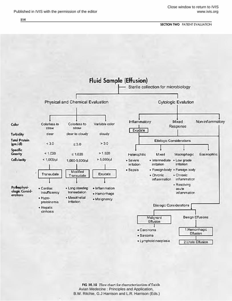
Abdominal effusions can be classified based uponcellularity, types of cells present, protein content,specific gravity and gross appearance. Abdominalfluids are classified as transudates, modified tran-sudates, exudates, hemorrhage and malignant effu-sion.14 Transudates are odorless, transparent fluidscharacterized by a low cellularity (total cell counts
SECTION TWO PATIENT EVALUATION
208
Published in IVIS with the permission of the editorClose window to return to IVIS www.ivis.org
Avian Medecine : Principles and Application, B.W. Ritchie, G.J Harrison and L.R. Harrison (Eds.)
usually less than 1000 /mm3), a specific gravity lessthan 1.020 and a total protein less than 3.0 g/dl.Transudates are typically colorless or have a strawcolor resembling diluted serum. Transudative effu-sions do not clot. These poorly cellular fluids containprimarily macrophages and occasional mesothelialcells. Transudates occur as a result of oncotic pres-sure changes or other circulatory disturbances. Thesame causes for abdominal transudative effusions inmammals most likely occur in birds. These includehepatic cirrhosis, cardiac insufficiency and hypopro-teinemia.
Modified transudates resemble transudative effu-sions; however, they have an increased cellularity(total cell counts usually less than 5000 /mm3 butgreater than 1000 /mm3). The mononuclear leuko-cytes predominate in this type of effusion with occa-sional mesothelial cells and rare heterophils. Themesothelial cells usually appear reactive. Reactivemesothelial cells tend to be round or oval with in-creased cytoplasmic basophilia (Color 10.1). The cellmargins often have a scalloped or villus-like appear-ance. The nuclei have coarse chromatin and promi-nent nucleoli. Multinucleation, cytoplasmic vacuola-tion and mitotic activity are often associated withreactive mesothelial cells. Proliferation of mesothe-lial cells results in the exfoliation of mesothelial cellaggregates that appear as cellular sheets, balls orrosettes (Color 10.2). Care should be taken not tomistake these cells for malignant neoplasia. Modi-fied transudates result from hydrostatic pressurechanges or irritation of long-standing transudativeeffusions. Transudative and modified transudativeeffusions are commonly found in the abdominal cav-ity of mynah birds suffering from hemochromatosis.
Exudative effusions are characterized by high cellu-larity (total cell counts usually greater than 5000/mm3), a specific gravity greater than 1.020 and aprotein content greater than 3.0 g/dl. The majority ofthe cells found in exudative effusions are inflamma-tory cells (Color 10.3). Acute exudative effusionsdemonstrate primarily a heterophilic inflammatoryresponse; however, macrophages quickly move intothe fluid, creating a mixed-cell inflammatory re-sponse within a few hours of onset. Lymphocytes andplasma cells are often seen in long-standing exuda-tive effusions. Exudative effusions vary in color andturbidity. They are frequently viscous, have a foulodor and tend to clot. Abdominal lesions often associ-ated with exudative effusions include septic peritoni-tis, egg-related peritonitis and abdominal malignan-cies.
Hemorrhagic effusions are identified by the presenceof erythrocytic phagocytosis in the fluid sample(Color 10.4). Without demonstration of erythro-phagocytosis, one cannot differentiate hemorrhagefrom peripheral blood contamination of the sample.If thrombocytes are present, the sample was mostlikely contaminated with peripheral blood during thesampling procedure. Thrombocytes disappear rap-idly in hemorrhagic effusions. Proof of erythrophago-cytosis is made by the detection of macrophages thathave phagocytized erythrocytes (suggestive of recenthemorrhage), or that contain iron pigment or he-mosiderin crystals resulting from erythrocyte degra-dation (implying a duration greater than 48 hours).Iron pigment appears as gray to blue-black pigmentin the cytoplasm of macrophages using Wright’sstain. Hemosiderin appears as diamond-shaped,golden crystals within the macrophage cytoplasm.
Malignant effusions have features of either exuda-tive or hemorrhagic effusions, but contain cells com-patible with malignant neoplasia (Color 10.5, 10.6).Abdominal effusions caused by neoplasms are theresult of blockage of blood or lymphatic vessels. Cyst-adenocarcinomas of the ovary of older female birdsare a common cause of malignant effusions. Theseeffusions can resemble hemorrhagic or exudative ef-fusions that contain epithelial cells with features ofmalignant neoplasia. These cells often form cellularaggregates of balls or rosettes and have cytoplasmicsecretory vacuolation.
Urate peritonitis is a rare effusion that can occur inthe abdomen of birds when urinary fluids leak intothe abdominal cavity. The cytology of the acute lesionis poorly cellular but contains a marked number ofsodium and potassium urate crystals. These crystalsare the same ones found in the urate portion of thebird’s droppings. Urate crystals are spherical (2 to 8mm) and have a spoke-wheel appearance. They arealso birefringent under polarized light. The milkyappearance of this type of abdominal effusion resem-bles that of the urate portion of avian droppings. Ifthe bird survives this condition long enough, inflam-matory cells will migrate into the fluid.
Cytology of the Alimentary Tract
The oral cavity, esophagus, ingluvies (crop) and clo-aca are often sampled for cytologic examination. Le-sions in the oral cavity may have different etiologiesbut similar gross appearance. Therefore, sampling oforal lesions for cytologic examination is a quick andsimple procedure for differentiation of these etiolo-
CHAPTER 10 CYTOLOGY
209
Published in IVIS with the permission of the editorClose window to return to IVIS www.ivis.org
Avian Medecine : Principles and Application, B.W. Ritchie, G.J Harrison and L.R. Harrison (Eds.)
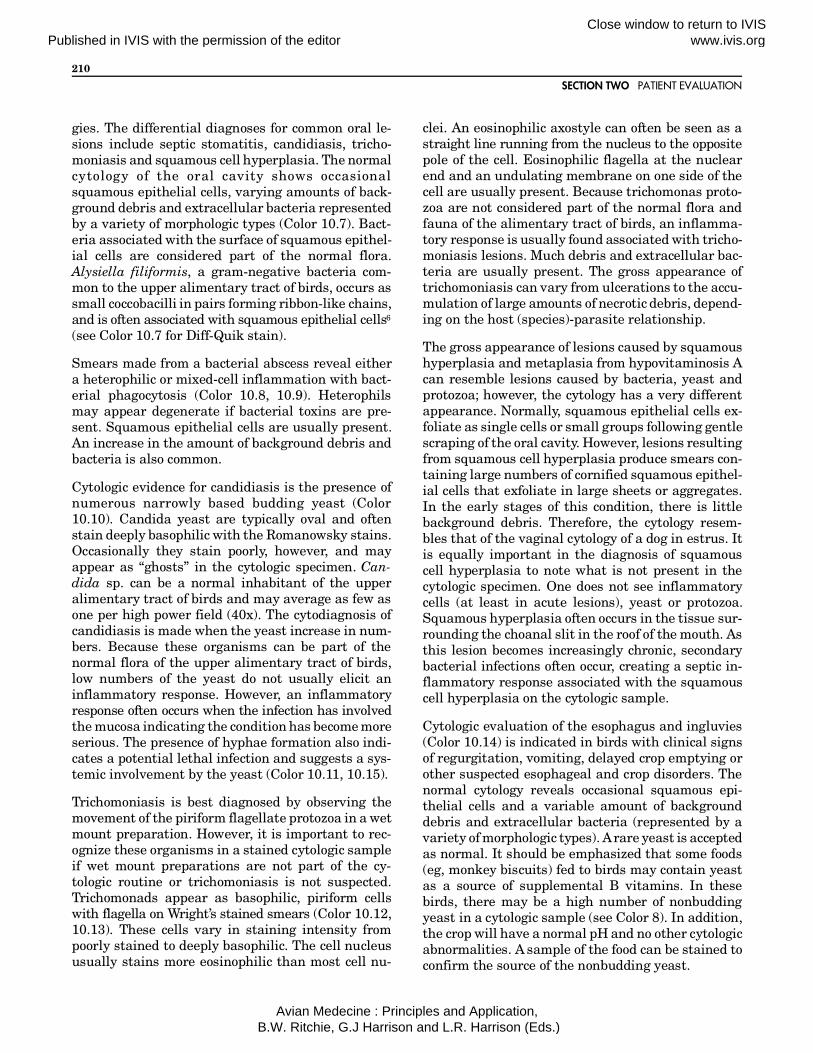
gies. The differential diagnoses for common oral le-sions include septic stomatitis, candidiasis, tricho-moniasis and squamous cell hyperplasia. The normalcytology of the oral cavity shows occasionalsquamous epithelial cells, varying amounts of back-ground debris and extracellular bacteria representedby a variety of morphologic types (Color 10.7). Bact-eria associated with the surface of squamous epithel-ial cells are considered part of the normal flora.Alysiella filiformis, a gram-negative bacteria com-mon to the upper alimentary tract of birds, occurs assmall coccobacilli in pairs forming ribbon-like chains,and is often associated with squamous epithelial cells6
(see Color 10.7 for Diff-Quik stain).
Smears made from a bacterial abscess reveal eithera heterophilic or mixed-cell inflammation with bact-erial phagocytosis (Color 10.8, 10.9). Heterophilsmay appear degenerate if bacterial toxins are pre-sent. Squamous epithelial cells are usually present.An increase in the amount of background debris andbacteria is also common.
Cytologic evidence for candidiasis is the presence ofnumerous narrowly based budding yeast (Color10.10). Candida yeast are typically oval and oftenstain deeply basophilic with the Romanowsky stains.Occasionally they stain poorly, however, and mayappear as “ghosts” in the cytologic specimen. Can-dida sp. can be a normal inhabitant of the upperalimentary tract of birds and may average as few asone per high power field (40x). The cytodiagnosis ofcandidiasis is made when the yeast increase in num-bers. Because these organisms can be part of thenormal flora of the upper alimentary tract of birds,low numbers of the yeast do not usually elicit aninflammatory response. However, an inflammatoryresponse often occurs when the infection has involvedthe mucosa indicating the condition has become moreserious. The presence of hyphae formation also indi-cates a potential lethal infection and suggests a sys-temic involvement by the yeast (Color 10.11, 10.15).
Trichomoniasis is best diagnosed by observing themovement of the piriform flagellate protozoa in a wetmount preparation. However, it is important to rec-ognize these organisms in a stained cytologic sampleif wet mount preparations are not part of the cy-tologic routine or trichomoniasis is not suspected.Trichomonads appear as basophilic, piriform cellswith flagella on Wright’s stained smears (Color 10.12,10.13). These cells vary in staining intensity frompoorly stained to deeply basophilic. The cell nucleususually stains more eosinophilic than most cell nu-
clei. An eosinophilic axostyle can often be seen as astraight line running from the nucleus to the oppositepole of the cell. Eosinophilic flagella at the nuclearend and an undulating membrane on one side of thecell are usually present. Because trichomonas proto-zoa are not considered part of the normal flora andfauna of the alimentary tract of birds, an inflamma-tory response is usually found associated with tricho-moniasis lesions. Much debris and extracellular bac-teria are usually present. The gross appearance oftrichomoniasis can vary from ulcerations to the accu-mulation of large amounts of necrotic debris, depend-ing on the host (species)-parasite relationship.
The gross appearance of lesions caused by squamoushyperplasia and metaplasia from hypovitaminosis Acan resemble lesions caused by bacteria, yeast andprotozoa; however, the cytology has a very differentappearance. Normally, squamous epithelial cells ex-foliate as single cells or small groups following gentlescraping of the oral cavity. However, lesions resultingfrom squamous cell hyperplasia produce smears con-taining large numbers of cornified squamous epithel-ial cells that exfoliate in large sheets or aggregates.In the early stages of this condition, there is littlebackground debris. Therefore, the cytology resem-bles that of the vaginal cytology of a dog in estrus. Itis equally important in the diagnosis of squamouscell hyperplasia to note what is not present in thecytologic specimen. One does not see inflammatorycells (at least in acute lesions), yeast or protozoa.Squamous hyperplasia often occurs in the tissue sur-rounding the choanal slit in the roof of the mouth. Asthis lesion becomes increasingly chronic, secondarybacterial infections often occur, creating a septic in-flammatory response associated with the squamouscell hyperplasia on the cytologic sample.
Cytologic evaluation of the esophagus and ingluvies(Color 10.14) is indicated in birds with clinical signsof regurgitation, vomiting, delayed crop emptying orother suspected esophageal and crop disorders. Thenormal cytology reveals occasional squamous epi-thelial cells and a variable amount of backgrounddebris and extracellular bacteria (represented by avariety of morphologic types). A rare yeast is acceptedas normal. It should be emphasized that some foods(eg, monkey biscuits) fed to birds may contain yeastas a source of supplemental B vitamins. In thesebirds, there may be a high number of nonbuddingyeast in a cytologic sample (see Color 8). In addition,the crop will have a normal pH and no other cytologicabnormalities. A sample of the food can be stained toconfirm the source of the nonbudding yeast.
SECTION TWO PATIENT EVALUATION
210
Published in IVIS with the permission of the editorClose window to return to IVIS www.ivis.org
Avian Medecine : Principles and Application, B.W. Ritchie, G.J Harrison and L.R. Harrison (Eds.)

The same lesions and cytodiagnoses described for theoral cavity also apply to the normal cytologies of theesophagus and crop. Another cytologic indication of adisorder involving the esophagus and crop is thepresence of many bacteria represented by one mor-phologic type (as compared to the normal variety oftypes), even though there is no apparent inflamma-tory response (Color 10.15). This condition is typicalof a peracute ingluvitis, and the disorder is oftenreferred to as “sour crop.” It is indicative of a per-acute bacterial infection, and an inflammatory re-sponse has either not been established or the re-sponse has been overwhelmed and the degenerateheterophils cannot be recognized in the backgrounddebris. The pH is often greater than 7, whereasnormal crop pH is 6.5 to 7. Capillaria ova may bedetected in cytologic samples from the esophagus orcrop of some birds with capillariasis. These ova aredouble operculated and may not stain (see Chapter36).
Examination of the cloacal cytology is indicatedwhenever a disorder of the lower intestinal tract,reproductive tract, urinary tract or cloaca is sus-pected. The normal cytology of the cloaca reveals afew epithelial cells (noncornified squamous or colum-nar), extracellular bacteria (variety of morphologictypes), background debris and urate crystals. Abnor-mal findings would include the presence of inflam-matory cells, large numbers of yeast and a uniformpopulation of bacteria. Because the cloaca is a com-mon opening to the intestinal tract, urinary tract andreproductive tract, cells found in cloacal samplesmay have originated from any of these systems or thecloacal tissue. Therefore, if inflammatory cells arefound, for example, one cannot determine which sys-tem is involved based upon cytologic findings alone.
The use of a speculum and a swab or tube may allowcollection of cytologic samples at the cloacal openingof the intestinal tract, urinary tract or reproductivetract. Uterine samples may be obtained through thecervix, especially in hens that have recently laideggs. Abnormal post-parturient hens (usually show-ing uterine inflammation) may require flushing ofthe uterus with lactated Ringer’s solution until theinflammatory cells disappear from the wash fluid.Cytology of the lower intestinal tract is usuallypoorly cellular with occasional epithelial cells, back-ground debris and a variety of extracellular bacteria.Special stains may be required for the detection ofpathogens, such as Mycobacterium and Giardia spp(see Table 10.1).
The normal fluid excreted from the urinary tract ofbirds is a poorly cellular, cream-colored, thick, mu-coid semisolid (see Color 8). The cytology reveals amarked amount of sodium and potassium urate crys-tals. Abnormal urinary fluid is watery and may con-tain cellular elements such as inflammatory cellsand cellular casts.
Cytology of the Respiratory Tract
The normal cytology of the nasal and infraorbitalsinuses of birds reveals occasional noncornifiedsquamous epithelial cells and low numbers of ex-tracellular bacteria with little background debris.The normal cytology of tracheal wash samples con-sists of a few ciliated respiratory epithelial cells andgoblet cells (Color 10.16, 10.17). An occasionalsquamous epithelial cell may be found. These cellsmay represent cellular contamination from the oralcavity if the end of the tube is not passed directly intothe glottis or they may originate from the syrinx,which contains bistratified squamous epithelium insome birds. Ciliated respiratory epithelial cells arecolumnar or prismatic in shape and have an eccentricnucleus at the small pole of the cell. Eosinophilic ciliaare located at the opposite, larger pole of the cell.Goblet cells are columnar cells with eccentric nuclei.They lack cilia but contain eosinophilic cytoplasmicgranules and vacuoles.
Cytologic evidence for periorbital sinusitis is pro-vided by the presence of inflammatory cells in theaspirate. Lesions with a bacterial etiology are indi-cated by a septic, heterophilic or mixed-cell inflam-mation. Mycotic lesions often reveal either a mixed-cell or macrophagic inflammation with the presenceof fungal elements, such as yeast, hyphae or spores.Sinus infections associated with chlamydia often re-veal a mixed-cell or macrophagic inflammation(Color 10.18). Chlamydial inclusions appear assmall, blue-to-purple spherules, often in dense clus-ters, within the cytoplasm of macrophages whenstained with Wright’s stain. Chlamydial stains, suchas Gimenez or Macchiavello’s stains, may be used toaid in the detection of chlamydia (see Color 10.33).The chlamydial inclusions appear red, and the hostcells appear blue-green with Gimenez stain. Thechlam-ydial elementary bodies stain red, and thelarger initial bodies stain blue with Macchiavello’sstain (see Color 10.34).
A septic tracheobronchitis is identified from a tra-cheal wash sample by the presence of inflammatorycells showing bacterial phagocytosis. An endoscope is
CHAPTER 10 CYTOLOGY
211
Published in IVIS with the permission of the editorClose window to return to IVIS www.ivis.org
Avian Medecine : Principles and Application, B.W. Ritchie, G.J Harrison and L.R. Harrison (Eds.)
excellent for collecting cytologic samples from thetrachea of psittacine birds the size of an Amazonparrot or larger. In severe cases, the ciliated respira-tory epithelial cells appear degenerate. Degeneraterespiratory epithelial cells show loss of cilia, cyto-plasmic vacuolation and karyolysis. Degenerationand fragmentation of the ciliated respiratory epi-thelial cells in association with a macrophagic andlymphocytic inflammation are suggestive of a viraletiology. Inflammation of the trachea and bronchiusually results in an increase in goblet cells andmucin formation, which causes an increased thick-ness to the noncellular background.
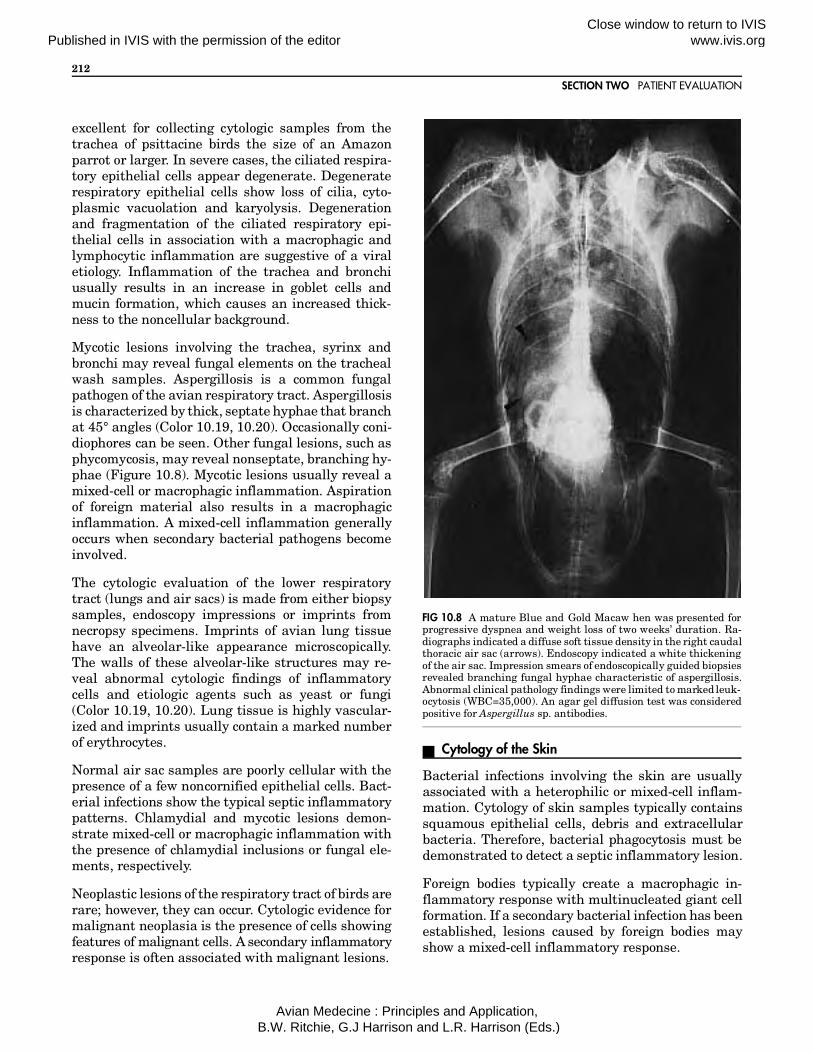
Mycotic lesions involving the trachea, syrinx andbronchi may reveal fungal elements on the trachealwash samples. Aspergillosis is a common fungalpathogen of the avian respiratory tract. Aspergillosisis characterized by thick, septate hyphae that branchat 45° angles (Color 10.19, 10.20). Occasionally coni-diophores can be seen. Other fungal lesions, such asphycomycosis, may reveal nonseptate, branching hy-phae (Figure 10.8). Mycotic lesions usually reveal amixed-cell or macrophagic inflammation. Aspirationof foreign material also results in a macrophagicinflammation. A mixed-cell inflammation generallyoccurs when secondary bacterial pathogens becomeinvolved.
The cytologic evaluation of the lower respiratorytract (lungs and air sacs) is made from either biopsysamples, endoscopy impressions or imprints fromnecropsy specimens. Imprints of avian lung tissuehave an alveolar-like appearance microscopically.The walls of these alveolar-like structures may re-veal abnormal cytologic findings of inflammatorycells and etiologic agents such as yeast or fungi(Color 10.19, 10.20). Lung tissue is highly vascular-ized and imprints usually contain a marked numberof erythrocytes.
Normal air sac samples are poorly cellular with thepresence of a few noncornified epithelial cells. Bact-erial infections show the typical septic inflammatorypatterns. Chlamydial and mycotic lesions demon-strate mixed-cell or macrophagic inflammation withthe presence of chlamydial inclusions or fungal ele-ments, respectively.
Neoplastic lesions of the respiratory tract of birds arerare; however, they can occur. Cytologic evidence formalignant neoplasia is the presence of cells showingfeatures of malignant cells. A secondary inflammatoryresponse is often associated with malignant lesions.
Cytology of the Skin
Bacterial infections involving the skin are usuallyassociated with a heterophilic or mixed-cell inflam-mation. Cytology of skin samples typically containssquamous epithelial cells, debris and extracellularbacteria. Therefore, bacterial phagocytosis must bedemonstrated to detect a septic inflammatory lesion.
Foreign bodies typically create a macrophagic in-flammatory response with multinucleated giant cellformation. If a secondary bacterial infection has beenestablished, lesions caused by foreign bodies mayshow a mixed-cell inflammatory response.
FIG 10.8 A mature Blue and Gold Macaw hen was presented forprogressive dyspnea and weight loss of two weeks’ duration. Ra-diographs indicated a diffuse soft tissue density in the right caudalthoracic air sac (arrows). Endoscopy indicated a white thickeningof the air sac. Impression smears of endoscopically guided biopsiesrevealed branching fungal hyphae characteristic of aspergillosis.Abnormal clinical pathology findings were limited to marked leuk-ocytosis (WBC=35,000). An agar gel diffusion test was consideredpositive for Aspergillus sp. antibodies.
SECTION TWO PATIENT EVALUATION
212
Published in IVIS with the permission of the editorClose window to return to IVIS www.ivis.org
Avian Medecine : Principles and Application, B.W. Ritchie, G.J Harrison and L.R. Harrison (Eds.)

Cutaneous xanthomatosis is a unique condition ofbirds caused by an excessive accumulation of lipidsin the skin (see Color 25). A macrophagic inflamma-tory response with multinucleated giant cells andcholesterol crystals is observed on the cytologic speci-men (Color 10.21, 10.22). Cholesterol crystals appearas angular, translucent crystals that vary in size andshape. They often appear stacked upon each other.Skin affected with xanthomatosis is thickened, yel-low and friable. It is often found in areas whereprevious hemorrhage (eg, feather cysts and skintrauma) or pressure from underlying tumors (eg,lipomas) has occurred (see Color 25).
Subcutaneous lipomas produce a cytologic specimenthat appears “greasy” on the unstained slide. Thecytology reveals numerous lipocytes, which vary insize (Color 10.23). Avian lipocytes often have largecytoplasmic vacuoles in association with clusters ofsmall vacuoles. The vacuoles tend to be round. Thecell nucleus appears pyknotic and pushed to one edgeof the cell, often appearing as if pushed beyond thecell margin. The background material in slides fromlipomas resembles the cytoplasm of the lipocytes andcontains numerous fat droplets. These clear, round,fat droplets usually partially dissolve in the alcohol-based stains (eg, Wright’s stain) but are easily seenin the water-soluble stains such as new methyleneblue. Special fat stains such as Sudan IV can be usedto demonstrate the fat droplets.
The cytology of feather cysts varies, depending uponthe chronicity of the lesion (see Color 24). Earlystages of feather cyst development reveal a markednumber of red blood cells in the sample. Oftenerythrophagocytosis can be found. As the lesion be-comes more chronic and caseous exudation develops,the cytology resembles that of mixed-cell inflamma-tion with a marked amount of background debris andoccasional multinucleated giant cell formation.Feather fragments may also be found.
Cutaneous and subcutaneous malignant neoplasmsare rare in birds, but can be detected on cytologicexamination. Lymphoid neoplasia produces a highlycellular sample of immature lymphocytes (Color10.24). These lymphoblasts and prolymphocytes arelarge, round cells that exfoliate as single cells. Theyhave large nuclei with fine chromatin and multipleor large prominent nucleoli. The cytoplasm stainsbasophilic. Bizarre-appearing lymphocytes and mi-totic figures may also be present. The background oflymphoid tissue, such as lymphoid neoplasms, typi-cally contains small, irregular, blue cytoplasmic frag-
ments. Finding these fragments may be helpful inthe cytologic identification of lymphoid tissue.
Cutaneous melanosarcomas have also been found inbirds. Poorly differentiated melanosarcomas revealmesenchymal cells that contain few cytoplasmicmelanin granules. The gross appearance of the in-volved skin shows dark pigmentation. The malignantcells usually exfoliate as single cells, and the back-ground may contain melanin granules from rupturedcells. The round melanin granules vary from black todark brown to golden in color.
Avian poxvirus lesions reveal clusters of squamousepithelial cells that contain large cytoplasmic vacu-oles (Color 10.25). The large cytoplasmic vacuolesfound in the affected squamous cell push the cellnucleus to the cell margin. These vacuoles representthe ballooning degeneration of the squamous epithe-lium typical of pox lesions. These cytoplasmic vacu-oles often contain small, pale eosinophilic inclusionswith oil immersion examination of Wright’s stainedsmears. A secondary septic inflammatory response isoften associated with ulcerated pox lesions.
Cytology of the Cornea and Conjunctiva
Normal conjunctival scrapings provide poorly cellu-lar samples with little background material. Thecells normally found are epithelial cells that maycontain intracytoplasmic pigment granules. The nor-mal cytology of the cornea is also poorly cellular andconsists of occasional noncornified squamous epithel-ial cells. Inflammatory lesions involving the corneaand conjunctiva reveal inflammatory cells and in-creased numbers of exfoliated epithelial cells. Theepithelial cells often demonstrate degenerativechanges, such as cytoplasmic vacuolation, karyolysisor karyorrhexis. Chronic inflammatory lesions mayshow an increase in the number of epithelial cellsthat contain pigment granules. Chronic lesions mayalso reveal the presence of cornified squamous epi-thelial cells that are not normally found in the con-junctiva or cornea (Figure 10.9).
Cytology of Synovial Fluid
The amount of fluid in synovial joints of most birds isnormally too small for sampling; however, an abnor-mal accumulation of joint fluid may provide enoughsample for evaluation. Normal synovial fluid ispoorly cellular. The cells are mononuclear cells, rep-resenting either synovial lining cells or mononuclearleukocytes. The background of normal synovial fluid
CHAPTER 10 CYTOLOGY
213
Published in IVIS with the permission of the editorClose window to return to IVIS www.ivis.org
Avian Medecine : Principles and Application, B.W. Ritchie, G.J Harrison and L.R. Harrison (Eds.)

cytology consists of a heavy, granular, eosinophilicsubstance representing the mucin in the fluid.
An increase in the inflammatory cells and change inthe color, clarity, and viscosity of the fluid is indica-tive of inflammatory joint lesions (see Figure 12.77).There may be a decrease in the granular eosinophilicbackground material, suggesting a decrease in mucincontent. Erosion of the articular cartilage may resultin the presence of multinucleated osteoclasts in thesynovial fluid. Spindle-shaped fibroblasts suggesterosion into the fibrous layer of the articular capsule.Septic joint lesions may demonstrate bacterialphagocytosis by leukocytes (primarily heterophils).An increase in the number of inflammatory cells,especially heterophils, is also seen with traumaticarthritis. The presence of erythrocytes and erythro-phagocytosis is supportive of a cytodiagnosis of hem-arthrosis.
Articular gout produces a cream-to-yellow-coloreddeposit in affected joints (see Color 21). The cytologyof this material reveals numerous, needle-shapedcrystals (monosodium urate) (Color 10.26). Thesecrystals are birefringent under polarized light. Theyoccasionally stain eosinophilic with Wright’s stain.Inflammatory cells are often present and the mucincontent is often reduced, as reflected in the reductionin the amount of eosinophilic granular background.
Cytology of Internal Organs
The liver, kidney and spleen are occasionally sam-pled by biopsy to achieve an antemortem diagnosis inbirds. Cytologic evaluation should also be performedwhenever lesions involving these organs are found onpostmortem examinations.
Birds typically do not have lymph nodes as found inmammals. Avian lymphoid tissue appears as lym-phoid aggregates in the walls of the intestines, inter-nal organs (especially the spleen and liver) and skin.The cloacal bursa of young birds is a sac-like lym-phoid nodule found in the dorsal wall of the proc-todeum of the cloaca (see Figure 5.6). The cytology ofnormal lymphoid tissue shows a predominance ofsmall mature lymphocytes (greater than 90 percentof the lymphoid cells) (Color 10.27). The larger pro-lymphocytes, lymphoblasts and plasma cells nor-mally occur in low numbers. Reactive lymphoid tis-sue demonstrates an increase in the number ofimmature lymphocytes (prolymphocytes and lym-phoblast) and plasma cells (Color 10.28). Reactivityof the lymphoid tissue is suggestive of antigenicstimulation of the immune system. Lymphoid hyper-plasia causes an increase in the lymphoid tissuemass; however, the cytology appears normal with theexception of a slight increase in the number of pro-lymphocytes. Lymphoid neoplasia produces amarked increase in the number of immature lympho-cytes, especially lymphoblasts, in the cytologic speci-men. The neoplastic cells may show varying degreesof cellular features of malignant neoplasia. There isusually an increase in the number of mitotic figuresin samples obtained from lymphoid neoplasia.
Cytologic samples of the liver are usually highlycellular with a predominance of hepatocytes, eryth-rocytes and free nuclei. Depending upon the locationof sampling, there may be numerous lymphocytespresent. Hepatocytes are large epithelial cells thatoccur in sheets or clusters or as single cells. Normalhepatic cytology reveals uniform-appearing hepato-cytes. These cells have an abundant, basophilic,finely granular cytoplasm and a round-to-oval,slightly eccentric nucleus. Binucleation is occasion-ally seen. Hepatocytes are easily ruptured duringslide preparation; therefore, the background of he-patic tissue resembles that of the hepatocyte cyto-plasm, and many free nuclei are commonly seen.Normal hematopoiesis is occasionally found becausethe liver is a common location for ectopic hematopoi-esis. Also, macrophages containing iron pigment (he-mosiderin) are occasionally seen.
FIG 10.9 An adult canary was presented with bilateral epiphoraand mild conjunctivitis. Cytologic evaluation of conjunctiva scrap-ings may have been helpful in determining an etiology for thisbird’s problems. This bird responded to treatment with tylosin(courtesy of Michael Murray).
SECTION TWO PATIENT EVALUATION
214
Published in IVIS with the permission of the editorClose window to return to IVIS www.ivis.org
Avian Medecine : Principles and Application, B.W. Ritchie, G.J Harrison and L.R. Harrison (Eds.)

Inflammatory lesions of the liver reveal numerousmature heterophils and an increase in the number ofmacrophages and plasma cells (Color 10.29). It isimportant not to confuse normal ectopic granulopoi-esis with heterophilic inflammation. If developingstages of the heterophils can be found, the cytology isrepresentative of granulocytopoiesis (see Chapter 9).If the heterophils are mature cells, however, then thecytology indicates inflammation. The hepatocytesmay demonstrate degenerative changes in the pres-ence of hepatic inflammation.
Avian tuberculosis produces a macrophagic inflam-matory response in the liver (see Color 20). Thecytology reveals numerous macrophages and mult-inucleated giant cells. When stained with Roma-nowsky stain, the background of the smear containsnumerous large bacterial rods that do not stain.Likewise, macrophages may contain numerous bact-erial rods that do not stain (Color 10.30). Becausemycobacterium have a waxy cell wall, they do notstain with routine cytology stains. Therefore, an acid-fast stain is required to demonstrate the tuberclebacilli, which stain red (Color 10.31). However, thepresence of a macrophagic inflammation with mult-inucleated giant cells and “ghost-like” bacterial rodsprovides a presumptive diagnosis for tuberculosis.
Avian chlamydiosis often results in a mixed-cell ormacrophagic inflammation in the spleen or liver witha marked increase in the number of plasma cells(Color 10.28). Small, blue-to-purple, intracytoplas-mic inclusions suggestive of chlamydial elementaryand initial bodies may be seen in macrophages (Color10.32). Confirmation of chlamydial inclusions isaided by special stains (eg, Gimenez or Macchiavello’s).
Hepatic lipidosis produces cytologic specimens thatappear “greasy” on gross examination. The stainedsmears reveal enlarged hepatocytes that containround, cytoplasmic vacuoles (Color 10.35). The back-ground material also contains these round vacuolessuggestive of lipid material.
Primary neoplasm of the liver reveals hepatocytesshowing features of malignant neoplasia. Affectedcells are usually pleomorphic with deep, cytoplasmicbasophilia and immature-appearing nuclei (eg,smooth nuclear chromatin and multiple or largeprominent nucleoli). Ectopic cells that show featuresof malignant neoplasia may also be found and areindicative of a metastatic lesion in the liver.
Occasionally, parasites may be found on splenic he-patic imprints (Color 10.36). Those commonly seenare schizogony of Haemoproteus and Leukocytozoon,sporozoites of Atoxoplasma and microfilaria.
Normally, cytology of the spleen shows a markednumber of erythrocytes and lymphocytes, reflectingthe cytology of a lymphoid tissue. Macrophages arealso present and occasionally contain iron pigmentfrom erythrophagocytosis of senescent red cells. Ex-cessive splenic iron pigment is seen in birds withhemolytic anemia owing to increased red cell degra-dation by the spleen (Color 10.37). Chlamydial infec-tions often cause a marked increase in the number ofsplenic plasma cells. Macrophages often demon-strate intracytoplasmic chlamydial inclusions. De-velopmental stages of blood parasites may also befound in splenic samples (see Color 9). Systemic bac-terial or fungal infections may result in an increasein the number of inflammatory cells, especially ma-ture heterophils, in the spleen. Often, the etiologicagent can be found either within the leukocytes or inthe noncellular background.
The normal kidney produces a highly cellular samplethat contains numerous epithelial cells with anabundant, slightly basophilic cytoplasm and slightlyeccentric, round-to-oval nuclei. Numerous erythro-cytes and free cell nuclei are usually present. Uratecrystals are also common. Abnormal cytology in-cludes an increase in the number of inflammatorycells or the presence of cells having features of neo-plasia. Epithelial cells from renal adenomas showincreased cytoplasmic basophilia, slight pleomor-phism and occasional mitotic figures. Renal adeno-carcinomas produce epithelial cells having featuresof malignant neoplasia. Nephroblastomas (em-bryonal nephroma) produce poorly differentiated epi-thelial and mesenchymal cells. The cuboidal epi-thelial cells are associated with spindle-shaped cellsof the fibrous stroma, and the background may con-tain a heavy, eosinophilic substance. This back-ground material is suggestive of a cellular attempt toproduce a matrix (eg, chondroid or osteoid).
Products Mentioned in the Texta. Cytospin, Shandon Southern Instruments, Sewickley, PAb. Calgiswab, Inolex, Glenwood, IL
CHAPTER 10 CYTOLOGY
215
Published in IVIS with the permission of the editorClose window to return to IVIS www.ivis.org
Avian Medecine : Principles and Application, B.W. Ritchie, G.J Harrison and L.R. Harrison (Eds.)
SECTION TWO PATIENT EVALUATION
216
Published in IVIS with the permission of the editorClose window to return to IVIS www.ivis.org
Avian Medecine : Principles and Application, B.W. Ritchie, G.J Harrison and L.R. Harrison (Eds.)
CHAPTER 10 CYTOLOGY
217
Published in IVIS with the permission of the editorClose window to return to IVIS www.ivis.org
Avian Medecine : Principles and Application, B.W. Ritchie, G.J Harrison and L.R. Harrison (Eds.)
Cytology
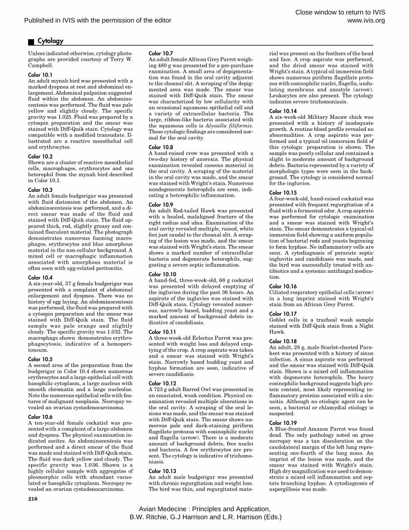
Unless indicated otherwise, cytology photo-graphs are provided courtesy of Terry W.Campbell.
Color 10.1An adult mynah bird was presented with amarked dyspnea at rest and abdominal en-largement. Abdominal palpation suggestedfluid within the abdomen. An abdomino-centesis was performed. The fluid was paleyellow and slightly cloudy. The specificgravity was 1.025. Fluid was prepared by acytospin preparation and the smear wasstained with Diff-Quik stain. Cytology wascompatible with a modified transudate. Il-lustrated are a reactive mesothelial celland erythrocytes.
Color 10.2Shown are a cluster of reactive mesothelialcells, macrophages, erythrocytes and oneheterophil from the mynah bird describedin Color 10.1.
Color 10.3An adult female budgerigar was presentedwith fluid distension of the abdomen. Anabdominocentesis was performed, and a di-rect smear was made of the fluid andstained with Diff-Quik stain. The fluid ap-peared thick, red, slightly greasy and con-tained flocculent material. The photographdemonstrates numerous foaming macro-phages, erythrocytes and blue amorphousmaterial in the non-cellular background. Amixed cell or macrophagic inflammationassociated with amorphous material isoften seen with egg-related peritonitis.
Color 10.4A six-year-old, 37 g female budgerigar waspresented with a complaint of abdominalenlargement and dyspnea. There was nohistory of egg laying. An abdominocentesiswas performed, the fluid was prepared witha cytospin preparation and the smear wasstained with Diff-Quik stain. The fluidsample was pale orange and slightlycloudy. The specific gravity was 1.032. Themacrophage shown demonstrates erythro-phagocytosis, indicative of a hemoperi-toneum.
Color 10.5A second area of the preparation from thebudgerigar in Color 10.4 shows numerouserythrocytes and a large epithelial cell withbasophilic cytoplasm, a large nucleus withsmooth chromatin and a large nucleolus.Note the numerous epithelial cells with fea-tures of malignant neoplasia. Necropsy re-vealed an ovarian cystadenocarcinoma.
Color 10.6A ten-year-old female cockatiel was pre-sented with a complaint of a large abdomenand dyspnea. The physical examination in-dicated ascites. An abdominocentesis wasperformed and a direct smear of the fluidwas made and stained with Diff-Quik stain.The fluid was dark yellow and cloudy. Thespecific gravity was 1.036. Shown is ahighly cellular sample with aggregates ofpleomorphic cells with abundant vacuo-lated or basophilic cytoplasm. Necropsy re-vealed an ovarian cystadenocarcinoma.
Color 10.7An adult female African Grey Parrot weigh-ing 480 g was presented for a pre-purchaseexamination. A small area of depigmenta-tion was found in the oral cavity adjacentto the choanal slit. A scraping of the depig-mented area was made. The smear wasstained with Diff-Quik stain. The smearwas characterized by low cellularity withan occasional squamous epithelial cell anda variety of extracellular bacteria. Thelarge, ribbon-like bacteria associated withthe squamous cells is Alysiella filiformis.These cytologic findings are considered nor-mal for the oral cavity.
Color 10.8A hand-raised crow was presented with atwo-day history of anorexia. The physicalexamination revealed caseous material inthe oral cavity. A scraping of the materialin the oral cavity was made, and the smearwas stained with Wright’s stain. Numerousnondegenerate heterophils are seen, indi-cating a heterophilic inflammation.
Color 10.9An adult Red-tailed Hawk was presentedwith a healed, malaligned fracture of theright radius and ulna. Examination of theoral cavity revealed multiple, raised, whitefoci just caudal to the choanal slit. A scrap-ing of the lesion was made, and the smearwas stained with Wright’s stain. The smearshows a marked number of extracellularbacteria and degenerate heterophils, sug-gesting a severe septic inflammation.
Color 10.10A hand-fed, three-week-old, 68 g cockatielwas presented with delayed emptying ofthe ingluvies during the past 36 hours. Anaspirate of the ingluvies was stained withDiff-Quik stain. Cytology revealed numer-ous, narrowly based, budding yeast and amarked amount of background debris in-dicative of candidiasis.
Color 10.11A three-week-old Eclectus Parrot was pre-sented with weight loss and delayed emp-tying of the crop. A crop aspirate was takenand a smear was stained with Wright’sstain. Narrowly based budding yeast andhyphae formation are seen, indicative ofsevere candidiasis.
Color 10.12A 723 g adult Barred Owl was presented inan emaciated, weak condition. Physical ex-amination revealed multiple ulcerations inthe oral cavity. A scraping of the oral le-sions was made, and the smear was stainedwith Diff-Quik stain. The smear shows nu-merous pale and dark-staining piriformflagellate protozoa with eosinophilic nucleiand flagella (arrow). There is a moderateamount of background debris, free nucleiand bacteria. A few erythrocytes are pre-sent. The cytology is indicative of trichomo-niasis.
Color 10.13An adult male budgerigar was presentedwith chronic regurgitation and weight loss.The bird was thin, and regurgitated mate-
rial was present on the feathers of the headand face. A crop aspirate was performed,and the dried smear was stained withWright’s stain. A typical oil immersion fieldshows numerous piriform flagellate proto-zoa with eosinophilic nuclei, flagella, undu-lating membrane and axostyle (arrow).Leukocytes are also present. The cytologyindicates severe trichomoniasis.
Color 10.14A six-week-old Military Macaw chick waspresented with a history of inadequategrowth. A routine blood profile revealed noabnormalities. A crop aspirate was per-formed and a typical oil immersion field ofthis cytologic preparation is shown. Thesample was poorly cellular and contained aslight to moderate amount of backgrounddebris. Bacteria represented by a variety ofmorphologic types were seen in the back-ground. The cytology is considered normalfor the ingluvies.
Color 10.15A four-week-old, hand-raised cockatiel waspresented with frequent regurgitation of afluid with a fermented odor. A crop aspiratewas performed for cytologic examinationand a smear was stained with Wright’sstain. The smear demonstrates a typical oilimmersion field showing a uniform popula-tion of bacterial rods and yeasts beginningto form hyphae. No inflammatory cells areseen. A cytodiagnosis of peracute septicingluvitis and candidiasis was made, andthe bird was successfully treated with an-tibiotics and a systemic antifungal medica-tion.
Color 10.16Ciliated respiratory epithelial cells (arrow)in a lung imprint stained with Wright’sstain from an African Grey Parrot.
Color 10.17Goblet cells in a tracheal wash samplestained with Diff-Quik stain from a NightHawk.
Color 10.18An adult, 28 g, male Scarlet-chested Para-keet was presented with a history of sinusinfection. A sinus aspirate was performedand the smear was stained with Diff-Quikstain. Shown is a mixed cell inflammationwith degenerate heterophils. The heavyeosinophilic background suggests high pro-tein content, most likely representing in-flammatory proteins associated with a sin-usitis. Although no etiologic agent can beseen, a bacterial or chlamydial etiology issuspected.
Color 10.19A Blue-fronted Amazon Parrot was founddead. The only pathology noted on grossnecropsy was a tan discoloration on thecaudolateral margin of the left lung repre-senting one-fourth of the lung mass. Animprint of the lesion was made, and thesmear was stained with Wright’s stain.High dry magnification was used to demon-strate a mixed cell inflammation and sep-tate branching hyphae. A cytodiagnosis ofaspergillosis was made.
218
Published in IVIS with the permission of the editorClose window to return to IVIS www.ivis.org
Avian Medecine : Principles and Application, B.W. Ritchie, G.J Harrison and L.R. Harrison (Eds.)

Published in IVIS with the permission of the editorClose window to return to IVIS www.ivis.org
Avian Medecine : Principles and Application, B.W. Ritchie, G.J Harrison and L.R. Harrison (Eds.)

Published in IVIS with the permission of the editorClose window to return to IVIS www.ivis.org
Avian Medecine : Principles and Application, B.W. Ritchie, G.J Harrison and L.R. Harrison (Eds.)

Cytology
Color 10.20An adult, 520 g African Grey Parrot on anall-seed diet was presented because it nolonger growled, a typical behavior whenapproached. A tracheal wash sample wascollected, and a smear was prepared by acytospin preparation and stained with Diff-Quik stain. A highly cellular sample con-taining numerous erythrocytes is illus-trated. There are multinucleated giant cellsand septate fungal hyphae, indicative of amycotic infection involving the respiratorytract.
Color 10.21An adult, 1342 g Green-winged Macaw waspresented with a complaint of feather losson the distal end of the right wing. A feathercyst had been removed from this area threemonths earlier. Physical examination re-vealed thickened, yellow, friable skin on thedorsal aspect of the right metacarpus. Acontact smear of the excisional biopsy of theabnormal skin was obtained and stainedwith Diff-Quik stain. Multinucleated giantcells and macrophages (arrow) on a heavygranular background are demonstrated.
Color 10.22Multinucleated giant cells and cholesterolcrystals (arrow) from the bird described inColor 10.21. These findings are compatiblewith xanthomatosis, which typically re-veals a macrophagic inflammation withmultinucleated giant cell formation.
Color 10.23The physical examination of a 30 g, adult,male budgerigar (fed an all-seed diet) re-vealed a large, firm, subcutaneous massoverlying the keel. A fine-needle aspirationbiopsy of the mass was made, and a smearwas stained with Diff-Quik stain. Typicalavian lipocytes (arrow) are shown. The cy-tology is compatible with a lipoma.
Color 10.24A 23-year-old, 320 g Spectacled AmazonParrot was presented with a marked swel-ling around the right eye and feather losson the head. A fine-needle aspiration biopsyof the mass on the head was performed, andthe smear was stained with Diff-Quik stain.The highly cellular sample shown containsnumerous lymphocytes. The majority of thelymphocytes are large, immature and fre-quently show mytotic activity. These find-ings are indicative of lymphoid neoplasia.
Color 10.25An adult, 112 g Ring Dove was presented ina morbid condition and died soon after thephysical examination. The bird was housedin an outdoor aviary, and was presented formarked depression and multiple, raised,irregular cutaneous lesions on the head,legs and feet. A fine-needle aspiration bi-opsy of a raised lesion near the right eyewas made, and the smear was stained withDiff-Quik stain. Shown are epithelial cellswith large cytoplasmic vacuoles (arrow)typical of avian poxvirus lesions.
Color 10.26An adult, 160 g Nanday Conure was pre-sented with a swollen left tibiotarsal and
tarsometatarsal joint and left leg lameness.Arthrocentesis of the affected joint was per-formed and the smear was stained withDiff-Quik stain. The sample contains nu-merous free nuclei, possibly from rupturederythrocytes, and needle-like crystals. Thecytology is compatible with articular gout.
Color 10.27An imprint of a normal spleen from a KingPenguin that was euthanatized because ofa severe skeletal deformity. Note the pre-dominance of small-to-medium, maturelymphocytes, one lymphoblast and twoplasma cells.
Color 10.28A splenic imprint from the Yellow-napedAmazon Parrot described in Color 10.32-10.34 shows a marked increase in plasmacells, indicative of reactive lymphoid tissue.
Color 10.29A four-year-old male budgerigar was pre-sented for bilateral leg paralysis. Wholebody radiographs revealed a large mass inthe area of the kidneys, and a presumptivediagnosis of renal neoplasia was made. Atthe owner’s request, the bird was euthana-tized. Necropsy revealed a large, locallyinvasive mass that appeared to involveboth kidneys. A histologic diagnosis ofnephroblastoma was made. Necropsy alsorevealed a slight discoloration of the liver,which appeared pale. The imprint of theliver shown here reveals normal-appearinghep-atocytes and erythrocytes. There isalso an increased number of mature hetero-phils present, suggesting a mild hetero-philic inflammation and hepatitis. Histol-ogy confirmed the hepatitis; however, noetiology could be determined.
Color 10.30An adult, female, 370 g Blue-fronted Ama-zon Parrot was presented for necropsy. Thebird had shown no previous signs of illness.Necropsy revealed multiple, raised lesionsthroughout the liver parenchyma. The le-sions were varied in size and tended to beround. No other gross lesions were seen. Acontact smear of the liver was made, andthe smear was stained with Diff-Quikstain. Shown is a typical oil immersion fielddemonstrating numerous macrophagesand bacterial rods in the background thatdid not stain.
Color 10.31Acid-fast-positive reaction (arrow) of thebacteria in the Blue-fronted Amazon Parrotdescribed in Color 10.30, supportive of adiagnosis of avian tuberculosis.
Color 10.32An adult, 270 g Yellow-naped Amazon Par-rot was presented in critical condition witha history of anorexia, weight loss, frequentregurgitation and loose droppings withgreen urates. Whole body radiographs re-vealed hepatomegaly and splenomegaly.The blood profile indicated marked leuko-cytosis, toxic heterophilia and marked ele-vations in serum AST. The bird died withinone hour of presentation. Necropsy re-vealed a marked reduction of the pectoral
muscle mass, sinusitis, cloudy air sacs,hep-atomegaly and splenomegaly. An im-print of the enlarged spleen was made, andthe smear was stained with Diff-Quikstain. A marked number of plasma cellswas noted in the splenic imprint (see Color10.28). Also shown is a macrophage thatcontains small, intracytoplasmic inclu-sions suggestive of chlamydia.
Color 10.33Chlamydial inclusions stained with Gime-nez stain.
Color 10.34Chlamydial inclusions stained with Mac-chiavello’s stain.
Color 10.35An obese, five-year-old, 125 g female cocka-tiel was presented for marked lethargy anddyspnea. Whole body radiographs revealedhepatomegaly. The blood profile revealed alipemic serum sample with a normal CBCand chemistry profile. A biopsy of the liverwas performed and the smear was stainedwith Diff-Quik stain. Shown is the typicalappearance of the hepatocyte, which wasenlarged and contained numerous vacu-oles. The background contained round, fatdroplets. The cytology is compatible withhepatic lipidosis.
Color 10.36A severely debilitated, adult, male Ameri-can Kestrel was presented with an openfracture of the right proximal humerus.The peripheral blood smear revealed amarked number of Haemoproteus gameto-cytes. The bird died 24 hours after presen-tation and an imprint of the spleen wasmade and stained with Diff-Quik stain.Round Haemoproteus schizonts (arrow)were found throughout the splenic imprintas shown here. There is also a large amountof dark-blue iron pigment present.
Color 10.37An adult African Grey Parrot housed in apet store was presented with a history oflethargy and anorexia. A blood profile re-vealed a moderate degenerative anemia(PCV=21%). The bird died within six hoursof presentation. Gross necropsy revealed aslightly enlarged spleen. A splenic imprintwas made and stained with Diff-Quikstain. Illustrated is a typical oil immersionfield of the splenic imprint showing amarked amount of dark-blue iron pigment.The large amount of iron pigment was alsoseen histologically. This finding is sugges-tive of a hemolytic anemia; however, theetiology could not be found.
Color 10.38Impression smear from the spleen of a my-nah bird. Wright’s stain was used to dem-onstrate Atoxoplasma sp. in macrophages.Note that the organism is causing indenta-tion of the nucleus of the infected macro-phages (courtesy of Carol Partington).
221
Published in IVIS with the permission of the editorClose window to return to IVIS www.ivis.org
Avian Medecine : Principles and Application, B.W. Ritchie, G.J Harrison and L.R. Harrison (Eds.)

TABLE 10.3 Staining Procedures
Acid-Fast Stain1. Air-dry then gently heat fix2. Cover with carbol fuchsin3. Steam over water bath (3 to 5 min.)4. Rinse with tap water5. Decolorize with acid alcohol until most red color is removed6. Rinse twice in tap water7. Cover with methylene blue stain (1 min.)8. Gently rinse with tap water (air dry)
Gram’s Stain1. Air dry and gently heat fix slide2. Cover with crystal violet (1 min.)3. Gently rinse in tap water4. Cover with Gram’s iodine (1 min.)5. Gently rinse in tap water6. Decolorize with 95% ethyl alcohol (15 to 30 sec.)7. Gently rinse in tap water8. Cover with safranin (1 min.)9. Gently rinse in tap water (air dry)
Macchiavello’s Stain1. Air dry then heat fix2. Cover with basic fuchsin (5 min.)3. Quickly rinse in tap water4. Dip in citric acid one to ten times (1 to 3 sec.)5. Rinse in tap water6. Cover with methylene blue (20 to 30 sec.)7. Rinse in tap water (air dry)
Modified Gimenez Stain1. Air dry then heat fix2. Cover with carbol fuchsin (1 to 2 min.)3. Rinse in tap water4. Cover with malachite green (6 to 9 sec.)5. Rinse in tap water6. Recover with malachite green (6 to 9 sec.)7. Rinse with tap water (air dry)
Sudan III Stain1. Apply stain to wet or dry smear2. Apply coverslip
New Methylene Blue Stain1. Completely air dry or use as a wet mount2. Apply small drop of stain3. Add coverslip
Stamp Stain1. Air dry smear then heat fix2. Cover for 10 min. with carbolated fuchsin as used for Gram’s stain diluted 1:4 with water3. Rinse with tap water4. Differentiate in 0.5% H2SO4 until the preparation looks gray; time according to thickness of the smear5. Counterstain with 5% malachite green or methylene blue (15 sec.)6. Rinse with tap water (air dry)
Wright’s Stain1. Air dry slide2. Flood with Wright’s stain (stand 1 to 3 min.)3. Add equal amount of Wright’s buffer4. Gently mix by blowing until a metallic green sheen is formed5. Allow to stand twice as long as step two (2 to 6 min.)6. Rinse with tap water (air dry)
Diff-Quik Stain1. Air dry slide2. Dip in fixative five times (1 sec. each)3. Dip in solution one to five times (1 sec. each)4. Dip in solution two to five times (1 sec. each)5. Rinse in distilled water (air dry)
Giemsa Stain1. Air dry slide2. Fix in methyl or ethyl alcohol (2 to 7 min.)3. Air dry4. Immerse in Giemsa stain (15 to 40 min.)5. Rinse in tap water (air dry)
References and Suggested Reading
1.Awandhiya RP, Vegad JL, Kolte GN: Amicroscopic study of increased vascu-lar permeability and leukocyte emi-gration in the chicken wing web. ResVet Sci 31:231-235, 1981.
2.Awandhiya RP, Vegad JL, Kolte GN:Eosinophilic leukocytic response indinitrochlorobenzene skin hypersensi-tivity reaction in the chicken. AvianPathol 11:187-194, 1982.
3.Banks WJ: Histology and Compara-tive Organology: A Text Atlas. Balti-more, Williams and Wilkins, 1974.
4.Campbell TW: Avian Hematology andCytology. Ames, Iowa State Univer-sity Press, 1988.
5.Jortner BS, Adams WR: Turpentine-in-duced inflammation in the chicken: Alight and electronmicroscopy studywith emphasis on the macrophage,epithelioid cell, and multinucleatedgiant cell reaction. Avian Dis 15:533-550, 1971.
6.Kaiser GE, Starzyk MJ: Ultrastructureand cell division of an oral bacteriumresembling Alysiella filiformis. Can JMicrobiol 19:325-327, 1972.
7.Maxwell MH, Siller WG, Mackenzie GM:Eosinophilia associated with facial oe-dema in fowls. Vet Record 105:232-233, 1979.
8.Maxwell MH: Attempted induction ofan avian eosinophilia using variousagents. Res Vet Sci 29:293-297, 1980.
9.Maxwell MH: Histochemical identifi-cation of tissue eosinophils in the in-flammatory response of the fowl Gal-lus domesticus. Res Vet Sci 37:7-11,1984.
10.Maxwell MH, Burns RB: Experimentalstimulation of eosinophil stimulationand of eosinophil production in thedomestic fowl. Res Vet Sci 41:114-123, 1986.
11.Montali RJ: Comparative pathology ofinflammation in the higher verte-
brates (reptiles, birds, and mam-mals). J Comp Path 99(1):1-26, 1988.
12.Olah I, McCorkle F, Glick V: Lectin-in-duced giant cell formation in thechicken wattle. Poult Sci 59:2142-2157, 1980.
13.Perman V, Alsaker RD, Riis RC: Cytol-ogy of the Dog and Cat. South Bend,American Animal Hospital Associa-tion, 1979, pp 4-5.
14.Rebar AH: Handbook of VeterinaryCytology. Saint Louis, Ralston Pu-rina Co, 1979, pp 15-50.
SECTION TWO PATIENT EVALUATION
222
Published in IVIS with the permission of the editorClose window to return to IVIS www.ivis.org
Avian Medecine : Principles and Application, B.W. Ritchie, G.J Harrison and L.R. Harrison (Eds.)