Embed Size (px)
DESCRIPTION
jurnal
Citation preview
1
Bell’s Palsy – Medical Treatment and Influence of Prognostic Factors
Sara Axelsson
DOCTORAL DISSERTATION
by due permission of the Faculty of Medicine Lund University, Sweden. To be defended at Belfragesalen, BMC, Lund. Date 18 may 2013 at 9:00 am.
Faculty opponent
Professor Sten Hellström,

2
3
Bell’s Palsy – Medical Treatment and Influence of Prognostic Factors
Sara Axelsson
4
Department of Otorhinolaryngology, Head and Neck Surgery, Clinical Sciences, Lund University
Copyright © Sara Axelsson
Lund University, Faculty of Medicine Doctoral Dissertation Series 2013:49 ISBN 978-91-87449-19-2 ISSN 1652-8220 Printed in Sweden by Media-Tryck, Lund University Lund 2013
A part of FTI (the Packaging and A part of FTI (the Packaging and Newspaper Collection Service)Newspaper Collection Service)

5
To Lars, Axel and Filip

6
7
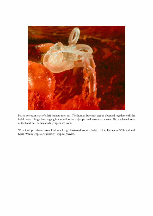
Plastic corrosion cast of a left human inner ear. The human labyrinth can be observed together with the facial nerve. The geniculate ganglion as well as the major petrosal nerve can be seen. Also the lateral knee of the facial nerve and chorda tympani are seen. With kind permission from Professor Helge Rask-Andersson, Christer Bäck, Hermann Wilbrand and Karin Wadin Uppsala University Hospital Sweden.
8
Table of Contents
Abbreviations 11
List of Papers 12
Introduction 13 History 13 Anatomy 14 Peripheral facial palsy 18 Bell’s palsy 19
Aetiology 19 Grading 21 Treatment 25
Surgical treatment 25 Medical treatment 25 Eye care 27 Physical therapy, Botulinum toxin, acupuncture and electrical stimulation 27 Reconstructive surgery 27 Multi-disciplinary facialteam 28
Prognostic factors 28 Age 28 Severity 28 Time to start of recovery 29 Treatment start 29 Pain 29 Stapedial reflex 30 Electrophysiology 30
Recovery rates 31 Sequelae 31
Aims of the thesis 33
9
Materials and Methods 35 Patients 35
Paper I 35 Papers II–IV 35
Methods 36 Paper I 36 Papers II–IV 36
Statistical analysis 37 Paper I 37 Papers II–IV 37
Results 39
Discussion 49
Conclusions 55
Populärvetenskaplig sammanfattning 57
Acknowledgements 59
References 61

10
11
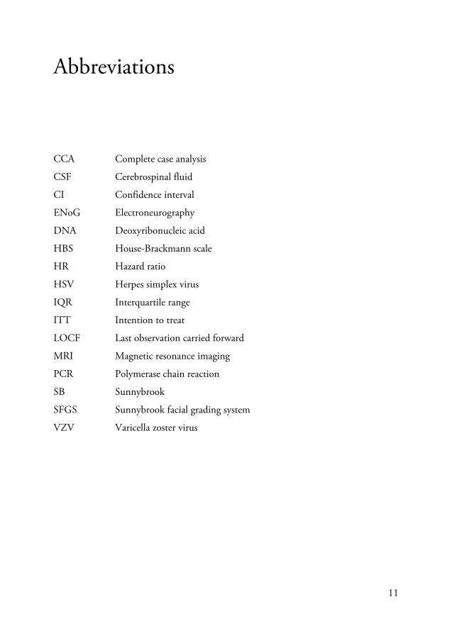
Abbreviations
CCA Complete case analysis
CSF Cerebrospinal fluid
CI Confidence interval
ENoG Electroneurography
DNA Deoxyribonucleic acid
HBS House-Brackmann scale
HR Hazard ratio
HSV Herpes simplex virus
IQR Interquartile range
ITT Intention to treat
LOCF Last observation carried forward
MRI Magnetic resonance imaging
PCR Polymerase chain reaction
SB Sunnybrook
SFGS Sunnybrook facial grading system
VZV Varicella zoster virus
12
List of Papers
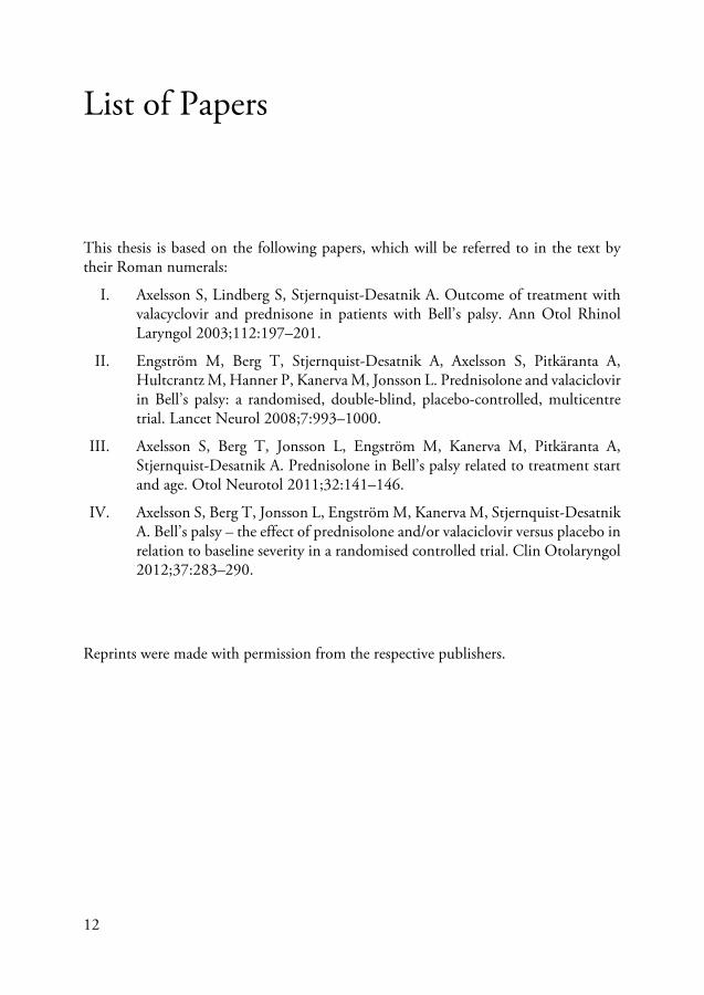
This thesis is based on the following papers, which will be referred to in the text by their Roman numerals:
I. Axelsson S, Lindberg S, Stjernquist-Desatnik A. Outcome of treatment with valacyclovir and prednisone in patients with Bell’s palsy. Ann Otol Rhinol Laryngol 2003;112:197–201.
II. Engström M, Berg T, Stjernquist-Desatnik A, Axelsson S, Pitkäranta A, Hultcrantz M, Hanner P, Kanerva M, Jonsson L. Prednisolone and valaciclovir in Bell’s palsy: a randomised, double-blind, placebo-controlled, multicentre trial. Lancet Neurol 2008;7:993–1000.
III. Axelsson S, Berg T, Jonsson L, Engström M, Kanerva M, Pitkäranta A, Stjernquist-Desatnik A. Prednisolone in Bell’s palsy related to treatment start and age. Otol Neurotol 2011;32:141–146.
IV. Axelsson S, Berg T, Jonsson L, Engström M, Kanerva M, Stjernquist-Desatnik A. Bell’s palsy – the effect of prednisolone and/or valaciclovir versus placebo in relation to baseline severity in a randomised controlled trial. Clin Otolaryngol 2012;37:283–290.
Reprints were made with permission from the respective publishers.
13
Introduction
History
Sir Charles Bell (1774–1842), a British anatomist and surgeon, in the 19th century published his discovery that the facial nerve innervates the facial muscles that give facial expression. He further more reported that the trigeminal nerve mainly is responsible for facial sensation and described several cases of facial paralysis due to trauma or infection (Bell 1821, Bell 1829). Before Bell’s discovery the functions of the facial and trigeminal nerve were poorly understood. Many believed that both the facial nerve and the trigeminal nerve are responsible for facial sensation and facial movement. In the mid-19th century the eponym “Bell’s palsy” was established (van de Graaf et al 2009).
Only a few documents on idiopathic facial paralysis from the pre-Bell era have been found (van de Graaf et al 2009, van de Graaf and Nicolai 2005). In 1686 Cornelis Stalpart van der Wiel (1620–1702) from the Netherlands reported a case of idiopathic peripheral facial paralysis in his treatise One Hundred Rare Observations in Medicine, Surgery and Anatomy. He described a female patient “who fourteen days post partem got a twisting of her mouth, the affected side was weak and numb, she chewed inefficiently, her lips did not close straightly, one eye could not be closed properly and she drooled”. She was treated by Stalpart van der Wiel with several “anti-rheumatic ointments” and was cured within 3 weeks (Stalpart van der Wiel 1686). The second description of Bell’s palsy was a handwritten note made by James Douglas (1675–1742) from Scotland in 1704 (van de Graaf et al 2009). Nicolaus Anton Friedreich (1761–1836) from Germany in 1797 reported three cases of idiopathic peripheral facial paralysis, which he called “rheumatic paralysis of the facial muscles” (Friedreich 1797, van de Graaf et al 2009).
The first extensive study on idiopathic peripheral facial paralysis was performed in 1804 and 1805 by Professor Evert Jan Thomassen à Thuessink (1762–1832) from the Netherlands. He noted that local inflammatory signs, including pain around the ear, mastoid and neck, co-occur with facial paralysis. Almost all cases in his study developed acute or subacute paralysis after exposure to cold or rain. Most patients recovered within several weeks to months. He also located the problem to the seventh cranial nerve and briefly described the peripheral course of the nerve through the parotid gland. Thomassen à Thuessink believed that cold results in “precipitation of rheumatic
14
substance on the facial nerve”, thereby causing facial palsy. Treatment consisted of “anti-inflammatory means and refrigerant and relaxing diaphoretics”. He also believed that the prognosis was less favourable when treatment was delayed “as the changes in the affected nerve become irreversible” (Thomassen à Thuessink 1804, Thomassen à Thuessink 1805). His pupil Willem Hendrik Forsten Verschuir did a literature study and wrote an extensive review on the topic in 1804, De Paralysi Musculorum Faciei, sic Dicta Rheumatica. He concluded that the main difference between idiopathic peripheral facial paralysis and central facial paralysis was the absence, in the former, of sensory loss, loss of sight, impaired memory, headache, vertigo, paralysis of the limbs and paralysis of the tongue. However, at the time many cases of idiopathic peripheral paralysis were probably diagnosed as stroke. There was in his review no description of the difference in involvement of the upper third of the face between peripheral facial paralysis and central paralysis. Forsten Verschuir also differentiated idiopathic peripheral paralysis from trigeminal neuralgia (Forsten Verschuir 1804). In the pre-Bell era, since it was commonly believed that both facial and trigeminal nerves were responsible for facial sensation and facial movements, many surgeons unfortunately divided the facial nerve instead of the trigeminal nerve in cases with severe facial pain (van de Graaf et al 2009).
The “cold theory” is the oldest hypothesis on the aetiology of Bell’s palsy and despite all research efforts in this field the aetiology is still not fully clarified today (see “Aetiology” under “Bell’s Palsy”).
Anatomy
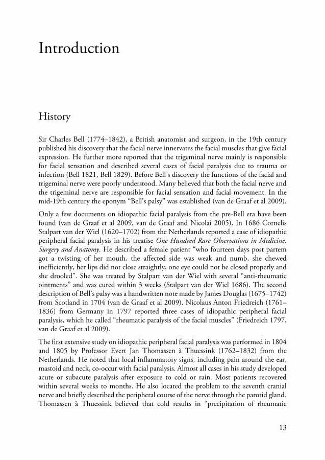
The course of the facial nerve through the Fallopian canal is unique. No other nerve in the body covers such a long distance through a bony canal (May and Schaitkin 2000) The facial nerve, cranial nerve VII, is composed of motor fibres, parasympathetic fibres and sensory fibres. The motor fibres innervate the facial muscles and are responsible for facial expression. Efferent parasympathetic fibres innervate the submandibular, sublingual and lacrimal glands. Taste from the anterior two-thirds of the tongue, and skin sensations in the region of the external ear are carried by sensory fibres (Fig 1) (May and Schaitkin 2000).
15
Figure 1. The facial nerve and its different branches. Drawings by Lena Lyons. The cortical motor face region is located in the lower part of the precentral gyrus. The nerve fibres from the cortex to the facial nucleus pass the internal capsule and the basal part of the pons. Most fibres cross over in the caudal pons and reach the facial nucleus on the opposite side but some diverge to the ipsilateral facial nucleus. The superior portion of the nucleus innervates the occipitofrontal muscle, the upper part of the orbicularis oculi and the corrugator supercilii. These neurons receive bilateral cortical input. Facial muscles in the lower face receive only contralateral input from the motor cortex. This is why a central lesion results in contralateral paralysis of the lower face but
N. FACIALIS AND INTERMEDIUS (VII)
Ganglion pterygopalatinum
Ganglion submandibulare
Nucleus nervi facialis
Nucleus salivatoriussuperior
Nuclei tractussolitarii
Rami zygomatici
Rami temporales
Rami buccales
Rami marginalis mandibularis
Rami cervicalis
Glandula submandibularis
Foramen stylomastoideum
Glandulalacrimalis
N. petrosussuperficialismajor
Glandulasublingualis Motor fibres
Sensory fibresParasympathetic fibres
Ganglion geniculi
Chorda tympani
16
spares the function in the forehead on the same side (Malone and Maisel 1988, May and Schaitkin 2000).
When the nerve fibres emerge from the facial nucleus they make a loop around the nucleus of the abducens nerve to form the internal genu of the facial nerve before they leave the brain stem. When the nerve reaches the internal acoustic canal it joins together with N. intermedius, which emerges from the superior salivatory nucleus and the nucleus solitarius. The facial nerve in the internal acoustic canal is also accompanied by the vestibulocochlear nerve. The facial nerve and the intermediate nerve separate from the eighth nerve at the lateral fundus of the internal acoustic canal and run through the skull bone via a bony canal, the Fallopian canal, named after an Italian anatomist, Gabriele Falloppio (1523–1562) (Malone and Maisel 1988, May and Schaitkin 2000).
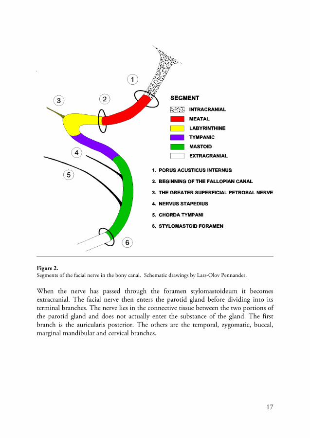
The Fallopian canal is approximately 30 mm long and is subdivided into three segments, the labyrinthine, the tympanic and the mastoid segment (Fig 2). The labyrinthine segment is the narrowest and also shortest segment (3–4 mm) of the facial nerve in the Fallopian canal. It extends from the fundus of the internal auditory canal to the distal portion of the geniculate ganglion. Its narrowest part is at the entrance of the Fallopian canal where the nerve takes up 83% of the available space compared with 73% in the tympanic portion and 64% in the mastoid area (May and Schaitkin 2000). There is no epineurium covering the nerve in the labyrinthine segment. The blood supply in this region is also unique since there are no anastomosing arterial arcades. The nerve is therefore probably more vulnerable in the labyrinthine segment. When it reaches the geniculate ganglion the nerve makes a sharp bend, the external genu, to enter the tympanic, or horizontal, portion of the nerve (12–13 mm). The autonomic fibres joining the motor fibres leave at the level of the geniculate ganglion to form the greater superficial petrosal nerve, which innervates the lacrimal gland. The bony canal in the tympanic segment is thin and often has small dehiscences, particularly above the oval window. The second genu marks the beginning of the mastoid segment and continues vertically to the stylomastoid foramen. This is the longest part of the facial nerve’s intratemporal course (15–20 mm). In this portion the facial nerve has branches to the stapedius muscle and the chorda tympani. The chorda tympani nerve contains secretory motor fibres to the submaxillary and sublingual glands. It also carries special sensory fibres from the anterior two-thirds of the tongue (taste) and fibres from the posterior wall of the external auditory meatus (pain) (May and Schaitkin 2000). There is no proof of topographic location of the branches of the facial nerve within the nerve trunk in the temporal bone (Malone and Maisel 1988).
17
Figure 2. Segments of the facial nerve in the bony canal. Schematic drawings by Lars-Olov Pennander. When the nerve has passed through the foramen stylomastoideum it becomes extracranial. The facial nerve then enters the parotid gland before dividing into its terminal branches. The nerve lies in the connective tissue between the two portions of the parotid gland and does not actually enter the substance of the gland. The first branch is the auricularis posterior. The others are the temporal, zygomatic, buccal, marginal mandibular and cervical branches.
18
Peripheral facial palsy
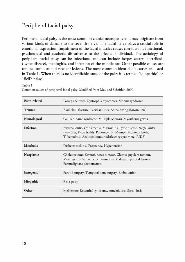
Peripheral facial palsy is the most common cranial neuropathy and may originate from various kinds of damage to the seventh nerve. The facial nerve plays a crucial role in emotional expression. Impairment of the facial muscles causes considerable functional, psychosocial and aesthetic disturbance to the affected individual. The aetiology of peripheral facial palsy can be infectious, and can include herpes zoster, borreliosis (Lyme disease), meningitis, and infection of the middle ear. Other possible causes are trauma, tumours and vascular lesions. The most common identifiable causes are listed in Table 1. When there is no identifiable cause of the palsy it is termed “idiopathic” or “Bell’s palsy”. Table 1 Common causes of peripheral facial palsy. Modified from May and Schaitkin 2000.
Birth-related Forceps delivery, Dystrophia myotonica, Möbius syndrome
Trauma Basal skull fracture, Facial injuries, Scuba diving (barotrauma)
Neurological Guillian-Barré syndrome, Multiple sclerosis, Myasthenia gravis
Infection External otitis, Otitis media, Mastoiditis, Lyme disease, Herpes zoster cephalicus, Encephalitis, Poliomyelitis, Mumps, Mononucleosis, Tuberculosis, Acquired immunodeficiency syndrome (AIDS)
Metabolic Diabetes mellitus, Pregnancy, Hypertension
Neoplastic Cholesteatoma, Seventh nerve tumour, Glomus jugulare tumour, Meningioma, Sarcoma, Schwannoma, Malignant parotid lesions, Paramalignant phenomenon
Iatrogenic Parotid surgery, Temporal bone surgery, Embolisation
Idiopathic Bell’s palsy
Other Melkersson-Rosenthal syndrome, Amyloidosis, Sarcoidosis
19
Bell’s palsy
Bell’s palsy is an abrupt onset of unilateral weakness or paralysis of the face due to acute peripheral facial nerve dysfunction, with no readily identifiable cause, and with some recovery of function within 6 months (May and Hughes 1987). It is a diagnosis of exclusion and causative conditions in the temporal bone, central nervous system and parotid gland have to be ruled out. Bell’s palsy accounts for about 70% of all cases of peripheral facial palsy and the annual incidence is about 30/100 000 population with a peak incidence between the second and fourth decades of life (Katusic et al 1986, Peitersen 2002, Yanagihara 1988). There is no difference in gender or side of the face, and no seasonal clustering. In most cases, the natural course of Bell’s palsy is favourable but at least 30% of patients will have some sequelae and 4% have severe residual paresis, synkinesis and/or contracture (Peitersen 2002). A positive family history of Bell’s palsy has been reported in 4–17% (Adour et al 1978, May and Schaitkin 2000, Peitersen 2002). Recurrence of Bell’s palsy has been reported in about 6–13% of cases (Adour et al 1978, Devriese et al 1990, Katusic et al 1986, May and Schaitkin 2000, Peitersen 2002). In recurrent cases, the palsy can either involve the same side or alternate between sides.
Aetiology The aetiology and pathogenic mechanism of Bell’s palsy have not yet been clarified despite extensive research. Immunological reactions, viral infections, ischaemia and genetic theories have been postulated (Roob et al 1999). Post-mortem examinations of the facial nerve have been performed in deceased patients who had Bell’s palsy. One showed infiltration by lymphocytes of the nerve in the internal auditory meatus and proximal Fallopian canal and revealed that the inflammation induces oedema and mechanical compression of the nerve in the narrow Fallopian canal (Michaels 1990). Another found that inflammatory cells had infiltrated the entire nerve and that the perineurium was thickened and oedematous. It found normal blood vessels in the perineurium and no signs of venous obstruction, but evidence of myelin breakdown, axonal changes and oedema, suggesting viral neuritis (Liston and Kleid 1989). Fisch and Felix propose that in Bell’s palsy the nerve is entrapped at the meatal foramen as a result of neural oedema (Fisch and Felix 1983). Intraoperative conduction studies have shown an electrophysiological blockage at the meatal foramen (Gantz et al 1982). Together histopathological findings in Bell’s palsy show demyelinisation and axonal loss with lymphocytic or phagocytic infiltration, findings that seem to be most pronounced at the labyrinthine segment.
Magnetic resonance imaging (MRI) studies of the facial nerve in Bell’s palsy patients have demonstrated pathological gadolinium enhancement of the nerve in the distal internal auditory canal and labyrinthine/geniculate segment (Engström et al 1993,
20
Schwaber et al 1990). The contrast enhancement of the facial nerve has been explained by inflammatory damage to the blood-nerve barrier, with increased vascular permeability and/or venous congestion (Sartoretti-Schefer et al 1994).
Of the various possible mechanisms for Bell’s palsy, the virus theory has gained the most support. The prevailing theory is that viruses such as Herpes simplex virus type 1 (HSV-1) or Varicella zoster virus (VZV) can be reactivated and may be the cause of Bell’s palsy. In 1972, McCormick suggested that HSV-1 was present in a latent state in the geniculate ganglion and could be reactivated by upper respiratory tract infections, emotional disturbances, sunburn, etc. The virus then travels down the axon, causing inflammation in the facial nerve, with swelling/oedema and entrapment and/or ischaemia of the nerve in the narrow Fallopian canal (McCormick 1972). The virus theory was also suggested by Adour et al (Adour et al 1975). Another possible mechanism proposed was that viral infection directly disturbs nerve function by immune mechanisms and not by compression (Adour 2002).
The HSV-1 genome was detected post-mortem in the geniculate ganglion from a patient with Bell’s palsy (Burgess et al 1994). Herpes simplex virus type 1 deoxyribonucleic acid (DNA) has been detected in the geniculate ganglion in 15 of 17 autopsies without history of Bell’s palsy, supporting the theory that viruses can be present in a latent state in the geniculate ganglion (Takasu et al 1992). Several studies have reported stationary or slightly elevated antibody titres against HSV-1 but very rarely a titre rise in patients with Bell’s palsy (Adour et al 1975, Jonsson et al 1988, Mulkens et al 1980, Vahlne et al 1981). Animal experiments have shown the ability of HSV-1 to induce acute and transient facial paralysis in mice after being inoculated with virus into the auricles or tongue. The histopathological and immunocytochemical examination showed nerve swelling, inflammatory cell infiltration and vascular degeneration of the facial nerve, and HSV antigen was also detected along the facial nerve (Sugita et al 1995).
The virus theory was further supported by a Japanese polymerase chain reaction (PCR) study by Murakami et al who detected HSV-1 DNA in endoneural fluid and/or tissue from posterior auricular muscle in 11/14 (79%) patients with Bell’s palsy. They detected VZV virus DNA in 8/9 (89%) patients with Rumsey-Hunt syndrome. In a control group no HSV-1 or VZV DNA was detected (Murakami et al 1996). However, other studies could not reproduce the findings of Murakami et al. Stjernquist-Desatnik et al analysed biopsy specimens from the posterior auricular muscle and cerebrospinal fluid (CSF) for HSV-1 and VZV DNA using PCR in 20 patients with Bell’s palsy within 72 hours of onset of palsy. They found HSV-1 DNA in only 1/20 (5%) patients and VZV DNA in 1/20 (5%) patients with Bell’s palsy (Stjernquist-Desatnik et al 2006). They speculated whether viral replication may already have declined in many cases after onset of palsy. One reason for the discrepancy between the results may be epidemiological difference and furthermore, the patients in the study by Murakami et
21
al were selected and all had long-standing, total paralysis. Perhaps there is prolonged replication in cases of total and long-standing paralysis. Linder et al analysed biopsy specimens from the orbicularis oris muscle in 13 patients with Bell’s palsy, and could not detect HSV-1, HSV-2 or VZV DNA (Linder et al 2005), thus also contradicting the results of Murakami et al (1996).
Varicella zoster virus can give rise to facial nerve palsy and simultaneous affection of the vestibulocochlear nerve, leading to hearing loss, vertigo and facial palsy, accompanied by severe pain and ipsilateral herpetic vesicles – Ramsey Hunt syndrome. The symptoms are more severe and the prognosis is worse than in Bell’s palsy. Recent studies have presented evidence that VZV may be responsible for many cases of facial palsy in patients diagnosed as Bell’s palsy. These patients have no herpetic vesicular rash and their condition is termed “zoster sine herpete”. In a study by Furuta, using PCR and serological assays, VZV in saliva was detected in 35/121 (29%) patients clinically diagnosed as Bell’s palsy. He suggested that VZV is a major aetiologic agent of Bell’s palsy (Furuta et al 2005, Furuta et al 2000). However, others have shown that VZV may be reactivated and found in saliva samples of healthy persons without facial palsy after being exposed to stress (Mehta et al 2004). So far the aetiology of Bell’s palsy is still unclear.
Grading
It is fundamental to have a reliable and valid method of evaluating facial palsy and be able to assess the course of recovery and the effect of treatment over time. The evaluation should be sensitive enough to detect clinically important changes over time. It should also be easy to administer, and require little time and equipment (Ross et al 1996).
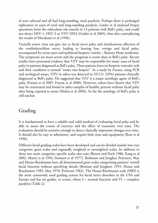
Different facial grading scales have been developed and can be divided mainly into two categories, gross scales and regionally weighted or unweighted scales. In addition to these two main categories, specific scales also exist (Burres and Fisch 1986, Kang et al 2002, Murty et al 1994, Stennert et al 1977). Bothman and Jongkes, Peitersen, May and House-Brackmann have all demonstrated gross scales categorising patients’ overall facial function without specifying details (Botman and Jongkees 1955, House and Brackmann 1985, May 1970, Peitersen 1982). The House-Brackmann scale (HBS) is the most commonly used grading system for facial nerve disorders in the USA and Europe and has six grades, or scores, where I = normal function and VI = complete paralysis (Table 2).
22
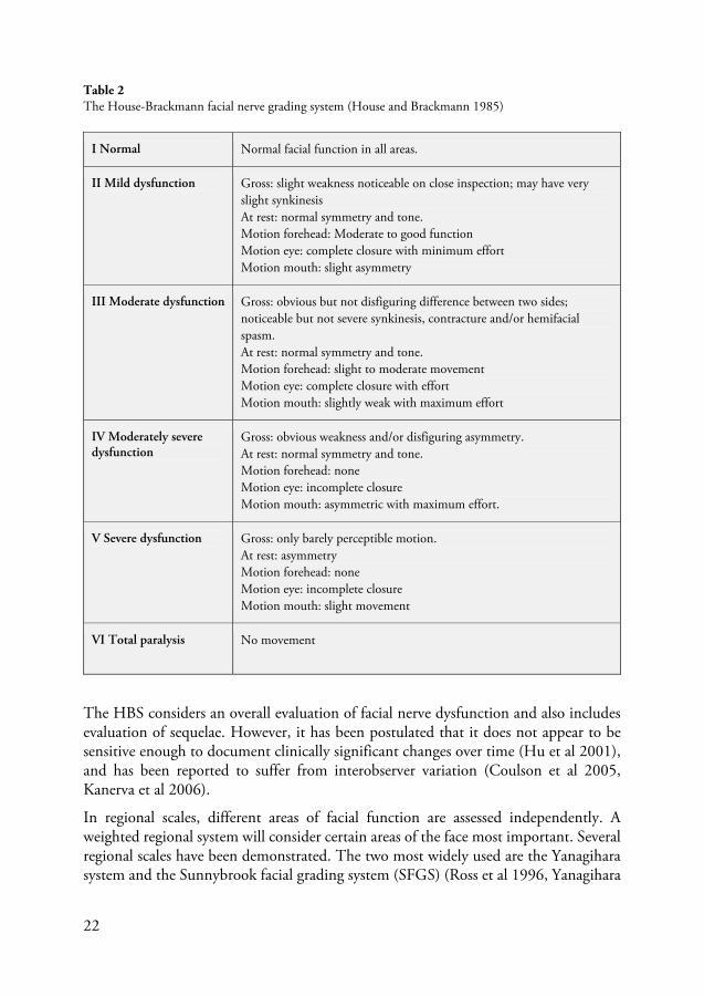
Table 2 The House-Brackmann facial nerve grading system (House and Brackmann 1985)
I Normal Normal facial function in all areas.
II Mild dysfunction
Gross: slight weakness noticeable on close inspection; may have very slight synkinesis At rest: normal symmetry and tone. Motion forehead: Moderate to good function Motion eye: complete closure with minimum effort Motion mouth: slight asymmetry
III Moderate dysfunction
Gross: obvious but not disfiguring difference between two sides; noticeable but not severe synkinesis, contracture and/or hemifacial spasm. At rest: normal symmetry and tone. Motion forehead: slight to moderate movement Motion eye: complete closure with effort Motion mouth: slightly weak with maximum effort
IV Moderately severe dysfunction
Gross: obvious weakness and/or disfiguring asymmetry.At rest: normal symmetry and tone. Motion forehead: none Motion eye: incomplete closure Motion mouth: asymmetric with maximum effort.
V Severe dysfunction
Gross: only barely perceptible motion.At rest: asymmetry Motion forehead: none Motion eye: incomplete closure Motion mouth: slight movement
VI Total paralysis No movement
The HBS considers an overall evaluation of facial nerve dysfunction and also includes evaluation of sequelae. However, it has been postulated that it does not appear to be sensitive enough to document clinically significant changes over time (Hu et al 2001), and has been reported to suffer from interobserver variation (Coulson et al 2005, Kanerva et al 2006).
In regional scales, different areas of facial function are assessed independently. A weighted regional system will consider certain areas of the face most important. Several regional scales have been demonstrated. The two most widely used are the Yanagihara system and the Sunnybrook facial grading system (SFGS) (Ross et al 1996, Yanagihara
23
1977). The Yanagihara system is the most commonly used facial grading system in Japan. It assesses ten aspects of facial muscle function, each scored 0–4, to give a possible maximum score of 40. It does not include evaluation of sequelae, such as synkinesis and contracture (Satoh et al 2000, Yanagihara 1977).
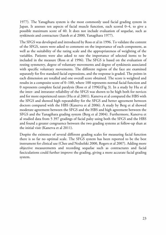
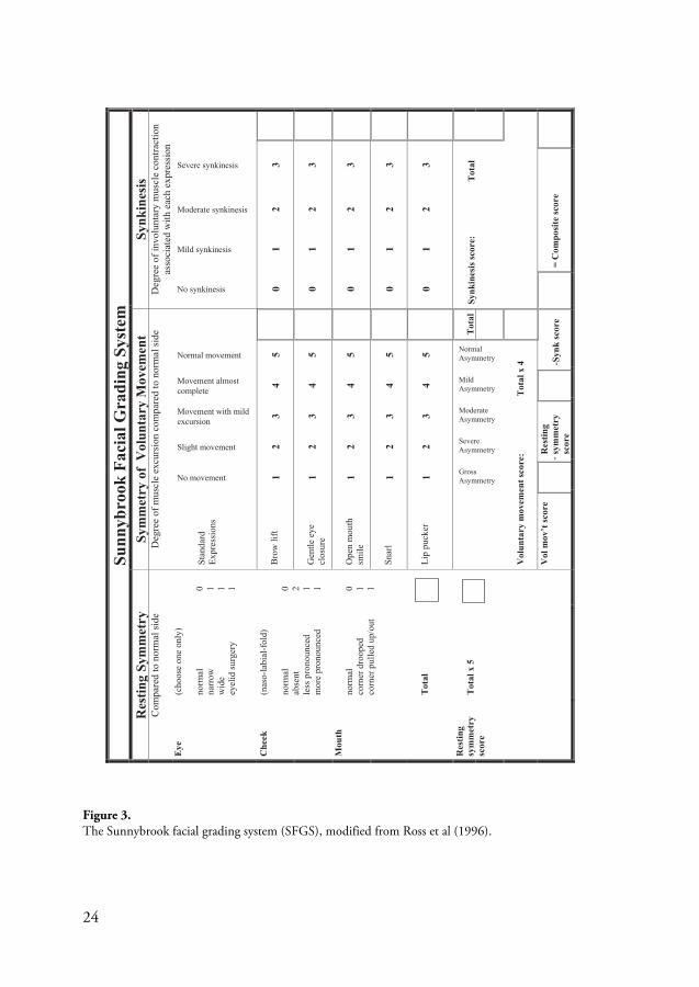
The SFGS was developed and introduced by Ross et al in 1996. To validate the content of the SFGS, raters were asked to comment on the importance of each component, as well as the suitability of the rating scale and the appropriateness of weighting of the variables. Patients were also asked to rate the importance of selected items to be included in the measure (Ross et al 1996). The SFGS is based on the evaluation of resting symmetry, degree of voluntary movements and degree of synkinesis associated with specific voluntary movements. The different regions of the face are examined separately for five standard facial expressions, and the response is graded. The points in each dimension are totalled and one overall score obtained. The score is weighted and results in a composite score of 0–100, where 100 represents normal facial function and 0 represents complete facial paralysis (Ross et al 1996)(Fig 3). In a study by Hu et al the inter- and intrarater reliability of the SFGS was shown to be high both for novices and for more experienced raters (Hu et al 2001). Kanerva et al compared the HBS with the SFGS and showed high repeatability for the SFGS and better agreement between doctors compared with the HBS (Kanerva et al 2006). A study by Berg et al showed moderate agreement between the SFGS and the HBS and high agreement between the SFGS and the Yanagihara grading system (Berg et al 2004). Furthermore, Kanerva et al studied data from 5 397 gradings of facial palsy using both the SFGS and the HBS and found a greater congruence between the two grading systems at follow-up than at the initial visit (Kanerva et al 2011).
Despite the existence of several different grading scales for measuring facial function there is so far no optimal scale. The SFGS system has been reported to be the best instrument for clinical use (Chee and Nedzelski 2000, Rogers et al 2007). Adding more objective measurements and recording sequelae such as contractures and facial fasciculations could further improve the grading, giving a more accurate facial grading system.
24
Figure 3. The Sunnybrook facial grading system (SFGS), modified from Ross et al (1996).
-
Sunn
ybro
ok F
acia
l Gra
ding
Sys
tem
R
estin
g Sy
mm
etry
Sy
mm
etry
of
Vol
unta
ry M
ovem
ent
Synk
ines
is
Com
pare
d to
nor
mal
side
D
egre
e of
mus
cle
excu
rsio
n co
mpa
red
to n
orm
al si
de
Deg
ree
of in
volu
ntar
y m
uscl
e co
ntra
ctio
n as
soci
ated
with
eac
h ex
pres
sion
E
ye(c
hoos
e on
e on
ly)
norm
al
narr
oww
ide
eyel
id su
rger
y
0 1 1 1
Stan
dard
Expr
essi
ons
No movement
Slight movement
Movement with mild excursion
Movement almost complete
Normal movement
No synkinesis
Mild synkinesis
Moderate synkinesis
Severe synkinesis
Che
ek
(nas
o-la
bial
-fol
d)
norm
al
abse
ntle
ss p
rono
unce
d m
ore
pron
ounc
ed
0 2 1 1
Bro
w li
ft 1
23
45
01
23
Gen
tle e
ye
clos
ure
12
34
50
12
3
Mou
thno
rmal
co
rner
dro
oped
co
rner
pul
led
up/o
ut
0 1 1
Ope
n m
outh
sm
ile
12
34
50
12
3
Snar
l1
23
45
01
23
Tot
alLi
p pu
cker
1
23
45
01
23
Res
ting
sym
met
rysc
ore
Tot
al x
5
GrossAsymmetry
SevereAsymmetry
ModerateAsymmetry
MildAsymmetry
NormalAsymmetry
Tot
alSy
nkin
esis
scor
e:T
otal
V
olun
tary
mov
emen
t sco
re:
Tot
al x
4
V
ol m
ov’t
scor
e
Res
ting
sym
met
rysc
ore
-Syn
k sc
ore
= C
ompo
site
scor
e
25
Treatment
Since the aetiology of Bell’s palsy is not clarified, different kinds of treatment including heat, massage, galvanic stimulation, ointments, diaphoretics, passive exercise, wire splints, and stellate ganglion blockage have been used.
The most prevalent aetiology of Bell’s palsy appears to be inflammation and swelling with entrapment of the facial nerve in the Fallopian canal. Different treatments have been proposed, all aimed to reduce the inflammation, swelling/oedema and compression of the nerve.
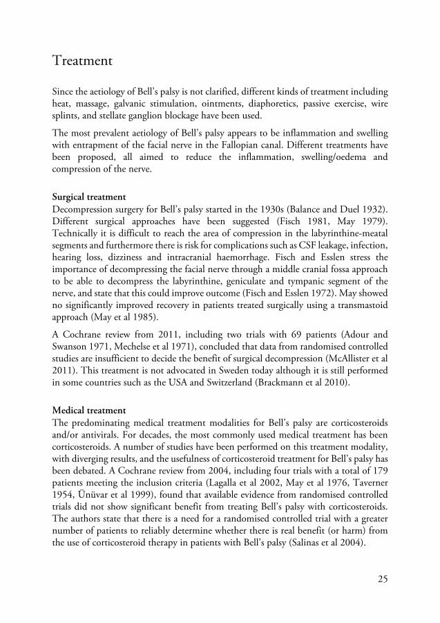
Surgical treatment Decompression surgery for Bell’s palsy started in the 1930s (Balance and Duel 1932). Different surgical approaches have been suggested (Fisch 1981, May 1979). Technically it is difficult to reach the area of compression in the labyrinthine-meatal segments and furthermore there is risk for complications such as CSF leakage, infection, hearing loss, dizziness and intracranial haemorrhage. Fisch and Esslen stress the importance of decompressing the facial nerve through a middle cranial fossa approach to be able to decompress the labyrinthine, geniculate and tympanic segment of the nerve, and state that this could improve outcome (Fisch and Esslen 1972). May showed no significantly improved recovery in patients treated surgically using a transmastoid approach (May et al 1985).
A Cochrane review from 2011, including two trials with 69 patients (Adour and Swanson 1971, Mechelse et al 1971), concluded that data from randomised controlled studies are insufficient to decide the benefit of surgical decompression (McAllister et al 2011). This treatment is not advocated in Sweden today although it is still performed in some countries such as the USA and Switzerland (Brackmann et al 2010).
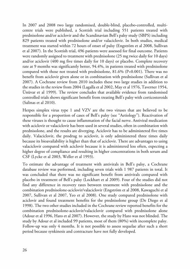
Medical treatment The predominating medical treatment modalities for Bell’s palsy are corticosteroids and/or antivirals. For decades, the most commonly used medical treatment has been corticosteroids. A number of studies have been performed on this treatment modality, with diverging results, and the usefulness of corticosteroid treatment for Bell’s palsy has been debated. A Cochrane review from 2004, including four trials with a total of 179 patients meeting the inclusion criteria (Lagalla et al 2002, May et al 1976, Taverner 1954, Ünüvar et al 1999), found that available evidence from randomised controlled trials did not show significant benefit from treating Bell’s palsy with corticosteroids. The authors state that there is a need for a randomised controlled trial with a greater number of patients to reliably determine whether there is real benefit (or harm) from the use of corticosteroid therapy in patients with Bell’s palsy (Salinas et al 2004).
26
In 2007 and 2008 two large randomised, double-blind, placebo-controlled, multi-centre trials were published, a Scottish trial including 551 patients treated with prednisolone and/or aciclovir and the Scandinavian Bell’s palsy study (SBPS) including 829 patients treated with prednisolone and/or valaciclovir. In both studies, medical treatment was started within 72 hours of onset of palsy (Engström et al 2008, Sullivan et al 2007). In the Scottish trial, 496 patients were assessed for final outcome. Patients were randomly assigned to treatment with prednisolone (25 mg twice daily for 10 days) and/or aciclovir (400 mg five times daily for 10 days) or placebo. Complete recovery rate at 9 months was significantly better, 94.4%, in patients treated with prednisolone compared with those not treated with prednisolone, 81.6% (P<0.001). There was no benefit from aciclovir given alone or in combination with prednisolone (Sullivan et al 2007). A Cochrane review from 2010 includes these two large studies in addition to the studies in the review from 2004 (Lagalla et al 2002, May et al 1976, Taverner 1954, Ünüvar et al 1999). The review concludes that available evidence from randomised controlled trials shows significant benefit from treating Bell’s palsy with corticosteroids (Salinas et al 2010).
Herpes simplex virus type 1 and VZV are the two viruses that are believed to be responsible for a proportion of cases of Bell’s palsy (see “Aetiology”). Reactivation of these viruses is thought to cause inflammation of the facial nerve. Antiviral medication with aciclovir or valaciclovir has been used in several studies, often in combination with prednisolone, and the results are diverging. Aciclovir has to be administered five times daily. Valaciclovir, the prodrug to aciclovir, is only administered three times daily because its bioavailability is higher than that of aciclovir. There are advantages to using valaciclovir compared with aciclovir because it is administered less often, expecting a higher degree of compliance and resulting in higher concentrations in both serum and CSF (Lycke et al 2003, Weller et al 1993).
To estimate the advantage of treatment with antivirals in Bell’s palsy, a Cochrane database review was performed, including seven trials with 1 987 patients in total. It was concluded that there was no significant benefit from antivirals compared with placebo in treatment of Bell’s palsy (Lockhart et al 2009). Four of the studies did not find any difference in recovery rates between treatment with prednisolone and the combination prednisolone-aciclovir/valaciclovir (Engström et al 2008, Kawaguchi et al 2007, Sullivan et al 2007, Yeo et al 2008). One study compared prednisolone with aciclovir and found treatment benefits for the prednisolone group (De Diego et al 1998). The two other studies included in the Cochrane review reported benefits for the combination prednisolone-aciclovir/valaciclovir compared with prednisolone alone (Adour et al 1996, Hato et al 2007). However, the study by Hato was not blinded. The study by Adour et al included 99 patients, most of them (80%) with incomplete palsy. Follow-up was only 4 months. It is not possible to assess sequelae after such a short period because synkinesis and contracture have not fully developed.
27
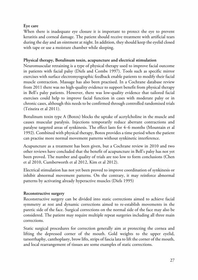
Eye care When there is inadequate eye closure it is important to protect the eye to prevent keratitis and corneal damage. The patient should receive treatment with artificial tears during the day and an ointment at night. In addition, they should keep the eyelid closed with tape or use a moisture chamber while sleeping.
Physical therapy, Botulinum toxin, acupuncture and electrical stimulation Neuromuscular retraining is a type of physical therapy used to improve facial outcome in patients with facial palsy (Diels and Combs 1997). Tools such as specific mirror exercises with surface electromyographic feedback enable patients to modify their facial muscle contraction. Massage has also been practised. In a Cochrane database review from 2011 there was no high-quality evidence to support benefit from physical therapy in Bell’s palsy patients. However, there was low-quality evidence that tailored facial exercises could help to improve facial function in cases with moderate palsy or in chronic cases, although this needs to be confirmed through controlled randomised trials (Teixeira et al 2011).
Botulinum toxin type A (Botox) blocks the uptake of acetylcholine in the muscle and causes muscular paralysis. Injections temporarily reduce aberrant contractions and paralyse targeted areas of synkinesis. The effect lasts for 4–6 months (Mountain et al 1992). Combined with physical therapy, Botox provides a time period when the patient can practise more normal movement patterns without synkinetic interference.
Acupuncture as a treatment has been given, but a Cochrane review in 2010 and two other reviews have concluded that the benefit of acupuncture in Bell’s palsy has not yet been proved. The number and quality of trials are too low to form conclusions (Chen et al 2010, Cumberworth et al 2012, Kim et al 2012).
Electrical stimulation has not yet been proved to improve coordination of synkinesis or inhibit abnormal movement patterns. On the contrary, it may reinforce abnormal patterns by activating already hyperactive muscles (Diels 1995)
Reconstructive surgery Reconstructive surgery can be divided into static corrections aimed to achieve facial symmetry at rest and dynamic corrections aimed to re-establish movements in the paretic side of the face. Surgical corrections on the normal side of the face may also be considered. The patient may require multiple repeat surgeries including all three main corrections.
Static surgical procedures for correction generally aim at protecting the cornea and lifting the depressed corner of the mouth. Gold weights to the upper eyelid, tarsorrhaphy, canthoplasty, brow lifts, strips of fascia lata to lift the corner of the mouth, and local rearrangement of tissues are some examples of static corrections.
28
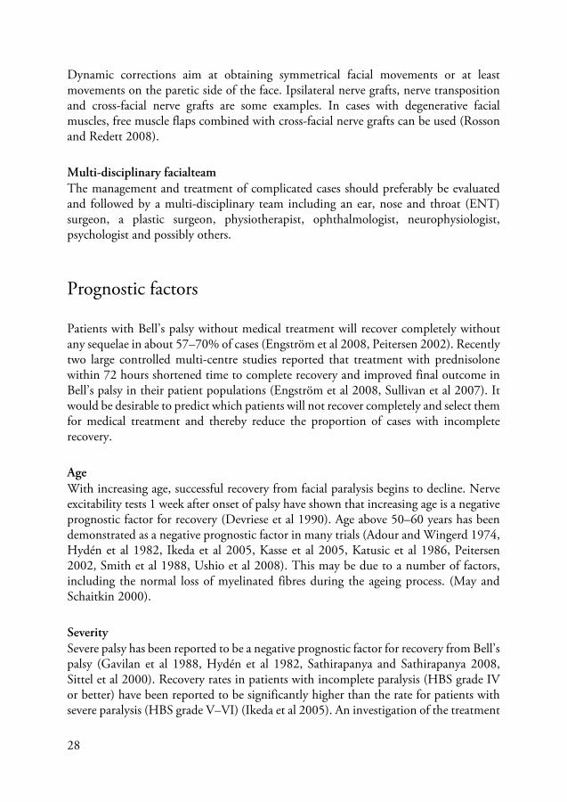
Dynamic corrections aim at obtaining symmetrical facial movements or at least movements on the paretic side of the face. Ipsilateral nerve grafts, nerve transposition and cross-facial nerve grafts are some examples. In cases with degenerative facial muscles, free muscle flaps combined with cross-facial nerve grafts can be used (Rosson and Redett 2008).
Multi-disciplinary facialteam The management and treatment of complicated cases should preferably be evaluated and followed by a multi-disciplinary team including an ear, nose and throat (ENT) surgeon, a plastic surgeon, physiotherapist, ophthalmologist, neurophysiologist, psychologist and possibly others.
Prognostic factors
Patients with Bell’s palsy without medical treatment will recover completely without any sequelae in about 57–70% of cases (Engström et al 2008, Peitersen 2002). Recently two large controlled multi-centre studies reported that treatment with prednisolone within 72 hours shortened time to complete recovery and improved final outcome in Bell’s palsy in their patient populations (Engström et al 2008, Sullivan et al 2007). It would be desirable to predict which patients will not recover completely and select them for medical treatment and thereby reduce the proportion of cases with incomplete recovery.
Age With increasing age, successful recovery from facial paralysis begins to decline. Nerve excitability tests 1 week after onset of palsy have shown that increasing age is a negative prognostic factor for recovery (Devriese et al 1990). Age above 50–60 years has been demonstrated as a negative prognostic factor in many trials (Adour and Wingerd 1974, Hydén et al 1982, Ikeda et al 2005, Kasse et al 2005, Katusic et al 1986, Peitersen 2002, Smith et al 1988, Ushio et al 2008). This may be due to a number of factors, including the normal loss of myelinated fibres during the ageing process. (May and Schaitkin 2000).
Severity Severe palsy has been reported to be a negative prognostic factor for recovery from Bell’s palsy (Gavilan et al 1988, Hydén et al 1982, Sathirapanya and Sathirapanya 2008, Sittel et al 2000). Recovery rates in patients with incomplete paralysis (HBS grade IV or better) have been reported to be significantly higher than the rate for patients with severe paralysis (HBS grade V–VI) (Ikeda et al 2005). An investigation of the treatment
29
effect of corticosteroids and/or antivirals in relation to severity at baseline has been called for (Linder et al 2010, Lockhart et al 2009).
Time to start of recovery Start of remission is highly significant for prognosis (Peitersen 2002). Peitersen reported a significantly better outcome in study patients who started recovery in the first and second week compared with patients who started recovery in the third week after palsy onset. Ikeda found severity of palsy 1 month after palsy onset to be an important factor for predicting the prognosis in Bell’s palsy patients. If HBS was IV or worse at 1 month the recovery rate was poor in 79% of patients (Ikeda et al 2005).
In the SBPS including 829 patients with Bell’s palsy, early deterioration in facial function, measured by SFGS, between baseline and first follow-up 11–17 days later was shown to be a negative prognostic factor for recovery at 12 months (Marsk et al 2010). The Sunnybrook (SB) score at 1 month after onset of palsy was reported to accurately predict non-recovery (SB score <70) at 12 months (Marsk et al 2012).
Treatment start There are few controlled studies regarding the prognostic value of the time of the start of treatment with corticosteroids in Bell’s palsy. Treatment with prednisolone within 3 days has been associated with higher recovery rates than achieved with later treatment (Yeo et al 2008). Early treatment within 24 hours has been reported to result in better outcome in a study of patients with complete or near complete paralysis. However, this study was not double-blinded (Shafshak et al 1994).
Uncontrolled studies show diverging results. In one study, patients treated with intravenous high-dose prednisolone, dextran and pentoxifylline within 3 days had better outcome compared with those who started treatment between 4 and 12 days (Sittel et al 2000). In another, corticosteroid treatment within 7 days of palsy onset achieved a significantly better outcome compared with therapy initiated after this period (Sathirapanya and Sathirapanya 2008). In another uncontrolled study, no significant difference was reported in facial outcome scores between patients starting treatment within 3 days and patients starting treatment 4–7 days after onset of palsy (Adour et al 1972). Kawaguchi et al report no significant difference in recovery rates between patients treated within 3 days and patients treated 4–7 days after onset (Kawaguchi et al 2007).
Pain Ipsilateral pain around the ear or in the neck is a frequent symptom in the early stages of Bell’s palsy. The pain may precede the paralysis by a few days (Roob et al 1999). The aethiopathological background of the pain is unclear. Pain has been correlated to worse prognosis in some studies (Gavilan et al 1988, Hydén et al 1982, Katusic et al 1986,
30
Peitersen 2002). Other studies have not found this correlation (Adour et al 1978, May et al 1976, Smith et al 1988). Presence of pain on day 11–17 after onset of palsy has been found to be a negative prognostic factor (Berg et al 2009). Herpes zoster cephalicus (Ramsey-Hunt syndrome) is often associated with more severe pain around the ear and also with vesicles following a dermatome. Cases of facial palsy accompanied by severe pain but without vesicles may be referred to as “zoster sine herpete” if antibody titres to herpes zoster are simultaneously elevated (Furuta et al 2005, Furuta et al 2000).
Stapedial reflex Presence of stapedial reflex during the first 10 days of Bell’s palsy indicates a good prognosis. However, absence of stapedial reflex cannot be used on its own for prognostic purposes (Ekstrand and Glitterstam 1979). The absence of stapedial reflex response during the first days of development of Bell’s palsy has been considered a sign of poor prognosis (Gavilan et al 1988). In a study by Ikeda et al, patients who had lost the stapedial muscle reflex showed a significantly worse outcome than seen in patients with a reflex. However, 80% of patients with loss of stapedial reflex recovered satisfactorily and the absence of stapedial reflex was therefore considered to be a low prognostic risk factor (Ikeda et al 2005). Portmann likewise shows that the stapedial reflex alone was not sufficient to be of prognostic value in Bell’s palsy patients studied (Portmann et al 1990). A confounding factor is that Bell’s palsy is progressive for 7–10 days and the stapedial reflex may be present in the early days and then vanish.
Electrophysiology Electrophysiology tests can be used to predict outcome in Bell’s palsy. Electroneurography (ENoG) is the most frequently used test and has been claimed to be the most reliable test to assess facial nerve degeneration in Bell’s palsy (Fisch 1984, May and Hughes 1987). During ENoG the nerve is stimulated percutaneously over the stylomastoid foramen and the compound muscle action potential is recorded in the facial muscle and reported as a percentage of the non-affected side. Facial nerve degeneration of ≥90% has been shown to predict long-term outcome of facial weakness (Dumitru et al 1988, Mantsopoulos et al 2011). However, the test cannot be used in the early stage of palsy since Wallerian degeneration of motor nerve fibres takes some days after axonal injury (Gilchrist 2009) Regarding use of ENoG, the period between 10 and 14 days after onset was found to be the most valuable for prediction of recovery (Chow et al 2002, Qiu et al 1996).
31
Recovery rates
In Bell’s palsy there is a high rate of spontaneous recovery; however, the figures for spontaneous recovery and recovery after medical treatment vary across different studies. One of the problems when comparing different studies is how recovery is defined since different scales have been used to assess recovery, e.g. the House-Brackmann, Yanagihara and Sunnybrook grading scales. The inclusion/exclusion criteria, follow-up time, choice of analysis method and definition of recovery will all affect the results. In the SBPS, recovery rates were estimated using the last observation carried forward (LOCF) method and the SB score of 100 was used to assess complete recovery at 12 months.
Berg et al examined how recovery rates in the SBPS would have been affected with use of a different analysis method and a different definition of “recovery”. They showed a range of 72–96% in recovery rates for patients treated with prednisolone and a range of 57–88% in recovery rates for patients not treated with prednisolone depending on which method was used (Berg et al 2009). They applied four different definitions of “recovery”:
1. “Recovery” defined as an SB score of 100 at 12 months: use of the LOCF method, giving a recovery rate of 57% for untreated patients and 72% for patients treated with prednisolone.
2. “Recovery” defined as HBS grade I at 12 months: use of the LOCF method, giving a recovery rate of 64% for untreated patients and 78% for patients treated with prednisolone.
3. “Recovery” defined as HBS grade I at 12 months; and use of complete case analysis, giving a recovery rate of 70% for untreated patients and 86% for patients treated with prednisolone.
4. “Recovery” defined as HBS grade II at 12 months; and use of complete case analysis, giving a recovery rate of 88% for untreated patients and 96% for patients treated with prednisolone.
Sequelae
Sequelae can be troublesome for the patient and have probably been underestimated by clinicians (Smith et al 1994). When recovery from the palsy is incomplete there are different grades of sequelae that may become troublesome to the patient, such as synkinesis, hemi-facial spasm, contractures and fasciculations. Peitersen reports that 12% of patients in his study had slight, 13% had moderate, and 4% had severe sequelae
32
(Peitersen 2002). In addition to functional problems there is also a psychological aspect. Many serious diseases are only known to the patient and the people in the patient’s closest confidence. Facial paresis, however, cannot be concealed and the patient cannot control the way they wish to present themselves to the world. This often results in serious emotional consequences. This, together with impairment of eating and drinking and eye problems, may seriously reduce the quality of life in patients with facial paresis.
33
Aims of the thesis
The general aim of this thesis was to study the treatment effect of prednisolone and/or valaciclovir and the influence of time to treatment start, age and severity at baseline in patients with Bell’s palsy.
Specific aims were:
• to find out whether treatment with the antiviral drug valaciclovir in combination with prednisolone is more effective than no medical treatment at all;
• to study the short-term and long-term effects of treatment with prednisolone and/or valaciclovir on the recovery of the facial nerve in a large number of patients with Bell’s palsy. Also, to study the side effects of the drugs and their effects on synkinesis;
• to evaluate the effect of prednisolone in relation to time of treatment start within the first 72 hours of Bell’s palsy and the effect of the drug in relation to patients’ age; and
• to study the treatment effect of prednisolone and/or valaciclovir on outcome of Bell’s palsy in relation to severity of palsy at baseline.

34
35
Materials and Methods
Patients
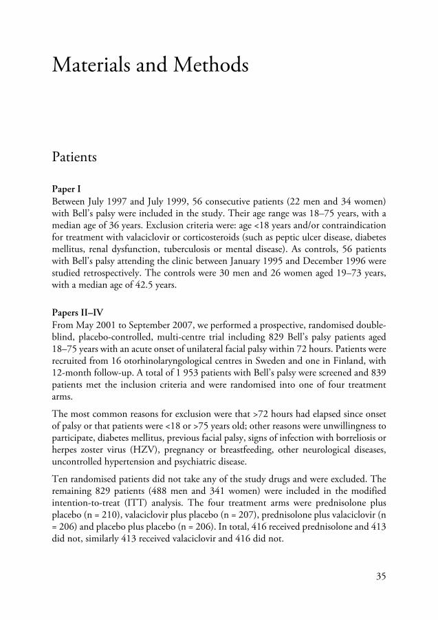
Paper I Between July 1997 and July 1999, 56 consecutive patients (22 men and 34 women) with Bell’s palsy were included in the study. Their age range was 18–75 years, with a median age of 36 years. Exclusion criteria were: age <18 years and/or contraindication for treatment with valaciclovir or corticosteroids (such as peptic ulcer disease, diabetes mellitus, renal dysfunction, tuberculosis or mental disease). As controls, 56 patients with Bell’s palsy attending the clinic between January 1995 and December 1996 were studied retrospectively. The controls were 30 men and 26 women aged 19–73 years, with a median age of 42.5 years.
Papers II–IV From May 2001 to September 2007, we performed a prospective, randomised double-blind, placebo-controlled, multi-centre trial including 829 Bell’s palsy patients aged 18–75 years with an acute onset of unilateral facial palsy within 72 hours. Patients were recruited from 16 otorhinolaryngological centres in Sweden and one in Finland, with 12-month follow-up. A total of 1 953 patients with Bell’s palsy were screened and 839 patients met the inclusion criteria and were randomised into one of four treatment arms.
The most common reasons for exclusion were that >72 hours had elapsed since onset of palsy or that patients were <18 or >75 years old; other reasons were unwillingness to participate, diabetes mellitus, previous facial palsy, signs of infection with borreliosis or herpes zoster virus (HZV), pregnancy or breastfeeding, other neurological diseases, uncontrolled hypertension and psychiatric disease.
Ten randomised patients did not take any of the study drugs and were excluded. The remaining 829 patients (488 men and 341 women) were included in the modified intention-to-treat (ITT) analysis. The four treatment arms were prednisolone plus placebo (n = 210), valaciclovir plus placebo (n = 207), prednisolone plus valaciclovir (n = 206) and placebo plus placebo (n = 206). In total, 416 received prednisolone and 413 did not, similarly 413 received valaciclovir and 416 did not.
36
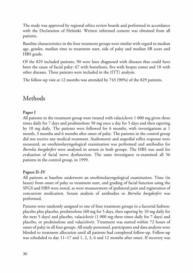
The study was approved by regional ethics review boards and performed in accordance with the Declaration of Helsinki. Written informed consent was obtained from all patients.
Baseline characteristics in the four treatment groups were similar with regard to median age, gender, median time to treatment start, side of palsy and median SB score and HBS grade.
Of the 829 included patients, 90 were later diagnosed with diseases that could have been the cause of facial palsy: 67 with borreliosis, five with herpes zoster and 18 with other diseases. These patients were included in the (ITT) analysis.
The follow-up visit at 12 months was attended by 743 (90%) of the 829 patients.
Methods
Paper I All patients in the treatment group were treated with valaciclovir 1 000 mg given three times daily for 7 days and prednisolone 50 mg once a day for 5 days and then tapering by 10 mg daily. The patients were followed for 6 months, with investigations at 1 month, 3 months and 6 months after onset of palsy. The patients in the control group did not receive any medical treatment. Audiometry and stapedial reflex response were measured, an otorhinolaryngological examination was performed and antibodies for Borrelia burgdorferi were analysed in serum in both groups. The HBS was used for evaluation of facial nerve dysfunction. The same investigator re-examined all 56 patients in the control group, in 1999.
Papers II–IV All patients at baseline underwent an otorhinolaryngological examination. Time (in hours) from onset of palsy to treatment start, and grading of facial function using the SFGS and HBS were noted, as were measurement of ipsilateral pain and registration of concurrent medication. Serum analysis of antibodies to Borrelia burgdorferi was performed.
Patients were randomly assigned to one of four treatment groups in a factorial fashion: placebo plus placebo, prednisolone (60 mg for 5 days, then tapering by 10 mg daily for the next 5 days) and placebo, valaciclovir (1 000 mg three times daily for 7 days) and placebo, or prednisolone and valaciclovir. Treatment was started within 72 hours of onset of palsy in all four groups. All study personnel, participants and data analysts were blinded to treatment allocation until all patients had completed follow-up. Follow-up was scheduled to day 11–17 and 1, 2, 3, 6 and 12 months after onset. If recovery was
37
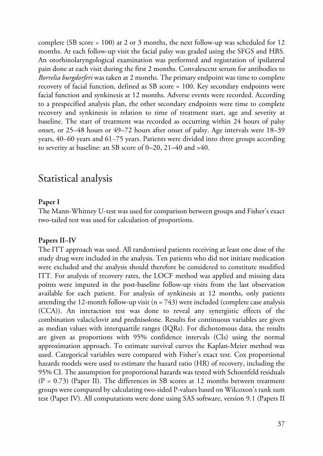
complete (SB score = 100) at 2 or 3 months, the next follow-up was scheduled for 12 months. At each follow-up visit the facial palsy was graded using the SFGS and HBS. An otorhinolaryngological examination was performed and registration of ipsilateral pain done at each visit during the first 2 months. Convalescent serum for antibodies to Borrelia burgdorferi was taken at 2 months. The primary endpoint was time to complete recovery of facial function, defined as SB score = 100. Key secondary endpoints were facial function and synkinesis at 12 months. Adverse events were recorded. According to a prespecified analysis plan, the other secondary endpoints were time to complete recovery and synkinesis in relation to time of treatment start, age and severity at baseline. The start of treatment was recorded as occurring within 24 hours of palsy onset, or 25–48 hours or 49–72 hours after onset of palsy. Age intervals were 18–39 years, 40−60 years and 61−75 years. Patients were divided into three groups according to severity at baseline: an SB score of 0–20, 21–40 and >40.
Statistical analysis
Paper I The Mann-Whitney U-test was used for comparison between groups and Fisher’s exact two-tailed test was used for calculation of proportions.
Papers II–IV The ITT approach was used. All randomised patients receiving at least one dose of the study drug were included in the analysis. Ten patients who did not initiate medication were excluded and the analysis should therefore be considered to constitute modified ITT. For analysis of recovery rates, the LOCF method was applied and missing data points were imputed in the post-baseline follow-up visits from the last observation available for each patient. For analysis of synkinesis at 12 months, only patients attending the 12-month follow-up visit (n = 743) were included (complete case analysis (CCA)). An interaction test was done to reveal any synergistic effects of the combination valaciclovir and prednisolone. Results for continuous variables are given as median values with interquartile ranges (IQRs). For dichotomous data, the results are given as proportions with 95% confidence intervals (CIs) using the normal approximation approach. To estimate survival curves the Kaplan-Meier method was used. Categorical variables were compared with Fisher’s exact test. Cox proportional hazards models were used to estimate the hazard ratio (HR) of recovery, including the 95% CI. The assumption for proportional hazards was tested with Schoenfeld residuals (P = 0.73) (Paper II). The differences in SB scores at 12 months between treatment groups were compared by calculating two-sided P-values based on Wilcoxon’s rank sum test (Paper IV). All computations were done using SAS software, version 9.1 (Papers II

38
and III) and 9.2 (Paper IV) (SAS Inc, Cary, NC, USA). The study was registered with ClinicalTrials.gov (number, NCT00510263).
39
Results
Paper I Outcome of treatment with valaciclovir and prednisone in patients with Bell’s palsy
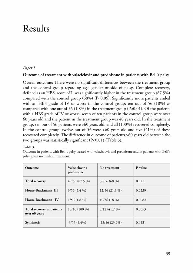
Overall outcome; There were no significant differences between the treatment group and the control group regarding age, gender or side of palsy. Complete recovery, defined as an HBS score of I, was significantly higher in the treatment group (87.5%) compared with the control group (68%) (P<0.05). Significantly more patients ended with an HBS grade of IV or worse in the control group: ten out of 56 (18%) as compared with one out of 56 (1,8%) in the treatment group (P<0.01). Of the patients with a HBS grade of IV or worse, seven of ten patients in the control group were over 60 years old and the patient in the treatment group was 40 years old. In the treatment group, ten out of 56 patients were >60 years old, and all (100%) recovered completely. In the control group, twelve out of 56 were >60 years old and five (41%) of these recovered completely. The difference in outcome of patients >60 years old between the two groups was statistically significant (P<0.01) (Table 3). Table 3. Outcome in patients with Bell´s palsy treated with valaciclovir and prednisone and in patients with Bell´s palsy given no medical treatment.
Outcome Valaciclovir + prednisone
No treatment P-value
Total recovery 49/56 (87.5 %) 38/56 (68 %) 0.0211
House-Brackmann III 3/56 (5.4 %) 12/56 (21.3 %) 0.0239
House-Brackmann IV 1/56 (1.8 %) 10/56 (18 %) 0.0082
Total recovery in patients over 60 years
10/10 (100 %) 5/12 (41.7 %) 0.0053
Synkinesis 3/56 (5.4%) 13/56 (23.2%) 0.0131
40
Stapedial reflex response; Of the 56 patients in the control group, 47 underwent a stapedial reflex response test. Of the 24 with normal reflex response, 18 had a complete recovery. Among the 23 with no measurable reflex response, 14 had a complete recovery. The difference was not significant. In the treatment group 53 of 56 patients had a stapedial reflex test performed, all of the 27 patients with normal reflex response recovered completely and 19 of 26 with no measurable reflex response recovered completely (P<0.005). In both groups, 45 of the 51 with normal reflex response had a complete recovery as compared with 33 of the 49 with no measurable reflex response (P<0.05).
Time to treatment start; In the treatment group, 48 of 56 patients were treated within 72 hours of onset of palsy. The other eight patients started treatment between 4 and 16 days, and all eight recovered completely.
Synkinesis; Synkinesis was present in 13 of the 56 patients in the control group as compared with three of the 56 in the treatment group (P<0.05). Four patients in the control group suffered from contracture, but none in the treatment group did.
Paper II Prednisolone and valaciclovir in Bell’s palsy: a randomised, double-blind, placebo-controlled, multicentre trial
Time to complete recovery (SB score = 100) was significantly shorter for patients treated with prednisolone (n = 416) compared with patients not treated with prednisolone (n = 413) (HR 1.40; 95% CI 1.18–1.64; P<0.0001). There was no difference in time to complete recovery between patients (n = 413) who received valaciclovir and patients (n = 416) not treated with valaciclovir (HR 1.01; 95% CI 0.85–1.19; P = 0.9). We found no interaction between the effects of prednisolone and valaciclovir (P = 0.59).
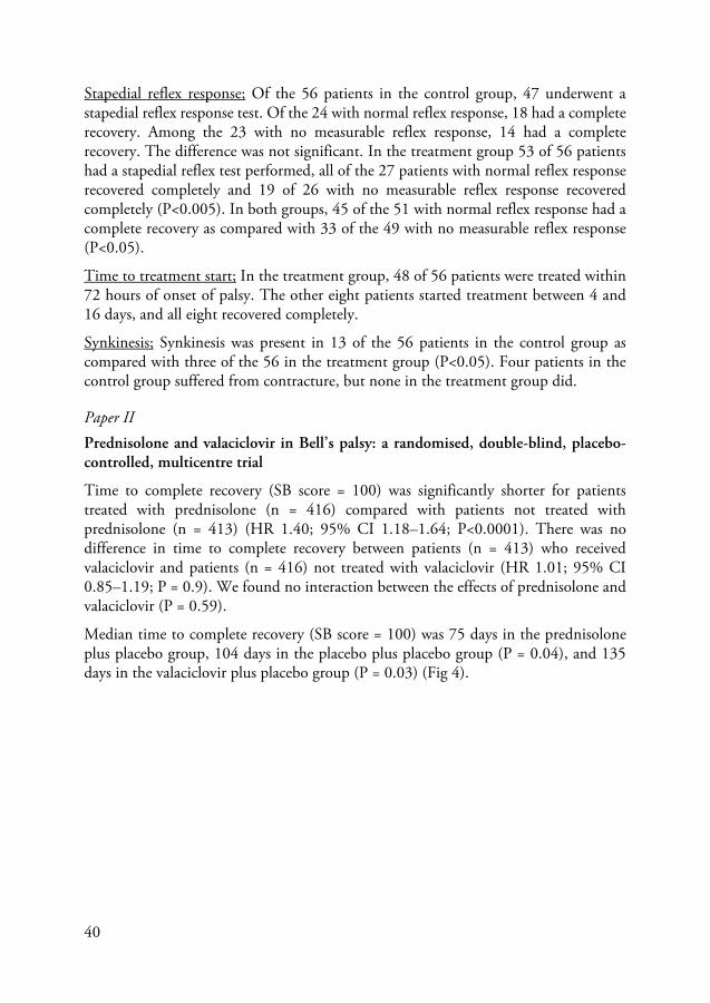
Median time to complete recovery (SB score = 100) was 75 days in the prednisolone plus placebo group, 104 days in the placebo plus placebo group (P = 0.04), and 135 days in the valaciclovir plus placebo group (P = 0.03) (Fig 4).
41
Figure 4. Kaplan-Meier estimates of patients who made a complete recovery (Sunnybrook (SB) score = 100 points) in the four treatment groups (n = 829). The median time (MD) to complete recovery for each treatment group is also illustrated. Time points: t1 = day 11−17; t2 = 1 month; t3 = 2 months; t4 = 3 months; t5 = 6 months; t6 = 12 months. Patients receiving prednisolone had significantly higher recovery rates at 3, 6 and 12 months than patients not receiving prednisolone. At 12 months, 300/416 (72%) in the prednisolone group recovered completely compared with 237/413 (57%) in the no prednisolone group (P<0.0001). The outcome in 413 patients treated with valaciclovir compared with 416 patients not treated with valaciclovir did not differ (P = 0.66).
Of the 743 patients with 12-month follow-up, synkinesis was present in 51 out of 370 patients (14%) treated with prednisolone compared with 107 out of 373 (29%) not
0 100 200 300 400
0
20
40
60
80
100
Days
Com
plet
e R
ecov
ery
(%)
Placebo (PL)Prednisolone (P)Valacyclovir (V)Prednisolone plus valacyclovir (PV)
50
Md PV
Md P
Md PL
Md V
194 180 143 113 98 77 (PL)200 184 130 99 73 57 (P)202 181 138 115 102 84 (V)200 174 123 94 77 54 (PV)
Numbers at risk (time points)
t1 t2 t3 t4 t5 t6
42
treated with prednisolone (difference -15%, 95% CI -21–-9%; P < 0.0001). Of the 369 patients who received valaciclovir, 73 (20%) had synkinesis at 12 months compared with 85 out of 374 (23%) patients not receiving valaciclovir (difference -3%, 95% CI -9–3%; P = 0.37).
Of the 829 patients included in the study, 728 patients (88%) took the study drugs as expected, indicating complete compliance. There were 114 non-serious adverse events reported by 92 patients, which were equally distributed among the four treatment groups. The most common reported adverse events were gastrointestinal complaints, headache, fatigue and insomnia.
Paper III Prednisolone in Bell’s palsy related to treatment start and age.
Treatment start
Treatment was started within 24 hours of onset in 308 (37%) of the 829 patients. In the time interval 25−48 hours 345 patients (42%) initiated treatment and 175 patients (21%) started medication between 49 and 72 hours after palsy. The median SB score was 42 (IQR 29−58) in patients who started treatment within 24 hours, compared with 37 (IQR 22−55) in patients treated between 25–48 hours, and 32 (IQR 19−50) in patients receiving medication between 49–72 hours after onset. The patients’ age did not differ between the three time interval groups.
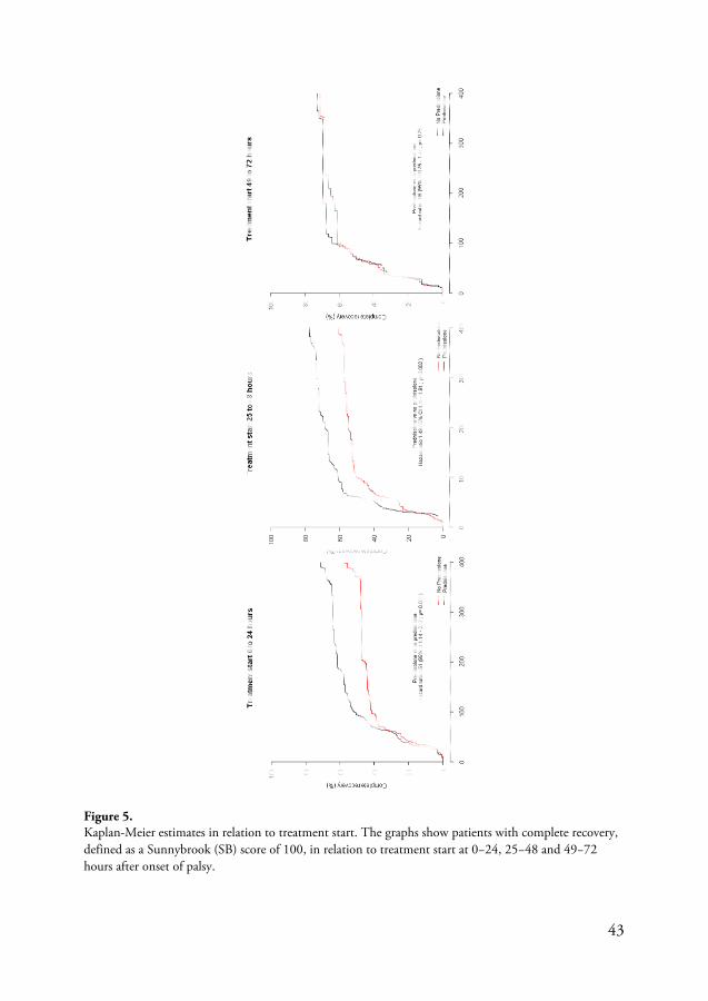
In patients treated with prednisolone within 24 hours, 103 out of 156 (66%) had a complete recovery at 12 months compared with 77 out of 152 (51%) patients not treated with prednisolone (difference -15%, 95% CI -26–-4%; P = 0.008). The corresponding recovery rates for patients treated between 25−48 hours was 128 out of 168 (76%) in prednisolone-treated patients compared with 102 out of 177 patients (58%) not treated with prednisolone (difference -19%, 95% CI -28–-9%; P = 0.0003). In the interval 49−72 hours, 69 out of 92 (75%) prednisolone-treated patients compared with 58 out of 83 (70%) non-prednisolone-treated patients achieved complete recovery (difference -5%, 95% CI -19–8%; P = 0.5).
Time to complete recovery was significantly shorter in patients treated with prednisolone within 24 hours and 25−48 hours, respectively, compared with patients not treated with prednisolone (HR 1.51 and 1.49; P = 0.004 and P = 0.002, respectively) (Fig 5).
43
Figure 5. Kaplan-Meier estimates in relation to treatment start. The graphs show patients with complete recovery, defined as a Sunnybrook (SB) score of 100, in relation to treatment start at 0−24, 25−48 and 49−72 hours after onset of palsy.
44
Of the 743 patients with a 12-month follow-up, synkinesis was significantly less in patients treated with prednisolone within 48 hours compared with patients who did not receive prednisolone. Where treatment was started within 24 hours, synkinesis was noted in 17% of the prednisolone-treated patients compared with 31% of patients in the non-prednisolone group (P = 0.008). Where the treatment was started between 25 and 49 hours post-onset of palsy, synkinesis was noted in 10% of patients receiving prednisolone compared with 32% of patients not receiving prednisolone (P<0.0001). Prednisolone treatment that was started between 49–72 hours post-palsy onset did not show any effect on synkinesis, compared with no prednisolone (14% v. 18%; P = 0.66).
Age
In total 392 patients were 18–39 years of age, and their median SB score at baseline was 42 (IQR 27–58). The 317 patients aged 40–60 years had a median SB score of 37 (IQR 22–53) and the 119 patients aged 61–75 years had a median SB score of 33 (IQR 17–46).
In the 18–39-year age group, complete recovery at 12 months was achieved by 137 out of 191 patients (72%) among those treated with prednisolone, compared with 129 out of 201 patients (64%) not treated with prednisolone (P = 0.13). In subjects 40–60 years old, the corresponding values were 122 out of 159 (77%) prednisolone-treated patients and 86 out of 158 patients (54%) not treated with prednisolone (P<0.0001), while in patients aged 61–75 years, 41 out of 66 (62%) and 21 out of 53 (40%), respectively, achieved full recovery (P = 0.02).
Synkinesis at 12 months, among patients aged 18−39 years, was present in 18 (11%) of the 160 patients in the prednisolone group, compared with 44 (25%) of 178 in the group not treated with prednisolone (P = 0.002). In the group aged 40−60 years, synkinesis occurred in 20 (13%) out of 150 patients treated with prednisolone and in 44 (31%) out of 144 patients not receiving prednisolone (P = 0.0004). Finally, the corresponding values for patients 61−75 years old were 13/60 (22%) for prednisolone and 19/51 (37%) for no prednisolone (P = 0.09).
We found no interaction, positive or detrimental, between the effects of prednisolone and valaciclovir regarding complete recovery or synkinesis, in relation to age (P = 0.51 and P = 0.09, respectively) or start of treatment (P = 0.65 and P = 0.11, respectively).
45
Paper IV Bell’s palsy – the effect of prednisolone and/or valaciclovir versus placebo in relation to baseline severity in a randomised controlled trial.
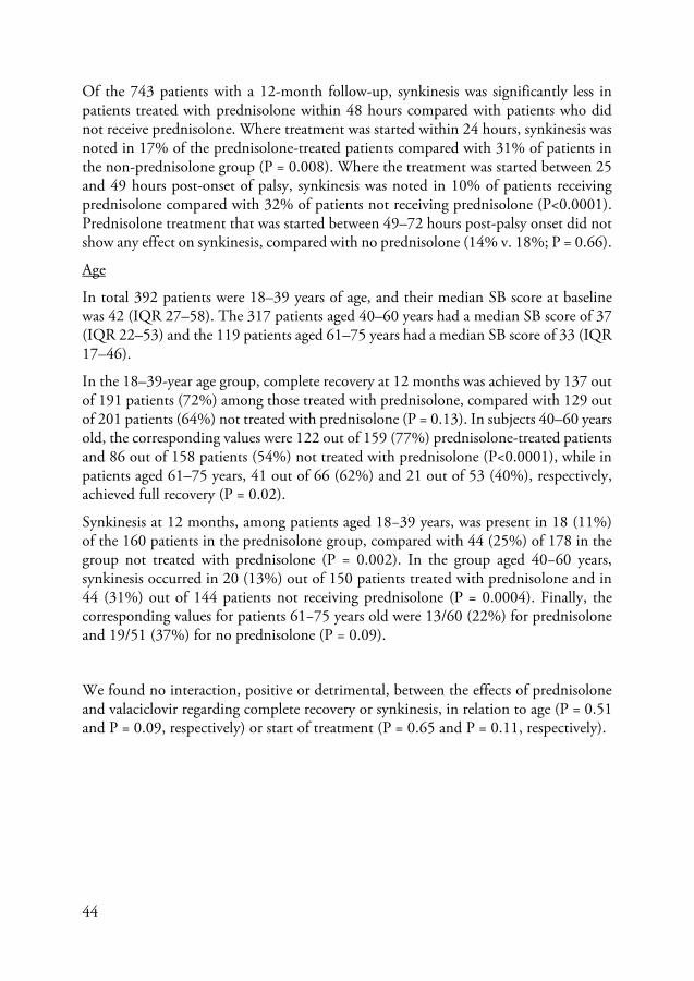
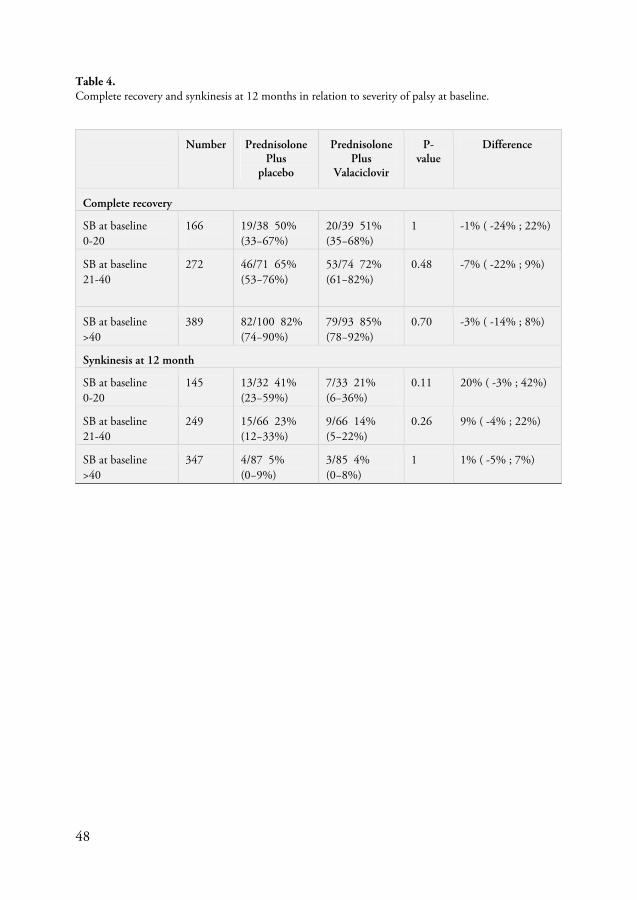
Age and treatment start were similar in patients given prednisolone compared with no prednisolone across the three severity groups. Out of 166 patients with severe palsy (SB score ≤20) at baseline, 77 received prednisolone. Of these, 39 (51%) had recovered completely by 12 months compared with 28 (31%) out of 89 who did not receive prednisolone (P = 0.02). In 272 patients with moderate palsy (SB score 21–40) at baseline, 99 (68%) out of 145 subjects treated with prednisolone had recovered completely by 12 months, compared with 65 (51%) out of 127 not treated with prednisolone (P = 0.004). Among 389 patients with mild palsy (SB scale score >40) at baseline, 193 received prednisolone. Of these, 161 (83%) had complete recovery at 12 months, in comparison with 144 (73%) of 196 not receiving prednisolone (P = 0.02). Patients with very mild palsy (SB scale score >60) at baseline did not show any significantly better recovery in the prednisolone group (61/68; 90%) compared with the no prednisolone group (51/65; 78%) (P = 0.1).
The mean SB score at 12 months differed significantly between patients treated with prednisolone and patients not treated with prednisolone in all three severity groups (Fig 6).
Figure 6. Mean Sunnybrook (SB) score at 12 months in patients in different severity groups treated with prednisolone compared with no prednisolone.
46
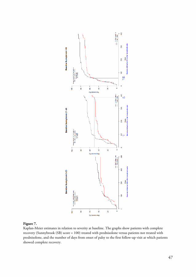
In patients with moderate palsy (SB score 21-40) at baseline, time to complete recovery was significantly shorter for patients treated with prednisolone compared with patients not treated with prednisolone (P<0.001). Difference in time to complete recovery between prednisolone treatment and no prednisolone was not seen in patients with mild palsy (SB score >40) or severe palsy (SB score ≤20) (Fig 7).
Synkinesis was present at 12 months in 157 (21%) out of the 743 patients who had a 12-month follow-up. In patients with severe palsy at baseline, the presence of synkinesis at 12 months did not differ significantly between those given prednisolone (31%) and those not given prednisolone (46%). Patients with moderate (18% and 30%, respectively), mild (4% and 19%, respectively) and very mild palsy at baseline (2% and 13%, respectively) had significantly less synkinesis at 12 months if treated with prednisolone compared with no prednisolone.
Valaciclovir had no effect on recovery except in patients with moderate palsy. Of 272 patients with moderate palsy at baseline, 83 out of 122 patients (68%) who received valaciclovir had a complete recovery at 12 months compared with 81 out of 150 patients (54%) who did not receive valaciclovir (P = 0.02). There was no significant effect of valaciclovir on the presence of synkinesis at 12 months in any of the three severity groups.
There was no significant additive effect of valaciclovir to prednisolone, regardless of severity of palsy at baseline. This was shown both for complete recovery rates and for the presence of synkinesis at 12 months (Table 4).
47
Figure 7. Kaplan-Meier estimates in relation to severity at baseline. The graphs show patients with complete recovery (Sunnybrook (SB) score = 100) treated with prednisolone versus patients not treated with prednisolone, and the number of days from onset of palsy to the first follow-up visit at which patients showed complete recovery.
48
Table 4. Complete recovery and synkinesis at 12 months in relation to severity of palsy at baseline.
Number Prednisolone Plus
placebo
Prednisolone Plus
Valaciclovir
P-value
Difference
Complete recovery
SB at baseline 0-20
166
19/38 50% (33−67%)
20/39 51% (35−68%)
1 -1% ( -24% ; 22%)
SB at baseline 21-40
272 46/71 65% (53−76%)
53/74 72% (61−82%)
0.48 -7% ( -22% ; 9%)
SB at baseline >40
389 82/100 82% (74−90%)
79/93 85% (78−92%)
0.70 -3% ( -14% ; 8%)
Synkinesis at 12 month
SB at baseline 0-20
145 13/32 41% (23−59%)
7/33 21% (6−36%)
0.11 20% ( -3% ; 42%)
SB at baseline 21-40
249 15/66 23% (12−33%)
9/66 14% (5−22%)
0.26 9% ( -4% ; 22%)
SB at baseline >40
347 4/87 5% (0−9%)
3/85 4% (0−8%)
1 1% ( -5% ; 7%)
49
Discussion
The first paper in this thesis can be seen as a pilot study where patients treated with a combination of valaciclovir and prednisolone had significantly better outcome than did control patients given no medical treatment, as was the standard in Sweden at the time. Significantly more patients with a severe outcome were from the control group (18%) than from the treatment group (1.8%). Among elderly patients >60 years, treatment seemed to prevent sequelae; all of >60-year-old patients in the treatment group had complete recovery as compared with only 41.7% of >60-year-olds in the control group. Furthermore, fewer patients in the treatment group compared with the control group had synkinesis and contractures. However, this finding may be due to the fact that the patients in the control group were followed for a longer time after onset of palsy compared with the patients in the treatment group and sequelae such as synkinesis and contractures develop over time. These findings are in accordance with the findings of Adour et al (Adour et al 1996).
The stapedial reflex has been used as a prognostic tool in Bell’s palsy (Ekstrand and Glitterstam 1979, Gavilan et al 1988). In our study, as in other studies, presence of the stapedial reflex indicated a good prognosis although 25% of patients in the control group with a stapedial reflex did not recover completely.
A major limitation was that our study was retrospective and not placebo-controlled or double-blind. Cochrane reports call for the need of adequately powered clinical trials with sufficient follow-up to detect the benefits (or harm) of corticosteroid and/or antiviral therapy in Bell’s palsy (Allen and Dunn 2004, Salinas et al 2004).
The SBPS has so far been the largest randomised, double-blind, placebo-controlled multi-centre trial assessing corticosteroid and antiviral treatment for Bell’s palsy (Engström et al 2008). Patients who received prednisolone had a shorter time to complete recovery of facial function and a more favourable outcome at 12 months compared with patients not receiving prednisolone. Synkinesis was also less common in prednisolone-treated patients. Valaciclovir alone was not proved to be effective, and did not give any additive effect to prednisolone.
A Scottish trial of 551 patients randomly assigned within 72 hours of onset to 10 days of 25 mg prednisolone twice daily and placebo, aciclovir 400 mg five times daily and placebo, both prednisolone and aciclovir or placebo, reported similar results. The investigators of the study concluded that early treatment with prednisolone
50
significantly improved the chances of complete recovery at 3 and 9 months. There was no benefit from aciclovir (Sullivan et al 2007).
The nerve damage in Bell’s palsy is consistent with oedema and inflammation of the nerve in the Fallopian canal (Engström et al 1997, Fisch and Esslen 1972, Song et al 2008). Early administration of anti-inflammatory corticosteroids may reduce oedema and subsequent spreading of conduction block or axonotmesis (Liston and Kleid 1989). The effect of early prednisolone treatment in Bell’s palsy is in accordance with the reported effect of corticosteroids in vestibular neuritis (Strupp et al 2004), a disease which may have the same pathogenesis as Bell’s palsy (Gianoli et al 2005, Strupp et al 2004).
This is the first large double-blind, placebo-controlled study testing valaciclovir alone and in combination with prednisolone in Bell’s palsy patients. Valaciclovir has a three-fold to five-fold higher bioavailability compared with aciclovir. Therefore valaciclovir gives higher compliance and higher concentrations in both serum and CSF, and the dose used, 1 000 mg three times daily, is well above the inhibitory level for herpes simplex but may be only partially inhibitory for VZV (Lycke et al 2003, Weller et al 1993). The ineffectiveness of valaciclovir may be due to the fact that the rate of virus replication had declined before treatment started or that herpes viruses are not the main cause of Bell’s palsy (Linder et al 2005, Stjernquist-Desatnik et al 2006).
Prednisolone plus valaciclovir was not more effective than prednisolone alone. This result is in accordance with previous findings involving valaciclovir (Kawaguchi et al 2007) and aciclovir (De Diego et al 1998, Sullivan et al 2007) plus corticosteroids. Nevertheless, an additional effect of aciclovir (Adour et al 1996) or valaciclovir (Hato et al 2007) on corticosteroid treatment has been reported. However, the trial by Adour and colleagues followed up patients for only 4 months, and the study by Hato and co-workers was not double-blinded; therefore, the results of both trials should be interpreted with caution.
In most trials of treatments for Bell’s palsy (Lagalla et al 2002, Sullivan et al 2007, Ünüvar et al 1999), facial function is evaluated using the HBS (House and Brackmann 1985). The SFGS reports facial function in a continuous manner and has a wider response range compared with the HBS (Ross et al 1996). The intrarater and inter-rater reliability of the SB scale is high when applied by either a novice or an expert assessor (Hu et al 2001). The SB scale is more reliable than the HBS (Kanerva et al 2006) and has been reported to be the best instrument for clinical use (Chee and Nedzelski 2000, Rogers et al 2007). We therefore used the SFGS as the main scale to assess facial nerve function.
A limitation of use of the SBPS was that for practical reasons, grading was performed by several different physicians. However, most follow-up evaluations were performed by one or two experienced physicians at each centre. Altogether 79% of assessments at
51
the 1–12-month follow-ups were done by 49 assessors at 17 centres. Another limitation is that subjective gradings are still a weak point of facial palsy treatment studies and will remain so until a good objective system is available.
Our recovery rates were lower than those previously reported (Peitersen 2002, Sullivan et al 2007). Although the overall results were similar between the two grading systems, we found lower complete recovery rates with the SFGS than with the HBS. This probably demonstrates the higher sensitivity of the SFGS for sequelae. The LOCF method which we used likewise gives lower recurrence rates, compared with CCA (Berg et al 2009). Furthermore, inclusion/exclusion criteria, follow-up time and grading done on the basis of interviews or photos rather than on the basis of a clinical examination affect final recovery rates (Adour et al 1996, Hato et al 2007, Peitersen 2002, Sullivan et al 2007).
The effect of prednisolone in Bell’s palsy was assessed in relation to prognostic factors such as treatment start, age, and severity at baseline. The present controlled trial is the largest trial, thus far, to study the effect of prednisolone given in the very early stage of Bell’s palsy. Prednisolone administered within 48 hours of palsy onset resulted in significantly higher recovery rates and less synkinesis compared with no prednisolone treatment. Prednisolone treatment within 49−72 hours did not significantly affect recovery rates or synkinesis. This is in accordance with findings in a controlled study by Yeo et al, where treatment with prednisolone within 3 days was associated with higher recovery rates compared with later treatment (Yeo et al 2008). Furthermore, Shefshak et al also report better outcome with early, within-24 hour treatment, but their study was not double-blinded and only included 160 patients, all with complete or near-complete paralysis (Shafshak et al 1994). The results of uncontrolled studies diverge concerning the relation between time of treatment start with corticosteroids and outcome of palsy (Adour et al 1972, Sathirapanya and Sathirapanya 2008, Sittel et al 2000).
Unexpectedly, patients not treated with prednisolone and included 49−72 hours after onset of palsy had a higher recovery rate (70%) and less synkinesis (18%) compared with those included within 24 hours (recovery 51%, synkinesis 32%) and 25–48 hours (recovery 58%, synkinesis 32%). The median SB score at baseline was 42 in the 0–24 hour group, 37 in the 25–48 hour group and 32 in the 49–72 hour group. The better outcome in patients not treated with prednisolone and included between 49 and 72 hours cannot be explained by differences in severity at baseline. It may be speculated that patients with a mild progression of palsy, and consequently more beneficial outcome, seek medical care later than those with more progressive palsy. Furthermore, subgroup analysis reduced the number of patients and made the statistical comparison more hazardous.
In patients aged 40 years or older, treatment with prednisolone gave significantly higher recovery rates compared with no treatment with this medication. Yeo et al recently
52
reported that prednisolone-treated patients older than 60 years showed a tendency to have a higher degree of recovery and a similar final status compared with younger age groups (Yeo et al 2007). Our findings support the statement by Yeo et al, that otolaryngologists should encourage older patients to participate in more active and intensive treatment.
In patients younger 40 years old, the recovery rate was not significantly higher in patients treated with prednisolone (72%) compared with patients not treated with prednisolone (64%). However, synkinesis at 12 months was less common in patients younger than 40 years old in the prednisolone treatment group. Altogether the results show that with regard to recovery rates and/or synkinesis, prednisolone was effective in all age groups.
In patients not treated with prednisolone, the recovery rate was 64% for patients aged 18–39, compared with 54% for patients 40–60 years old and only 40% for patients >60 years old. The finding that higher age is a negative prognostic factor for outcome, accords with the results of previous reports (Devriese et al 1990, Hydén et al 1982, Ikeda et al 2005, Peitersen 2002).
Regardless of severity at baseline, all severity groups (severe, moderate and mild) had a significantly higher complete recovery rate if treated with prednisolone compared with no prednisolone. Synkinesis at 12 months was also significantly less common in patients with moderate and mild palsy at baseline when treated with prednisolone compared with no prednisolone. In accordance with previous studies (Hato et al 2007, Hydén et al 1982, Sathirapanya and Sathirapanya 2008, Sittel et al 2000), a low SB score at baseline was correlated to a less favourable outcome. In patients with severe, moderate and mild palsy, complete recovery was recorded in 51%, 68% and 83%, respectively, in the prednisolone groups and in 31%, 51% and 73% in the no prednisolone groups. Valaciclovir treatment gave a significantly higher complete recovery rate in patients with moderate palsy (SB score 21–40) at baseline compared with no valaciclovir treatment. The recovery rate for this subgroup treated with valaciclovir, however, did not exceed that for the prednisolone group.
At the initial visit, the physician cannot predict if the palsy will deteriorate or not. Because the disease is progressive during the first weeks we do not know which patients will develop a more severe palsy. In a study by Linder et al, 36% of patients, almost all treated with steroids, deteriorated to a total paralysis within 2 weeks (Linder et al 2010). Marsk et al found that 28% deteriorated within the first 11–17 days after onset of palsy and that early treatment with prednisolone reduced the number of patients who did deteriorate (Marsk et al 2010).
The combination corticosteroid and antiviral treatment has been suggested to be more powerful in patients with severe palsy (Hato et al 2007, Minnerop et al 2008). In our study, valaciclovir did not add any significant effect to prednisolone regarding recovery
53
rates or synkinesis in any of the severity groups. In the study by Hato et al, the patients were treated with a lower dose of valaciclovir (1 000 mg/day compared with 3 000 mg/day in our study), although they reported better outcome with the combination therapy prednisolone and valaciclovir in cases of complete or severe palsy (Hato et al 2007). In the study by Minnerop et al, the combination of prednisolone and famciclovir was reported to be more effective in cases with severe palsy (Minnerop et al 2008). As these studies were not double-blinded and the study by Minnerop et al included <20 patients with severe palsy in each treatment group, the results can be questioned. Our study is in accordance with the studies included in the Cochrane review titled “Antiviral treatment for Bell’s palsy”, which concludes that antivirals are significantly less likely to achieve complete recovery compared with corticosteroids (Lockhart et al 2009).
The subgroup analysis is a limitation of our studies comparing how prognostic factors such as time to treatment start, age, and severity at baseline will influence outcome of treatment in patients with Bell’s palsy. Subgroup analysis reduces the number of patients and therefore makes statistical comparison more hazardous.
Clinical applicability of the findings of this thesis
Corticosteroid treatment may reduce inflammation and thus prevent further nerve damage in patients with Bell’s palsy. As at present we do not have any diagnostic tool that can, at the initial visit, predict the course and outcome for the individual palsy patient, it seems adequate to treat the adult patient regardless of severity of palsy and age. Even patients with mild palsy benefit from early treatment with corticosteroids, as do patients between 18 and 39 years. Treatment should be started as early as possible within 48 hours of onset of palsy as long as no contraindications for corticosteroid treatment are present.

54
55
Conclusions
• Better recovery rates were seen in patients treated with the combination prednisolone and valaciclovir compared with patients given no treatment. Especially elderly patients had a better outcome in the treatment group.
• Prednisolone treatment in patients with Bell’s palsy shortened the time to complete recovery and gave higher recovery rates and less synkinesis at 12 months compared with no prednisolone. Valaciclovir, either alone or in combination with prednisolone, did not affect facial recovery.
• Treatment start with prednisolone within 48 hours of onset of palsy resulted in significantly higher recovery rates and less synkinesis compared with no prednisolone treatment.
• In patients aged ≥40 years, prednisolone gave higher recovery rates than seen in those not treated with prednisolone. In subjects <40 years, prednisolone was not shown to significantly improve recovery rate, but it did significantly reduce synkinesis.
• Prednisolone treatment resulted in higher recovery rates compared with no prednisolone, regardless of severity of palsy at baseline. In patients with mild and moderate palsy, significantly fewer patients had synkinesis when treated with prednisolone. Valaciclovir had no additional effect to prednisolone.

56
57
Populärvetenskaplig sammanfattning
Plötsligt debuterande ansiktsförlamning utan känd bakomliggande orsak benämns Bell’s pares och drabbar ca 30/100 000 invånare/år. Förlamningen ger en försvagning eller total förlamning av ena ansiktshalvan. Ofta föregås förlamningen av smärta runt örat och kan även ge påverkan på smak, ljudupplevande och tårproduktion. Det har länge misstänkts att det rör sig om en inflammatorisk sjukdom där herpes virus kan vara en tänkbar orsak. Spontanutläkningen är relativt god och ca 60-70% blir helt återställda utan behandling. Resterande får mer eller mindre svåra resttillstånd, dessa leder till svårigheter att äta och dricka, dessutom är förlamningen väl synlig för omgivningen, vilket kan ge patienten psykosociala problem.Vid insjuknandet går det inte att förutspå vem som kommer att få bestående besvär.
Flera olika behandlingar har provats för att minska risken för resttillstånd, både kirurgiska och medicinska behandlingar har använts. De vanligaste medicinska behandlingarna har varit kortison och antivirala medel. Tidigare studier har varken iform av upplägg eller patientantal kunnat besvara frågan om kortison och/eller antivirala medels effektivitet vid behandling av Bell’s pares. Det hade varit önskvärt att hitta prognostiska faktorer som indikerar ökad risk för defektläkning. Behandlingen kan då erbjudas de patienter som löper risk att defektläka och man undviker överbehandling.
Avhandlingens första delarbete är en retrospektiv studie där patienter som insjuknat i Bell’s pares under åren 1997-1999 fick behandling med kortison (prednisolon) och antivirala medel (valaciclovir), denna grupp av patienter jämfördes med en grupp av patienter som insjuknat 1995-1996 och ej givits någon behandling. Resultaten visade att de som fått behandling läkte ut i högre grad än de som inte fick behandling. Speciellt tydligt var det hos patienter >60 år.
Andra delarbetet, Skandinaviska Bell’s Pares Studien (SBPS), är en randomiserad, dubbel-blind, placebo-kontrollerad multicenterstudie. Studien löpte mellan åren 2001-2007. Sexton svenska öron-, näs- och halskliniker och en i Finland deltog. Patienter 18-75 år med Bell’s pares inkluderades i studien och följdes i 12 månader. Max 72 timmar från paresdebut skulle ha förflutit innan behandling sattes in. Paresens svårighetsgrad graderades med två olika skalor (Sunnybrook och House-Brackmann). I studien inkluderades 829 patienter. Patienterna randomiserades till en av 4 behandlingsgrupper: prednisolon plus placebo, valaciclovir plus placebo, prednisolon
58
plus valaciclovir eller placebo plus placebo. Resultaten visade att prednisolon signifikant förkortade tiden till utläkning samt ökade andelen patienter som läkte ut helt. Förekomsten av synkinesier (ofrivilliga medrörelser) var också signifikant lägre i prednisolongruppen. Valaciclovir hade ingen egen eller additiv effekt på utläkningen.
Tredje delarbetet baseras på data från SBPS. Studien utvärderar prognostiska faktorers, såsom tid till insatt behandling samt ålder hos patienten, betydelse för utläkning. Resultaten visade att prednisolon insatt inom 48 timmar resulterade i signifikant högre utläkningsgrad och lägre förekomst av synkinesier jämfört med icke prednisolon behandling. Patienter som behandlades inom 49-72 timmar efter paresdebut hade ingen signifikant effekt av prednisolonbehandlingen. Patienter äldre än 40 år uppvisade signifikant högre utläkningsgrad i prednisolongruppen. Hos patienter yngre än 40 år sågs ingen högre utläkningsgrad men väl lägre förekomst av synkinesier i prednisolongruppen.
I fjärde delarbetet, som även det baseras på data från SBPS, studeras huruvida svårighetsgraden av pares vid inkluderingen är av prognostisk betydelse. Det har tidigare hävdats att milda pareser alltid läker ut och därför kanske inte skulle behöva behandlas. Patienterna delades upp i tre grupper, svår pares, måttlig pares och lindrig pares. I samtliga grupper var utläkningen signifikant bättre hos de som behandlats med prednisolon jämfört med de som inte fått prednisolon. Enstaka tidigare studier har visat bättre utläkning av de svåra pareserna om de behandlats med en kombination av prednisolon och valaciclovir. Någon sådan additiv effekt av valaciclovir kunde inte verifieras i detta delarbete.
Klinisk applicerbarhet/Nyhetsvärde
Resultaten från SBPS ligger till grund för en Cochranerapport (systematisk översikt av randomiserade studier som undersöker effekten av en specifik behandling) från 2010 som förordar kortisonbehandling av patienter med Bell’s pares medan antivirala medels effekt inte kunnat styrkas. Tredje och fjärde delarbetet visar på vikten av att sätta in kortison snabbt, inom 48 timmar, efter paresdebut om inga kontraindikationer föreligger samt att man inte bör avstå från behandling vid mild pares eller hög ålder.
59
Acknowledgements
This thesis had not been written without the 17 otorhinolaryngological centres that participated in the Scandinavian Bell’s Palsy Study. I want to express my greatest gratitude to everyone who has contributed to these studies. In particular I want to thank:
My supervisor Anna Stjernquist-Desatnik for being my dedicated mentor. You introduced me into the research of Bell’s palsy and have guided me with patience the whole way from our first clinical study in 1999. I am grateful for your enthusiasm and all support during these years and for your assurance that this thesis will be real inspite of my doubt.
Lars Jonsson and Mats Engström for all work you put into making the Scandinavian Bell’s Palsy Study possible and your true dedication to the topic. Lasse, for your invaluable advice and help with revising the papers, you have always been disposable and interested in my research.
Thomas Berg, co-author, for good collaboration, valuable advice and help with Endnote.
Sven Lindberg, co author, for helpful advice and your real interest in research.
Mervi Kanerva, Anne Pitkäranta, Malou Hultcrantz and the late Per Hanner, co-authors and members of the steering committee for the Scandinavian Bell’s Palsy Study, for valuable collaboration and helpful advice with the manuscripts.
Peter Olsson, head of the department of Otorhinolaryngology, Helsingborg Hospital, for your support and for giving me the opportunity to combine clinical work with research and be able to write this thesis.
Charlotte Cervin-Hoberg for help with monitoring and input of data from the case report forms to the database.
Doctors, nurses and staff in the Scandinavian Bell’s Palsy Study, especially Mikael Karlberg and Inga-Lisa Sjölin for examining and registering a lot of patients in the study in Lund and Helsingborg.
Nermin Hadziosmanovic, biostatistician, for most valuable help with the statistics.
Lena Lyons for nice drawing of the facial nerve and its branches in this thesis.
60
Professor Helge Rask-Andersson, Uppsala University Hospital, for providing the fantastic picture of the inner ear and the facial nerve.
Colleagues and friends at the department of Otorhinolaryngology, Head and Neck Surgery, Lund for friendship, for fruitful discussions and a very good atmosphere in “drängkammaren”.
Colleagues and friends at the department of Otorhinolaryngology, Hospital of Helsingborg for your friendship, support and encouragement to complete this thesis.
My dear Lars, for invaluable computer support around the clock, for a nice drawing in my thesis and, your support and encouragement when I most needed it. Axel and Filip, my lovely sons, for reminding me of what is important in life.
Special thanks for the financial support from Stig and Ragna Gorthon Foundation, Thelma Zoega Foundation and Acta Otolaryngologica Foundation.
61
References
Adour KK. Decompression for Bell's palsy: why I don't do it. Eur Arch Otorhinolaryngol. 2002;259(1):40-7.
Adour KK, Bell DN, Hilsinger RL, Jr. Herpes simplex virus in idiopathic facial paralysis (Bell palsy). JAMA. 1975;233(6):527-30.
Adour KK, Byl FM, Hilsinger RL, Jr., et al. The true nature of Bell's palsy: analysis of 1,000 consecutive patients. Laryngoscope. 1978;88(5):787-801.
Adour KK, Ruboyianes JM, Von Doersten PG, et al. Bell's palsy treatment with acyclovir and prednisone compared with prednisone alone: a double-blind, randomized, controlled trial. Ann Otol Rhinol Laryngol. 1996;105(5):371-8.
Adour KK, Swanson PJ, Jr. Facial paralysis in 403 consecutive patients: emphasis on treatment response in patients with Bell's palsy. Trans Am Acad Ophthalmol Otolaryngol. 1971;75(6):1284-301.
Adour KK, Wingerd J. Idiopathic facial paralysis (Bell's palsy): factors affecting severity and outcome in 446 patients. Neurology. 1974;24(12):1112-6.
Adour KK, Wingerd J, Bell DN, et al. Prednisone treatment for idiopathic facial paralysis (Bell's palsy). N Engl J Med. 1972;287(25):1268-72.
Allen D, Dunn L. Aciclovir or valaciclovir for Bell's palsy (idiopathic facial paralysis). Cochrane Database Syst Rev. 2004(3):CD001869.
Balance C, Duel AB. The operative treatment of facial palsy. Arch Otolaryngol. 1932;15:1-70.
Bell C. On the nerves; giving an account of some experiments on their structure and functions, which lead to new arrangement of the system. Philos Trans R Soc. 1821;111:398-424.
Bell C. On the nerves of the face; being a second paper on that subject. Philos Trans R Soc. 1829;119:317-30.
Berg T, Axelsson S, Engstrom M, et al. The course of pain in Bell's palsy: treatment with prednisolone and valacyclovir. Otol Neurotol. 2009;30(6):842-6.
Berg T, Jonsson L, Engstrom M. Agreement between the Sunnybrook, House-Brackmann, and Yanagihara facial nerve grading systems in Bell's palsy. Otol Neurotol. 2004;25(6):1020-6.
62
Berg T, Marsk E, Engstrom M, et al. The effect of study design and analysis methods on recovery rates in Bell's palsy. Laryngoscope. 2009;119(10):2046-50.
Botman JW, Jongkees LB. The result of intratemporal treatment of facial palsy. Pract Otorhinolaryngol (Basel). 1955;17(2):80-100.
Brackmann D, Shelton S, Arriaga M. Otologic Surgery. 3 ed: Saunders; 2010.
Burgess RC, Michaels L, Bale JF, Jr., et al. Polymerase chain reaction amplification of herpes simplex viral DNA from the geniculate ganglion of a patient with Bell's palsy. Ann Otol Rhinol Laryngol. 1994;103(10):775-9.
Burres S, Fisch U. The comparison of facial grading systems. Arch Otolaryngol Head Neck Surg. 1986;112(7):755-8.
Chee GH, Nedzelski JM. Facial nerve grading systems. Facial Plast Surg. 2000;16(4):315-24.
Chen N, Zhou M, He L, et al. Acupuncture for Bell's palsy. Cochrane Database Syst Rev. 2010(8):CD002914.
Chow LC, Tam RC, Li MF. Use of electroneurography as a prognostic indicator of Bell's palsy in Chinese patients. Otol Neurotol. 2002;23(4):598-601.
Coulson SE, Croxson GR, Adams RD, et al. Reliability of the "Sydney," "Sunnybrook," and "House Brackmann" facial grading systems to assess voluntary movement and synkinesis after facial nerve paralysis. Otolaryngol Head Neck Surg. 2005;132(4):543-9.
Cumberworth A, Mabvuure NT, Norris JM, et al. Is acupuncture beneficial in the treatment of Bell's palsy?: best evidence topic (BET). Int J Surg. 2012;10(6):310-2.
De Diego JI, Prim MP, De Sarria MJ, et al. Idiopathic facial paralysis: a randomized, prospective, and controlled study using single-dose prednisone versus acyclovir three times daily. Laryngoscope. 1998;108(4 Pt 1):573-5.
Devriese PP, Schumacher T, Scheide A, et al. Incidence, prognosis and recovery of Bell's palsy. A survey of about 1000 patients (1974-1983). Clin Otolaryngol Allied Sci. 1990;15(1):15-27.
Diels H. Advances in Otolaryngology-Head and Neck Surgery. Chicago: Mosby; 1995.
Diels HJ, Combs D. Neuromuscular retraining for facial paralysis. Otolaryngol Clin North Am. 1997;30(5):727-43.
Dumitru D, Walsh NE, Porter LD. Electrophysiologic evaluation of the facial nerve in Bell's palsy. A review. Am J Phys Med Rehabil. 1988;67(4):137-44.
Ekstrand T, Glitterstam K. Bell's palsy--prognostic value of the stapedius reflex with contralateral stimulation. J Laryngol Otol. 1979;93(3):271-5.
63
Engström M, Abdsaleh S, Ahlström H, et al. Serial gadolinium-enhanced magnetic resonance imaging and assessment of facial nerve function in Bell's palsy. Otolaryngol Head Neck Surg. 1997;117(5):559-66.
Engström M, Berg T, Stjernquist-Desatnik A, et al. Prednisolone and valaciclovir in Bell's palsy: a randomised, double-blind, placebo-controlled, multicentre trial. Lancet Neurol. 2008;7(11):993-1000.
Engström M, Thuomas K-Å, Naeser P, et al. Facial nerve enhancement in Bell's palsy demonstrated by different gadolinium-enhanced magnetic resonance imaging techniques. Arch Otolaryngol Head Neck Surg. 1993;119(2):221-5.
Fisch U. Surgery for Bell's palsy. Arch Otolaryngol. 1981;107(1):1-11.
Fisch U. Prognostic value of electrical tests in acute facial paralysis. Am J Otol. 1984;5(6):494-8.
Fisch U, Esslen E. Total intratemporal exposure of the facial nerve. Pathologic findings in Bell's palsy. Arch Otolaryngol. 1972;95(4):335-41.
Fisch U, Felix H. On the pathogenesis of Bell's palsy. Acta Otolaryngol. 1983;95(5-6):532-8.
Forsten Verschuir W. De Paralysi Musculorum Faciei, sic dicta Rheumatica [On Rheumatic Paralysis of the Facial Muscles]. . Groningen: Abraham Groenewolt (English translation, Dijkers H, van de Graaf RC). 1804.
Friedreich N. De paralysi Musculorum Faciei Rheumatica [ On rheumatic paralysis of the facial muscles ]. Würzburg: Rienner. 1797.
Furuta Y, Ohtani F, Aizawa H, et al. Varicella-zoster virus reactivation is an important cause of acute peripheral facial paralysis in children. Pediatr Infect Dis J. 2005;24(2):97-101.
Furuta Y, Ohtani F, Kawabata H, et al. High prevalence of varicella-zoster virus reactivation in herpes simplex virus-seronegative patients with acute peripheral facial palsy. Clin Infect Dis. 2000;30(3):529-33.
Gantz BJ, Gmur A, Fisch U. Intraoperative evoked electromyography in Bell's palsy. Am J Otolaryngol. 1982;3(4):273-8.
Gavilan C, Gavilan J, Rashad M, et al. Discriminant analysis in predicting prognosis of Bell's palsy. Acta Otolaryngol. 1988;106(3-4):276-80.
Gianoli G, Goebel J, Mowry S, et al. Anatomic differences in the lateral vestibular nerve channels and their implications in vestibular neuritis. Otol Neurotol. 2005;26(3):489-94.
Gilchrist JM. Seventh cranial neuropathy. Semin Neurol. 2009;29(1):5-13.
64
Hato N, Yamada H, Kohno H, et al. Valacyclovir and prednisolone treatment for Bell's palsy: a multicenter, randomized, placebo-controlled study. Otol Neurotol. 2007;28(3):408-13.
House JW, Brackmann DE. Facial nerve grading system. Otolaryngol Head Neck Surg. 1985;93(2):146-7.
Hu WL, Ross B, Nedzelski J. Reliability of the Sunnybrook Facial Grading System by novice users. J Otolaryngol. 2001;30(4):208-11.
Hydén D, Sandstedt P, Ödkvist LM. Prognosis in Bell's palsy based on symptoms, signs and laboratory data. Acta Otolaryngol. 1982;93(5-6):407-14.
Ikeda M, Abiko Y, Kukimoto N, et al. Clinical factors that influence the prognosis of facial nerve paralysis and the magnitudes of influence. Laryngoscope. 2005;115(5):855-60.
Jonsson L, Sundqvist VA, Thomander L. Anti-herpes IgG and IgG subclass antibodies in Bell's palsy. Acta Otolaryngol. 1988;106(1-2):1-9.
Kanerva M, Jonsson L, Berg T, et al. Sunnybrook and House-Brackmann systems in 5397 facial gradings. Otolaryngol Head Neck Surg. 2011;144(4):570-4.
Kanerva M, Poussa T, Pitkäranta A. Sunnybrook and House-Brackmann Facial Grading Systems: intrarater repeatability and interrater agreement. Otolaryngol Head Neck Surg. 2006;135(6):865-71.
Kang TS, Vrabec JT, Giddings N, et al. Facial nerve grading systems (1985-2002): beyond the House-Brackmann scale. Otol Neurotol. 2002;23(5):767-71.
Kasse CA, Cruz OL, Leonhardt FD, et al. The value of prognostic clinical data in Bell's palsy. Braz J Otorhinolaryngol. 2005;71(4):454-8.
Katusic SK, Beard CM, Wiederholt WC, et al. Incidence, clinical features, and prognosis in Bell's palsy, Rochester, Minnesota, 1968-1982. Ann Neurol. 1986;20(5):622-7.
Kawaguchi K, Inamura H, Abe Y, et al. Reactivation of herpes simplex virus type 1 and varicella-zoster virus and therapeutic effects of combination therapy with prednisolone and valacyclovir in patients with Bell's palsy. Laryngoscope. 2007;117(1):147-56.
Kim JI, Lee MS, Choi TY, et al. Acupuncture for Bell's palsy: a systematic review and meta-analysis. Chin J Integr Med. 2012;18(1):48-55.
Lagalla G, Logullo F, Di Bella P, et al. Influence of early high-dose steroid treatment on Bell's palsy evolution. Neurol Sci. 2002;23(3):107-12.
Linder T, Bossart W, Bodmer D. Bell's palsy and Herpes simplex virus: fact or mystery? Otol Neurotol. 2005;26(1):109-13.
65
Linder TE, Abdelkafy W, Cavero-Vanek S. The management of peripheral facial nerve palsy: "paresis" versus "paralysis" and sources of ambiguity in study designs. Otol Neurotol. 2010;31(2):319-27.
Liston SL, Kleid MS. Histopathology of Bell's palsy. Laryngoscope. 1989;99(1):23-6.
Lockhart P, Daly F, Pitkethly M, et al. Antiviral treatment for Bell's palsy (idiopathic facial paralysis). Cochrane Database Syst Rev. 2009(4):CD001869.
Lycke J, Malmestrom C, Stahle L. Acyclovir levels in serum and cerebrospinal fluid after oral administration of valacyclovir. Antimicrob Agents Chemother. 2003;47(8):2438-41.
Malone B, Maisel RH. Chapter 2. Anatomy of the facial nerve. Am J Otol. 1988;9(6):494-504.
Mantsopoulos K, Psillas G, Psychogios G, et al. Predicting the long-term outcome after idiopathic facial nerve paralysis. Otol Neurotol. 2011;32(5):848-51.
Marsk E, Bylund N, Jonsson L, et al. Prediction of nonrecovery in Bell's palsy using Sunnybrook grading. Laryngoscope. 2012;122(4):901-6.
Marsk E, Hammarstedt L, Berg T, et al. Early deterioration in Bell's palsy: prognosis and effect of prednisolone. Otol Neurotol. 2010;31(9):1503-7.
May M. Facial paralysis, peripheral type: a proposed method of reporting. (Emphasis on diagnosis and prognosis, as well as electrical and chorda tympani nerve testing). Laryngoscope. 1970;80(3):331-90.
May M. Total facial nerve exploration: transmastoid, extralabyrinthine, and subtemporal indications and results. Laryngoscope. 1979;89(6 Pt 1):906-17.
May M, Hughes GB. Facial nerve disorders: update 1987. Am J Otol. 1987;8(2):167-80.
May M, Klein SR, Taylor FH. Idiopathic (Bell's) facial palsy: natural history defies steroid or surgical treatment. Laryngoscope. 1985;95(4):406-9.
May M, Schaitkin BM. The facial nerve. New York: Thieme; 2000.
May M, Wette R, Hardin WB, Jr., et al. The use of steroids in Bell's palsy: a prospective controlled study. Laryngoscope. 1976;86(8):1111-22.
McAllister K, Walker D, Donnan PT, et al. Surgical interventions for the early management of Bell's palsy. Cochrane Database Syst Rev. 2011;2:CD007468.
McCormick DP. Herpes-simplex virus as a cause of Bell's palsy. Lancet. 1972;1(7757):937-9.
Mechelse K, Goor G, Huizing EH, et al. Bell's palsy: prognostic criteria and evaluation of surgical decompression. Lancet. 1971;2(7715):57-9.
Mehta SK, Cohrs RJ, Forghani B, et al. Stress-induced subclinical reactivation of varicella zoster virus in astronauts. J Med Virol. 2004;72(1):174-9.
66
Michaels L. Histopathological changes in the temporal bone in Bell's palsy. Acta Otolaryngol Suppl. 1990;470:114-7; discussion 8.
Minnerop M, Herbst M, Fimmers R, et al. Bell's palsy: combined treatment of famciclovir and prednisone is superior to prednisone alone. J Neurol. 2008;255(11):1726-30.
Mountain RE, Murray JA, Quaba A. Management of facial synkinesis with Clostridium botulinum toxin injection. Clin Otolaryngol Allied Sci. 1992;17(3):223-4.
Mulkens PS, Bleeker JD, Schroder FP. Acute facial paralysis: a virological study. Clin Otolaryngol Allied Sci. 1980;5(5):303-10.
Murakami S, Mizobuchi M, Nakashiro Y, et al. Bell palsy and herpes simplex virus: identification of viral DNA in endoneurial fluid and muscle. Ann Intern Med. 1996;124(1 Pt 1):27-30.
Murty GE, Diver JP, Kelly PJ, et al. The Nottingham System: objective assessment of facial nerve function in the clinic. Otolaryngol Head Neck Surg. 1994;110(2):156-61.
Peitersen E. The natural history of Bell's palsy. Am J Otol. 1982;4(2):107-11.
Peitersen E. Bell's palsy: the spontaneous course of 2,500 peripheral facial nerve palsies of different etiologies. Acta Otolaryngol Suppl. 2002(549):4-30.
Portmann M, Dauman R, Negrevergne M, et al. The prognostic value of the stapedius reflex in peripheral facial palsy. Ear Nose Throat J. 1990;69(10):696-7.
Qiu WW, Yin SS, Stucker FJ, et al. Time course of Bell palsy. Arch Otolaryngol Head Neck Surg. 1996;122(9):967-72.
Rogers CR, Schmidt KL, VanSwearingen JM, et al. Automated facial image analysis: detecting improvement in abnormal facial movement after treatment with botulinum toxin A. Ann Plast Surg. 2007;58(1):39-47.
Roob G, Fazekas F, Hartung HP. Peripheral facial palsy: etiology, diagnosis and treatment. Eur Neurol. 1999;41(1):3-9.
Ross BG, Fradet G, Nedzelski JM. Development of a sensitive clinical facial grading system. Otolaryngol Head Neck Surg. 1996;114(3):380-6.
Rosson GD, Redett RJ. Facial palsy: anatomy, etiology, grading, and surgical treatment. J Reconstr Microsurg. 2008;24(6):379-89.
Salinas RA, Alvarez G, Daly F, et al. Corticosteroids for Bell's palsy (idiopathic facial paralysis). Cochrane Database Syst Rev. 2010(3):CD001942.
Salinas RA, Alvarez G, Ferreira J. Corticosteroids for Bell's palsy (idiopathic facial paralysis). Cochrane Database Syst Rev. 2004(4):CD001942.
67
Sartoretti-Schefer S, Wichmann W, Valavanis A. Idiopathic, herpetic, and HIV-associated facial nerve palsies: abnormal MR enhancement patterns. AJNR Am J Neuroradiol. 1994;15(3):479-85.
Sathirapanya P, Sathirapanya C. Clinical prognostic factors for treatment outcome in Bell's palsy: a prospective study. J Med Assoc Thai. 2008;91(8):1182-8.
Satoh Y, Kanzaki J, Yoshihara S. A comparison and conversion table of 'the House-Brackmann facial nerve grading system' and 'the Yanagihara grading system'. Auris Nasus Larynx. 2000;27(3):207-12.
Schwaber MK, Larson TC, III, Zealear DL, et al. Gadolinium-enhanced magnetic resonance imaging in Bell's palsy. Laryngoscope. 1990;100(12):1264-9.
Shafshak TS, Essa AY, Bakey FA. The possible contributing factors for the success of steroid therapy in Bell's palsy: a clinical and electrophysiological study. J Laryngol Otol. 1994;108(11):940-3.
Sittel C, Sittel A, Guntinas-Lichius O, et al. Bell's palsy: a 10-year experience with antiphlogistic-rheologic infusion therapy. Am J Otol. 2000;21(3):425-32.
Smith IM, Heath JP, Murray JA, et al. Idiopathic facial (Bell's) palsy: a clinical survey of prognostic factors. Clin Otolaryngol Allied Sci. 1988;13(1):17-23.
Smith IM, Mountain RE, Murray JA. An out-patient review of facial palsy in the community. Clin Otolaryngol Allied Sci. 1994;19(3):198-200.
Song MH, Kim J, Jeon JH, et al. Clinical significance of quantitative analysis of facial nerve enhancement on MRI in Bell's palsy. Acta Otolaryngol. 2008;128(11):1259-65.
Stalpart van der Wiel C. Eerste deel, van het tweede hondert-getal der saldsame Aanmerkingen, soo in de Genees- als Heel- en Snykonst, meest by eygen ondervinding van tijt tot tijt, vergadert, en opgestelt [ First part of the second hundred rare observations in medicine, surgery and anatomy, mainly collected and reported by myself ]. Gravenhage: Daniel Geselle. 1686.
Stennert E, Limberg CH, Frentrup KP. [An index for paresis and defective healing-an easily applied method for objectively determining therapeutic results in facial paresis (author's transl)]. HNO. 1977;25(7):238-45.
Stjernquist-Desatnik A, Skoog E, Aurelius E. Detection of herpes simplex and varicella-zoster viruses in patients with Bell's palsy by the polymerase chain reaction technique. Ann Otol Rhinol Laryngol. 2006;115(4):306-11.
Strupp M, Zingler VC, Arbusow V, et al. Methylprednisolone, valacyclovir, or the combination for vestibular neuritis. N Engl J Med. 2004;351(4):354-61.
68
Sugita T, Murakami S, Yanagihara N, et al. Facial nerve paralysis induced by herpes simplex virus in mice: an animal model of acute and transient facial paralysis. Ann Otol Rhinol Laryngol. 1995;104(7):574-81.
Sullivan FM, Swan IR, Donnan PT, et al. Early treatment with prednisolone or acyclovir in Bell's palsy. N Engl J Med. 2007;357(16):1598-607.
Takasu T, Furuta Y, Sato KC, et al. Detection of latent herpes simplex virus DNA and RNA in human geniculate ganglia by the polymerase chain reaction. Acta Otolaryngol. 1992;112(6):1004-11.
Taverner D. Cortisone treatment of Bell's palsy. Lancet. 1954;267(6847):1052-4.
Teixeira LJ, Valbuza JS, Prado GF. Physical therapy for Bell's palsy (idiopathic facial paralysis). Cochrane Database Syst Rev. 2011(12):CD006283.
Thomassen à Thuessink E. Verhandeling over de paralysis rheumatica musculorom faciei [ Report on rheumatic paralysis of yhe facial muscles ]. Het Medisch Magazijn Leijden. 1804;3:91-9.
Thomassen à Thuessink E. Paralysi musculorum faciei rheumatica, en prosopalgia (verlamming der aangezichtsspieren en gezichtspijn) [rheumatic facial paralysis and facial pain]. . Groningen: Waarnemingen. 1805.
Ushio M, Kondo K, Takeuchi N, et al. Prediction of the prognosis of Bell's palsy using multivariate analyses. Otol Neurotol. 2008;29(1):69-72.
Vahlne A, Edstrom S, Arstila P, et al. Bell's palsy and herpes simplex virus. Arch Otolaryngol. 1981;107(2):79-81.
van de Graaf RC, FF IJ, Nicolai JP, et al. Bell's palsy before Bell: Evert Jan Thomassen a Thuessink and idiopathic peripheral facial paralysis. J Laryngol Otol. 2009;123(11):1193-8.
van de Graaf RC, Nicolai JP. Bell's palsy before Bell: Cornelis Stalpart van der Wiel's observation of Bell's palsy in 1683. Otol Neurotol. 2005;26(6):1235-8.
Weller S, Blum MR, Doucette M, et al. Pharmacokinetics of the acyclovir pro-drug valaciclovir after escalating single- and multiple-dose administration to normal volunteers. Clin Pharmacol Ther. 1993;54(6):595-605.
Yanagihara N. Grading of facial palsy. Proceedings: Third International Symposium on Facial Nerve Surgery, Zurich, 1976. In: Fisch U, editor. Facial Nerve Surgery. Birmingham: Kugler Medical Publica-tions, Amstelveen, Netherlands; Aesculapius Publishing Co; 1977. p. 533-5.
Yanagihara N. Incidence of Bell's palsy. Ann Otol Rhinol Laryngol Suppl. 1988;137:3-4.
69
Yeo SG, Lee YC, Park DC, et al. Acyclovir plus steroid vs steroid alone in the treatment of Bell's palsy. Am J Otolaryngol. 2008;29(3):163-6.
Yeo SW, Lee DH, Jun BC, et al. Analysis of prognostic factors in Bell's palsy and Ramsay Hunt syndrome. Auris Nasus Larynx. 2007;34(2):159-64.
Ünüvar E, Oğuz F, Sidal M, et al. Corticosteroid treatment of childhood Bell's palsy. Pediatr Neurol. 1999;21(5):814-6.