Embed Size (px)
Citation preview
Avascular Necrosis of Femoral Head
C.RANGANATH
Bengaluru
Bone School @ Bangalore
Overview
• Introduction
• History
• Anatomy
• Epidemiology
• Etiology
• Pathogenesis
• Diagnosis and staging
• Management
Bone School @ Bangalore
Introduction
• Osteonecrosis, also referred to as avascular necrosis
(AVN), aseptic necrosis, and ischemic necrosis, is not a
specific disease but rather a condition in which a
circumscribed area of bone becomes necrotic as a
result of a loss of its blood supply
• Currently, 18% of all total hip arthroplasties performed
in the United States are done for osteonecrosis
Bone School @ Bangalore
ONFH
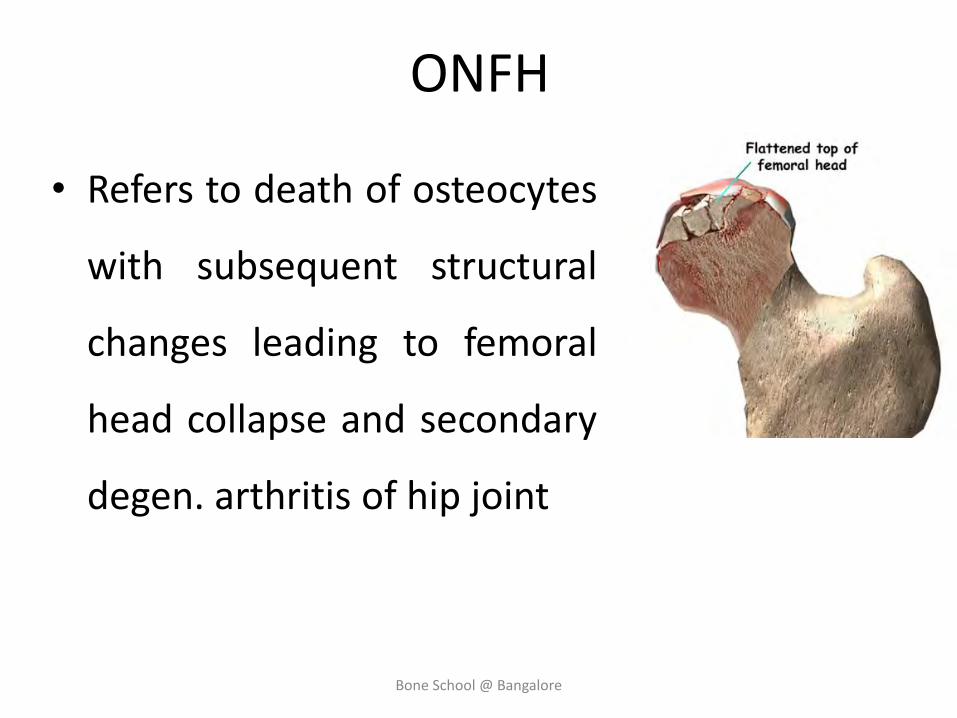
• Refers to death of osteocytes
with subsequent structural
changes leading to femoral
head collapse and secondary
degen. arthritis of hip joint
Bone School @ Bangalore
History
• First described by Munro (1738)
• Curveilhier (1835) depicted femoral head morphological changes secondary to interruption of blood flow
• Ficat (1985) stated this condition resulted from blockage of osseous microcirculation with intramedullary stasis and increased pressure
Bone School @ Bangalore
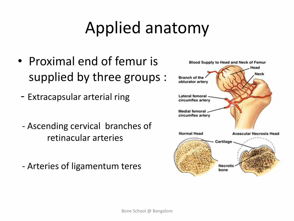
Applied anatomy
• Proximal end of femur is supplied by three groups :
- Extracapsular arterial ring
- Ascending cervical branches of retinacular arteries
- Arteries of ligamentum teres
Bone School @ Bangalore
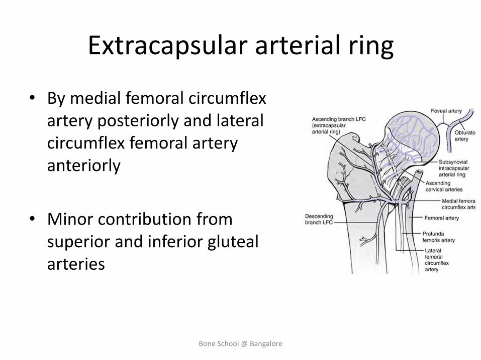
Extracapsular arterial ring
• By medial femoral circumflex artery posteriorly and lateral circumflex femoral artery anteriorly
• Minor contribution from superior and inferior gluteal arteries
Bone School @ Bangalore
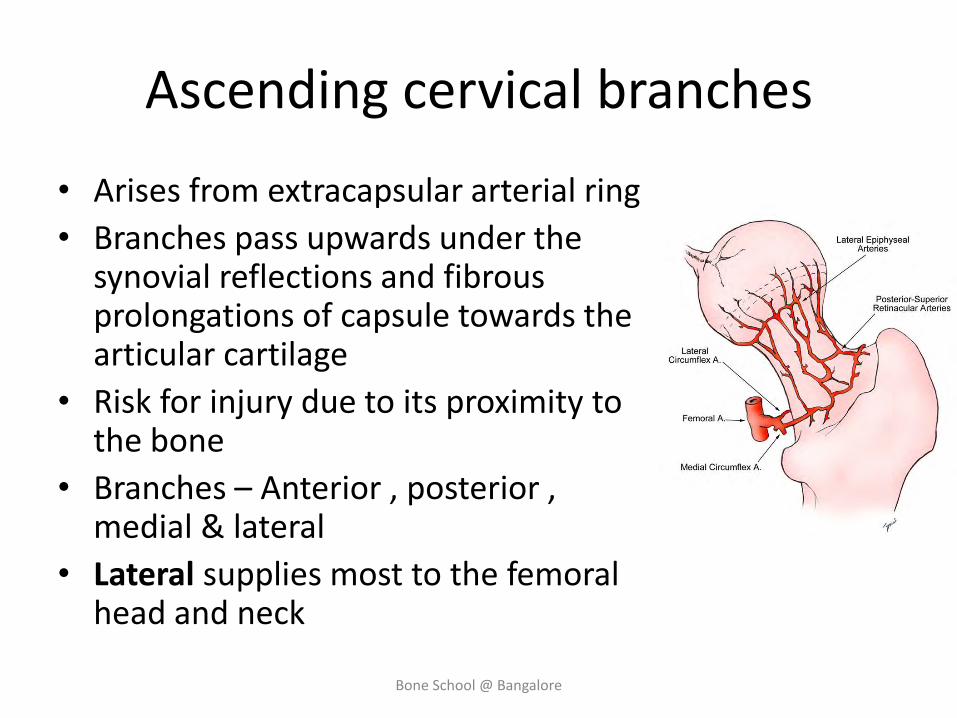
Ascending cervical branches
• Arises from extracapsular arterial ring
• Branches pass upwards under the synovial reflections and fibrous prolongations of capsule towards the articular cartilage
• Risk for injury due to its proximity to the bone
• Branches – Anterior , posterior , medial & lateral
• Lateral supplies most to the femoral head and neck
Bone School @ Bangalore
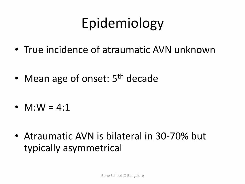
Epidemiology
• True incidence of atraumatic AVN unknown
• Mean age of onset: 5th decade
• M:W = 4:1
• Atraumatic AVN is bilateral in 30-70% but typically asymmetrical
Bone School @ Bangalore
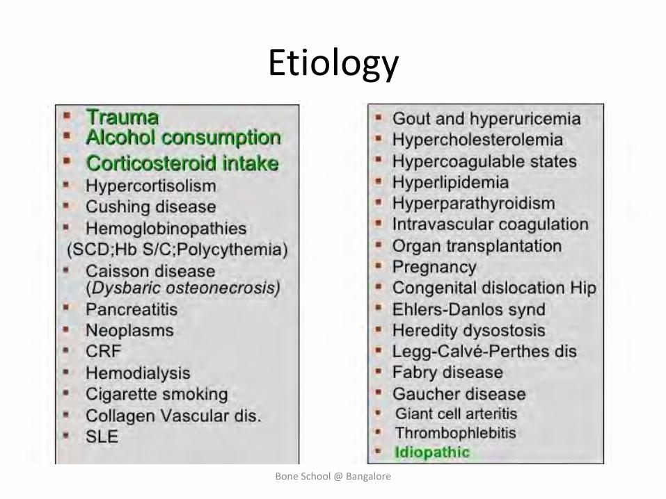
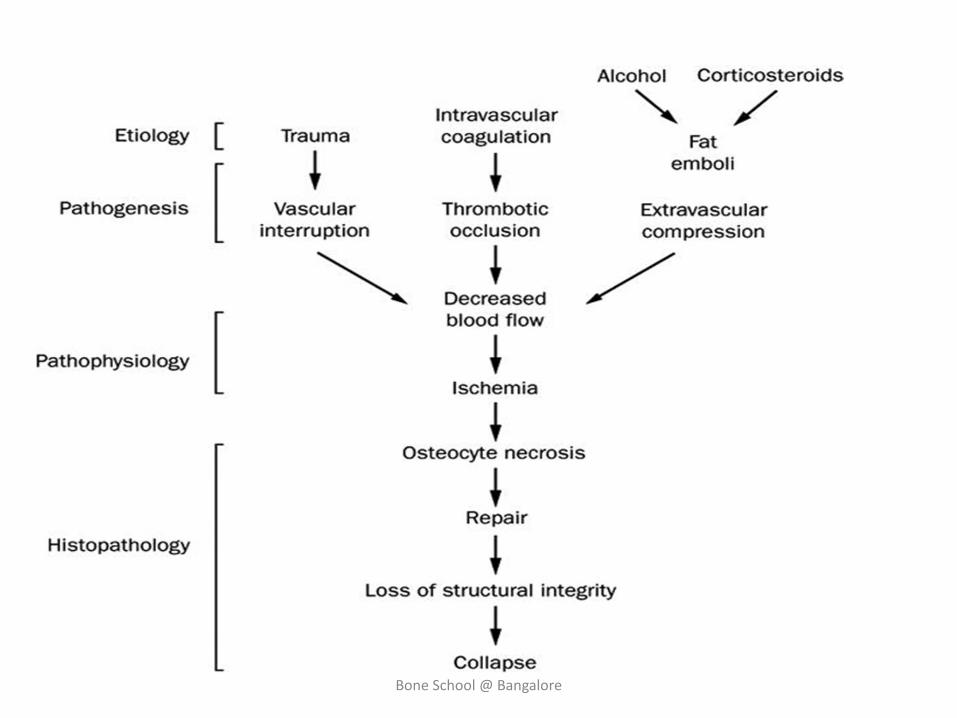
Etiology
Bone School @ Bangalore
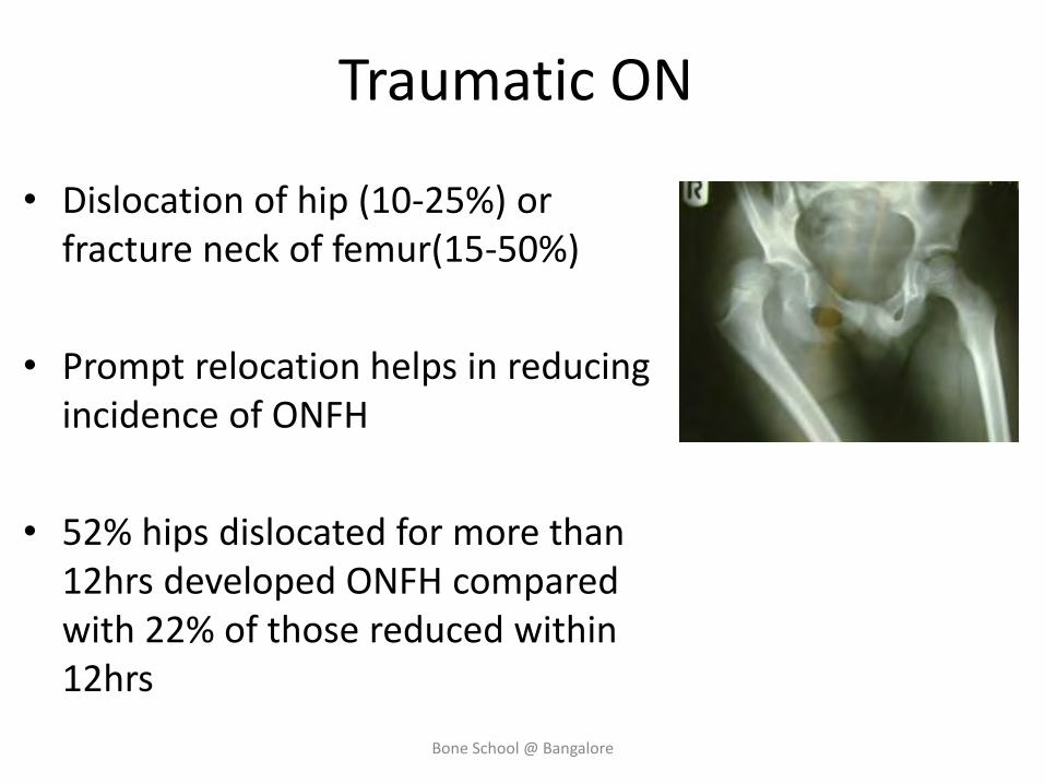
Traumatic ON
• Dislocation of hip (10-25%) or fracture neck of femur(15-50%)
• Prompt relocation helps in reducing incidence of ONFH
• 52% hips dislocated for more than 12hrs developed ONFH compared with 22% of those reduced within 12hrs
Bone School @ Bangalore
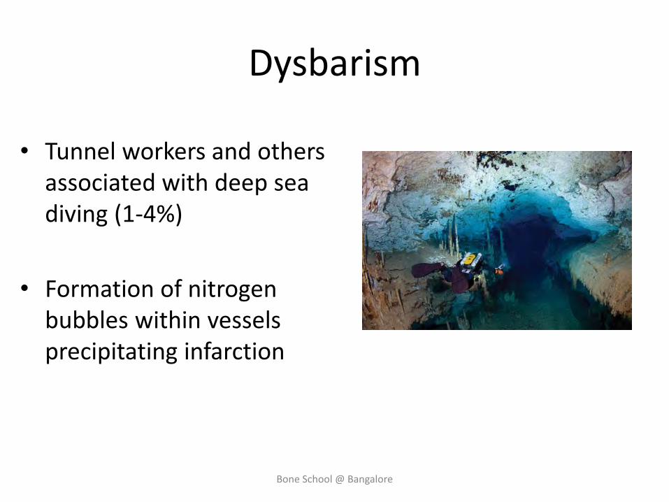
Dysbarism
• Tunnel workers and others associated with deep sea diving (1-4%)
• Formation of nitrogen bubbles within vessels precipitating infarction
Bone School @ Bangalore
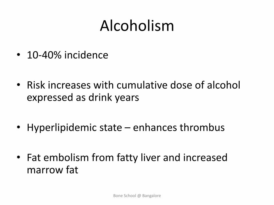
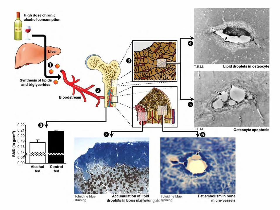
Alcoholism
• 10-40% incidence
• Risk increases with cumulative dose of alcohol expressed as drink years
• Hyperlipidemic state – enhances thrombus
• Fat embolism from fatty liver and increased marrow fat
Bone School @ Bangalore
Bone School @ Bangalore
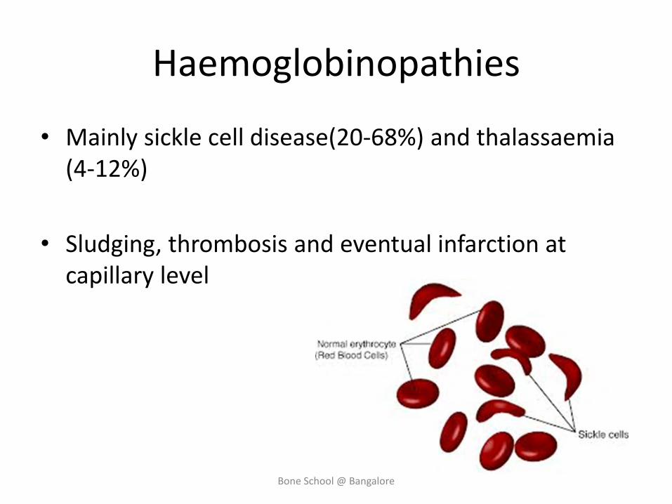
Haemoglobinopathies
• Mainly sickle cell disease(20-68%) and thalassaemia (4-12%)
• Sludging, thrombosis and eventual infarction at capillary level
Bone School @ Bangalore
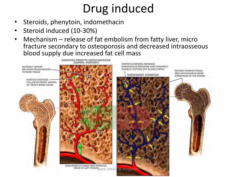
Drug induced • Steroids, phenytoin, indomethacin • Steroid induced (10-30%) • Mechanism – release of fat embolism from fatty liver, micro
fracture secondary to osteoporosis and decreased intraosseous blood supply due increased fat cell mass
Bone School @ Bangalore
Collagen disease
• RA and systemic lupus erythematosus
• Inflammation of small peripheral blood vessels promotes the formation of vascular thrombosis and tissue infarction
• Therapeutic use corticosteroids also causes ON
Bone School @ Bangalore
Radiation
• Mechanism is unknown
• Apparent result is a combination of obliterative endarteritis and cellular death
• A threshold dose of 3000 rads
Bone School @ Bangalore
Gout
Sodium urate crystals enhance clotting by
activation of Hageman factor, an initial protein
component in the intrinsic coagulation
mechanism
Bone School @ Bangalore
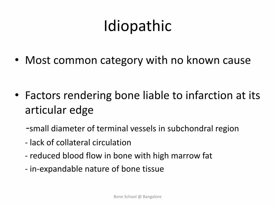
Idiopathic
• Most common category with no known cause
• Factors rendering bone liable to infarction at its articular edge
-small diameter of terminal vessels in subchondral region
- lack of collateral circulation
- reduced blood flow in bone with high marrow fat
- in-expandable nature of bone tissue
Bone School @ Bangalore
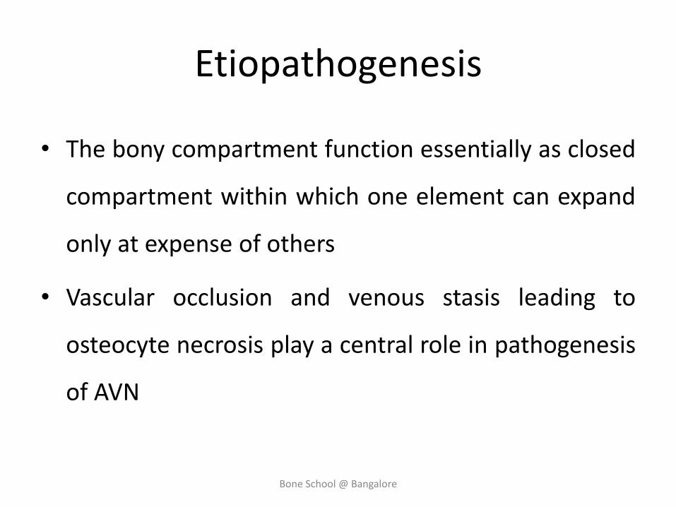
Etiopathogenesis
• The bony compartment function essentially as closed
compartment within which one element can expand
only at expense of others
• Vascular occlusion and venous stasis leading to
osteocyte necrosis play a central role in pathogenesis
of AVN
Bone School @ Bangalore
Bone School @ Bangalore
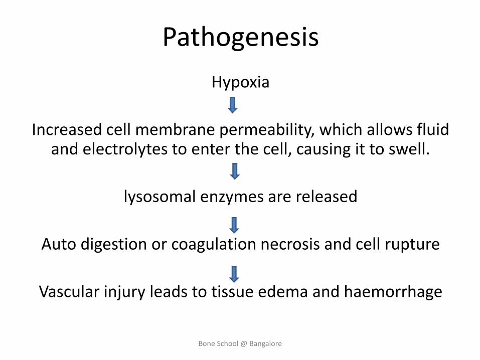
Pathogenesis
Hypoxia
Increased cell membrane permeability, which allows fluid and electrolytes to enter the cell, causing it to swell.
lysosomal enzymes are released
Auto digestion or coagulation necrosis and cell rupture
Vascular injury leads to tissue edema and haemorrhage
Bone School @ Bangalore
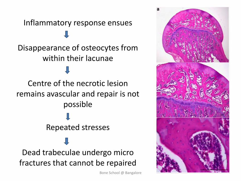
Inflammatory response ensues
Disappearance of osteocytes from within their lacunae
Centre of the necrotic lesion remains avascular and repair is not
possible
Repeated stresses
Dead trabeculae undergo micro fractures that cannot be repaired
Bone School @ Bangalore
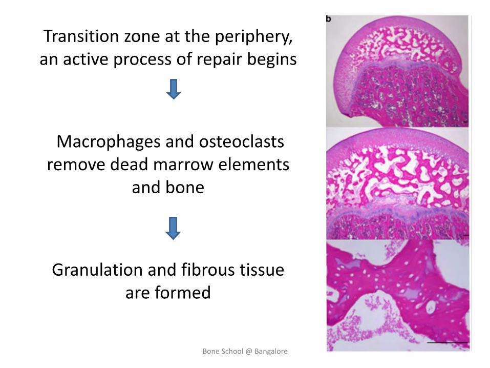
Transition zone at the periphery, an active process of repair begins
Macrophages and osteoclasts remove dead marrow elements
and bone
Granulation and fibrous tissue are formed
Bone School @ Bangalore
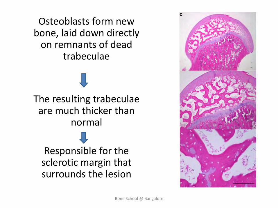
Osteoblasts form new bone, laid down directly
on remnants of dead trabeculae
The resulting trabeculae are much thicker than
normal
Responsible for the sclerotic margin that surrounds the lesion
Bone School @ Bangalore
• Small lesion (not in a major weight-bearing region) - Revascularization and completely replaced with viable bone
• Larger lesions, particularly those in a region of major weight bearing, have a poor prognosis - Gradual collapse
Bone School @ Bangalore
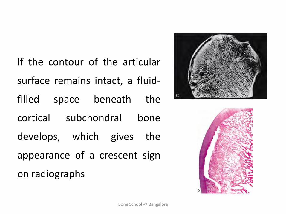
If the contour of the articular
surface remains intact, a fluid-
filled space beneath the
cortical subchondral bone
develops, which gives the
appearance of a crescent sign
on radiographs
Bone School @ Bangalore
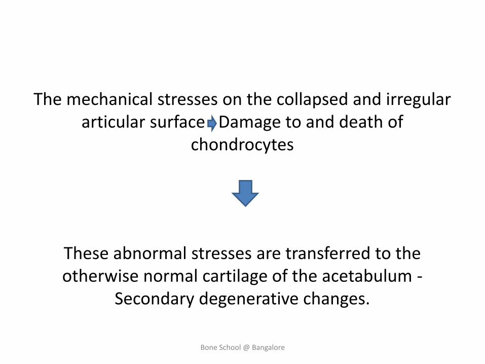
The mechanical stresses on the collapsed and irregular articular surface Damage to and death of
chondrocytes
These abnormal stresses are transferred to the otherwise normal cartilage of the acetabulum -
Secondary degenerative changes.
Bone School @ Bangalore
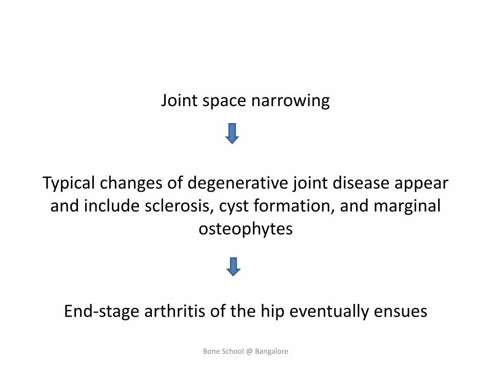
Joint space narrowing
Typical changes of degenerative joint disease appear and include sclerosis, cyst formation, and marginal
osteophytes
End-stage arthritis of the hip eventually ensues
Bone School @ Bangalore
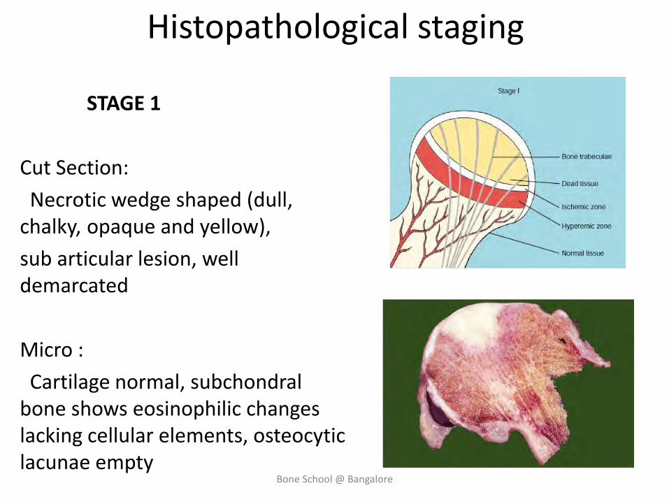
Histopathological staging
STAGE 1
Cut Section:
Necrotic wedge shaped (dull, chalky, opaque and yellow),
sub articular lesion, well demarcated
Micro :
Cartilage normal, subchondral bone shows eosinophilic changes lacking cellular elements, osteocytic lacunae empty
Bone School @ Bangalore
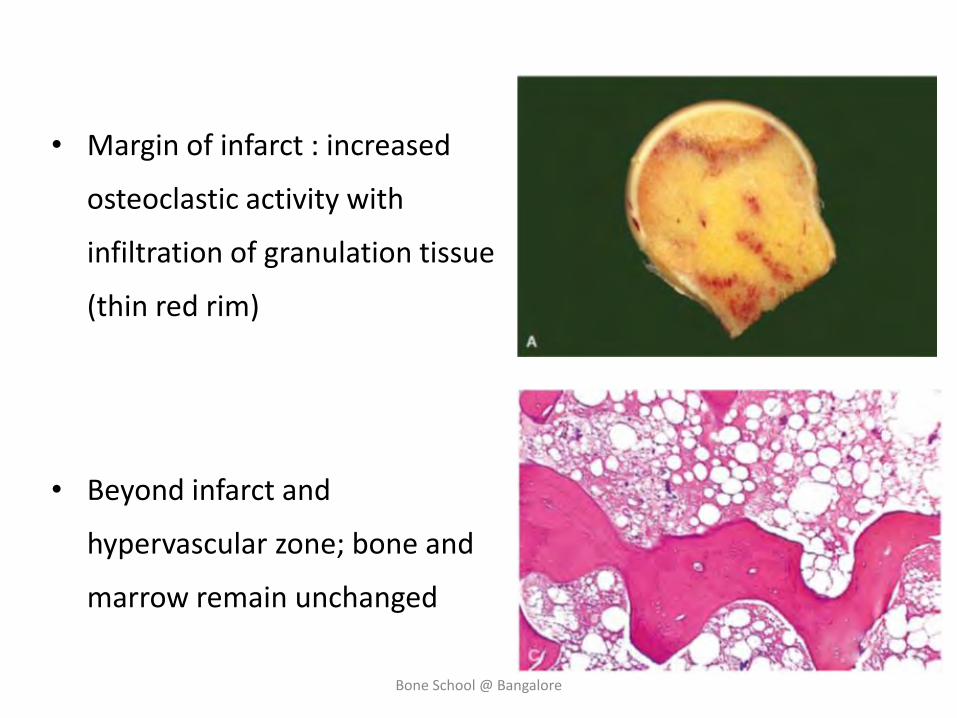
• Margin of infarct : increased
osteoclastic activity with
infiltration of granulation tissue
(thin red rim)
• Beyond infarct and
hypervascular zone; bone and
marrow remain unchanged
Bone School @ Bangalore
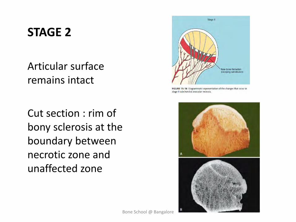
STAGE 2
Articular surface remains intact
Cut section : rim of bony sclerosis at the boundary between necrotic zone and unaffected zone
Bone School @ Bangalore
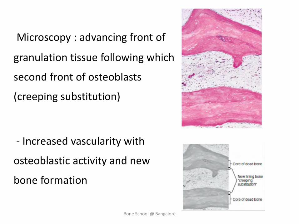
Microscopy : advancing front of
granulation tissue following which
second front of osteoblasts
(creeping substitution)
- Increased vascularity with
osteoblastic activity and new
bone formation
Bone School @ Bangalore
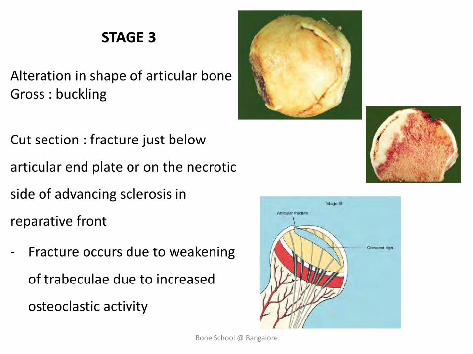
STAGE 3
Alteration in shape of articular bone Gross : buckling
Cut section : fracture just below
articular end plate or on the necrotic
side of advancing sclerosis in
reparative front
- Fracture occurs due to weakening
of trabeculae due to increased
osteoclastic activity
Bone School @ Bangalore
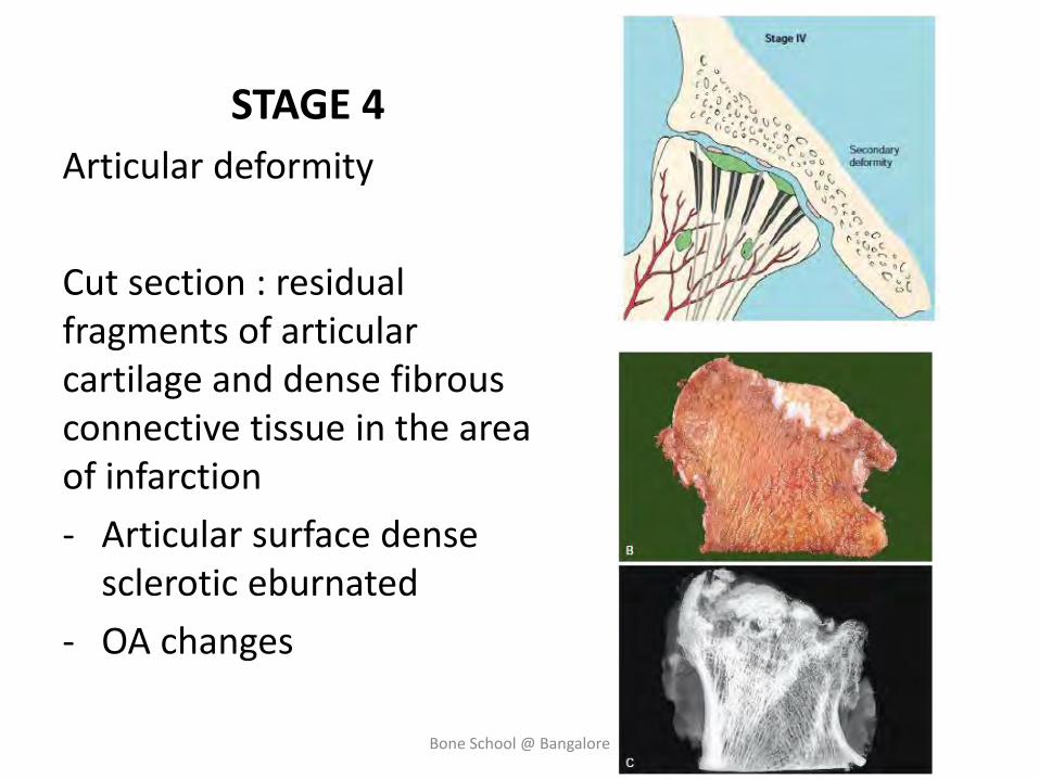
STAGE 4
Articular deformity
Cut section : residual fragments of articular cartilage and dense fibrous connective tissue in the area of infarction
- Articular surface dense sclerotic eburnated
- OA changes
Bone School @ Bangalore
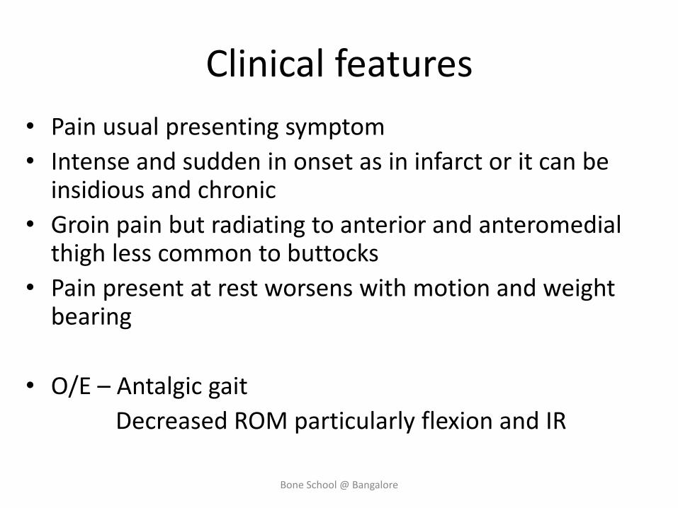
Clinical features
• Pain usual presenting symptom
• Intense and sudden in onset as in infarct or it can be insidious and chronic
• Groin pain but radiating to anterior and anteromedial thigh less common to buttocks
• Pain present at rest worsens with motion and weight bearing
• O/E – Antalgic gait
Decreased ROM particularly flexion and IR
Bone School @ Bangalore
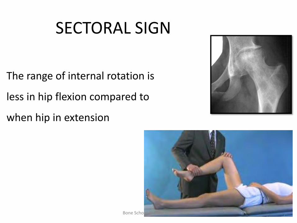
SECTORAL SIGN
The range of internal rotation is
less in hip flexion compared to
when hip in extension
Bone School @ Bangalore
Imaging
• RADIOGRAPHY (AP view and frog leg lateral views)
• Initial Radiographs will be normal
• Typical : mottled sclerosis and lucency usually in the anterosuperior segment of femoral head
• Progression into subchondral fracture and eventual collapse
• Advanced cases : secondary OA
Bone School @ Bangalore
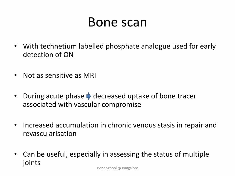
Bone scan
• With technetium labelled phosphate analogue used for early detection of ON
• Not as sensitive as MRI
• During acute phase decreased uptake of bone tracer associated with vascular compromise
• Increased accumulation in chronic venous stasis in repair and revascularisation
• Can be useful, especially in assessing the status of multiple joints
Bone School @ Bangalore

CT scan
• Can Visualize a small lesion not easily seen on routine radiographs, and it may demonstrate small areas of articular surface collapse that are not apparent on plain films
• It may also be used to help quantitate the extent of femoral head involvement
Bone School @ Bangalore
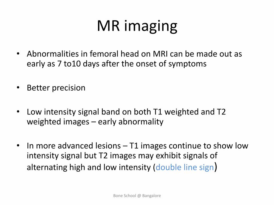
MR imaging
• Abnormalities in femoral head on MRI can be made out as early as 7 to10 days after the onset of symptoms
• Better precision
• Low intensity signal band on both T1 weighted and T2 weighted images – early abnormality
• In more advanced lesions – T1 images continue to show low intensity signal but T2 images may exhibit signals of
alternating high and low intensity (double line sign)
Bone School @ Bangalore
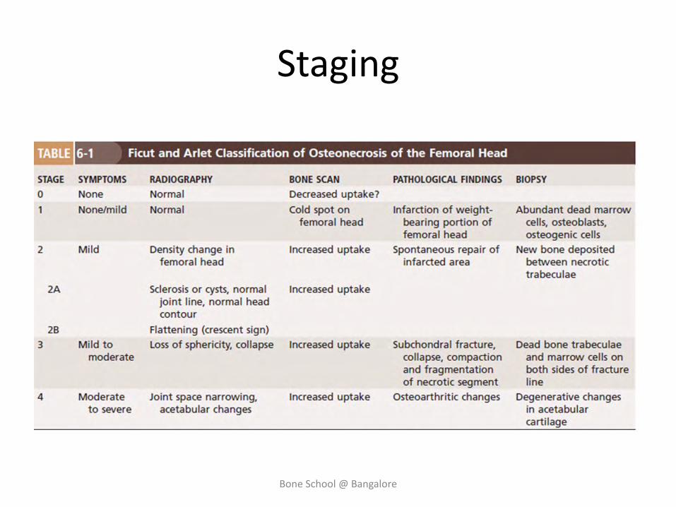
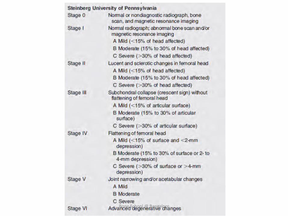
Staging
Bone School @ Bangalore
Bone School @ Bangalore
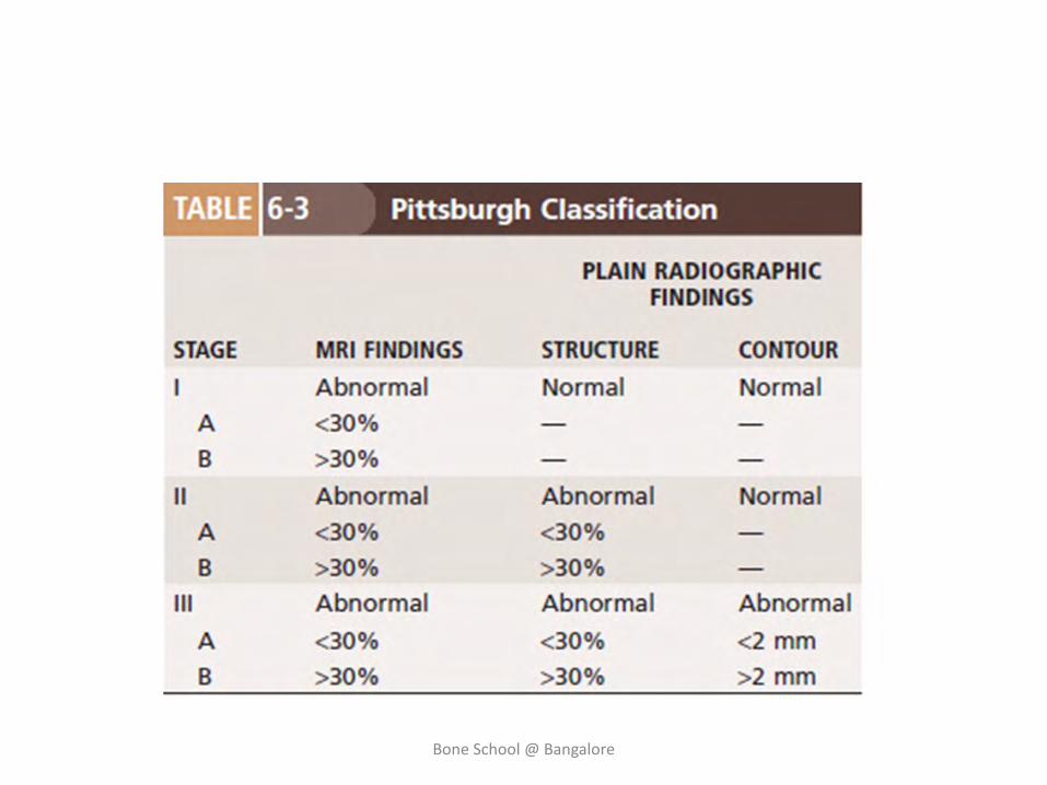
Bone School @ Bangalore
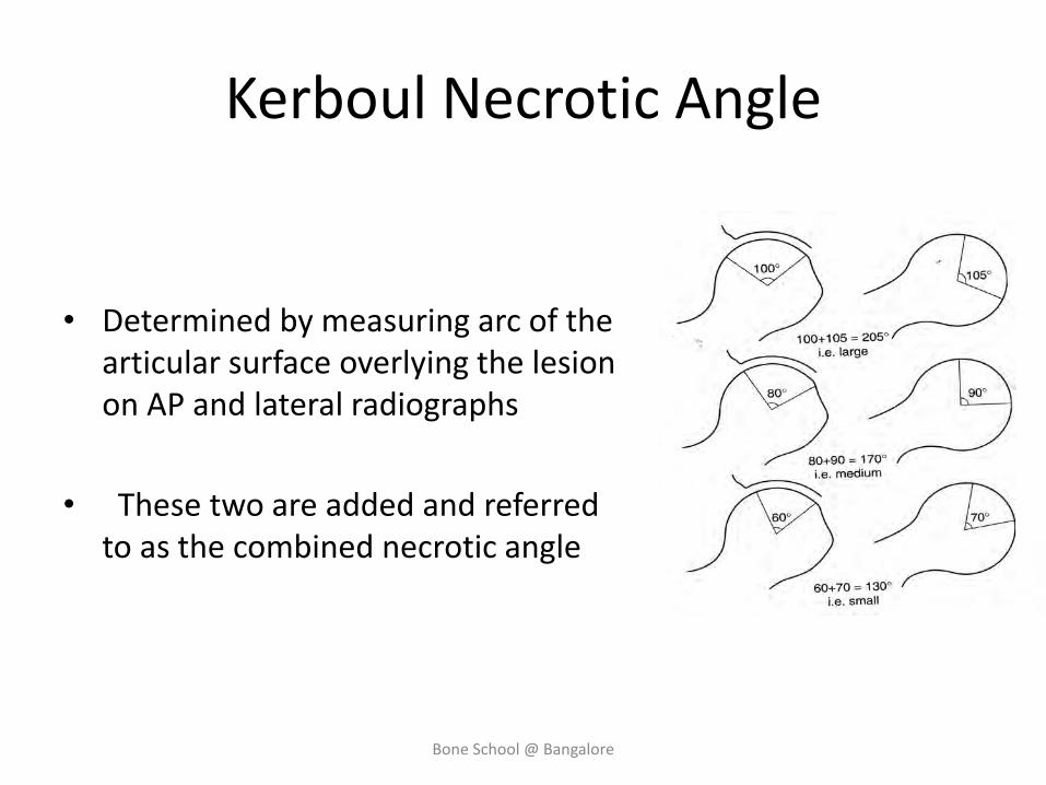
Kerboul Necrotic Angle
• Determined by measuring arc of the articular surface overlying the lesion on AP and lateral radiographs
• These two are added and referred to as the combined necrotic angle
Bone School @ Bangalore

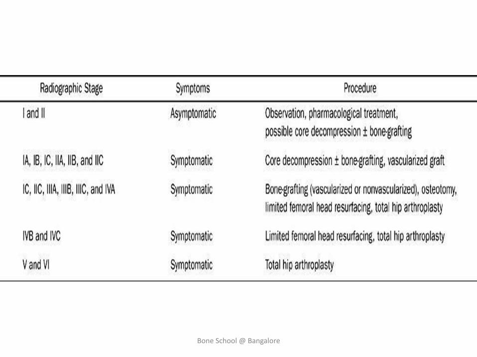
Goals of management
• Relief of pain
• Arrest the progression of disease
• Prevent the collapse of head
• Prevent secondary degenerative arthritis
Bone School @ Bangalore
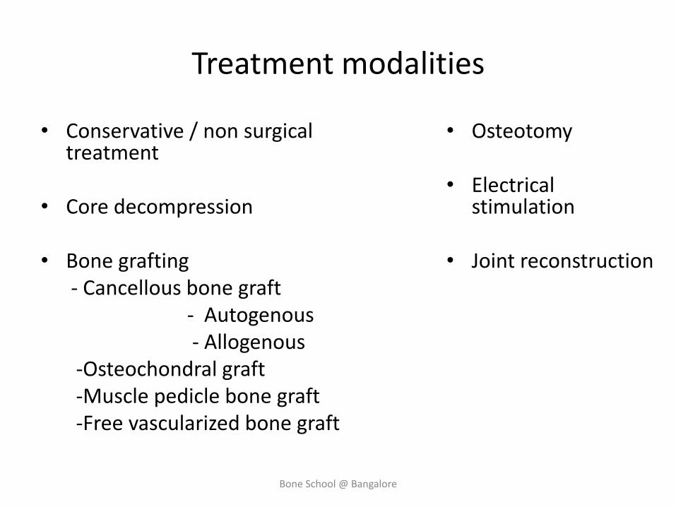
Treatment modalities
• Conservative / non surgical treatment
• Core decompression • Bone grafting - Cancellous bone graft - Autogenous - Allogenous -Osteochondral graft -Muscle pedicle bone graft -Free vascularized bone graft
• Osteotomy
• Electrical stimulation
• Joint reconstruction
Bone School @ Bangalore
Bone School @ Bangalore
Bone School @ Bangalore
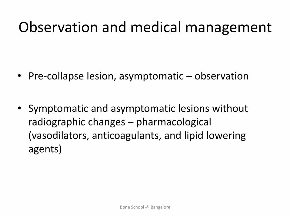
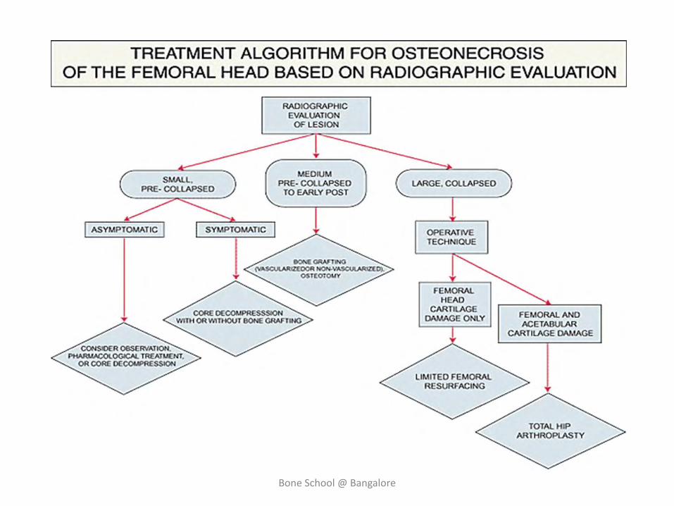
Observation and medical management
• Pre-collapse lesion, asymptomatic – observation
• Symptomatic and asymptomatic lesions without radiographic changes – pharmacological (vasodilators, anticoagulants, and lipid lowering agents)
Bone School @ Bangalore
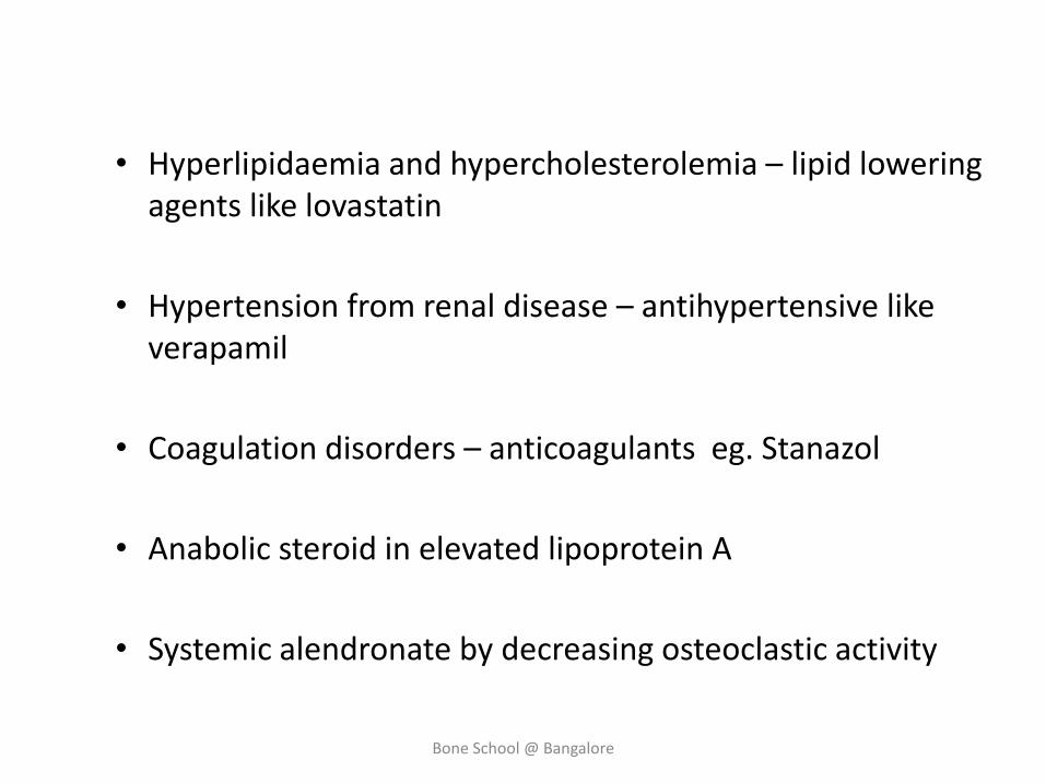
• Hyperlipidaemia and hypercholesterolemia – lipid lowering agents like lovastatin
• Hypertension from renal disease – antihypertensive like verapamil
• Coagulation disorders – anticoagulants eg. Stanazol
• Anabolic steroid in elevated lipoprotein A
• Systemic alendronate by decreasing osteoclastic activity
Bone School @ Bangalore
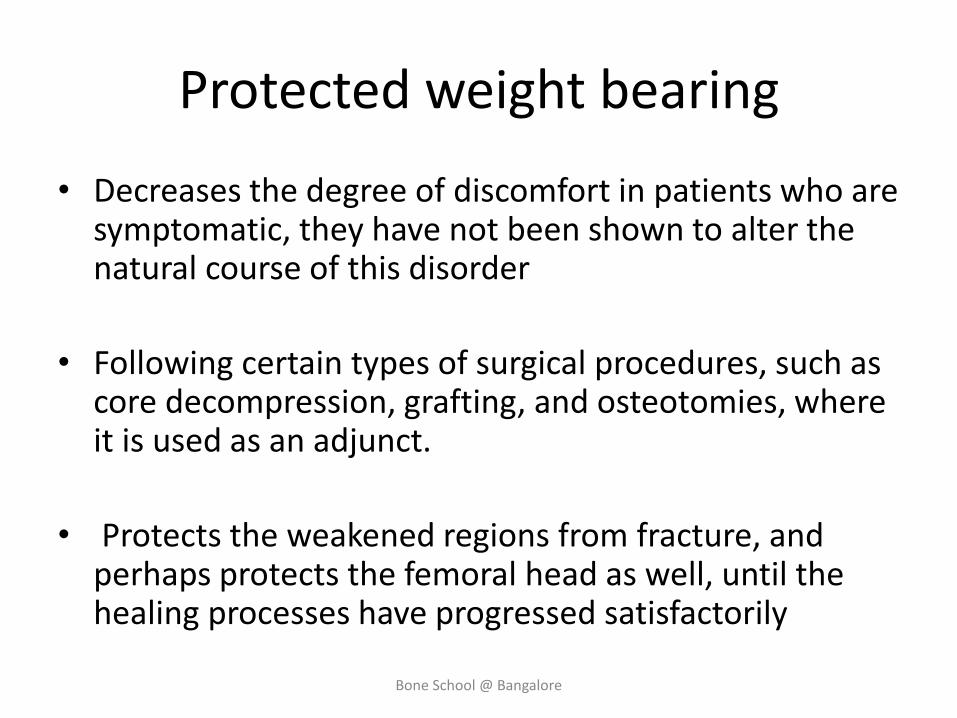
Protected weight bearing
• Decreases the degree of discomfort in patients who are symptomatic, they have not been shown to alter the natural course of this disorder
• Following certain types of surgical procedures, such as core decompression, grafting, and osteotomies, where it is used as an adjunct.
• Protects the weakened regions from fracture, and perhaps protects the femoral head as well, until the healing processes have progressed satisfactorily
Bone School @ Bangalore

Electrical stimulation
Two fundamental mechanisms of action:
1. Important role in control of local inflammation
2. Favours repair activity and can potentiate the healing process by stimulating neo-vascularization and new bone formation
Bone School @ Bangalore
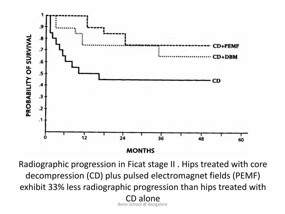
Radiographic progression in Ficat stage II . Hips treated with core decompression (CD) plus pulsed electromagnet fields (PEMF)
exhibit 33% less radiographic progression than hips treated with CD alone
Bone School @ Bangalore
Bone marrow infiltration
• The small no. of progenitor cells in the proximal extremity of the femur with ONFH causes insufficient creeping substitution after osteonecrosis
• Red bone marrow graft contains osteogenic precursors, which help in the reparative process
• Used in adjunct to Core Decompression
Bone School @ Bangalore
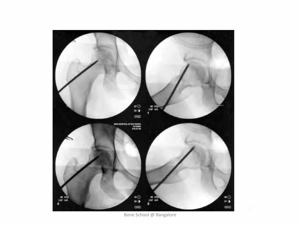
Core decompression
• Rationale – relieves intraosseous pressure , improves vascularity thus slows the disease progression
• Can be combined with placement of non vascularised, non structural bone grafts or graft substitutes
• Recently insertion of porous tantalum rods has been advocated
• Ficat stage 1 and 2a
• 30% patient even with early stage disease likely to end up with THR in 4-5yrs of core decompression
Bone School @ Bangalore
Core Decompression
• The best indications are hips with osteonecrosis without collapse
• In some patients who had Steinberg stage III (subchondral crescent, no collapse), successful outcomes have been obtained between 5 and 10 years
• Therefore, in selected patients, even more
advanced disease can be considered for core decompression
Bone School @ Bangalore
Bone School @ Bangalore
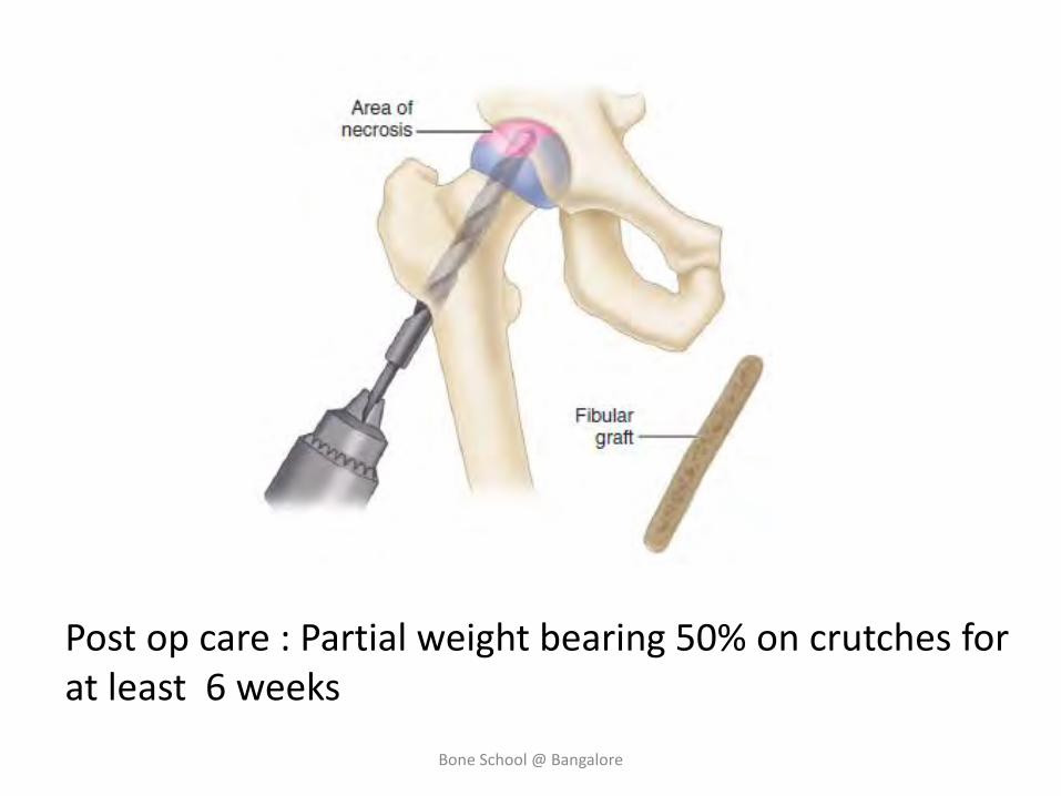
Post op care : Partial weight bearing 50% on crutches for at least 6 weeks
Bone School @ Bangalore
Bone grafting
• Various authors have claimed success rate of 50 – 80% after CD with structural BG
• Non vascularised BG – Ficat 1 and 2
• Accurate placement of the graft within the lesion and under the subchondral bone
• Standard core technique, Lightbulb technique, Trap door technique
Bone School @ Bangalore
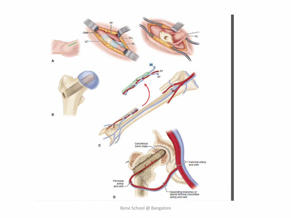
Vascularised fibular graft
• Rationale : - Decompression of femoral head - Excision of the sequestrum - Filling the defect with osteoinductive cancellous graft and viable cortical strut • Longer recovery period and less uniform and less
complete relief of pain
• Success rate 80-91% for younger symptomatic patients without collapse
Bone School @ Bangalore
Bone School @ Bangalore
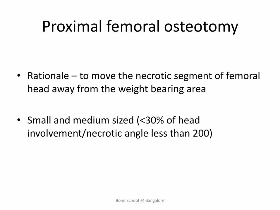
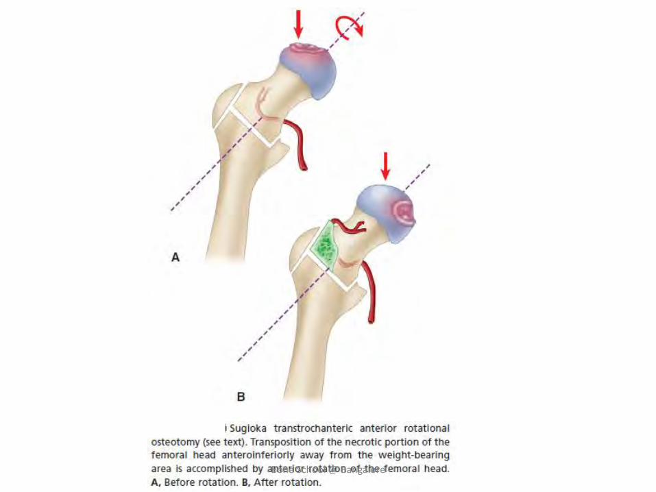
Proximal femoral osteotomy
• Rationale – to move the necrotic segment of femoral head away from the weight bearing area
• Small and medium sized (<30% of head involvement/necrotic angle less than 200)
Bone School @ Bangalore
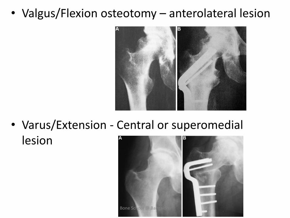
• Valgus/Flexion osteotomy – anterolateral lesion
• Varus/Extension - Central or superomedial lesion
Bone School @ Bangalore
Bone School @ Bangalore
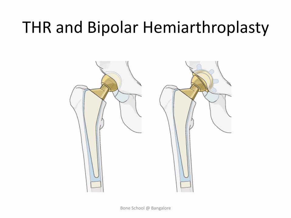
THR and Bipolar Hemiarthroplasty
Bone School @ Bangalore

Bone School @ Bangalore