Embed Size (px)
Citation preview

INTRODUCTION
A fundamental question in developmental biology is the originof complex patterns. Ornate yet eminently reproduciblesystems mold the form and function of every tissue and organ.One of the recurring players in patterning systems is thesymmetry-disrupting morphogen gradient. This gradient givesrise to broad regions that are refined through the action ofa variety of transcriptional regulators. In plants, manytranscriptional regulators that influence developmental patternshave been identified through molecular genetic studies;uncovering morphogens, on the other hand, has been farmore difficult. In this study, we provide evidence that thephytohormone auxin acts as a morphogen directing regionalpatterning in the developing gynoecium of Arabidopsisthaliana.
Auxin, historically the first plant hormone studied, has wide-ranging effects on growth and development throughout theplant. The major naturally occurring auxin in higher plants isindole-3-acetic acid (IAA), which is synthesized in the aerialportion of the plant and transported basipetally. According tothe canalization hypothesis, a positive feedback mechanismfocuses the flow of auxin into distinct cellular files, which willsubsequently differentiate as veins (Sachs, 1991). The self-organizing aspect of this process requires directed auxin flow,proposed to be mediated by polar localization of auxin effluxcarriers. Recently identified candidates for such carriers arerestricted within the cell membrane in a cytoskeleton-
dependent manner (Chen et al., 1998; Galweiler et al., 1998;Luschnig et al., 1998; Muller et al., 1998; Steinmann et al.,1999; Utsuno et al., 1998). Polar auxin transport (PAT)inhibitors have been utilized for decades to interfere with polarauxin flow, either by directly competing with auxin for carrier-binding or by otherwise rendering the cell incapable oftransport (Tavares, 1973). Application of such inhibitors resultsin accumulation of auxin near source cells and depletion ofauxin in cells normally downstream of the transport. Thus,treatment with PAT inhibitors increases the slope of existingauxin gradients in an inhibitor concentration-dependentmanner. Studies of plants germinated on PAT inhibitor-containing media support a substantial role for auxin fluxthroughout plant development, and reveal a particularly strongeffect of disrupted auxin flow on floral organ number andpatterning (Okada et al., 1991). These studies are limited,however, by the finding that floral meristem initiation is largelydependent on proper PAT. Inhibitor-treated plants rarely makeflowers. Studies of transgenic plants ubiquitously expressingIAA hydrolase, decreasing overall auxin levels, alsodemonstrate the dependence of flower meristem formation onIAA (Oka et al., 1999). The overall strong effects on plantmorphology combined with the scarcity of flowers in thesetreatments makes assessment of auxin-transport-related floralorgan defects difficult.
Although much work has been done to define the timing andmechanisms controlling the partitioning of the floral meristeminto its constituent organs, the process of organ morphogenesis
3877Development 127, 3877-3888 (2000)Printed in Great Britain © The Company of Biologists Limited 2000DEV0315
The phytohormone auxin has wide-ranging effects ongrowth and development. Genetic and physiologicalapproaches implicate auxin flux in determination of floralorgan number and patterning. This study uses a noveltechnique of transiently applying a polar auxin transportinhibitor, N-1-naphthylphthalamic acid (NPA), todeveloping Arabidopsisflowers to further characterize therole of auxin in organogenesis. NPA has marked effects onfloral organ number as well as on regional specification inwild-type gynoecia, as defined by morphological andhistological landmarks for regional boundaries, as well astissue-specific reporter lines. NPA’s effects on gynoeciumpatterning mimic the phenotype of mutations in ETTIN, amember of the auxin response factor family oftranscription factors. In addition, application of different
concentrations of NPA reveal an increased sensitivity ofweak ettin alleles to disruptions in polar auxin transport.In contrast, the defects found in spatula gynoecia arepartially rescued by treatment with NPA. A model isproposed suggesting an apical-basal gradient of auxinduring gynoecium development. This model provides amechanism linking ETTIN’s putative transcriptionalregulation of auxin-responsive genes to the establishmentor elaboration of tissue patterning during gynoecialdevelopment.
Key words: Arabidopsis thaliana, Gynoecium, Patterning, Polarauxin transport, N-1-naphthylphthalamic acid, NPA, ETTIN,SPATULA, Flower development
SUMMARY
Auxin and ETTIN in Arabidopsis gynoecium morphogenesis
Jennifer L. Nemhauser*, Lewis J. Feldman and Patricia C. Zambryski*
Department of Plant and Microbial Biology, 111 Koshland Hall, University of California, Berkeley, CA 94720-3102, USA*Authors for correspondence (e-mail: [email protected] and [email protected])
Accepted 3 July; published on WWW 22 August 2000

3878
remains poorly understood. Arabidopsisflowers consist of fourorgan types: the leaf-like perianth organs, sepals and petals,and the gametophyte-producing stamens and gynoecia. Thegynoecium, the female reproductive structure, is perhaps themost complex organ formed by Arabidopsis, and has beenmeticulously observed throughout its ontogeny (Sessions,1997). Genetic studies have yielded important insights into theunderlying mechanism of patterning in the gynoecium. Severalmutants affect specific subsets of the morphologically andfunctionally distinct tissues apparent in the mature gynoecium,suggesting that the development of individual tissues may belargely uncoupled (Alvarez and Smyth, 1999; Bowman andSmyth, 1999; Roe et al., 1997; Sessions and Zambryski, 1995).
Phenotypic analyses of one mutant, ettin (ett), facilitatethe dissection of regional patterning during gynoeciumorganogensis. ett mutants display an allele-strength-dependentloss of valve tissue with a concomitant increase in apical/adaxial style and stigma and basal internode (Sessions andZambryski, 1995). It has been proposed that the morphologicaldefects in ettare caused by destabilized regional boundariespositioned at the apical and basal limits of the valves, resultingin an overall loss of valve and gain of flanking tissues(Sessions, 1997). The molecular nature of the ETT protein hasimportant implications for the mechanism underlying theprocess of regional patterning. ETT shares a large amino-terminal DNA-binding domain with the auxin response factor(ARF) family of transcriptional regulators (Sessions et al.,1997). The founding member of this family, ARF1, wasidentified by its ability to bind a synthetic auxin responseelement (AuxRE) derived from conserved elements in theregulatory regions of many auxin-responsive genes (Ulmasovet al., 1997a). Ten ARFs have been characterized to date, withsome acting as powerful activators of transcription and othersas repressors (Ulmasov et al., 1999a,b). All but ETT displaytwo islands of carboxy-terminal similarity which allow forprotein-protein interactions both between ARF familymembers and with one group of auxin responsive genes, theAux/IAAs (Kim et al., 1997; Ulmasov et al., 1999a). DNA-binding by some ARFs is disrupted by interaction withAux/IAA proteins (Ulmasov et al., 1997b). None of the ARFstested to date, including ETT, are inducible by exogenousapplication of auxin (Ulmasov et al., 1999b; J. L. N., L. J. F.and P. C. Z., unpublished results). Another developmentalmutant, spatula (spt), is epistatic to ettin double mutantanalysis and has been proposed as a potential downstreamtarget of ETT (Alvarez and Smyth, 1998). sptis characterizedby reduction of style and stigma, absence of transmitting tractcells and incomplete fusion of the apical end of the gynoecium.The recent cloning and characterization of the SPTpromoterhas revealed the presence of several putative AuxREs(M. Heisler, M. Groszmann and D. Smyth, personalcommunication), suggesting a mechanism for the regulation ofSPTby ETT.
Genetic evidence implicates auxin flux in determination offloral organ number and patterning. pin-formed1 flowers,defective in auxin efflux, show decreased number of floralorgans and a dramatic loss of valve tissue in the gynoecium ofthe few flowers formed (Bennett et al., 1995). These effects aresimilar to those seen in plants exposed to PAT inhibitors.Mutations in PINOID, a kinase implicated in auxin transportor perception (Bennett et al., 1995; Christensen et al., 2000),
and MONOPTEROS(ARF5) (Hardtke and Berleth, 1998;Przemeck et al., 1996) also cause substantial reduction ingynoecium valves. These mutants, as well as the PAT inhibitorstudies, strongly implicate auxin in the specification of regionalpatterning early in gynoecium development.
Our study uses a novel technique of transiently applying aPAT inhibitor, N-1-naphthylphthalamic acid, to developingflowers to further characterize the role of auxin in gynoeciumdevelopment. PAT inhibitors have marked effects on regionalspecification in wild-type gynoecia, as defined bymorphological and histological landmarks for regionalboundaries, as well as tissue-specific reporter lines. In addition,application of different concentrations of the inhibitor revealan increased sensitivity of weak ettalleles to disruptions inPAT. A model suggesting an apical-basal gradient of auxinduring gynoecium development is proposed. This modelprovides a mechanism linking ETT’s putative transcriptionalregulation of auxin-responsive genes to the establishment orelaboration of tissue patterning during gynoecial development.
MATERIALS AND METHODS
StocksThe ett-1, ett-2, spt-2 mutations have been described previously(Sessions and Zambryski, 1995; Alvarez and Smyth, 1999). Both ettalleles are in the Wassilewskija ecotype. spt-2is in the Landsbergerecta background. Wild-type Wassilewskija plants were used inmock treatments. The GT142 (Landsberg erecta) (Sundaresan et al.,1995) and SLG::GUS (Columbia) (Toriyama et al., 1991) lines havebeen described previously.
Polar auxin transport inhibitor treatmentsPlants were grown under long day conditions (16 hour light, 8 hourdark) until flowering was visibly apparent (approximately 3.5 weeks).Plants were sprayed with a heavy mist of 10 or 100 µM N-1-naphthylphthalamic acid (Uniroyal Chemical Co., Inc.) with 0.01%Silwet L-77 (a surfactant; Lehle Seeds) at 9 am. Plants were thenplaced in humid chambers, returned to their regular light regime,resprayed at 4 pm, and returned to the humid chambers overnight. At10 am the following day, plants were washed with a saturating sprayof distilled water before being returned to the greenhouse. Mocktreatments were performed with distilled water containing 0.01%Silwet L-77. Flowers were observed directly and fixed for histologicaland ultrastructutral analyses approximately 10-14 days aftertreatment.
Scanning electron microscopySamples for scanning electron microscopy analysis were fixed in FAA(3.7% formaldehyde, 5% glacial acetic acid, 50% ethanol) containing1% Triton X-100, dehydrated in a graded ethanol series, and criticalpoint dried with liquid CO2. Tissues were coated with gold orgold/palladium and viewed using an ISI-DS130 microscope(International Scientific Instruments, Inc.) with an acceleratingvoltage of 10 kV.
β-glucuronidase (GUS) stainingLines were grown under long day conditions, and control or NPAtreatments were performed once flowering was evident (as describedabove). Apices were removed and placed immediately in an ice-coldsolution containing, 0.1 M sodium phosphate (pH 7), 0.3% Triton X-100, 5 mM K3Fe(CN)6, 5 mM K4Fe(CN)6, 20% methanol, 0.3% X-glucuronic acid. After 5 minutes of vacuum infiltration, samples wereplaced at 37°C for 12 hours. Apices were then fixed in ice-cold FAAovernight and subsequently dehydrated in a graded ethanol series.
J. L. Nemhauser, L. J. Feldman and P. C. Zambryski
3879Arabidopsis gynoecium morphogenesis
Light microscopyWhole apices were fixed in FAA overnight at 4°C, washed for 2 hoursin 0.1 M potassium phosphate buffer (pH 7.5), and then transferredto 8 N NaOH for 16 hours with agitation. Tissues were washed for 1hour with 0.1 M potassium phosphate buffer (pH 7.5) and stained in0.1% Aniline Blue in the same buffer solution for 15 minutes.Gynoecia were mounted on slides and viewed with Nomarski opticson a Leica DMRB microscope.
RESULTS
Transient disruption of polar auxin transport in thefloral apexN-1-naphthylphthalamic acid (NPA) is a phytotropin. Thisclass of polar auxin transport inhibitors is defined by theirstructure, benzoic acid derivatives ortho-linked to aromaticring systems (Lomax et al., 1995). Unlike other PAT inhibitors,which compete directly with auxin for transporter binding,phytotropins bind to another plasma membrane-localizedprotein thought to regulate the efflux carrier by an unknownmechanism. Several candidate targets for NPA-binding havebeen isolated biochemically, but the identity of these proteinsand their connection to auxin transport are unknown (Lomaxet al., 1995). tir3, an NPA resistant mutant, has fewer NPAbinding sites (Ruegger et al., 1997) and may be a mutation inthe NPA-binding protein or a factor necessary for its properexpression, localization, or stabilization. NPA has dramaticeffects on plant growth and development and displays noauxin-like properties (Mattsson et al., 1999; Lomax et al.,1995).
NPA is not trafficked by the auxin efflux carrier, and appearsto remain in the initial cells where it is introduced (Reed et al.,1998; Thomson et al., 1973). Several studies have successfullyused NPA to disrupt PAT and analyze development (Mattssonet al., 1999; Okada et al., 1991). Flower development can bedivided into three phases: initiation of floral meristems,initiation and patterning of floral organs within the floralmeristem, and postfertilization seed set and fruit development.Assays directly addressing auxin’s role in the first (Reinhardtet al., 2000) and last (Vivian-Smith and Koltunow, 1999) ofthese stages have been described. While effects throughoutflower development are observed following the NPA treatmentsdescribed in this work, the focus is to define the effects of NPAtreatment on the second stage of flower development –morphogenesis of floral organs. Since prolonged NPAexposure severely restricts the number of flowers formed, inthis investigation NPA was applied transiently to plants thathave already initiated flowering. Plants were exposed to NPAfor a single day and then washed thoroughly. Using thetransient treatment described here, approximately ten flowersshow defects in floral organ patterning.
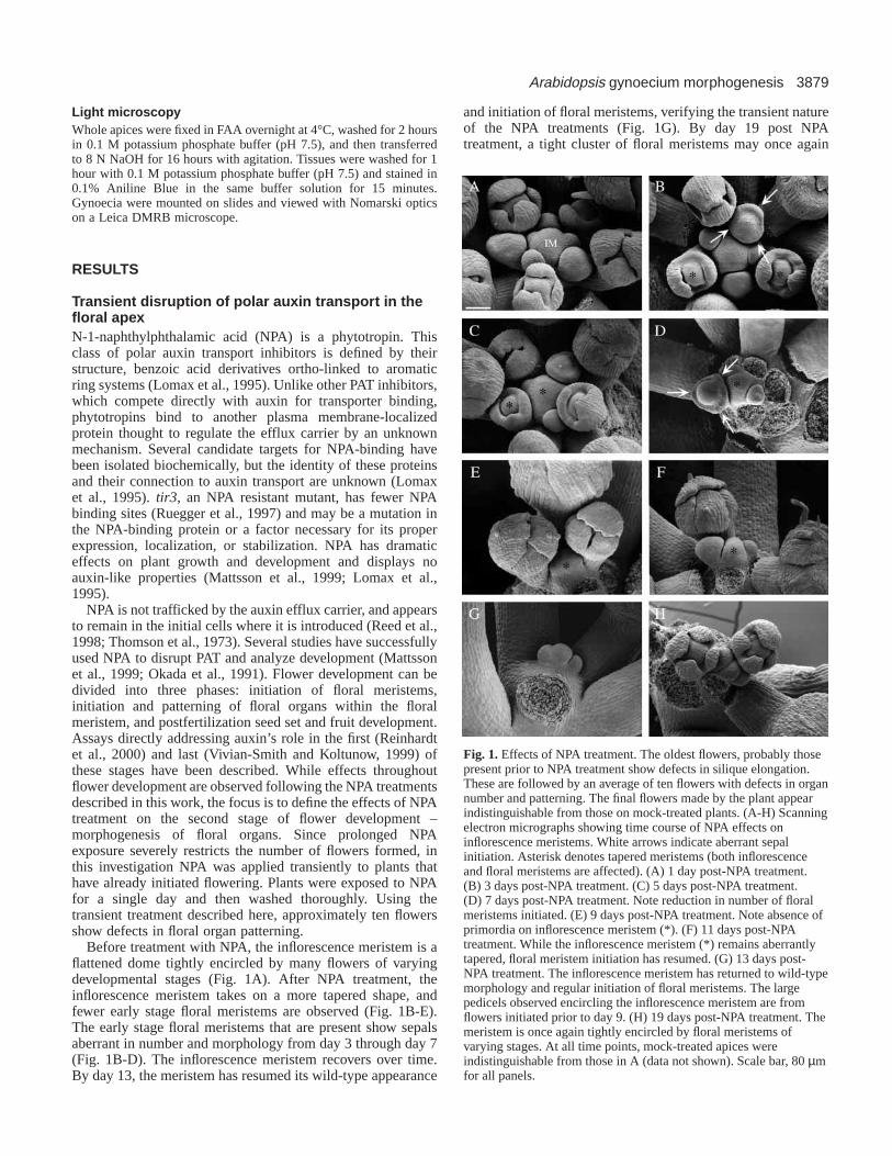
Before treatment with NPA, the inflorescence meristem is aflattened dome tightly encircled by many flowers of varyingdevelopmental stages (Fig. 1A). After NPA treatment, theinflorescence meristem takes on a more tapered shape, andfewer early stage floral meristems are observed (Fig. 1B-E).The early stage floral meristems that are present show sepalsaberrant in number and morphology from day 3 through day 7(Fig. 1B-D). The inflorescence meristem recovers over time.By day 13, the meristem has resumed its wild-type appearance
and initiation of floral meristems, verifying the transient natureof the NPA treatments (Fig. 1G). By day 19 post NPAtreatment, a tight cluster of floral meristems may once again
Fig. 1.Effects of NPA treatment. The oldest flowers, probably thosepresent prior to NPA treatment show defects in silique elongation.These are followed by an average of ten flowers with defects in organnumber and patterning. The final flowers made by the plant appearindistinguishable from those on mock-treated plants. (A-H) Scanningelectron micrographs showing time course of NPA effects oninflorescence meristems. White arrows indicate aberrant sepalinitiation. Asterisk denotes tapered meristems (both inflorescenceand floral meristems are affected). (A) 1 day post-NPA treatment.(B) 3 days post-NPA treatment. (C) 5 days post-NPA treatment.(D) 7 days post-NPA treatment. Note reduction in number of floralmeristems initiated. (E) 9 days post-NPA treatment. Note absence ofprimordia on inflorescence meristem (*). (F) 11 days post-NPAtreatment. While the inflorescence meristem (*) remains aberrantlytapered, floral meristem initiation has resumed. (G) 13 days post-NPA treatment. The inflorescence meristem has returned to wild-typemorphology and regular initiation of floral meristems. The largepedicels observed encircling the inflorescence meristem are fromflowers initiated prior to day 9. (H) 19 days post-NPA treatment. Themeristem is once again tightly encircled by floral meristems ofvarying stages. At all time points, mock-treated apices wereindistinguishable from those in A (data not shown). Scale bar, 80 µmfor all panels.
3880
be observed (Fig. 1H). Another PAT inhibitor, 2,3,5-triiodobenzoic acid, was also used to confirm that the effectsseen were a result of decreased polar auxin transport and werenot NPA specific (data not shown). All subsequent analysesreported below were performed on flowers formed in the periodafter day 1 (Fig. 1B) and prior to day 11 (Fig. 1F) followingNPA treatment.
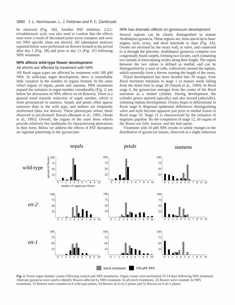
NPA affects wild-type flower developmentAll whorls are affected by treatment with NPAAll floral organ types are affected by treatment with 100 µMNPA. In wild-type organ development, there is remarkablylittle variation in the number of organs formed. In the outerwhorl organs of sepals, petals and stamens, NPA treatmentsexpand the variation in organ number considerably (Fig. 2; seebelow for discussion of NPA effects on ett flowers). There is ageneral trend towards reduction of organ number, which ismost pronounced in stamens. Sepals and petals often appearnarrower than in the wild type, and anthers are frequentlymisformed (data not shown). These phenotypes mimic thoseobserved in pin-formed1flowers (Bennett et al., 1995; Okadaet al., 1991). Overall, the organs of the outer three whorlsprovide relatively few landmarks for characterizing aberrationsin their form. Below we address the effects of PAT disruptionon regional patterning in the gynoecium.
NPA has dramatic effects on gynoecium development Several regions can be clearly distinguished in matureArabidopsis gynoecia. These regions are, from apical tip to base:stigma, style, ovary, and short internode or stipe (Fig. 3A).Ovules are enclosed by the ovary wall, or valve, and connectedto it through the placenta. Arabidopsisgynoecia comprise twocongenitally fused carpels, forming two locules, each containingtwo strands of intercalating ovules along their length. The regionbetween the two valves is defined as medial, and can bedistinguished by a zone of cells, collectively termed the replum,which externally form a furrow running the length of the ovary.
Floral development has been divided into 20 stages, fromfloral meristem initiation in stage 1 to mature seeds fallingfrom the dried fruit in stage 20 (Smyth et al., 1990). In floralstage 6, the gynoecium emerges from the center of the floralmeristem as a slotted cylinder. During development, thecylinder grows upward (apically) and also inward (adaxially),initiating replum development. Ovules begin to differentiate infloral stage 8. Regional epidermal differences distinguishingvalve and style become apparent just prior to medial fusion infloral stage 10. Stage 11 is characterized by the initiation ofstigmatic papillae. By the completion of stage 12, all organs ofthe flower are fully mature, and the bud opens.
Treatment with 10 µM NPA results in subtle changes in thedistribution of gynoecial tissues, observed as a slight reduction
J. L. Nemhauser, L. J. Feldman and P. C. Zambryski
25
50
75
100
0 1 2 3 4 5 6 7 8 9 10
0 1 2 3 4 5 6 7 8 9 10
25
50
75
100
0 1 2 3 4 5 6 7 8 9 10
25
50
75
100
number of organs per flower0 1 2 3 4 5 6 7 8 9 10
25
50
75
100
0 1 2 3 4 5 6 7 8 9 10
25
50
75
100
0 1 2 3 4 5 6 7 8 9 10
25
50
75
100
0 1 2 3 4 5 6 7 8 9 10
25
50
75
100
0 1 2 3 4 5 6 7 8 9 10
25
50
75
100
0 1 2 3 4 5 6 7 8 9 10
25
50
75
100
perc
ent o
f fl
ower
s
ett-1
ett-2
wild-type
stamenspetalssepals
mock treatment 100 µM NPA
Fig. 2.Floral organ number counts following control and NPA treatments. Organ counts were performed 10-14 days following NPA treatment.Aberrant gynoecia were used to identify flowers affected by NPA treatment. In all mock treatments, 25 flowers were counted. In NPAtreatments, 53 flowers were counted on 6 wild-type plants, 54 flowers on 6 ett-2 plants and 52 flowers on 6 ett-1 plants.

3881Arabidopsis gynoecium morphogenesis
in ovary length and a concomitant increase in stigma and style(Fig. 3B). With 100 µM NPA treatment, effects are moredramatic, revealing reduction of ovaries, including shrinkageof the valve in both the longitudinal and radial axes (Fig. 3C).In addition, stigmatic papillae, style, and stipe show increasedelongation.
The epidermal cells of each region of the gynoecium displaya distinct morphology (Fig. 4A,C). Style cells are rectangularand elongated along the apical/basal axis. Valve cells aresmaller and more square. Medial furrow cells are still smallerthan valve cells. Treatment with NPA does not affect theidentity of the cells from each region (Fig. 4B). However, whilethe medial furrow in wild-type gynoecia is indented relative tothe valves, the region between the valves protrudes outward ingynoecia treated with 100 µM NPA (Fig. 4A compared withB). Cells in this region appear more axialized, and theirprotrusion from between the valves may result from changesin cell shape and diminished size of the valve. Medialoutgrowths are also observed in untreated weak ettin mutants;however these outgrowths consist of cells displayingcharacteristics of the stylar transmitting tract (Fig. 4C; see alsoSessions, 1995).
NPA induced changes in epidermal morphology aremirrored in vascular patternsWild-type gynoecia have four internal veins, two medial and
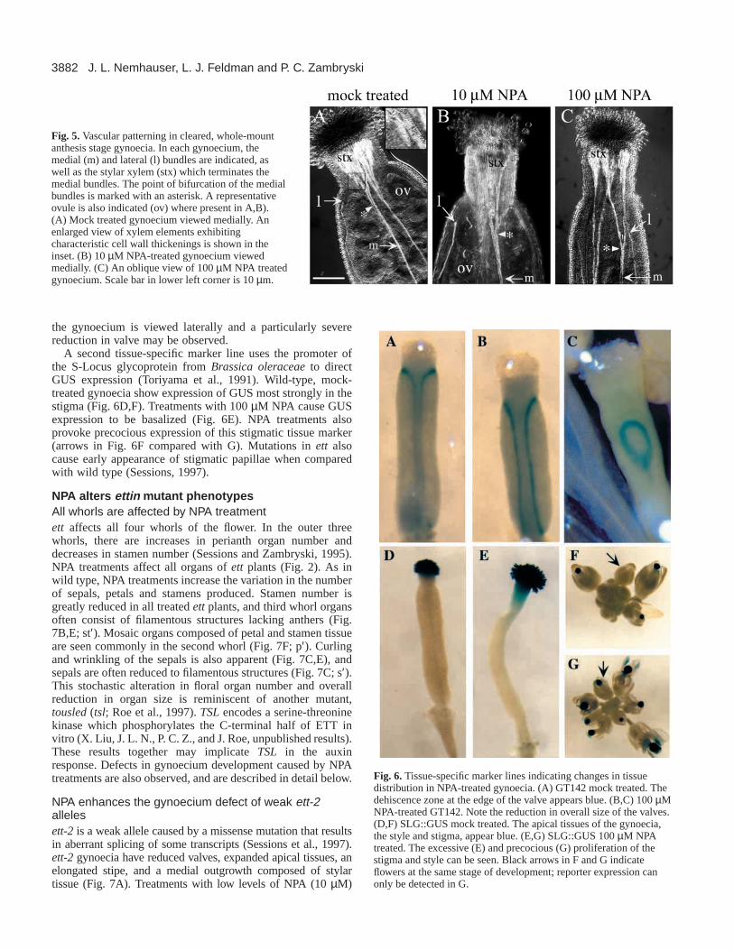
two lateral. At the style/ovary boundary, two effects onvascular traces may be observed which serve as internallandmarks for this regional transition. First, the lateral veinsterminate. Second, the medial veins bifurcate at this junctionand large arrays of xylem elements are apparent within thestylar region (Fig. 5A). The proliferation of stylar xylemsuggests a high concentration of auxin during its development.10 µM NPA treatments produce only mild effects on thebifurcation of the medial vasculature and the termination of thelateral strands (Fig. 5B), consistent with the subtle effects onstyle and valve observed by SEM. Treatments with 100 µMNPA result in both of the vascular markers of style/ovaryboundary becoming more deeply basalized (Fig. 5C). Asignificant loss of ovules is also seen with high level NPAtreatments (Fig. 5C).
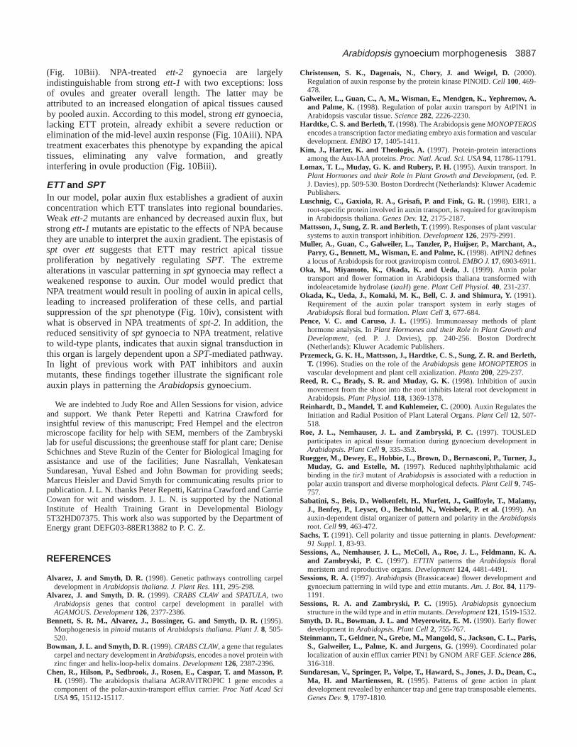
NPA also alters expression of gynoecial tissue-specificmarkersGT142 is an enhancer line that marks the dehiscence zonealong the margins of the valves, and visually delimits the valvearea (Fig. 6A; Sundaresan et al., 1995). Wild-type, untreatedgynoecia show stronger expression at their apical ends,although staining can be detected ringing the entire valve.Plants treated with 100 µM NPA and stained for β-glucuronidase (GUS) expression illustrate the decrease in valvein both the radial and longitudinal axes (Fig. 6B,C). In Fig. 6C,
Fig. 3.Scanning electron micrographsshowing NPA effects on tissuedistribution in the wild-type gynoecium.Valve extent is indicated for the right sideof each gynoecium as the space betweenthe two white arrows. Late stage 12gynoecia are shown for each treatment.(A) Mock-treated flower with stigma(sg), style (st), ovary (o), valve (v),adaxial replum (r), and stipe (i) indicated.(B) 10 µM NPA treatment. Note that thestigma and style are slightly elongated.(C) 100 µM NPA treatment. Note thereduced height of the gynoecium; thevalves are smaller than in mock-treatedplants, and there is a marked increase instyle and stigma proliferation. Scale bar,(A) 165 µm, (B) 200 µm, (C) 140 µm.
Fig. 4.Scanning electron micrographs of adaxial replum. (A) Junction of style (st), adaxial replum (r), and valve (v) in mock treatedgynoecium. (B) Junction of style (st), adaxial replum (r), and valve (v) in 100 µM NPA-treated gynoecium. Note that cells are more axializedand grow out from between the valves, but retain other features of the adaxial replum. (C) Junction of style (st), adaxial replum (r), and valve(v) in ett-2gynoecium. Note medial outgrowth consists of bulbous cells, unlike any normally found on the surface of the gynoecium. Scale bar,20 µm in all panels.
3882
the gynoecium is viewed laterally and a particularly severereduction in valve may be observed.
A second tissue-specific marker line uses the promoter ofthe S-Locus glycoprotein from Brassica oleraceaeto directGUS expression (Toriyama et al., 1991). Wild-type, mock-treated gynoecia show expression of GUS most strongly in thestigma (Fig. 6D,F). Treatments with 100 µM NPA cause GUSexpression to be basalized (Fig. 6E). NPA treatments alsoprovoke precocious expression of this stigmatic tissue marker(arrows in Fig. 6F compared with G). Mutations in ett alsocause early appearance of stigmatic papillae when comparedwith wild type (Sessions, 1997).
NPA alters ettin mutant phenotypesAll whorls are affected by NPA treatmentett affects all four whorls of the flower. In the outer threewhorls, there are increases in perianth organ number anddecreases in stamen number (Sessions and Zambryski, 1995).NPA treatments affect all organs of ett plants (Fig. 2). As inwild type, NPA treatments increase the variation in the numberof sepals, petals and stamens produced. Stamen number isgreatly reduced in all treated ettplants, and third whorl organsoften consist of filamentous structures lacking anthers (Fig.7B,E; st′). Mosaic organs composed of petal and stamen tissueare seen commonly in the second whorl (Fig. 7F; p′). Curlingand wrinkling of the sepals is also apparent (Fig. 7C,E), andsepals are often reduced to filamentous structures (Fig. 7C; s′).This stochastic alteration in floral organ number and overallreduction in organ size is reminiscent of another mutant,tousled(tsl; Roe et al., 1997). TSLencodes a serine-threoninekinase which phosphorylates the C-terminal half of ETT invitro (X. Liu, J. L. N., P. C. Z., and J. Roe, unpublished results).These results together may implicate TSLin the auxinresponse. Defects in gynoecium development caused by NPAtreatments are also observed, and are described in detail below.
NPA enhances the gynoecium defect of weak ett-2allelesett-2 is a weak allele caused by a missense mutation that resultsin aberrant splicing of some transcripts (Sessions et al., 1997).ett-2gynoecia have reduced valves, expanded apical tissues, anelongated stipe, and a medial outgrowth composed of stylartissue (Fig. 7A). Treatments with low levels of NPA (10 µM)
J. L. Nemhauser, L. J. Feldman and P. C. Zambryski
Fig. 5.Vascular patterning in cleared, whole-mountanthesis stage gynoecia. In each gynoecium, themedial (m) and lateral (l) bundles are indicated, aswell as the stylar xylem (stx) which terminates themedial bundles. The point of bifurcation of the medialbundles is marked with an asterisk. A representativeovule is also indicated (ov) where present in A,B).(A) Mock treated gynoecium viewed medially. Anenlarged view of xylem elements exhibitingcharacteristic cell wall thickenings is shown in theinset. (B) 10 µM NPA-treated gynoecium viewedmedially. (C) An oblique view of 100 µM NPA treatedgynoecium. Scale bar in lower left corner is 10 µm.
Fig. 6.Tissue-specific marker lines indicating changes in tissuedistribution in NPA-treated gynoecia. (A) GT142 mock treated. Thedehiscence zone at the edge of the valve appears blue. (B,C) 100 µMNPA-treated GT142. Note the reduction in overall size of the valves.(D,F) SLG::GUS mock treated. The apical tissues of the gynoecia,the style and stigma, appear blue. (E,G) SLG::GUS 100 µM NPAtreated. The excessive (E) and precocious (G) proliferation of thestigma and style can be seen. Black arrows in F and G indicateflowers at the same stage of development; reporter expression canonly be detected in G.

3883Arabidopsis gynoecium morphogenesis
largely eliminate any apparent differentiation of valve tissue, andthe remaining structure, composed exclusively of stigma, styleand stipe, resembles that of strong ett alleles (Fig. 7B). Thisdramatic phenotype reveals ett’s increased sensitivity to NPAtreatments, as low level NPA treatments have only subtle effectson development of wild-type gynoecia. 100 µM NPA treatmentsdo not appear to further enhance these defects (Fig. 7C).
Gynoecial defects produced by strong ett alleles arelargely unaffected by NPA treatmentett-1makes no detectable etttranscript, as a result of a T-DNAinsertion near the beginning of the coding region (Sessions etal., 1997). ett-1gynoecia rarely make valves and consist solelyof apical tissues supported by an elongated stipe (Sessions andZambryski, 1995). Ovules are often exposed at the apical endof the structure. Treatments with 10 or 100 µm NPA result inonly subtle enhancement of this exterior phenotype (Fig. 7E,F).The major effect of NPA treatments on ett-1is the severereduction in the number of ovules produced (notice absence ofexternal ovules in Fig. 7E,F compared with D). There are noapparent differences in the effects of low and high level NPAtreatments.
Altered vasculature reflects altered regional boundariesThe basalization of medial vein bifurcation and lateral veintermination in ettmutants has been described previously(Sessions and Zambryski, 1995). In 10 µM NPA treated ett-2mutants, all vascular bundles branch at the apical end of thegynoecium, making it difficult to distinguish lateral frommedial veins (Fig. 8B). If these veins are all medial, this would
reflect complete basalization of medial bundle bifurcation andlateral bundle termination. In this case, medial veins wouldbifurcate at the base of the gynoecium, appearing as fourstrands instead of two, and lateral veins would not form at all.Alternatively, the phenotype observed with NPA-treated ett-2gynoecia may be similar to what is observed with intermediateett alleles where lateral veins take on medial vein attributes,bifurcating in medial bundle-like fans of xylem (Sessions andZambryski, 1995). High level NPA treatments (100 µM) of ett-2 plants produce large numbers of parallel veins that terminateat the top of the organ in a dramatically enhanced fan of xylem(Fig. 8C). No ovules are seen in NPA-treated ett-2 gynoecia(Fig. 8B,C).
In ett-1, bifurcation of medial bundles and termination oflateral bundles is basalized, and the stylar xylem anastomosesat the apical end of the gynoecium (Sessions and Zambryski,1995). These phenotypes are largely unaffected by 10 or 100µM NPA treatments (Fig. 8E,F). As in treatedett-2 plants,lateral veins are rarely observed in NPA-treated ett-1plants,and a clear bifurcation of the medial vasculature is notapparent. This may indicate an extremely basalized style/ovaryboundary in these tissues. Overall, NPA-treated ett-1gynoeciashow a very similar phenotype to NPA-treated ett-2gynoecia.
spt mutants are partially rescued by NPA treatmentMutations in SPATULA(SPT) produce gynoecia with reducedapical tissues (Alvarez and Smyth, 1998; Alvarez and Smyth,1999; see also Fig. 9A). spt gynoecia lack transmitting trackcells and are often incompletely fused at the apical end. Thereis also reduced development of the stigmatic papillae. The
Fig. 7. Scanning electron micrographs showing NPAeffects on tissue distribution in ettgynoecia. Stage12/13 gynoecia are shown for each treatment. (A)ett-2mock-treated gynoecium with stigma (sg), style (st),valve (v), medial outgrowth (mo), and stipe (i)indicated. Valve extent is indicated on the left side ofthe gynoecium as the space between the two arrows.(B) ett-2gynoecium treated with 10 µM NPA.Stigmatic papillae are enlarged and are supported byelongated style and stipe tissue. No valve tissue isapparent. Note the increased height of the gynoeciumrelative to the petals, as compared with mock-treatedett-2(A). A misformed stamen lacking anther tissue inindicated (st′). (C) ett-2 gynoecium treated with 100µM NPA. The gynoecium is similar in appearance tothat observed in the low level treatment (10 µM).Curling of the sepals is also evident. s′ indicates abifurcated, filamentous sepal. (D)ett-1mock-treatedgynoecium. An external ovule (ov) is indicated. Novalve tissue is apparent. (E)ett-1gynoecium treatedwith 10 µM NPA. The overall structure is similar tomock-treated ett-1, except for the additional reductionin ovules. (F)ett-1gynoecium treated with 100 µMNPA. The high level treatment has no additional effecton ett-1. A mosaic second whorl organ composed ofpetal and stamen tissue is marked with a p′. Scale bar,(A) 250 µm, (B) 195 µm, (C) 205 µm, (D) 275 µm,(E) 190 µm, (F) 205 µm.

3884
strong spt allele, spt-2, causes strikingly aberrant gynoecialvasculature (Fig. 9B). Instead of bifurcating at the style/ovaryboundary, medial veins in spt gynoecia bifurcate at severalpoints along the apical half of the valve and fan out abaxially.The stylar xylem is more extensively elaborated than in wild-type gynoecia. The lateral veins terminate in knots of xylem(Fig. 9B; l’) and often curve adaxially. Discontinuous strandsas well as isolated islands of xylem are also observed (data notshown).
While valve and internode tissues of spt-2gynoecia are lessaffected by treatment with 100 µM NPA than either ettor wild-type gynoecia, NPA largely suppresses the effects of themutation on apical tissues (Fig. 9C). Stipe length is moderatelyincreased and valves are slightly reduced. A majority ofgynoecia show complete fusion of the apical end and greatlyenhanced stigma development (Fig. 9C). Vasculature of theNPA-treated spt-2 gynoecia is remarkably similar to thatobserved in untreated wild-type plants (Fig. 9D); thebifurcation of medial veins is more limited to the style/ovaryboundary, and the stylar xylem arrays are less dense. Lateralvein knots are also reduced. Ovule number is reduced as inNPA treatments of wild type.
DISCUSSION
This work documents the effects of disrupted polar auxintransport on floral organ development, particularly regionaldifferentiation in the Arabidopsisgynoecium. NPA treatmentsresult in increased concentration of auxin near source cells anddepauperation in outlying areas. In wild-typegynoecia, NPA treatment results in increasedapical tissue proliferation, a basalized style/ovaryboundary, decreased valve production and anelongated stipe. Ovule development also isnegatively affected by NPA treatments. Analysesvia scanning electron microscopy, tissue-specificGUS marker lines, and light microscopy oncleared tissue support these findings.
Low level NPA treatments are sufficient to strongly enhancethe gynoecial phenotype produced by the weak ett-2 allele –drastically reducing valve, increasing both proliferation anddistribution of apical tissues, and increasing the length of thestipe. The exterior morphology of plants with the strong ett-1allele is not dramatically affected by NPA treatments, thoughapical tissues proliferate more than in mock treated ett-1plants.As in wild type, ovule production in ett gynoecia is severelylimited by NPA treatment. In contrast, spt gynoecia arepartially rescued by NPA treatment. These results uncover anintimate link between the action of ETT and SPTand normalpolar auxin transport in the gynoecium.
The gynoecium as modified leaves: evolutionaryand developmental parallelsIn leaf development, the prevailing model for vein initiation isthat auxin is first synthesized in leaf margins and subsequentlydrains away towards the center of the organ. The flow of auxinis canalized into discrete channels which then differentiate intoa network of veins (Mattsson et al., 1999; Sachs, 1991). Lossof polar auxin transport in developing Arabidopsis leaves,resulting from mutations in PIN1or following NPA treatments(Mattsson et al., 1999), supports this model and shows strikingparallels with the results in the gynoecium described here.Young leaves treated with NPA exhibit increased density ofveins along their margins, as well as multiple parallel midveins.It is proposed that reduced PAT causes auxin to pool at themargins, leading to increased vein production there. Depletedauxin at the midvein would disrupt canalization and producemultiple smaller veins connecting the leaf to the petiole. This
J. L. Nemhauser, L. J. Feldman and P. C. Zambryski
Fig. 8.Vascular patterning in cleared, whole-mountanthesis stage gynoecia. In each gynoecium, themedial (m) and lateral (l) bundles are indicated wherepresent, as well as the stylar xylem (stx) whichterminates the medial bundles. The point ofbifurcation of the medial bundles is also indicatedwhere present (*). A representative ovule (ov) isindicated in A, the only case where ovules areobserved. (A)ett-2mock-treated gynoecium. (B)ett-2gynoecium treated with 10 µM NPA. Though fourbundles are present, it is difficult to distinguish medialfrom lateral as all veins terminate in medial bundle-like fans of xylem. (C)ett-2gynoecia treated with 100µM NPA. Notice many parallel veins terminating inlarge fans of xylem. (D)ett-1mock-treated gynoecia.The medial bundles anastomose at the apical end ofthe gynoecia. (E)ett-1 gynoecium treated with 10 µMNPA. Only two bundles are observed, both showingmedial attributes. (F)ett-1 gynoecium treated with100 µM NPA. These gynoecia closely resemble thoseof ett-1 gynoecia treated with 10 µM NPA. Scale bar,10 µm.

3885Arabidopsis gynoecium morphogenesis
is similar to NPA effects on the gynoecium where the veinslinking the gynoecium to the receptacle are multiplied. Thispattern of venation is consistent with cells along the style/ovaryboundary acting as a source of auxin. Veins in NPA-treatedgynoecia are strikingly similar to those observed in pinoid(Christensen et al., 2000), as well as ett. In light of the aberrantvenation found in spt gynoecia, these observations imply acentral role for auxin in gynoecium morphogenesis. TheAngiosperm carpel is proposed to have originated as modifiedleaves which fused to enclose and protect the gametophyte,suggesting that these parallel effects observed in leaf andgynoecium development may reflect a common evolutionaryprogram.
Auxin as a morphogen patterning the gynoecium Morphogens directly trigger differential responses in a short-range, concentration-dependent manner. Recent studies haveconcluded that auxin fits many of the criteria for a morphogen,including work in pine and aspen where a short-range auxingradient over the lateral meristem acts as a positional cue forcambial growth (Tuominen et al., 1997; Uggla et al., 1998;Uggla et al., 1996). Analyses of Arabidopsisroots stronglysuggests that auxin is a key organizer of cellular patterning inthat tissue (Sabatini et al., 1999). We propose a model whereauxin acts as a morphogen patterning the gynoecium (Fig. 10).The dramatic shifting of regional boundaries caused by NPAtreatment, may be explained by an apical-basal gradient ofauxin early in development. In this model, two boundaries inthe gynoecium primordium (a and b) mark threshold levels ofauxin. One boundary lies between presumptive style (darkgreen) and valve (light green); the other boundary separatespresumptive valve and stipe (yellow). Auxin-producing cellsare proposed to reside in the apical end of the gynoecium
primordium, represented as the wide base of the triangle shownto the left of each panel. Treatment with NPA results in poolingof auxin in these primordium cells, promoting precocious andexcessive proliferation of apical tissues in the maturegynoecium (shown as dark green in schematics belowprimordia). The depletion of auxin in more basal primordiumcells results in the loss of valve and ovules (light green) andlengthening of the stipe (yellow) in the mature gynoecium.This decrease in auxin transport is represented as a trianglewith decreased length in Fig. 10B, bringing ‘a’ and ‘b’threshold levels closer together. NPA studies and analyses ofauxin transport mutants show that valve development requiresproper PAT. In our model, valve formation is promoted by mid-level auxin concentrations. The stipe may be the defaultdevelopmental program resulting from little or no auxin.
The model described above addresses the effects of auxin onlongitudinal development of the gynoecium. It is clear,however, that auxin significantly affects radial development aswell, most clearly seen in the replum and valve. Severalgynoecium mutants display defects in both axes of thegynoecium, suggesting that their development is likelycoordinated. tousled, crabs clawand sptall show defects incarpel fusion and reduced apical tissue production (Alvarezand Smyth, 1999; Bowman and Smyth, 1999; Roe et al., 1997).ett gynoecia show an eversion of stylar transmitting tract, inaddition to the apical-basal defects previously mentioned(Sessions and Zambryski, 1995).
Another possible site of auxin action is during ovuledevelopment. The initiation or resumption of meristematicactivity in the placenta likely involves alterations in localhormone ratios. Ovules are more sensitive than other tissues ofthe gynoecium to disruptions in PAT, as valves produced intreated wild-type plants are largely devoid of ovules. This loss
Fig. 9.NPA effects on tissue distribution in sptgynoecia. Stage 12/13 gynoecia are shown in all panels. (A,C) Scanning electron micrographsare shown for both treatments. (B,D) Vascular patterning in cleared, whole-mount gynoecia. The medial (m) and lateral (l) bundles areindicated, as well as the stylar xylem (stx). The point(s) of bifurcation of the medial bundles is marked with an asterisk. (A) Mock-treated spt-2gynoecium with stigma (sg), style (st), valve (v), and stipe (i) indicated. Note the lack of fusion at the apical end and underdeveloped stigmaticpapillae. (B) Mock-treated spt-2gynoecium. Note the multiple bifurcations of the medial veins and the knot of xylem near the terminus of thelateral vein (l’). (C)spt-2gynoecium treated with 100 µM NPA. The internode is only slightly elongated and the valves show only modestreductions following NPA treatment. The apical tissue phenotypes are largely suppressed – the style appears completely fused and stigmaticpapillae are more elongated and abundant. (D)spt-2gynoecium treated with 100 µM NPA. The vascular defects are almost completelysuppressed by NPA treatment. The traces appear more focused overall, with fewer bifurcations, discontinuous strands, and isolated xylemelements. There are also dramatic reductions in the xylem knots at the termini of lateral veins. Medial vein bifurcation and lateral veintermination are somewhat basalized as in wild-type NPA treated gynoecia. Scale bar, (A) 375 µm, (B) 10 µm, (C) 300 µm, (D) 10 µm.

3886
of ovules appears to be position-dependent, most severelyaffecting ovules at the apical end of the gynoecium (data notshown), perhaps reflecting an upper limit of local auxinconcentrations for proper ovule development. However, ETTdoes not appear to play a large role in mediating the auxinresponse in the ovules, as these tissues are least affected by lossof ETT. ett tsl double mutants, in fact, leave a structureconsisting solely of placenta and ovules, suggesting neithergene is required for these tissues to differentiate. As ETT isone of a large family of proteins, another ARF may mediatethe auxin response in ovules.
As our model relies on auxin gradients for promotingregional patterning in the gynoecium, we attempted todetermine the presence of auxin, and auxin gradients withinthe developing organ. Unfortunately, the best tools currentlyavailable – a monoclonal auxin antibody (Pence and Caruso,1995) and the auxin-responsive reporter construct, DR5(Ulmasov et al., 1997b) – were inconclusive (data not shown).The small size of the Arabidopsisgynoecium primordium and
the limited sensitivity of available approaches make accuratequantitation of auxin levels difficult to determine.
ETT’s role ETT is proposed to establish or elaborate two regionalboundaries defining the apical and basal ends of valves duringgynoecium development, as illustrated in Fig. 10A (adaptedfrom Sessions, 1997). That NPA treatments phenocopy manyaspects of the ettphenotype, yet ETT is not itself auxininducible, demonstrates that polar auxin transport is requiredat these same boundaries. In this model, ETT mediates the mid-level auxin response, specifying valve development, whilerestricting high level auxin responses, perhaps by binding tothe promoters of auxin-responsive genes. Decreased level orfunction of ETT results in destabilized boundaries in thedeveloping gynoecium (represented by wavy lines aroundthreshold levels of auxin in Fig. 10Aii,iii). This destabilizationresults in a shrinkage of the presumptive valve region (lightgreen) and expansion of the presumptive apical (dark green)and basal (yellow) regions in the primordium. Moderatebasalization of the ‘a’ boundary and raising of the ‘b’ boundarycauses the phenotype observed in weak ett-2 gynoecia. Instrong ett-1 mutants, the shift in boundaries is more extreme,such that the middle region is eliminated.
The steeper auxin gradient resulting from NPA treatmentbrings the destabilized boundaries of ett-2 still closer,phenocopying ett-1, resulting in a stochastic loss of valve
J. L. Nemhauser, L. J. Feldman and P. C. Zambryski
ab
i. wild-type
iii. ett-1 (strong)
iv. spt-2 (strong)
ii. ett-2 (weak)
B. NPA treatmentA. no treatment Fig. 10. Model of gynoecium development with auxin acting as agradient-forming morphogen. Major regions of the gynoecium forwild type, weak ett-2and strong ett-1are indicated: dark green,style/stigma; light green, ovary; yellow, stipe. Triangles indicaterelative concentration of auxin, as well as the direction of auxin flux.Cylinders represent gynoecium primordia. Horizontal lines, a and b,within the primordium represent emerging regional boundariesdependent upon defined auxin concentrations. Wavy horizontal linesin the primordia of ettmutants represent destabilization of theseboundaries, as a result of reduced ETT function. Schematics belowthe primordia represent the mature structures resulting from theconditions present early on in development. (A) Models fordevelopment in the absence of NPA treatment in wild type (i), ett-2(ii), ett-1 (iii), and spt-2(iv). (B) With NPA treatment, the slope ofthe auxin gradient is increased. This is represented as a decreasedlength of the auxin concentration triangle. This alteration results inhigher auxin concentrations at the apical end of the structure anddepletion of auxin at the base. The increase in auxin concentration atthe apical ends results in increased apical tissue proliferation, mostnotably in wild type and ett-2. The change in the slope of auxinconcentration brings the regional boundaries dependant on thresholdlevels of auxin closer together. In the weak ett-2allele (ii), thisdecreased area of the presumptive valve region is furthercompromised by destabilized boundaries. As a result, the light greenregion of the primordium is greatly reduced and valves are rarelyobserved in mature gynoecia. This effect resembles strong ett-1gynoecia (Aiii). Strong ett-1gynoecia (iii) are relatively unaffectedby treatment with NPA. sptmutants are defective in apical tissuedevelopment, represented as a wavy apical line in the primordia. Thepresumed pooling of auxin in the apical tissues caused by NPAtreatments is able to partially compensate for the effects of the spt-2mutation, perhaps by bypassing a weakened auxin response (iv). Thereduced sensitivity of sptgynoecia to NPA treatment, relative towild-type plants, is evident in the mild effects on valve and internodelength. This may indicate that auxin signal transduction in thegynoecium is largely dependent upon a SPT-mediated pathway.
3887Arabidopsis gynoecium morphogenesis
(Fig. 10Bii). NPA-treated ett-2 gynoecia are largelyindistinguishable from strong ett-1 with two exceptions: lossof ovules and greater overall length. The latter may beattributed to an increased elongation of apical tissues causedby pooled auxin. According to this model, strong ett gynoecia,lacking ETT protein, already exhibit a severe reduction orelimination of the mid-level auxin response (Fig. 10Aiii). NPAtreatment exacerbates this phenotype by expanding the apicaltissues, eliminating any valve formation, and greatlyinterfering in ovule production (Fig. 10Biii).
ETT and SPTIn our model, polar auxin flux establishes a gradient of auxinconcentration which ETT translates into regional boundaries.Weak ett-2mutants are enhanced by decreased auxin flux, butstrong ett-1mutants are epistatic to the effects of NPA becausethey are unable to interpret the auxin gradient. The epistasis ofspt over ett suggests that ETT may restrict apical tissueproliferation by negatively regulating SPT. The extremealterations in vascular patterning in sptgynoecia may reflect aweakened response to auxin. Our model would predict thatNPA treatment would result in pooling of auxin in apical cells,leading to increased proliferation of these cells, and partialsuppression of the sptphenotype (Fig. 10iv), consistent withwhat is observed in NPA treatments of spt-2. In addition, thereduced sensitivity of sptgynoecia to NPA treatment, relativeto wild-type plants, indicates that auxin signal transduction inthis organ is largely dependent upon a SPT-mediated pathway.In light of previous work with PAT inhibitors and auxinmutants, these findings together illustrate the significant roleauxin plays in patterning theArabidopsisgynoecium.
We are indebted to Judy Roe and Allen Sessions for vision, adviceand support. We thank Peter Repetti and Katrina Crawford forinsightful review of this manuscript; Fred Hempel and the electronmicroscope facility for help with SEM, members of the Zambryskilab for useful discussions; the greenhouse staff for plant care; DeniseSchichnes and Steve Ruzin of the Center for Biological Imaging forassistance and use of the facilities; June Nasrallah, VenkatesanSundaresan, Yuval Eshed and John Bowman for providing seeds;Marcus Heisler and David Smyth for communicating results prior topublication. J. L. N. thanks Peter Repetti, Katrina Crawford and CarrieCowan for wit and wisdom. J. L. N. is supported by the NationalInstitute of Health Training Grant in Developmental Biology5T32HD07375. This work also was supported by the Department ofEnergy grant DEFG03-88ER13882 to P. C. Z.
REFERENCES
Alvarez, J. and Smyth, D. R.(1998). Genetic pathways controlling carpeldevelopment in Arabidopsis thaliana. J. Plant Res.111, 295-298.
Alvarez, J. and Smyth, D. R.(1999). CRABS CLAWand SPATULA, twoArabidopsis genes that control carpel development in parallel withAGAMOUS. Development126, 2377-2386.
Bennett, S. R. M., Alvarez, J., Bossinger, G. and Smyth, D. R.(1995).Morphogenesis in pinoid mutants of Arabidopsis thaliana. Plant J.8, 505-520.
Bowman, J. L. and Smyth, D. R.(1999). CRABS CLAW, a gene that regulatescarpel and nectary development in Arabidopsis, encodes a novel protein withzinc finger and helix-loop-helix domains. Development126, 2387-2396.
Chen, R., Hilson, P., Sedbrook, J., Rosen, E., Caspar, T. and Masson, P.H. (1998). The arabidopsis thaliana AGRAVITROPIC 1 gene encodes acomponent of the polar-auxin-transport efflux carrier. Proc Natl Acad SciUSA95, 15112-15117.
Christensen, S. K., Dagenais, N., Chory, J. and Weigel, D.(2000).Regulation of auxin response by the protein kinase PINOID. Cell 100, 469-478.
Galweiler, L., Guan, C., A, M., Wisman, E., Mendgen, K., Yephremov, A.and Palme, K. (1998). Regulation of polar auxin transport by AtPIN1 inArabidopsis vascular tissue. Science282, 2226-2230.
Hardtke, C. S. and Berleth, T.(1998). The Arabidopsis gene MONOPTEROSencodes a transcription factor mediating embryo axis formation and vasculardevelopment. EMBO17, 1405-1411.
Kim, J., Harter, K. and Theologis, A. (1997). Protein-protein interactionsamong the Aux-IAA proteins. Proc. Natl. Acad. Sci. USA94, 11786-11791.
Lomax, T. L., Muday, G. K. and Rubery, P. H. (1995). Auxin transport. InPlant Hormones and their Role in Plant Growth and Development, (ed. P.J. Davies), pp. 509-530. Boston Dordrecht (Netherlands): Kluwer AcademicPublishers.
Luschnig, C., Gaxiola, R. A., Grisafi, P. and Fink, G. R.(1998). EIR1, aroot-specific protein involved in auxin transport, is required for gravitropismin Arabidopsis thaliana. Genes Dev.12, 2175-2187.
Mattsson, J., Sung, Z. R. and Berleth, T.(1999). Responses of plant vascularsystems to auxin transport inhibition. Development126, 2979-2991.
Muller, A., Guan, C., Galweiler, L., Tanzler, P., Huijser, P., Marchant, A.,Parry, G., Bennett, M., Wisman, E. and Palme, K.(1998). AtPIN2 definesa locus of Arabidopsis for root gravitropism control. EMBO J.17, 6903-6911.
Oka, M., Miyamoto, K., Okada, K. and Ueda, J.(1999). Auxin polartransport and flower formation in Arabidopsis thaliana transformed withindoleacetamide hydrolase (iaaH) gene. Plant Cell Physiol.40, 231-237.
Okada, K., Ueda, J., Komaki, M. K., Bell, C. J. and Shimura, Y.(1991).Requirement of the auxin polar transport system in early stages ofArabidopsisfloral bud formation. Plant Cell3, 677-684.
Pence, V. C. and Caruso, J. L. (1995). Immunoassay methods of planthormone analysis. In Plant Hormones and their Role in Plant Growth andDevelopment, (ed. P. J. Davies), pp. 240-256. Boston Dordrecht(Netherlands): Kluwer Academic Publishers.
Przemeck, G. K. H., Mattsson, J., Hardtke, C. S., Sung, Z. R. and Berleth,T. (1996). Studies on the role of the Arabidopsisgene MONOPTEROSinvascular development and plant cell axialization. Planta200, 229-237.
Reed, R. C., Brady, S. R. and Muday, G. K.(1998). Inhibition of auxinmovement from the shoot into the root inhibits lateral root development inArabidopsis. Plant Physiol.118, 1369-1378.
Reinhardt, D., Mandel, T. and Kuhlemeier, C.(2000). Auxin Regulates theInitiation and Radial Position of Plant Lateral Organs. Plant Cell12, 507-518.
Roe, J. L., Nemhauser, J. L. and Zambryski, P. C.(1997). TOUSLEDparticipates in apical tissue formation during gynoecium development inArabidopsis. Plant Cell9, 335-353.
Ruegger, M., Dewey, E., Hobbie, L., Brown, D., Bernasconi, P., Turner, J.,Muday, G. and Estelle, M. (1997). Reduced naphthylphthalamic acidbinding in the tir3 mutant of Arabidopsisis associated with a reduction inpolar auxin transport and diverse morphological defects. Plant Cell9, 745-757.
Sabatini, S., Beis, D., Wolkenfelt, H., Murfett, J., Guilfoyle, T., Malamy,J., Benfey, P., Leyser, O., Bechtold, N., Weisbeek, P. et al. (1999). Anauxin-dependent distal organizer of pattern and polarity in the Arabidopsisroot. Cell 99, 463-472.
Sachs, T.(1991). Cell polarity and tissue patterning in plants. Development:91 Suppl.1, 83-93.
Sessions, A., Nemhauser, J. L., McColl, A., Roe, J. L., Feldmann, K. A.and Zambryski, P. C. (1997). ETTINpatterns the Arabidopsisfloralmeristem and reproductive organs. Development124, 4481-4491.
Sessions, R. A.(1997). Arabidopsis(Brassicaceae) flower development andgynoecium patterning in wild type and ettin mutants. Am. J. Bot.84, 1179-1191.
Sessions, R. A. and Zambryski, P. C.(1995). Arabidopsisgynoeciumstructure in the wild type and in ettinmutants. Development121, 1519-1532.
Smyth, D. R., Bowman, J. L. and Meyerowitz, E. M.(1990). Early flowerdevelopment in Arabidopsis. Plant Cell2, 755-767.
Steinmann, T., Geldner, N., Grebe, M., Mangold, S., Jackson, C. L., Paris,S., Galweiler, L., Palme, K. and Jurgens, G.(1999). Coordinated polarlocalization of auxin efflux carrier PIN1 by GNOM ARF GEF. Science286,316-318.
Sundaresan, V., Springer, P., Volpe, T., Haward, S., Jones, J. D., Dean, C.,Ma, H. and Martienssen, R. (1995). Patterns of gene action in plantdevelopment revealed by enhancer trap and gene trap transposable elements.Genes Dev.9, 1797-1810.

3888
Thomson, K., Hertel, R., Muller, S. and Tavares, J.(1973). 1-N-Naphthylphthalamic Acid and 2,3,5-Triiodobenzoic Acid: in vitro bindingto particulate cell fractions and action on auxin transport in corn coleoptiles.Planta109, 337-352.
Toriyama, K., Thorsness, M. K., Nasrallah, J. B. and Nasrallah, M. E.(1991). A BrassicaS locus gene promoter directs sporophytic expression inthe anther tapetum of transgenic Arabidopsis. Dev. Biol.143, 427-431.
Tuominen, H., Puech, L., Fink, S. and Sundberg, B.(1997). A radialconcentration gradient of indole-3-acetic acid is related to secondary xylemdevelopment in hybrid aspen. Plant Physiol.115, 577-585.
Uggla, C., Mellerowicz, E. J. and Sundberg, B.(1998). Indole-3-acetic acidcontrols cambial growth in scots pine by positional signaling. Plant Physiol.117, 113-121.
Uggla, C., Moritz, T., Sandberg, G. and Sundberg, B.(1996). Auxin as apositional signal in pattern formation in plants. Proc. Natl. Acad. Sci. USA93, 9282-9286.
Ulmasov, T., Hagen, G. and Guilfoyle, T. J.(1997a). ARF1, a transcriptionfactor that binds to auxin response elements. Science276, 1865-1868.
Ulmasov, T., Hagen, G. and Guilfoyle, T. J.(1999a). Activation andrepression of transcription by auxin-response factors. Proc. Natl. Acad. Sci.USA96, 5844-5849.
Ulmasov, T., Hagen, G. and Guilfoyle, T. J.(1999b). Dimerization and DNAbinding of auxin response factors. Plant J.19, 309-319.
Ulmasov, T., Murfett, J., Hagen, G. and Guilfoyle, T. J.(1997b). Aux-IAAproteins repress expression of reporter genes containing natural and highlyactive synthetic auxin response elements. Plant Cell9, 1963-1971.
Utsuno, K., Shikanai, T., Yamada, Y. and Hashimoto, T.(1998). Agr, anAgravitropic locus of Arabidopsis thaliana, encodes a novel membrane-protein family member. Plant Cell Physiol.39, 1111-1118.
Vivian-Smith, A. and Koltunow, A. M. (1999). Genetic analysis of growth-regulator-induced parthenocarpy in Arabidopsis. Plant Physiol.121, 437-451.
J. L. Nemhauser, L. J. Feldman and P. C. Zambryski