Embed Size (px)
Citation preview
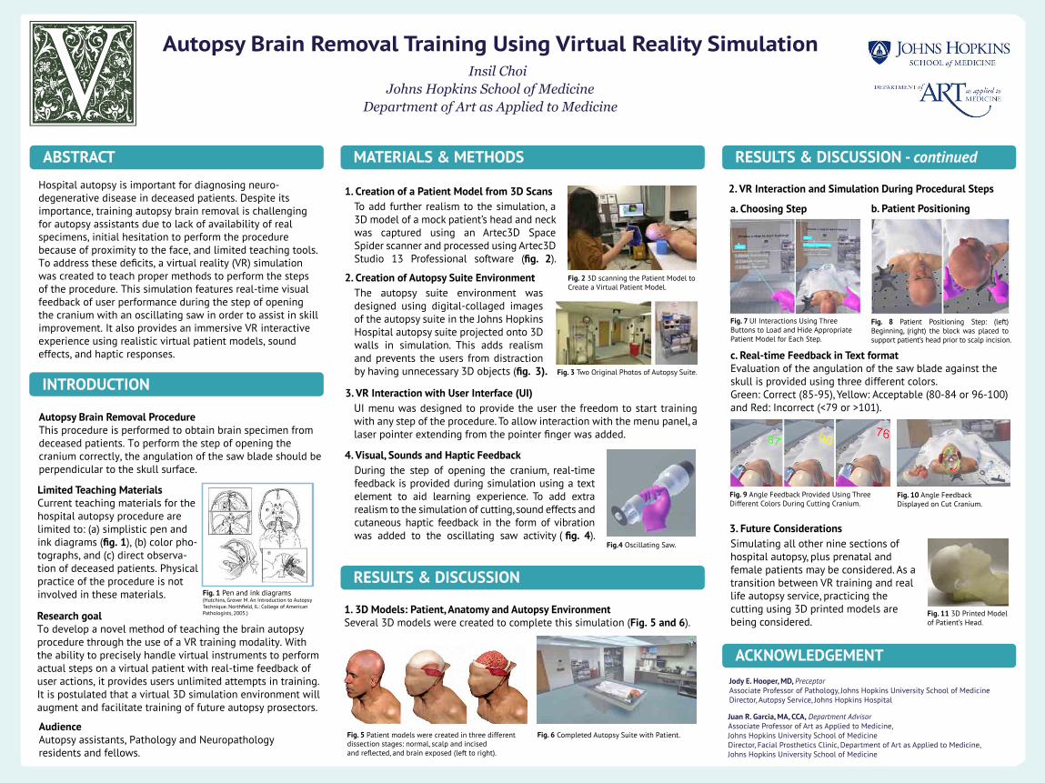
Fig. 2 3D scanning the Patient Model to Create a Virtual Patient Model.
Fig. 3 Two Original Photos of Autopsy Suite.
Fig. 8 Patient Positioning Step: (left) Beginning, (right) the block was placed to support patient’s head prior to scalp incision.
RESULTS & DISCUSSION
Fig. 7 UI Interactions Using Three Buttons to Load and Hide Appropriate Patient Model for Each Step.
Hospital autopsy is important for diagnosing neuro-degenerative disease in deceased patients. Despite itsimportance, training autopsy brain removal is challengingfor autopsy assistants due to lack of availability of realspecimens, initial hesitation to perform the procedurebecause of proximity to the face, and limited teaching tools.To address these deficits, a virtual reality (VR) simulationwas created to teach proper methods to perform the stepsof the procedure. This simulation features real-time visualfeedback of user performance during the step of openingthe cranium with an oscillating saw in order to assist in skillimprovement. It also provides an immersive VR interactiveexperience using realistic virtual patient models, soundeffects, and haptic responses.
1. Creation of a Patient Model from 3D Scans
Jody E. Hooper, MD, PreceptorAssociate Professor of Pathology, Johns Hopkins University School of MedicineDirector, Autopsy Service, Johns Hopkins Hospital
Juan R. Garcia, MA, CCA, Department AdvisorAssociate Professor of Art as Applied to Medicine, Johns Hopkins University School of MedicineDirector, Facial Prosthetics Clinic, Department of Art as Applied to Medicine,Johns Hopkins University School of Medicine
Research goalTo develop a novel method of teaching the brain autopsyprocedure through the use of a VR training modality. Withthe ability to precisely handle virtual instruments to performactual steps on a virtual patient with real-time feedback ofuser actions, it provides users unlimited attempts in training.It is postulated that a virtual 3D simulation environment willaugment and facilitate training of future autopsy prosectors.
RESULTS & DISCUSSION - continuedABSTRACT
INTRODUCTION
MATERIALS & METHODS
ACKNOWLEDGEMENT
Autopsy Brain Removal Training Using Virtual Reality SimulationInsil Choi
Johns Hopkins School of MedicineDepartment of Art as Applied to Medicine
Fig. 1 Pen and ink diagrams (Hutchins, Grover M. An Introduction to Autopsy Technique. Northfield, IL: College of American Pathologists, 2005.)
Fig. 9 Angle Feedback Provided Using Three Different Colors During Cutting Cranium.
Limited Teaching MaterialsCurrent teaching materials for thehospital autopsy procedure arelimited to: (a) simplistic pen andink diagrams (fig. 1), (b) color pho-tographs, and (c) direct observa-tion of deceased patients. Physicalpractice of the procedure is notinvolved in these materials.
c. Real-time Feedback in Text formatEvaluation of the angulation of the saw blade against the skull is provided using three different colors. Green: Correct (85-95), Yellow: Acceptable (80-84 or 96-100) and Red: Incorrect (<79 or >101).
To add further realism to the simulation, a 3D model of a mock patient’s head and neck was captured using an Artec3D Space Spider scanner and processed using Artec3D Studio 13 Professional software (fig. 2).
4. Visual, Sounds and Haptic Feedback During the step of opening the cranium, real-time feedback is provided during simulation using a text element to aid learning experience. To add extra realism to the simulation of cutting, sound effects and cutaneous haptic feedback in the form of vibration was added to the oscillating saw activity ( fig. 4).
3. VR Interaction with User Interface (UI)UI menu was designed to provide the user the freedom to start training with any step of the procedure. To allow interaction with the menu panel, a laser pointer extending from the pointer finger was added.
2. Creation of Autopsy Suite EnvironmentThe autopsy suite environment was designed using digital-collaged images of the autopsy suite in the Johns Hopkins Hospital autopsy suite projected onto 3D walls in simulation. This adds realism and prevents the users from distraction by having unnecessary 3D objects (fig. 3).
Autopsy Brain Removal ProcedureThis procedure is performed to obtain brain specimen fromdeceased patients. To perform the step of opening thecranium correctly, the angulation of the saw blade should beperpendicular to the skull surface.
AudienceAutopsy assistants, Pathology and Neuropathologyresidents and fellows.
Fig.4 Oscillating Saw.
2. VR Interaction and Simulation During Procedural Steps
1. 3D Models: Patient, Anatomy and Autopsy EnvironmentSeveral 3D models were created to complete this simulation (Fig. 5 and 6).
b. Patient Positioninga. Choosing Step
Fig. 5 Patient models were created in three different dissection stages: normal, scalp and incised and reflected, and brain exposed (left to right).
Fig. 6 Completed Autopsy Suite with Patient.
3. Future ConsiderationsSimulating all other nine sections of hospital autopsy, plus prenatal and female patients may be considered. As a transition between VR training and real life autopsy service, practicing the cutting using 3D printed models are being considered.
Fig. 10 Angle Feedback Displayed on Cut Cranium.
Fig. 11 3D Printed Model of Patient’s Head.