Embed Size (px)
Citation preview
Autophagy inhibitor Lys05 has single-agent antitumoractivity and reproduces the phenotype of a geneticautophagy deficiencyQuentin McAfeea, Zhihui Zhangb, Arabinda Samantaa, Samuel M. Levib, Xiao-Hong Maa, Shengfu Piaoa, John P. Lyncha,Takeshi Ueharac, Antonia R. Sepulvedac, Lisa E. Davisd, Jeffrey D. Winklerb,e,1, and Ravi K. Amaravadia,e,1
Departments of aMedicine and cPathology, Perelman School of Medicine, bDepartment of Chemistry, School of Arts and Sciences, and eAbramson CancerCenter, University of Pennsylvania, Philadelphia, PA 19104; and dDepartment of Pharmacy Practice & Pharmacy Administration, Philadelphia College ofPharmacy, University of the Sciences, Philadelphia, PA 19104
Edited by Dennis A. Carson, University of California at San Diego, La Jolla, CA, and approved April 10, 2012 (received for review November 9, 2011)
Autophagy is a lysosome-dependent degradative process thatprotects cancer cells from multiple stresses. In preclinical models,autophagy inhibition with chloroquine (CQ) derivatives augmentsthe efficacy of many anticancer therapies, but CQ has limitedactivity as a single agent. Clinical trials are underway combininganticancer agents with hydroxychloroquine (HCQ), but concen-trations of HCQ required to inhibit autophagy are not consistentlyachievable in the clinic. We report the synthesis and characteriza-tion of bisaminoquinoline autophagy inhibitors that potentlyinhibit autophagy and impair tumor growth in vivo. The structuralmotifs that are necessary for improved autophagy inhibitioncompared with CQ include the presence of two aminoquinolinerings and a triamine linker and C-7 chlorine. The lead compound,Lys01, is a 10-fold more potent autophagy inhibitor than HCQ.Compared with HCQ, Lys05, a water-soluble salt of Lys01, morepotently accumulates within and deacidifies the lysosome, resultingin impaired autophagy and tumor growth. At the highest doseadministered, some mice develop Paneth cell dysfunction thatresembles the intestinal phenotype ofmice and humanswith geneticdefects in the autophagy gene ATG16L1, providing in vivo evidencethat Lys05 targets autophagy. Unlike HCQ, significant single-agentantitumor activity is observed without toxicity in mice treated withlower doses of Lys05, establishing the therapeutic potential of thiscompound in cancer.
cell death | stress responses | cancer cell survival | drug resistance |antimalarials
Autophagy, the sequestration of organelles and proteins in au-tophagic vesicles (AVs) and degradation of this cargo through
lysosomal fusion (1), allows tumor cells to survive metabolic andtherapeutic stresses (2–5). Therapy-induced autophagy is a keyresistance mechanism to many anticancer agents (6), and autoph-agy levels are increased in most cancers (7). Chloroquine (CQ;Fig. 1, compound 1) derivatives block autophagy by impairing ly-sosomal function (3, 8, 9). Studies in multiple mouse models ofmalignancy have demonstrated that autophagy inhibition with CQderivatives augments the efficacy of a variety of anticancer agents.Clinical trials combining cancer therapies with hydroxychloroquine(HCQ; Fig. 1), have been launched, and preliminary results in-dicate these combinations have activity (6). However, pharmaco-kinetic (PK)-pharmacodynamic (PD) studies conducted in patientsreceiving HCQ for cancer therapy have indicated that the highmicromolar concentrations of HCQ required to inhibit autophagyin vitro are inconsistently achieved in humans (10). There is anunmet need to develop more potent inhibitors of autophagy.The design and synthesis of dimeric analogs of CQ that exploit
the thermodynamic advantages imparted by polyvalency (11, 12)has been previously studied in the context of malaria (13–15).The synthesis of heteroalkane-bridged bisquinolines did notproduce sufficient antimalarial activity to warrant further inves-tigation (14). Subsequently, a series of tetraquinolines was re-ported with potent antimalarial properties (13), confirming that
polyvalency could afford increased potency. Augmented cyto-toxicity was observed in cancer cell lines when Akt inhibitorswere combined with fluorinated quinolines (16), and CQ analogswith a piperazine connector had enhanced anticancer propertiescompared with CQ (17). These results suggest that the CQ scaf-fold could serve as the basis for the development of effectivecancer chemotherapeutics, but to date the properties of dimericCQ derivatives as anticancer therapeutics have not been in-vestigated. Here we report the synthesis of dimeric CQ analogsand a structure-activity analysis using autophagy and cytotoxicityassays as the biological reporters of activity. This work identifiesLys01 as a viable lead compound for development as an autophagyinhibitor and anticancer therapeutic.
ResultsStrategy for Synthesis of Bivalent Aminoquinoline Autophagy Inhibitors.To apply the strategy of polyvalency (11, 12) to the synthesis ofautophagy inhibitors, dimeric CQ (Fig. 1; 3: Lys01) was preparedfrom commercially available materials (compounds herein arelisted in Table S1). Based on literature precedent (14), we envi-sioned the preparation of 3 from two equivalents of 4 (14) andone equivalent of 5, as outlined in Fig. S1. Though 6 (R = H) isknown (14), the bisquinoline 3 (R = Me) has not been describedin the literature. Because of its putative lysosomotropism, werefer to 3 as Lys01 (Fig. 1). Reaction of two equivalents of 4 with5 led to the formation of a mixture of the desired product 3 alongwith some of the monoquinoline 7: Lys02 (Fig. 1 and Fig. S1), thesynthesis of which was previously reported (18). To examine therole of the C-7 chlorine substituent in 3, we prepared 9 (Fig. 1;9: Lys-03), the dimethoxy analog of compound 3, starting from4-bromo-7-methoxyquinoline 8. To determine the importance of thepolyamine connector of 3, we prepared the polyether analog 11:Lys04 of compound 3 from two equivalents of 8 and the commer-cially available 2,2′-(ethylenedioxy)bis(ethylamine) 10 (Fig. 2).
Lys01 Is a More Potent Autophagy Inhibitor than HCQ or CQ. LN229(glioblastoma) cells were treated with Lys01 and derivativesLys02, Lys03, Lys04, HCQ, and CQ. Near complete cell death ofcultured cells was observed in cells treated with Lys01 at con-
Author contributions: Q.M., Z.Z., T.U., A.R.S., L.E.D., J.D.W., and R.K.A. designed research;Q.M., Z.Z., A.S., S.M.L., X.-H.M., S.P., J.P.L., T.U., A.R.S., L.E.D., J.D.W., and R.K.A. per-formed research; Z.Z., S.M.L., J.P.L., T.U., A.R.S., L.E.D., J.D.W., and R.K.A. contributednew reagents/analytic tools; Q.M., A.S., S.M.L., X.-H.M., S.P., J.P.L., T.U., A.R.S., L.E.D.,J.D.W., and R.K.A. analyzed data; and Q.M., J.P.L., A.R.S., L.E.D., J.D.W., and R.K.A. wrotethe paper.
Conflict of interest statement: There is a patent pending that protects the compoundsdescribed in this paper and use of the compounds in cancer and other indications. Thispatent has not been licensed and has generated no revenue to date.
This article is a PNAS Direct Submission.1To whom correspondence may be addressed. E-mail: [email protected] [email protected].
This article contains supporting information online at www.pnas.org/lookup/suppl/doi:10.1073/pnas.1118193109/-/DCSupplemental.
www.pnas.org/cgi/doi/10.1073/pnas.1118193109 PNAS | May 22, 2012 | vol. 109 | no. 21 | 8253–8258
MED
ICALSC
IENCE
S
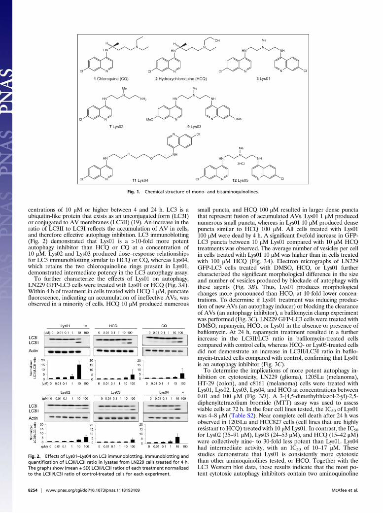
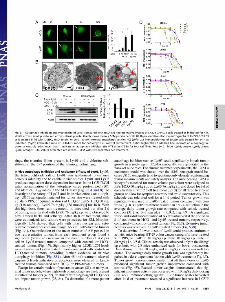
centrations of 10 μM or higher between 4 and 24 h. LC3 is aubiquitin-like protein that exists as an unconjugated form (LC3I)or conjugated to AV membranes (LC3II) (19). An increase in theratio of LC3II to LC3I reflects the accumulation of AV in cells,and therefore effective autophagy inhibition. LC3 immunoblotting(Fig. 2) demonstrated that Lys01 is a >10-fold more potentautophagy inhibitor than HCQ or CQ at a concentration of10 μM. Lys02 and Lys03 produced dose–response relationshipsfor LC3 immunoblotting similar to HCQ or CQ, whereas Lys04,which retains the two chloroquinoline rings present in Lys01,demonstrated intermediate potency in the LC3 autophagy assay.To further characterize the effects of Lys01 on autophagy,
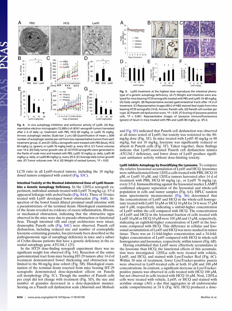
LN229 GFP-LC3 cells were treated with Lys01 or HCQ (Fig. 3A).Within 4 h of treatment in cells treated with HCQ 1 μM, punctatefluorescence, indicating an accumulation of ineffective AVs, wasobserved in a minority of cells. HCQ 10 μM produced numerous
small puncta, and HCQ 100 μM resulted in larger dense punctathat represent fusion of accumulated AVs. Lys01 1 μM producednumerous small puncta, whereas in Lys01 10 μM produced densepuncta similar to HCQ 100 μM. All cells treated with Lys01100 μM were dead by 4 h. A significant fivefold increase in GFP-LC3 puncta between 10 μM Lys01 compared with 10 μM HCQtreatments was observed. The average number of vesicles per cellin cells treated with Lys01 10 μM was higher than in cells treatedwith 100 μM HCQ (Fig. 3A). Electron micrographs of LN229GFP-LC3 cells treated with DMSO, HCQ, or Lys01 furthercharacterized the significant morphological difference in the sizeand number of vesicles produced by blockade of autophagy withthese agents (Fig. 3B). Thus, Lys01 produces morphologicalchanges more pronounced than HCQ, at 10-fold lower concen-trations. To determine if Lys01 treatment was inducing produc-tion of new AVs (an autophagy inducer) or blocking the clearanceof AVs (an autophagy inhibitor), a bafilomycin clamp experimentwas performed (Fig. 3C). LN229 GFP-LC3 cells were treated withDMSO, rapamycin, HCQ, or Lys01 in the absence or presence ofbafilomycin. At 24 h, rapamycin treatment resulted in a furtherincrease in the LC3II/LC3 ratio in bafilomycin-treated cellscompared with control cells, whereas HCQ- or Lys05-treated cellsdid not demonstrate an increase in LC3II/LC3I ratio in bafilo-mycin-treated cells compared with control, confirming that Lys01is an autophagy inhibitor (Fig. 3C).To determine the implications of more potent autophagy in-
hibition on cytotoxicity, LN229 (glioma), 1205Lu (melanoma),HT-29 (colon), and c8161 (melanoma) cells were treated withLys01, Lys02, Lys03, Lys04, and HCQ at concentrations between0.01 and 100 μM (Fig. 3D). A 3-(4,5-dimethylthiazol-2-yl)-2,5-diphenyltetrazolium bromide (MTT) assay was used to assessviable cells at 72 h. In the four cell lines tested, the IC50 of Lys01was 4–8 μM (Table S2). Near complete cell death after 24 h wasobserved in 1205Lu and HCC827 cells (cell lines that are highlyresistant to HCQ) treated with 10 μMLys01. In contrast, the IC50for Lys02 (35–91 μM), Lys03 (24–53 μM), and HCQ (15–42 μM)were collectively nine- to 30-fold less potent than Lys01. Lys04had intermediate activity, with an IC50 of 10–17 μM. Thesestudies demonstrate that Lys01 is consistently more cytotoxicthan other aminoquinolines tested, or HCQ. Together with theLC3 Western blot data, these results indicate that the most po-tent cytotoxic autophagy inhibitors contain two aminoquinoline
N
HNN
Cl
H
1 Chloroquine (CQ)
N
HNN
Cl
OHH
2 Hydroxychloroquine (HCQ)
N
HNN
NH
Cl
Me
N Cl
3 Lys01
N
HNN
NH
Cl
Me
N Cl12 Lys05
3HCl
N
HNN
NH2
Cl
Me
7 Lys02
N
HNN
NH
MeO
Me
N OMe
9 Lys03
N
HN
Cl
NH
N Cl
OO
11 Lys04
Fig. 1. Chemical structure of mono- and bisaminoquinolines.
Fig. 2. Effects of Lys01–Lys04 on LC3 immunoblotting. Immunoblotting andquantification of LC3II/LC3I ratio in lysates from LN229 cells treated for 4 h.The graphs show (mean ± SD) LC3II/LC3I ratios of each treatment normalizedto the LC3II/LC3I ratio of control-treated cells for each experiment.
8254 | www.pnas.org/cgi/doi/10.1073/pnas.1118193109 McAfee et al.
rings, the triamine linker present in Lys01 and a chlorine sub-stituent at the C-7 position of the aminoquinoline ring.
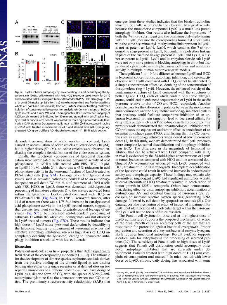
In Vivo Autophagy Inhibition and Antitumor Efficacy of Lys05. Lys05,the trihydrochloride salt of Lys01, was synthesized to enhanceaqueous solubility and to enable in vivo studies. Lys01 and Lys05produced equivalent dose-dependent increases in the LC3II/LC3Iratio, accumulation of the autophagy cargo protein p62 (20),and identical IC50 values in the MTT assay (Fig. S2 A and B). Toinvestigate the safety of Lys05 and its in vivo effects on autoph-agy, c8161 xenografts matched for tumor size were treated withi.p. daily PBS, or equimolar doses of HCQ or Lys05 [HCQ 60 mg/kg (138 nmols/g), Lys05 76 mg/kg (138 nmols/g)] for 48 h. Withthis high-dose, short-term treatment, no mice died, but after 2 dof dosing, mice treated with Lys05 76 mg/kg i.p. were observed tohave arched backs and lethargy. After 48 h of treatment, micewere euthanized, and tumors were processed for EM. Morpho-logically, EM showed that cells with intact nuclear and cyto-plasmic membranes contained large AVs in Lys05-treated tumors(Fig. 4A). Quantification of the mean number of AV per cell intwo representative tumors from each treatment group found asignificant (>twofold) increase in the mean number of AV percell in Lys05-treated tumors compared with control- or HCQ-treated tumors (Fig. 4B). Significantly higher LC3II/LC3I levelswere observed in Lys05-treated tumors compared with control- orHCQ-treated tumors, providing further evidence of in vivoautophagy inhibition (Fig. S3A). After 48 h of treatment, cleavedcaspase 3 levels indicative of apoptosis were elevated in Lys05-treated tumors compared with HCQ- or PBS-treated tumors.Except for certain models of pancreatic cancer (21), in many an-
imal tumormodels, where high levels of autophagy are likely presentin untreated tumors (4, 22), treatment with single-agent HCQ doesnot impair tumor growth (23, 24). To determine if a more potent
autophagy inhibitor such as Lys05 could significantly impair tumorgrowth as a single agent, 1205Lu xenografts were generated in theflanks of nudemice. For chronic treatment experiments, the 1205Lumelanoma model was chosen over the c8161 xenograft model be-cause c8161 xenografts tend to spontaneously ulcerate, confoundingtumor measurements and safety analysis. Ten mice bearing 1205Luxenografts matched for tumor volume per cohort were assigned toPBS, HCQ 60 mg/kg i.p., or Lys05 76 mg/kg i.p. and dosed for 3 d ofdaily treatment with 2 d off treatment (3/5 d) for all three treatmentgroups, to allow for symptom recovery and avoid excess toxicity. Thisschedule was tolerated well for a 14-d period. Tumor growth wassignificantly impaired in Lys05-treated tumors compared with con-trols (Fig. 4C). Lys05 treatment resulted in a 53% reduction in theaverage daily tumor growth rate compared with vehicle-treatedcontrols (31.2 vs. 14.6 mm3/d; P = 0.002; Fig. 4D). A significantthree- and sixfold accumulation ofAVwas observed at the end of 14d of treatment in HCQ- and Lys05-treated tumors, respectively,compared with control-treated tumors (Fig. S3B). Extensive tumornecrosis was observed in Lys05-treated tumors (Fig. S3B).To determine if lower doses of Lys05 could produce antitumor
activity, mice bearing HT-29 colon cancer xenografts were treatedwith PBS, or Lys05 at 10 mg/kg i.p. daily, 40 mg/kg i.p. daily, or80 mg/kg i.p. 3/5 d. Clinical toxicity was observed only in the 80 mg/kg cohort, with 2/8 mice euthanized early for bowel obstruction.Daily dosing for the 10 mg/kg and 40 mg/kg cohorts was well tol-erated. The average daily tumor growth rate was significantly im-paired in a dose-dependent fashion with Lys05 treatment (Fig. 4E).Tumor growth curves demonstrated that all three doses of Lys05produced significant tumor growth impairment compared withcontrol (Fig. 4F). Excised tumor weights demonstrated that sig-nificant antitumor activity was observed with 10 mg/kg daily dosing(Fig. 4G). Immunoblotting against LC3 in tumor lysates harvestedafter 14 d of treatment revealed a significant increase in LC3II/
A
Q
(μM) 0 1 10 100
0 75
1.00
1.25 Lys01Lys02Lys03Lys04m
alized)
D 1205Lu
30 *
01H
CQ
-7 -6 -5 -40.00
0.25
0.50
0.75 yHCQ
Log [M]
MTT
(norm
No.
punc
ta/c
ell
51015202530
Lys0
B0.25
0.50
0.75
1.00
1.25
TT(normalized)
LN229N
05
(μM) - 1 10 100 1 10 100HCQ Lys05
1.00
1.25
alized)
-7 -6 -5 -40.00
Log [M]
M
c8161
DMSO HCQ Lys01
Cio tro
l
2
2.5
R 1 MBafilomycinControl -7 -6 -5 -4
0.00
0.25
0.50
0.75
Log [M]
MTT(norma
LC3I
I/LC
3IR
ataf
ilom
ycin
/Con
t
0.5
1
1.5
2Rapa 1μM - + - - - + - -HCQ 10 μM - - + - - - + -Lys01 10 μM - - - + - - - +
LC3ILC3II
Log [M]
0 50
0.75
1.00
1.25
normalized)
HT29L
Ba 0Actin
-7 -6 -5 -40.00
0.25
0.50
Log [M]
MTT(n
Fig. 3. Autophagy inhibition and cytotoxicity of Lys01 compared with HCQ. (A) Representative images of LN229 GFP-LC3 cells treated as indicated for 4 h.White arrows: small puncta; red arrows: dense puncta. Graph shows mean ± SEM puncta per cell. (B) Representative electron micrographs of LN229-GFP-LC3cells treated (4 h) with DMSO, HCQ 10 μM, or Lys01 10 μM. Arrows: autophagic vesicles. (C) (Left) LC3 immunoblotting of LN229 cells treated for 24 h asindicated. (Right) Calculated ratio of LC3II/LC3I ratios for bafilomycin vs. control cotreatment. Ratios higher than 1 (dashed line) indicate an autophagy in-ducer or control; ratios lower than 1 indicate an autophagy inhibitor. (D) MTT assay (72 h) for four cell lines. Red: Lys01; blue: Lys02; purple: Lys03; green:Lys04; orange: HCQ. Values presented are means ± SEM with five replicates per treatment.
McAfee et al. PNAS | May 22, 2012 | vol. 109 | no. 21 | 8255
MED
ICALSC
IENCE
S
LC3I ratio in all Lys05-treated tumors, including the 10 mg/kg-dosed tumors compared with control (Fig. S3C).
Intestinal Toxicity at the Maximal Administered Dose of Lys05 Resem-bles a Genetic Autophagy Deficiency. In the 1205Lu xenograft ex-periment, individual animals treated with Lys05 76 mg/kg i.p. 3/5 dappeared lethargic with arched backs (Fig. S4A). Three of 10 micetreated with Lys05 developed bowel obstruction (Fig. S4B). In-spection of the bowel found dilated proximal small intestine witha pseudostricture of the terminal ileum. Histological examinationof the ileum revealed no evidence of excess inflammation, fibrosis,or mechanical obstruction, indicating that the obstructive signsobserved in the mice were due to pseudo-obstruction or functionalileus. Though intestinal villi and crypt architecture were intact,dysmorphic Paneth cells (Fig. S4C) were observed. Paneth celldysfunction, including reduced size and number of eosinophiliclysozyme-containing granules, has previously been described as thepathognomonic sign of autophagy deficiency in mice and a subsetof Crohn disease patients that have a genetic deficiency in the es-sential autophagy gene ATG16L1 (25).In the HT29 dose-finding xenograft experiment there was no
significant weight loss observed (Fig. 5A). Resection of the entiregastrointestinal tract from mice bearing HT-29 tumors after 14 d oftreatment demonstrated bowel thickening and obstruction waslimited to the 80-mg/kg dose cohort (Fig. 5B). Histological exam-ination of the terminal ileum resected from mice bearing HT-29xenografts demonstrated dose-dependent effects on Panethcell morphology (Fig. 5C). Though the number of Paneth cellsper crypt did not change with treatment (Fig. 5D), the size andnumber of granules decreased in a dose-dependent manner.Scoring on a Paneth cell dysfunction scale (Materials and Methods
and Fig. S5) indicated that Paneth cell dysfunction was observedat all doses tested of Lys05, but toxicity was restricted to the 80-mg/kg dose (Fig. 5E). In mice treated with Lys05 40 mg/kg or 80mg/kg, but not 10 mg/kg, lysozyme was significantly reduced orabsent in Paneth cells (Fig. 5F). Taken together, these findingsindicate that Lys05-associated Paneth cell dysfunction mimicsATG16L1 deficiency, and lower doses of Lys05 produce signifi-cant antitumor activity without dose-limiting toxicity.
Lys05 Inhibits Autophagy by Deacidifying the Lysosome. To comparethe relative lysosomal accumulation of Lys05 andHCQ, lysosomeswere subfractionated from 1205Lu cells treated with PBS, HCQ10μM, or Lys05 10 μM, and 1205Lu tumors harvested after 14 d oftreatment with PBS, HCQ 60 mg/kg i.p., or Lys05 76 mg/kg i.p.every 3/5 d. Immunoblotting against the lysosomalmarker LAMP2confirmed adequate separation of the lysosomal and whole-cellpopulation in cells and tumor samples (Fig. 6A). HPLC tandemmass spectrometry measurements (Fig. S6) determined thatthe concentrations of Lys05 and HCQ in the whole-cell homoge-nate treated with Lys05 10 μMorHCQ10 μM for 24 h were 57 μMand 8 μM, respectively, indicating a sixfold-higher concentrationof Lys05 within the cell compared with HCQ. The concentrationof Lys05 and HCQ in the lysosomal fraction of cells treated withLys05 10 μMorHCQ10 μMwere 105 μMand 13 μM, respectively,indicating an eightfold-higher concentration of Lys05 in the lyso-some compared with HCQ. This difference in cellular and lyso-somal accumulation of Lys05 andHCQwasmoremarked in tumortissue. There was an 11-fold-higher concentration and a 34-fold-higher concentration of Lys05 compared with HCQ in whole-cellhomogenates and lysosomes, respectively, within tumors (Fig. 6B).Having established that Lys05 more effectively accumulates in
the lysosome than HCQ, the functional effects of this accumula-tion were investigated. 1205Lu cells were treated with vehicle,Lys05, and HCQ, and stained with LysoTracker Red (Fig. 6C).Within 30 min of treatment, fewer LysoTracker-positive punctawere observed in Lys05-treated cells at both 10 μM and 100 μMconcentrations. In contrast, a significant decrease in LysoTracker-positive puncta was observed in cells treated with HCQ 100 μM,but not observed in cells treated with HCQ 10 μM. Next, 1205Lucells were treated with vehicle, Lys05, or HCQ and stained withacridine orange (AO; a dye that aggregates in all endovesicularacidic compartments) at 24 h (Fig. 6D). HCQ produced a dose-
Fig. 4. In vivo autophagy inhibition and antitumor activity of Lys05. (A) Rep-resentative electron micrographs (12,000×) of c8161 xenograft tumors harvestedafter 2 d of daily i.p. treatment with PBS, HCQ 60 mg/kg, or Lys05 76 mg/kg.Arrows: autophagic vesicles. (Scale bar: 2 μm.) (B) Quantification of mean ± SEMnumber of autophagic vesicles per cell from two representative tumors fromeachtreatment group. (C andD) 1205Lu xenografts were treatedwith PBS (blue), HCQ60 mg/kg i.p. (green), or Lys05 76 mg/kg (red) i.p. every 3/5 d. (C) Tumor volumesover 14 d. (D) Daily tumor growth rate. (E–G) HT29 xenografts were generated inthe flanks of nude mice and treated with PBS, Lys05 10 mg/kg i.p. daily, Lys05 40mg/kg i.p. daily, or Lys0580mg/kg i.p. every 3/5d. (E) Averagedaily tumorgrowthrate. (F) Tumor volumes over 14 d. (G) Weight of excised tumors, *P < 0.05.
Fig. 5. Lys05 treatment at the highest dose reproduces the intestinal pheno-type of a genetic autophagy deficiency. (A–F) Weight and intestines were ana-lyzed formice bearingHT29xenografts treatedwith PBS and Lys0510–80mg/kg.(A) Daily weight. (B) Representative excised gastrointestinal tracts after 14 d oftreatment. (C) Representative images (40×) of H&E-stained ileal crypts frommicebearing HT29 xenografts (14 d). Arrows: Paneth cells. (D) Paneth cell number percrypt. (E) Paneth cell dysfunction score, *P<0.05. (F) Scoringof lysozyme-positivecells, *P = 0.001. Representative images of lysozyme immunofluorescence(green) of ileum in mice treated with PBS and Lys05 80 mg/kg i.p. 3/5 d.
8256 | www.pnas.org/cgi/doi/10.1073/pnas.1118193109 McAfee et al.
dependent accumulation of acidic vesicles. In contrast, Lys05caused an accumulation of acidic vesicles at lower doses (10 μM),but at higher doses (50 μM), no acidic vesicles were observed, in-dicating the complete deacidification of the endovesicular system.Finally, the functional consequences of lysosomal deacidifi-
cation were investigated by measuring enzymatic activity of acidphosphatase. In 1205Lu cells treated with PBS, HCQ 10 μM,or Lys05 10 μM, within 24 h there was a 43% reduction in acidphosphatase activity in the lysosomal fraction of Lys05-treated vs.PBS-treated cells (Fig. S7A). Leakage of certain lysosomal en-zymes, such as activated cathepsins, could lead to an autophagy-independent cell death. Within 24 h of treatment of 1205Lu cellswith PBS, HCQ, or Lys05, there was decreased acid-dependentprocessing of immature cathepsin D to the mature activated formwithin the lysosome in Lys05-treated compared with HCQ- orPBS-treated cells (Fig. S7B), In 1205Lu xenograft tumors, after14 d of treatment there was a 1.75-fold increase in extralysosomalacid phosphatase activity in the Lys05-treated tumors, suggestingthat chronic treatment can lead to extralysosomal leakage of en-zymes (Fig. S7C), but increased acid-dependent processing ofcathepsin D within the whole-cell homogenate was not observedin Lys05-treated tumors (Fig. S7D). These results indicate thathigh doses of Lys05 cause lysosomal dysfunction by deacidifyingthe lysosome, leading to impairment of lysosomal enzymes andeffective autophagy inhibition, whereas high doses of HCQ in-completely deacidify the lysosome, leading to incomplete auto-phagy inhibition associated with less cell death.
DiscussionPolyvalent molecules can have properties that differ significantlyfrom those of the corresponding monomers (11, 12). The rationalefor the development of dimeric species as pharmaceuticals derivesfrom the possible binding of the dimeric ligand at two distinctbinding sites either on a single receptor or at defined sites on twoseparate monomers of a dimeric protein (26). We have designedLys01 as a dimeric form of CQ, with the spacer N,N-bis(2-ami-noethyl)methylamine 5 as the connector between two CQ moie-ties. The preliminary structure-activity relationship (SAR) that
emerges from these studies indicates that the bivalent quinolinestructure of Lys01 is critical to the observed biological activity,because the monomeric compound Lys02 is a much less potentautophagy inhibitor. Our results also indicate the importance ofboth the 7-chloro substituent and the bisaminoethyl–methylaminelinker in Lys01, because the corresponding bismethyl ether Lys03,with the same bisaminoethyl–methylamine linker present in Lys01,is not as potent as Lys01. Lys04, which contains the 7-chloro-quinoline rings present in Lys01, but contains a polyether linkagein place of the triamino linkage present in Lys01 and Lys03, is alsonot as potent as Lys01. Lys01 and its trihydrochloride salt Lys05were not only more potent at blocking autophagy in vitro, but alsoproduced cytotoxicity in multiple cancer cell lines and antitumoractivity in multiple human tumor xenograft models.The significant 3- to 10-fold difference between Lys05 and HCQ
in lysosomal concentration, autophagy inhibition, and cytotoxicityobserved with Lys01 compared with HCQ, cannot be attributed toa simple concentration effect, i.e., doubling of the concentration ofthe quinolone ring in Lys01. However, the enhanced basicity of thepentaamino structure of Lys01 compared with the structures ofboth CQ and HCQ, each of which contain only three nitrogenatoms, could lead to enhanced sequestration of Lys01 in the acidiclysosome relative to that of CQ and HCQ, respectively. Anotherpossible basis for the difference in potency between themonomericaminoquinolines and the bisquinoline autophagy inhibitor Lys01 isthat bivalency could facilitate cooperative inhibition of an un-known lysosomal protein target, or lead to decreased affinity fordrug efflux pumps such as ATP-binding cassette transporters. Ourprevious work demonstrated that pharmacological inhibition withCQ produces the equivalent antitumor effect as knockdown of anessential autophagy gene ATG5, establishing that the CQ deriva-tives act as autophagy inhibitors when dosed at low micromolardoses (3). In this study, we have demonstrated that Lys01 producesmore complete lysosomal deacidification and autophagy inhibitionthan HCQ. The difference in the magnitude of lysosomal in-hibition that can be achieved with Lys05 compared with HCQin vivo is evidenced by the 34-fold-higher concentrations of Lys05in tumor lysosomes compared with HCQ and the associated dou-bling of AV accumulation associated with Lys05 compared withHCQ treatment in 1205Lu xenografts. Incomplete deacidificationof the lysosome could result in rebound increase in endovesicularacidity and autophagic capacity. These findings may explain whyintermittent single-agent Lys05 had significant antitumor activity,whereas intermittent HCQ treatment resulted in acceleration oftumor growth in 1205Lu xenografts. Others have demonstratedthat, during effective distal autophagy inhibition, accumulation ofdysfunctional AV and eventual bursting of lysosomes (27, 28)conspire to increase reactive oxygen species, generating DNAdamage, followed by cell death by apoptosis or necrosis (21). Ourdata support the mechanism of action of lysosomal impairment forLys01, but identification of a molecular target within the lysosomefor Lys01 will be the focus of future research.The Paneth cell dysfunction observed at the highest dose of
Lys05 administered supports the proposed mechanism of actionof the drug. Paneth cells are secretory cells of the gut that areresponsible for protection against bacterial overgrowth. Properexpression and secretion of a key antibacterial enzyme lysozymelikely requires functional autophagy. Recent evidence supportsa critical role for autophagy in the processing of secreted pro-teins (29). The sensitivity of Paneth cells to high doses of Lys05suggests that Paneth cell dysfunction could accompany othernovel autophagy inhibitors that are currently under de-velopment. Patients treated with high doses of HCQ also com-plain of constipation and nausea.* In mice treated with lowerdoses of Lys05, chronic daily dosing was associated with some
Fig. 6. Lys05 inhibits autophagy by accumulating in and deacidifying the ly-sosome. (A) 1205Lu cells (treated with PBS, HCQ 10 μM, or Lys05 10 μM for 24 h)andharvested1205Lu xenograft tumors (treatedwithPBS,HCQ60mg/kg i.p. 3/5d, or Lys05 76mg/kg i.p. 3/5 d for 14 d) were homogenized and fractionated intowhole-cell (WC) and lysosomal (L) fractions. LAMP2 immunoblotting confirmedisolation of concentrated lysosomes for analysis. (B) Concentrations of HCQ orLys05 in cells and tumor WC and L homogenates. (C) Fluorescence imaging of1205Lu cells treated as indicated for 30 min and stained with LysoTracker Red.LysoTracker puncta (red) per cell was scored for three high-poweredfields. Blue:nuclear DAPI staining. Data presented is mean ± SEM. (D) Fluorescence imagingof c8161 cells treated as indicated for 24 h and stained with AO. Orange: ag-gregated AO; green: diffuse AO. Graph shows mean +/− SD %acidic vesicles.
*Algazy KM, et al. (2011) Combined mTOR inhibition and autophagy inhibition: Phase Itrial of temsirolimus and hydroxychloroquine in patients with advanced solid tumors.One Hundred Second Annual Meeting of the American Association for Cancer Research,April 2–6, 2011, Orlando, FL, abstr 4500.
McAfee et al. PNAS | May 22, 2012 | vol. 109 | no. 21 | 8257
MED
ICALSC
IENCE
S

degree of Paneth cell dysfunction but no clinical signs of gas-trointestinal toxicity. These findings indicate that Lys01 is a leadcompound with great potential to be optimized further for po-tency as an autophagy inhibitor. Because autophagy inhibitorswill likely be incorporated into combination regimens involvingone or more anticancer therapies, the antitumor activity ob-served with low doses and intermittent dosing bodes well for thedevelopment of Lys01 and its derivatives as cancer therapeutics.Finally, the enhanced lysosomal accumulation, autophagy in-hibition, and therapeutic activity observed with this bisaminoqui-noline in cancer models suggest that quinoline polyvalency mayalso be associated with improved therapeutic activity in diseasessuch as malaria and rheumatic disorders, in which CQ derivativeshave played an important therapeutic role for decades.
Materials and MethodsChemical Synthesis of Compounds Lys01–Lys05. See SI Materials and Methods.
Cell Lines and Plasmids. Cell lines C8161 and PC-9 were maintained in DMEM,LN229, and 1205Lu in RPMI-1640, and both supplemented with 10% (vol/vol)FBS (Sigma), in an atmosphere of 5% CO2 at 37 °C. LN229 GFP-LC3 wasgenerated as previously described (22).
Fluorescence Imaging. C8161 GFP-LC3 cells were treated and fixed with 4%(vol/vol) paraformaldehyde. Fluorescence imaging was captured at usinga Zeiss wide-fieldmicroscope. Twenty-five cells per treatment were scored forGFP-LC3 puncta using ImageJ software. For LysoTracker Red, cells weretreated for 30 min and labeled for 15 min with 25 nM LysoTracker Red. ForAO, cells were treated for 24 h and stained with 5 μMAO. Digital images werecaptured using a Zeiss Axioplan 2. Experiments were conducted in triplicates.Scoring of LysoTracker puncta and AO cells was conducted using AdobePhotoshop CS4 Extended.
MTT and Acid Phosphatase Assay. For the MTT assay (Roche) cells were treatedin five replicates and analyzed after 72 h per the manufacturer’s protocol.
The Acid Phsophatase Assay kit (Sigma) was used according to the manu-facturer’s protocol.
Immunoblotting. Cells were lysed in RIPA buffer with protease inhibitors(Roche) and phosphatase inhibitor (Sigma). Immunoblotting was performedas previously described using the following antibodies: LC3 (QCB Biologicals),P62/SQSTM1 (Santa Cruz), actin (Santa Cruz), LAMP2 (Santa Cruz), and ca-thepsin D (Santa Cruz). Band densities from Western blots were quantifiedusing Adobe Photoshop CS4 Extended.
Tumor Xenograft Experiments. Approval for animal care and use was providedby the Institutional Animal Care and Use Committee at the University ofPennsylvania. All experiments were carried out using 5- to 8-wk-old Nu/Nunude mice obtained from Charles River Labs. Cultured C8161, 1205Lu, orHT29 cells were harvested and suspended in ice-cold PBS and expandedin vivo by s.c. injection into the left flank of mice (1–2 × 10⁶ cells/flank).Tumor size was measured using digital calipers, and tumor volume wascalculated using the following formula: volume (mm3) = 1/2 A (length) × B(width)2. For tumor immunoblotting, tumor tissue manually crushed in 12vol of RIPA buffer.
Electron Microscopy, Histology, and Immunofluorescence. EM processingscoring of AV was performed as previously described (3, 34). See SI Materialsand Methods for histological scoring and lysozyme staining.
Lysosomal Subfractionation and HPLC Methods. Cell homogenates wereenriched for lysosomal fractions by differential centrifugation followed bydensity centrifugation (Lysosome Extraction Kit; Sigma-Aldrich). For mea-surement of Lys05 and HCQ using HPLCMS/MS, see SI Materials andMethods.
ACKNOWLEDGMENTS. We thank Dr. Donna Huryn for helpful comments,Daisy Flores for lysozyme immunostaining, and Ray Meade and the bio-medical imaging core for help with the microscopy. This work was supportedby an Abramson Cancer Center pilot award and National Cancer InstituteGrant 1K23CA120862 (to R.K.A.).
1. Lum JJ, DeBerardinis RJ, Thompson CB (2005) Autophagy in metazoans: Cell survivalin the land of plenty. Nat Rev Mol Cell Biol 6:439–448.
2. Amaravadi RK, Thompson CB (2007) The roles of therapy-induced autophagy andnecrosis in cancer treatment. Clin Cancer Res 13:7271–7279.
3. Amaravadi RK, et al. (2007) Autophagy inhibition enhances therapy-induced apo-ptosis in a Myc-induced model of lymphoma. J Clin Invest 117:326–336.
4. Degenhardt K, et al. (2006) Autophagy promotes tumor cell survival and restrictsnecrosis, inflammation, and tumorigenesis. Cancer Cell 10:51–64.
5. Amaravadi RK (2008) Autophagy-induced tumor dormancy in ovarian cancer. J ClinInvest 118:3837–3840.
6. Amaravadi RK, et al. (2011) Principles and current strategies for targeting autophagyfor cancer treatment. Clin Cancer Res 17:654–666.
7. Lazova R, et al. (2012) Punctate LC3B expression is a common feature of solid tumors andassociated with proliferation, metastasis, and poor outcome. Clin Cancer Res 18:370–379.
8. Carew JS, et al. (2007) Targeting autophagy augments the anticancer activity of thehistone deacetylase inhibitor SAHA to overcome Bcr-Abl-mediated drug resistance.Blood 110:313–322.
9. Degtyarev M, et al. (2008) Akt inhibition promotes autophagy and sensitizes PTEN-null tumors to lysosomotropic agents. J Cell Biol 183:101–116.
10. Rosenfeld MR, et al. (2010) Pharmacokinetic analysis and pharmacodynamic evidenceof autophagy inhibition in patients with newly diagnosed glioblastoma treated ona phase I trial of hydroxychloroquine in combination with adjuvant temozolomideand radiation (ABTC 0603). J Clin Oncol, 28: 15s (abstr).
11. Vance D, Shah M, Joshi A, Kane RS (2008) Polyvalency: A promising strategy for drugdesign. Biotechnol Bioeng 101:429–434.
12. Shrivastava A, Nunn AD, Tweedle MF (2009) Designer peptides: Learning from nature.Curr Pharm Des 15:675–681.
13. Girault S, et al. (2001) Antiplasmodial activity and cytotoxicity of bis-, tris-, and tet-raquinolines with linear or cyclic amino linkers. J Med Chem 44:1658–1665.
14. Vennerstrom JL, et al. (1998) Bisquinolines. 2. Antimalarial N,N-bis(7-chloroquinolin-4-yl)heteroalkanediamines. J Med Chem 41:4360–4364.
15. Burnett JC, et al. (2003) Novel small molecule inhibitors of botulinum neurotoxin Ametalloprotease activity. Biochem Biophys Res Commun 310:84–93.
16. Hu C, Raja Solomon V, Cano P, Lee H (2010) A 4-aminoquinoline derivative thatmarkedly sensitizes tumor cell killing by Akt inhibitors with a minimum cytotoxicity tonon-cancer cells. Eur J Med Chem 45:705–709.
17. Solomon VR, Hu C, Lee H (2010) Design and synthesis of chloroquine analogs withanti-breast cancer property. Eur J Med Chem 45:3916–3923.
18. Higuchi T, Omiya H, Umezawa M, Kim HS, Wataya Y (2007) Compound with an-timalarial activity and antimalarial drug containing the same as active ingredi-ent. Eur Patent Appl 07737389.2, Publ WO 2007/097450 (30.08.2007 Gazette2007/35).
19. Tanida I, Ueno T, Kominami E (2004) LC3 conjugation system in mammalian au-tophagy. Int J Biochem Cell Biol 36:2503–2518.
20. Pankiv S, et al. (2007) p62/SQSTM1 binds directly to Atg8/LC3 to facilitate degrada-tion of ubiquitinated protein aggregates by autophagy. J Biol Chem 282:24131–24145.
21. Yang S, et al. (2011) Pancreatic cancers require autophagy for tumor growth. GenesDev 25:717–729.
22. Ma XH, et al. (2011) Measurements of tumor cell autophagy predict invasiveness,resistance to chemotherapy, and survival in melanoma. Clin Cancer Res 17:3478–3489.
23. Fan QW, et al. (2010) Akt and autophagy cooperate to promote survival of drug-resistant glioma. Sci Signal 3:ra81.
24. Saleem A, et al. (2012) Effect of dual inhibition of apoptosis and autophagy inprostate cancer. Prostate, 10.1002/pros.22487.
25. Cadwell K, et al. (2008) A key role for autophagy and the autophagy gene Atg16l1 inmouse and human intestinal Paneth cells. Nature 456:259–263.
26. Chow LM, Chan TH (2009) Novel classes of dimer antitumour drug candidates. CurrPharm Des 15:659–674.
27. Boya P, Kroemer G (2008) Lysosomal membrane permeabilization in cell death. On-cogene 27:6434–6451.
28. Poole B, Ohkuma S (1981) Effect of weak bases on the intralysosomal pH in mouseperitoneal macrophages. J Cell Biol 90:665–669.
29. Manjithaya R, Anjard C, Loomis WF, Subramani S (2010) Unconventional secretion ofPichia pastoris Acb1 is dependent on GRASP protein, peroxisomal functions, andautophagosome formation. J Cell Biol 188:537–546.
8258 | www.pnas.org/cgi/doi/10.1073/pnas.1118193109 McAfee et al.