Embed Size (px)
Citation preview

Murray and Rose Anne KennyAndrew McLaren, Simon Kerr, Louise Allan, I. Nicholas Steen, Clive Ballard, John Allen, Alan
Autonomic Function Is Impaired in Elderly Stroke Survivors
Print ISSN: 0039-2499. Online ISSN: 1524-4628 Copyright © 2005 American Heart Association, Inc. All rights reserved.
is published by the American Heart Association, 7272 Greenville Avenue, Dallas, TX 75231Stroke doi: 10.1161/01.STR.0000160748.88374.ce
2005;36:1026-1030; originally published online March 31, 2005;Stroke.
http://stroke.ahajournals.org/content/36/5/1026World Wide Web at:
The online version of this article, along with updated information and services, is located on the
http://stroke.ahajournals.org//subscriptions/
is online at: Stroke Information about subscribing to Subscriptions:
http://www.lww.com/reprints Information about reprints can be found online at: Reprints:
document. Permissions and Rights Question and Answer process is available in the
Request Permissions in the middle column of the Web page under Services. Further information about thisOnce the online version of the published article for which permission is being requested is located, click
can be obtained via RightsLink, a service of the Copyright Clearance Center, not the Editorial Office.Strokein Requests for permissions to reproduce figures, tables, or portions of articles originally publishedPermissions:
at Tulane University on September 5, 2014http://stroke.ahajournals.org/Downloaded from at Tulane University on September 5, 2014http://stroke.ahajournals.org/Downloaded from

Autonomic Function Is Impaired in ElderlyStroke Survivors
Andrew McLaren, MRCP; Simon Kerr, MRCP; Louise Allan, MRCP; I. Nicholas Steen, PhD;Clive Ballard, MD, MRCPsych; John Allen, PhD; Alan Murray, PhD; Rose Anne Kenny, MD, FRCP
Background and Purpose—Impaired autonomic function is common in the acute poststroke phase but little is known aboutthe longer term effects, particularly in older people. We sought to determine if autonomic function is impaired afterstroke recovery in older patients.
Methods—A cross-sectional case-control study comparing autonomic function in 76 nondemented stroke patients with 70community-living controls aged older than 75 years.
Results—Cases were assessed on average 9 months after stroke. From power spectral analysis of heart rate variability,stroke patients had lower total (P�0.032) and low-frequency (P�0.014) spectral densities and impaired baroreflexsensitivity (� low-frequency baroreflex sensitivity, P�0.006). From a series of cardiovascular autonomic reflex tests,heart rate variation during forced respiration, Valsalva ratio, and blood pressure overshoot during Valsalva maneuverwere significantly lower in stroke patients (P�0.003, �0.001, and 0.027, respectively). Blood pressure response toisometric exercise was significantly exaggerated in stroke patients (P�0.007).
Conclusions—Cardiovascular autonomic function is impaired long after the index event in stroke survivors. Impairedautonomic function may increase the risk of all-cause mortality and cardiovascular mortality in older stroke survivors.(Stroke. 2005;36:1026-1030.)
Key Words: autonomic nervous system � stroke
Fatality rates 1 month after stroke are high, �23% forall-cause stroke.1 Long-term mortality is also higher than
the general population with �2-fold relative risk of death forthose surviving beyond 30 days.2 The majority of deathsbeyond 30 days are caused by nonstroke-related events, inparticular cardiac death.2 One-year fatality rate is �31% forcerebral infarction and �37% for all-stroke causes.3 From theDutch TIA Trial Study, sudden death accounted for 43% ofserious cardiac events (death or nonfatal myocardial infarc-tion) during long-term follow-up of patients after transientischemic attack or minor stroke.4
It is clearly established that abnormal autonomic control,measured by heart rate variability, is an independent predictorof death after myocardial infarction.5,6 Reduced heart ratevariability has been consistently associated with increasedrisk of cardiac and overall mortality, and it is hypothesizedthat this is because of sudden arrhythmic death caused byautonomic imbalance.7
Previous studies of younger stroke patients (mean ages 52to 69 years) indicate that autonomic function is derangedimmediately after stroke,8 –10 but little is known about
changes in autonomic function over the longer-term. In onestudy of 31 stroke survivors (mean age 52), autonomicfunction was impaired up to 6 months after stroke.9 However,there are no long-term studies of autonomic function in olderstroke survivors. We hypothesized that impaired autonomicfunction is persistently impaired after the index event in olderstroke patients.
Patients and MethodsStroke patients 75 years of age or older were recruited fromconsecutive patients on representative hospital-based stroke registersin Tyneside, UK. Patients had been discharged from hospital andwere free of dementia11 or any disabilities that would precludecompliance with autonomic function tests and in sinus rhythm.Stroke was defined using the World Health Organization defini-tion.12 The patient cohort was assessed at a minimum of 3 monthsafter stroke. Evaluation included a cardiovascular and neurologicalassessment (Oxfordshire Community Stroke Project classification,13
current Scandinavian Stroke Study long-term score14) and headcomputed tomography (CT) scan results from the index stroke. Casecontrols were volunteers, matched for age of older community-dwelling people. They had no previous history of transient ischemicattack or stroke, were not institutionalized, and had no evidence ofdementia.
Received June 7, 2004; final revision received September 26, 2004; accepted October 12, 2004.From the Institute for Ageing and Health (A.M., S.K., L.A., R.A.K.), Newcastle General Hospital, Westgate Road, Newcastle upon Tyne; the Centre
for Health Services Research (I.N.S.), School of Population and Health Sciences (Epidemiology and Public Health), University of Newcastle upon Tyne;the Wolfson Centre for Age Related Disorders (C.B.), Guy’s Campus, King’s College London; and the Regional Medical Physics Department (J.A.,A.M.), Freeman Hospital, Newcastle upon Tyne.
Correspondence to Dr Andrew McLaren, Institute for Ageing and Health, Newcastle General Hospital, Westgate Road, Newcastle upon Tyne NE46BE. E-mail [email protected]
© 2005 American Heart Association, Inc.
Stroke is available at http://www.strokeaha.org DOI: 10.1161/01.STR.0000160748.88374.ce
1026 at Tulane University on September 5, 2014http://stroke.ahajournals.org/Downloaded from
The investigation sequence was the same for stroke patients andcontrols. All subjects were asked to refrain from smoking andcaffeine ingestion on the day of the investigations and to eat only alight breakfast. All investigations were performed between 9:00 AM
to 1:00 PM in a warm, quiet room. There was a 10-minute supine restphase before investigations commenced and a 2-minute rest phaseafter individual tests. The study was approved by the local researchethics committee. All participants gave informed written and signedconsent to the study.
Heart Rate Variability and Baroreflex SensitivitySingle-lead electrocardiogram (ECG) and blood pressure pulses(Portapres TNO) were captured to computer for 5 minutes whilesubjects lay supine and breathing naturally (sampling rate 1 kHz).Beat-to-beat R wave interval and systolic blood pressure data wereextracted using in-house software,15 with artifact and nonsinus beatsremoved using a semi-automated interpolation technique. The powerspectral density was calculated using fast Fourier transformation-based techniques to obtain the power in 3 separate frequency bandsaccording to international guidelines: low-frequency, 0.04 to 0.15Hz; high-frequency, 0.15 to 0.40 Hz; and total power �0.40 Hz.16
The baroreflex sensitivity was determined by a validated techniqueof synchronization of systolic blood pressure and RR interval data.17
The � index of baroreflex sensitivity was calculated in the samelow-frequency and high-frequency bands using the cross-spectraldensities between RR and systolic pressure variability when mutualcoherence exceeded 0.5.15
Cardiovascular Reflex Autonomic TestsECG and blood pressure data were also recorded to computer duringactive stand, isometric exercise, Valsalva maneuver, and cold pressorand forced respiration tests. Any recording with excessive movementartifact or nonsinus activity was excluded from the analysis.
The 30:15 ratio was obtained from the maximum and minimumRR intervals after standing.18 Orthostatic blood pressure change wasthe change from mean systolic blood pressure for the 20 beatsimmediately before standing to nadir during active standing for 3minutes.19 Isometric exercise was performed by rising from thesupine to sitting position while legs remained flat on the couch, andremaining in that position for 3 minutes. The response was thedifference between the mean diastolic blood pressure values for the20 beats before sitting and 20 beats immediately before the end ofisometric exercise. Valsalva maneuver was performed by blowinginto a tube at 40 mm Hg for 15 seconds on 3 occasions. The Valsalvaratio was the ratio of maximum/minimum RR interval,20 and systolicblood pressure response was recorded from baseline to overshoot.Largest ratio and systolic blood pressure response were used foranalysis.21 For the cold pressor test, participants immersed the handin ice-cold water for 1 minute.22 Diastolic blood pressure responsewas calculated from the 20 beats before immersion to the 20 beatsduring the final phase of immersion. The mean E-I difference(change in heart rate from inspiration to expiration) was calculatedfrom 6 deep timed breaths over 1 minute.23
The 24-hour ambulatory blood pressure measurement was per-formed using Spacelabs 90207 monitors (Spacelabs Medical).
Statistical AnalysisDifferences in frequency of categorical data were analyzed usingPearson �2 test. RR interval and heart rate variability data weretransformed using the natural logarithm, and then group means werecompared using the t test to provide a confidence interval for theobserved differences. Blood pressure may influence heart ratevariability and baroreflex sensitivity.24,25 Analysis of covariance wasused to determine whether observed differences in autonomic func-tion could be explained by the difference in systolic ambulatoryblood pressure. Stepwise multiple linear regression was used todetermine if observed differences in autonomic function could beexplained by other potential confounding factors. History of hyper-tension, chronic obstructive pulmonary disease and/or asthma, myo-cardial infarction, peripheral vascular disease, diabetes or cardiac
failure, alcohol use, and prescription of thiazide, calcium channel-blocker or �-blocker, age, and ambulatory systolic blood pressurewere treated as explanatory variables for each autonomic outcomevariable. Least significant variables were removed until only signif-icant terms remained, then stroke or control status was entered toobtain the significance of group status as an explanatory variableafter adjustment for confounding factors. A significance level of 5%was adopted and, when appropriate, results are given in the form of95% CI.
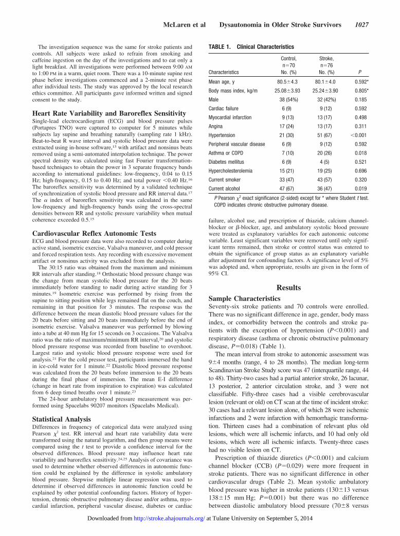
ResultsSample CharacteristicsSeventy-six stroke patients and 70 controls were enrolled.There was no significant difference in age, gender, body massindex, or comorbidity between the controls and stroke pa-tients with the exception of hypertension (P�0.001) andrespiratory disease (asthma or chronic obstructive pulmonarydisease, P�0.018) (Table 1).
The mean interval from stroke to autonomic assessment was9�4 months (range, 4 to 28 months). The median long-termScandinavian Stroke Study score was 47 (interquartile range, 44to 48). Thirty-two cases had a partial anterior stroke, 26 lacunar,13 posterior, 2 anterior circulation stroke, and 3 were notclassifiable. Fifty-three cases had a visible cerebrovascularlesion (relevant or old) on CT scan at the time of incident stroke:30 cases had a relevant lesion alone, of which 28 were ischemicinfarctions and 2 were infarction with hemorrhagic transforma-tion. Thirteen cases had a combination of relevant plus oldlesions, which were all ischemic infarcts, and 10 had only oldlesions, which were all ischemic infarcts. Twenty-three caseshad no visible lesion on CT.
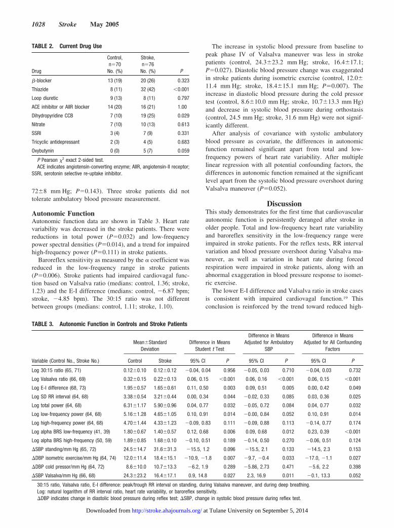
Prescription of thiazide diuretics (P�0.001) and calciumchannel blocker (CCB) (P�0.029) were more frequent instroke patients. There was no significant difference in othercardiovascular drugs (Table 2). Mean systolic ambulatoryblood pressure was higher in stroke patients (130�13 versus138�15 mm Hg; P�0.001) but there was no differencebetween diastolic ambulatory blood pressure (70�8 versus
TABLE 1. Clinical Characteristics
Characteristics
Control,n�70
No. (%)
Stroke,n�76
No. (%) P
Mean age, y 80.5�4.3 80.1�4.0 0.592*
Body mass index, kg/m 25.08�3.93 25.24�3.90 0.805*
Male 38 (54%) 32 (42%) 0.185
Cardiac failure 6 (9) 9 (12) 0.592
Myocardial infarction 9 (13) 13 (17) 0.498
Angina 17 (24) 13 (17) 0.311
Hypertension 21 (30) 51 (67) �0.001
Peripheral vascular disease 6 (9) 9 (12) 0.592
Asthma or COPD 7 (10) 20 (26) 0.018
Diabetes mellitus 6 (9) 4 (5) 0.521
Hypercholesterolemia 15 (21) 19 (25) 0.696
Current smoker 33 (47) 43 (57) 0.320
Current alcohol 47 (67) 36 (47) 0.019
P Pearson �2 exact significance (2-sided) except for * where Student t test.COPD indicates chronic obstructive pulmonary disease.
McLaren et al Dysautonomia in Older Stroke Survivors 1027
at Tulane University on September 5, 2014http://stroke.ahajournals.org/Downloaded from
72�8 mm Hg; P�0.143). Three stroke patients did nottolerate ambulatory blood pressure measurement.
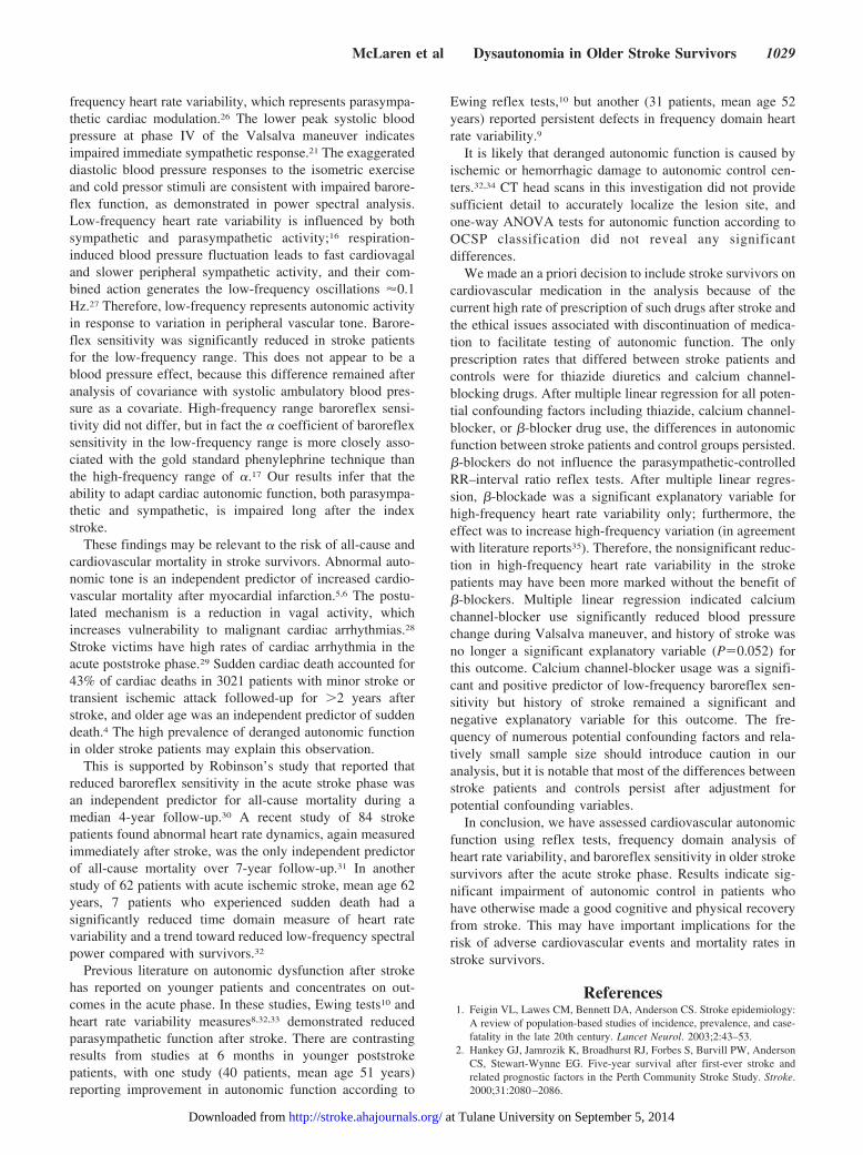
Autonomic FunctionAutonomic function data are shown in Table 3. Heart ratevariability was decreased in the stroke patients. There werereductions in total power (P�0.032) and low-frequencypower spectral densities (P�0.014), and a trend for impairedhigh-frequency power (P�0.111) in stroke patients.
Baroreflex sensitivity as measured by the � coefficient wasreduced in the low-frequency range in stroke patients(P�0.006). Stroke patients had impaired cardiovagal func-tion based on Valsalva ratio (medians: control, 1.36; stroke,1.23) and the E-I difference (medians: control, �6.87 bpm;stroke, �4.85 bpm). The 30:15 ratio was not differentbetween groups (medians: control, 1.11; stroke, 1.10).
The increase in systolic blood pressure from baseline topeak phase IV of Valsalva maneuver was less in strokepatients (control, 24.3�23.2 mm Hg; stroke, 16.4�17.1;P�0.027). Diastolic blood pressure change was exaggeratedin stroke patients during isometric exercise (control, 12.0�11.4 mm Hg; stroke, 18.4�15.1 mm Hg; P�0.007). Theincrease in diastolic blood pressure during the cold pressortest (control, 8.6�10.0 mm Hg; stroke, 10.7�13.3 mm Hg)and decrease in systolic blood pressure during orthostasis(control, 24.5 mm Hg; stroke, 31.6 mm Hg) were not signif-icantly different.
After analysis of covariance with systolic ambulatoryblood pressure as covariate, the differences in autonomicfunction remained significant apart from total and low-frequency powers of heart rate variability. After multiplelinear regression with all potential confounding factors, thedifferences in autonomic function remained at the significantlevel apart from the systolic blood pressure overshoot duringValsalva maneuver (P�0.052).
DiscussionThis study demonstrates for the first time that cardiovascularautonomic function is persistently deranged after stroke inolder people. Total and low-frequency heart rate variabilityand baroreflex sensitivity in the low-frequency range wereimpaired in stroke patients. For the reflex tests, RR intervalvariation and blood pressure overshoot during Valsalva ma-neuver, as well as variation in heart rate during forcedrespiration were impaired in stroke patients, along with anabnormal exaggeration in blood pressure response to isomet-ric exercise.
The lower E-I difference and Valsalva ratio in stroke casesis consistent with impaired cardiovagal function.19 Thisconclusion is reinforced by the trend toward reduced high-
TABLE 2. Current Drug Use
Drug
Control,n�70
No. (%)
Stroke,n�76
No. (%) P
�-blocker 13 (19) 20 (26) 0.323
Thiazide 8 (11) 32 (42) �0.001
Loop diuretic 9 (13) 8 (11) 0.797
ACE inhibitor or AIIR blocker 14 (20) 16 (21) 1.00
Dihydropyridine CCB 7 (10) 19 (25) 0.029
Nitrate 7 (10) 10 (13) 0.613
SSRI 3 (4) 7 (9) 0.331
Tricyclic antidepressant 2 (3) 4 (5) 0.683
Oxybutynin 0 (0) 5 (7) 0.059
P Pearson �2 exact 2-sided test.ACE indicates angiotensin-converting enzyme; AIIR, angiotensin-II receptor;
SSRI, serotonin selective re-uptake inhibitor.
TABLE 3. Autonomic Function in Controls and Stroke Patients
Variable (Control No., Stroke No.)
Mean�StandardDeviation
Difference in MeansStudent t Test
Difference in MeansAdjusted for Ambulatory
SBP
Difference in MeansAdjusted for All Confounding
Factors
Control Stroke 95% CI P 95% CI P 95% CI P
Log 30:15 ratio (65, 71) 0.12�0.10 0.12�0.12 �0.04, 0.04 0.956 �0.05, 0.03 0.710 �0.04, 0.03 0.732
Log Valsalva ratio (66, 69) 0.32�0.15 0.22�0.13 0.06, 0.15 �0.001 0.06, 0.16 �0.001 0.06, 0.15 �0.001
Log E-I difference (68, 73) 1.95�0.57 1.65�0.61 0.11, 0.50 0.003 0.09, 0.51 0.005 0.00, 0.42 0.049
Log SD RR interval (64, 68) 3.38�0.54 3.21�0.44 0.00, 0.34 0.044 �0.02, 0.33 0.085 0.03, 0.36 0.025
Log total power (64, 68) 6.31�1.17 5.90�0.96 0.04, 0.77 0.032 �0.05, 0.72 0.084 0.04, 0.77 0.032
Log low-frequency power (64, 68) 5.16�1.28 4.65�1.05 0.10, 0.91 0.014 �0.00, 0.84 0.052 0.10, 0.91 0.014
Log high-frequency power (64, 68) 4.70�1.44 4.33�1.23 �0.09, 0.83 0.111 �0.09, 0.88 0.113 �0.14, 0.77 0.174
Log alpha BRS low-frequency (41, 39) 1.80�0.67 1.40�0.57 0.12, 0.68 0.006 0.09, 0.68 0.012 0.23, 0.39 �0.001
Log alpha BRS high-frequency (50, 59) 1.89�0.85 1.68�0.10 �0.10, 0.51 0.189 �0.14, 0.50 0.270 �0.06, 0.51 0.124
�SBP standing/mm Hg (65, 72) 24.5�14.7 31.6�31.3 �15.5, 1.2 0.096 �15.5, 2.1 0.133 �14.5, 2.3 0.153
�DBP isometric exercise/mm Hg (64, 74) 12.0�11.4 18.4�15.1 �10.9, �1.8 0.007 �9.7, �0.4 0.033 �17.0, �1.1 0.027
�DBP cold pressor/mm Hg (64, 72) 8.6�10.0 10.7�13.3 �6.2, 1.9 0.289 �5.86, 2.73 0.471 �5.6, 2.2 0.398
�SBP Valsalva/mm Hg (66, 68) 24.3�23.2 16.4�17.1 0.9, 14.8 0.027 2.3, 16.9 0.011 �0.1, 13.3 0.052
30:15 ratio, Valsalva ratio, E-I difference: peak/trough RR interval on standing, during Valsalva maneuver, and during deep breathing.Log: natural logarithm of RR interval ratio, heart rate variability, or baroreflex sensitivity.�DBP indicates change in diastolic blood pressure during reflex test; �SBP, change in systolic blood pressure during reflex test.
1028 Stroke May 2005
at Tulane University on September 5, 2014http://stroke.ahajournals.org/Downloaded from
frequency heart rate variability, which represents parasympa-thetic cardiac modulation.26 The lower peak systolic bloodpressure at phase IV of the Valsalva maneuver indicatesimpaired immediate sympathetic response.21 The exaggerateddiastolic blood pressure responses to the isometric exerciseand cold pressor stimuli are consistent with impaired barore-flex function, as demonstrated in power spectral analysis.Low-frequency heart rate variability is influenced by bothsympathetic and parasympathetic activity;16 respiration-induced blood pressure fluctuation leads to fast cardiovagaland slower peripheral sympathetic activity, and their com-bined action generates the low-frequency oscillations �0.1Hz.27 Therefore, low-frequency represents autonomic activityin response to variation in peripheral vascular tone. Barore-flex sensitivity was significantly reduced in stroke patientsfor the low-frequency range. This does not appear to be ablood pressure effect, because this difference remained afteranalysis of covariance with systolic ambulatory blood pres-sure as a covariate. High-frequency range baroreflex sensi-tivity did not differ, but in fact the � coefficient of baroreflexsensitivity in the low-frequency range is more closely asso-ciated with the gold standard phenylephrine technique thanthe high-frequency range of �.17 Our results infer that theability to adapt cardiac autonomic function, both parasympa-thetic and sympathetic, is impaired long after the indexstroke.
These findings may be relevant to the risk of all-cause andcardiovascular mortality in stroke survivors. Abnormal auto-nomic tone is an independent predictor of increased cardio-vascular mortality after myocardial infarction.5,6 The postu-lated mechanism is a reduction in vagal activity, whichincreases vulnerability to malignant cardiac arrhythmias.28
Stroke victims have high rates of cardiac arrhythmia in theacute poststroke phase.29 Sudden cardiac death accounted for43% of cardiac deaths in 3021 patients with minor stroke ortransient ischemic attack followed-up for �2 years afterstroke, and older age was an independent predictor of suddendeath.4 The high prevalence of deranged autonomic functionin older stroke patients may explain this observation.
This is supported by Robinson’s study that reported thatreduced baroreflex sensitivity in the acute stroke phase wasan independent predictor for all-cause mortality during amedian 4-year follow-up.30 A recent study of 84 strokepatients found abnormal heart rate dynamics, again measuredimmediately after stroke, was the only independent predictorof all-cause mortality over 7-year follow-up.31 In anotherstudy of 62 patients with acute ischemic stroke, mean age 62years, 7 patients who experienced sudden death had asignificantly reduced time domain measure of heart ratevariability and a trend toward reduced low-frequency spectralpower compared with survivors.32
Previous literature on autonomic dysfunction after strokehas reported on younger patients and concentrates on out-comes in the acute phase. In these studies, Ewing tests10 andheart rate variability measures8,32,33 demonstrated reducedparasympathetic function after stroke. There are contrastingresults from studies at 6 months in younger poststrokepatients, with one study (40 patients, mean age 51 years)reporting improvement in autonomic function according to
Ewing reflex tests,10 but another (31 patients, mean age 52years) reported persistent defects in frequency domain heartrate variability.9
It is likely that deranged autonomic function is caused byischemic or hemorrhagic damage to autonomic control cen-ters.32,34 CT head scans in this investigation did not providesufficient detail to accurately localize the lesion site, andone-way ANOVA tests for autonomic function according toOCSP classification did not reveal any significantdifferences.
We made an a priori decision to include stroke survivors oncardiovascular medication in the analysis because of thecurrent high rate of prescription of such drugs after stroke andthe ethical issues associated with discontinuation of medica-tion to facilitate testing of autonomic function. The onlyprescription rates that differed between stroke patients andcontrols were for thiazide diuretics and calcium channel-blocking drugs. After multiple linear regression for all poten-tial confounding factors including thiazide, calcium channel-blocker, or �-blocker drug use, the differences in autonomicfunction between stroke patients and control groups persisted.�-blockers do not influence the parasympathetic-controlledRR–interval ratio reflex tests. After multiple linear regres-sion, �-blockade was a significant explanatory variable forhigh-frequency heart rate variability only; furthermore, theeffect was to increase high-frequency variation (in agreementwith literature reports35). Therefore, the nonsignificant reduc-tion in high-frequency heart rate variability in the strokepatients may have been more marked without the benefit of�-blockers. Multiple linear regression indicated calciumchannel-blocker use significantly reduced blood pressurechange during Valsalva maneuver, and history of stroke wasno longer a significant explanatory variable (P�0.052) forthis outcome. Calcium channel-blocker usage was a signifi-cant and positive predictor of low-frequency baroreflex sen-sitivity but history of stroke remained a significant andnegative explanatory variable for this outcome. The fre-quency of numerous potential confounding factors and rela-tively small sample size should introduce caution in ouranalysis, but it is notable that most of the differences betweenstroke patients and controls persist after adjustment forpotential confounding variables.
In conclusion, we have assessed cardiovascular autonomicfunction using reflex tests, frequency domain analysis ofheart rate variability, and baroreflex sensitivity in older strokesurvivors after the acute stroke phase. Results indicate sig-nificant impairment of autonomic control in patients whohave otherwise made a good cognitive and physical recoveryfrom stroke. This may have important implications for therisk of adverse cardiovascular events and mortality rates instroke survivors.
References1. Feigin VL, Lawes CM, Bennett DA, Anderson CS. Stroke epidemiology:
A review of population-based studies of incidence, prevalence, and case-fatality in the late 20th century. Lancet Neurol. 2003;2:43–53.
2. Hankey GJ, Jamrozik K, Broadhurst RJ, Forbes S, Burvill PW, AndersonCS, Stewart-Wynne EG. Five-year survival after first-ever stroke andrelated prognostic factors in the Perth Community Stroke Study. Stroke.2000;31:2080–2086.
McLaren et al Dysautonomia in Older Stroke Survivors 1029
at Tulane University on September 5, 2014http://stroke.ahajournals.org/Downloaded from
3. Thrift AG, Dewey HM, Macdonell RA, McNeil JJ, Donnan GA.Incidence of the major stroke subtypes: Initial findings from the NorthEast Melbourne stroke incidence study (NEMESIS). Stroke. 2001;32:1732–1738.
4. Pop GA, Koudstaal PJ, Meeder HJ, Algra A, van Latum JC, van Gijn J.Predictive value of clinical history and electrocardiogram in patients withtransient ischemic attack or minor ischemic stroke for subsequent cardiacand cerebral ischemic events. The Dutch TIA trial study group. Archivesof Neurology. 1994;51:333–341.
5. La Rovere MT, Bigger JT, Jr., Marcus FI, Mortara A, Schwartz PJ.Baroreflex sensitivity and heart-rate variability in prediction of totalcardiac mortality after myocardial infarction. ATRAMI (Autonomic Toneand Reflexes After Myocardial Infarction) Investigators. Lancet. 1998;351:478–484.
6. Kleiger RE, Miller JP, Bigger JT, Jr., Moss AJ. Decreased heart ratevariability and its association with increased mortality after acute myo-cardial infarction. Am J Cardiol. 1987;59:256–262.
7. Lombardi F, Makikallio TH, Myerburg RJ, Huikuri HV. Sudden cardiacdeath: Role of heart rate variability to identify patients at risk. CardiovascRes. 2001;50:210–217.
8. Barron SA, Rogovski Z, Hemli J. Autonomic consequences of cerebralhemisphere infarction. Stroke. 1994;25:113–116.
9. Korpelainen JT, Sotaniemi KA, Huikuri HV, Myllya VV. Abnormal heartrate variability as a manifestation of autonomic dysfunction in hemi-spheric brain infarction. Stroke. 1996;27:2059–2063.
10. Korpelainen JT, Sotaniemi KA, Suominen K, Tolonen U, Myllyla VV.Cardiovascular autonomic reflexes in brain infarction. Stroke. 1994;25:787–792.
11. American Psychiatric Association. Diagnostic and Statistical Manual ofMental Disorders, Fourth Edition. Washington DC: American PsychiatricAssociation; 1994.
12. Hatano S. Experience from a multicentre stroke register: a preliminaryreport. Bull World Health Organ. 1976;54:541–553.
13. Bamford J, Sandercock P, Dennis M, Burn J, Warlow C. Classificationand natural history of clinically identifiable subtypes of cerebralinfarction. Lancet. 1991;337:1521–1526.
14. Scandinavian Stroke Study Group. Multicenter trial of hemodilution inischemic stroke—background and study protocol. Scandinavian StrokeStudy Group. Stroke. 1985;16:885–890.
15. Clayton RH, Bowman AJ, Ford GA, Murray A. Measurement ofbaroreflex gain from heart rate and blood pressure spectra: a comparisonof spectral estimation techniques. Physiol Measurement. 1995;16:131–139.
16. Malik M, Bigger J, Camm A, Kleiger R, Malliani A. Heart rate vari-ability: Standards of measurement, physiological interpretation andclinical use. Task force of the European Society of Cardiology and theNorth Am Society of Pacing and Electrophysiology. Circulation. 1996;93:1043–1065.
17. Lord SW, Clayton RH, Hall MC, Gray JC, Murray A, McComb JM,Kenny RA. Reproducibility of three different methods of measuringbaroreflex sensitivity in normal subjects. Clin Sci (Lond). 1998;95:575–581.
18. Wieling W, van Brederode JF, de Rijk LG, Borst C, Dunning AJ. Reflexcontrol of heart rate in normal subjects in relation to age: a data base forcardiac vagal neuropathy. Diabetologia. 1982;22:163–166.
19. American Academy of Neurology Assessment. Clinical autonomic testingreport of the therapeutics and technology assessment subcommittee of theAmerican Academy of Neurology. Neurology. 1996;46:873–880.
20. Levin AB. A simple test of cardiac function based upon the heart ratechanges induced by the Valsalva maneuver. Am J Cardiol. 1966;18:90–99.
21. Sandroni P, Benarroch EE, Low PA. Pharmacological dissection of com-ponents of the Valsalva maneuver in adrenergic failure. J Appl Physiol.1991;71:1563–1567.
22. Fagius J, Karhuvaara S, Sundlof G. The cold pressor test: Effects onsympathetic nerve activity in human muscle and skin nerve fascicles. ActaPhysiol Scand. 1989;137:325–334.
23. Ewing DJ, Clarke BF. Diagnosis and management of diabetic autonomicneuropathy. BMJ (Clin Res Ed). 1982;285:916–918.
24. Harrington F, Murray A, Ford GA. Relationship of baroreflex sensitivityand blood pressure in an older population. J Hypertens. 2000;18:1629–1633.
25. Ylitalo A, Airaksinen KE, Tahvanainen KU, Kuusela TA, Ikaheimo MJ,Rantala A, Lilja M, Huikuri HV. Baroreflex sensitivity in drug-treatedsystemic hypertension. Am J Cardiol. 1997;80:1369–1372.
26. Pomeranz B, Macaulay RJ, Caudill MA, Kutz I, Adam D, Gordon D,Kilborn KM, Barger AC, Shannon DC, Cohen RJ. Assessment ofautonomic function in humans by heart rate spectral analysis. Am JPhysiol. 1985;248:H151–H153.
27. Bernardi L, Leuzzi S, Radaelli A, Passino C, Johnston JA, Sleight P.Low-frequency spontaneous fluctuations of r-r interval and bloodpressure in conscious humans: a baroreceptor or central phenomenon?Clin Sci (Lond). 1994;87:649–654.
28. Fei L, Statters DJ, Hnatkova K, Poloniecki J, Malik M, CammAJ. Change of autonomic influence on the heart immediately before theonset of spontaneous idiopathic ventricular tachycardia. J Am CollCardiol. 1994;24:1515–1522.
29. Mikolich JR, Jacobs WC, Fletcher GF. Cardiac arrhythmias in patientswith acute cerebrovascular accidents. JAMA. 1981;246:1314–1317.
30. Robinson TG, Dawson SL, Eames PJ, Panerai RB, Potter JF. Cardiacbaroreceptor sensitivity predicts long-term outcome after acute ischemicstroke. Stroke. 2003;34:705–712.
31. Makikallio AM, Makikallio TH, Korpelainen JT, Sotaniemi KA, HuikuriHV, Myllyla VV. Heart rate dynamics predict poststroke mortality. Neu-rology. 2004;62:1822–1826.
32. Tokgozoglu SL, Batur MK, Top uoglu MA, Saribas O, Kes S, Oto A.Effects of stroke localization on cardiac autonomic balance and suddendeath. Stroke. 1999;30:1307–1311.
33. Korpelainen JT, Huikuri HV, Sotaniemi KA, Myllyla VV. Abnormalheart rate variability reflecting autonomic dysfunction in brainsteminfarction. Acta Neurologica Scand. 1996;94:337–342.
34. Sander D, Klingelhofer J. Extent of autonomic activation followingcerebral ischemia is different in hypertensive and normotensive humans.Arch Neurol. 1996;53:890–894.
35. Lampert R, Ickovics JR, Viscoli CJ, Horwitz RI, Lee FA. Effects ofpropranolol on recovery of heart rate variability following acute myo-cardial infarction and relation to outcome in the beta-blocker heart attacktrial. Am J Cardiol. 2003;91:137–142.
1030 Stroke May 2005
at Tulane University on September 5, 2014http://stroke.ahajournals.org/Downloaded from