Embed Size (px)
Citation preview

Autologous Serum Eye Drops:
Fact or Fiction?
Grand Rounds March 2005
Jay C. Bradley, MD
Sandra M. Brown, MD

Nutrition of the Ocular Surface
• Aqueous– Glucose– Electrolytes– Amino acids
• Lacrimal gland– Growth factors– Vitamins
• Conjunctival vessels– Neuropeptides– Lactoferrin– Fibronectin and adhesion factors– Immunoglobulins

Nutrition-Related Problems
• Decreased epithelium trophic factors and their carrier molecules
• Non-healing epithelial defects• Surgical attempts to rehabilitate the ocular
surface in severely dry eyes fail frequently• Ideal tear substitute should provide
epithelio-trophic support in addition to lubrication

Properties of Tears
• Antimicrobial
• Nourishing
• Optical
• MechanicalPharmaceutical tear replacements
What about these 2 properties?

Concept of NaturalTear Substitutes
• Fibronectin, vitamins, and growth factors– In vitro and in vivo studies
• Single compound approach (eg substance P)– Problems with stability, expense– Limited clinical success
• Various bodily fluids including…
• AUTOLOGOUS SERUM
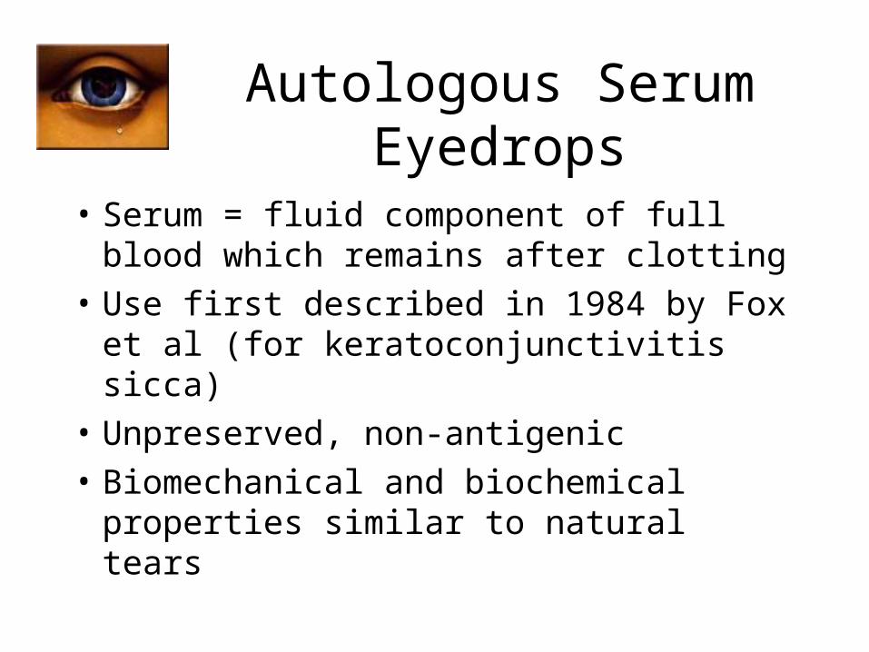
Autologous Serum Eyedrops
• Serum = fluid component of full blood which remains after clotting
• Use first described in 1984 by Fox et al (for keratoconjunctivitis sicca)
• Unpreserved, non-antigenic
• Biomechanical and biochemical properties similar to natural tears

Unstimulated Tears versus Serum
• pH = 7.4
• Osmolality = 298
• EGF (ng/ml) = 0.2 – 3.0
• TGF-b (ng/ml) = 2 – 10
• Vitamin A (mg/ml) = 0.02
• Lysozyme (mg/ml) = 1.4
• SIgA (ug/ml) = 1190
• Fibronectin (ug/ml) = 21
• pH = 7.4• Osmolality = 296• EGF (ng/ml) = 0.5• TGF-b (ng/ml) = 6 – 33• Vitamin A (mg/ml) = 46• Lysozyme (mg/ml) = 6• SIgA (ug/ml) = 2• Fibronectin (ug/ml) = 205• Hepatocyte GF, NGF, IGF-1,
Substance P, Complement, Fibroblast GF, cGRP, other Ig, etc.

AS – In Vitro Actions
• Contains epithelio-trophic / modulating factors
• Promotes growth and migration of ocular surface epithelial cells in vitro– Dose-dependent effect on SV40 transfected
human corneal epithelial cell line (Tsubota)– Expression of mucin-1 from immortalized
conjunctival epithelial cells (Tsubota)

AS – In Vitro Actions
• Maintains corneal epithelial cell morphology and function better than pharmaceutical tear substitutes (Geerling et al)
• Increases transcription of RNA for nerve growth factor and transforming growth factor-beta in cultured human keratocytes (Ebner et al)

Autologous Serum Eyedrops
• Renaissance after report of successful use in eyes with persistent epithelial defects (Tsubota et al 1999)– True epithelio-trophic potential for the ocular surface?
• Attempted treatment: keratoconjunctivitis sicca due to– Sjogren syndrome– Graft-vs-host disease– Neurotrophic keratitis– Superior limbic keratoconjunctivitis– Rheumatoid arthritis
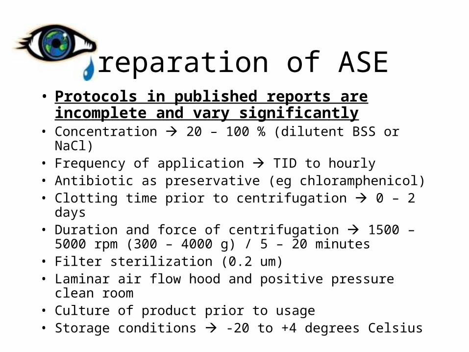
Preparation of ASE• Protocols in published reports are incomplete
and vary significantly• Concentration 20 – 100 % (dilutent BSS or NaCl)• Frequency of application TID to hourly• Antibiotic as preservative (eg chloramphenicol)• Clotting time prior to centrifugation 0 – 2 days• Duration and force of centrifugation 1500 – 5000 rpm
(300 – 4000 g) / 5 – 20 minutes• Filter sterilization (0.2 um)• Laminar air flow hood and positive pressure clean room• Culture of product prior to usage• Storage conditions -20 to +4 degrees Celsius

Preparation of ASE
• Standard operative procedure (SOP)– University of Lubeck, Germany (Geerling er al)– National Blood Service in England and Wales– Japanese group (Tsubota et al)
• No FDA-approved standardized protocol in United States

Clinical Results

Severe Dry Eye
• 7 Major Reports• N = 135 eyes• 20 – 100 % ASE at 4 X/day to hourly frequency• Overall success
– RB staining 33 – 68 %– F staining 39 – 61 %– Subjective improvement 30 – 100 %– Impression cytology 44 %
• Symptoms recurred upon ASE discontinuation and crossover to conventional therapy

SLK
• Prospective cohort study with 11 patients (Goto et al)– Used 20 % ASE as additional therapy 10 times daily
– Within 4 weeks discomfort improved in 9 of 11 and epitheliopathy improved in all patients
– Significantly increased TBUT and decreased conjunctival squamous metaplasia
– Discomfort recurred with discontinuation of ASE

Recurrent Erosion Syndrome
• Prospective cohort study of 11 patients with unilateral post-traumatic RES (Del Castillo et al)– Used NPAT and ASE 20 % tid for 3 months in tapered
fashion– Mean recurrence rate was reduced from 2.2 to
0.028/month of F/U (mean F/U 9.4 months)– Given self-healing nature of post-traumatic RES, the
fact the duration since trauma was not specified, and the failure to state if other previously used modalities were suspended during ASE use, these data have to be reviewed with care
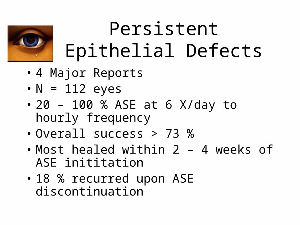
Persistent Epithelial Defects
• 4 Major Reports• N = 112 eyes• 20 – 100 % ASE at 6 X/day to hourly
frequency• Overall success > 73 %• Most healed within 2 – 4 weeks of ASE
inititation• 18 % recurred upon ASE discontinuation

Adjunctive Treatment in Ocular Surface Reconstruction
• 14 eyes of 10 patients receiving limbal stem cell transplant, amniotic membrane , and/or PK were treated with 20 % ASE (Tsubota et al)– OCP/SJS with Schirmer = 0– 12 of 14 had stable epithelium at 20 weeks
• 2 patients undergoing PK for PED (Poon et al)– Achieved stable epithelium with ASE– Epitheliopathy recurred upon ASE discontinuation
• Young patients with severe ocular surface disease and absolute dry eye (SJS) surface reconstruction failed despite ASE use (Tsubota et al)

Criticisms• Variations in the study populations such as degree of
aqueous deficiency
• Variations in production and treatment protocol for ASE
• Additive rather than substitutive therapy
• Therapeutic CTL or punctal occlusion
• Increasing fluid supply rather than the epithelio-trophic nature of ASE may have yielded the beneficial effect
• Comparison of published data is further limited by variations in reporting “success of treatment” as
– Number of patients improving
– Mean change in parameter
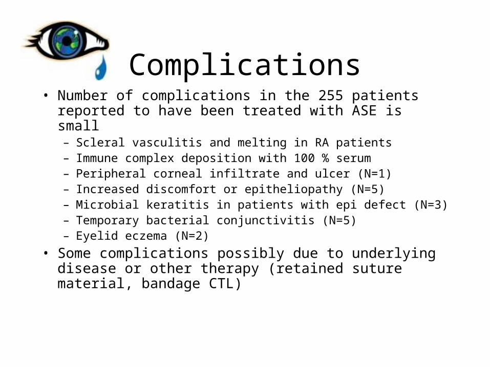
Complications• Number of complications in the 255 patients reported to
have been treated with ASE is small– Scleral vasculitis and melting in RA patients– Immune complex deposition with 100 % serum– Peripheral corneal infiltrate and ulcer (N=1)– Increased discomfort or epitheliopathy (N=5)– Microbial keratitis in patients with epi defect (N=3)– Temporary bacterial conjunctivitis (N=5)– Eyelid eczema (N=2)
• Some complications possibly due to underlying disease or other therapy (retained suture material, bandage CTL)

“Effect of Autologous Serum Eyedrops in the Treatment of Severe Dry Eye”
• Kojima et al. Am J Ophthalmol 2005;139:242-6.
• Prospective randomized case-control trial
• 37 eyes of 20 severe dry patients without punctal occlusion
• After 2 week wash-out, randomly assigned to two groups– A – only preservative-free artificial tears
– S – only autologous 20 % serum eyedrops 6 times a day
• Improved mean TBUT, F/RB staining scores, and subjective symptom scores improved

ASE Use in OSD: Hot Topic in Ophthalmology
• Over 40 PubMed articles in last 5 years• Subject of debate at scientific meetings and other
non-PubMed indexed journals• Almost all large-cohort ASE research occurring
outside of United States due to lack of FDA-approved standardized laboratory production protocol
• Primarily compassionate use exemptions for last resort additive therapy

Further Study Needed:• Development of FDA-approved manufacturing protocol
– Stability– Sterility– Storage
• Clinical trials– Tight inclusion and exclusion criteria to achieve homogeneous patient
populations– Dose-response investigation– Cross-over studies
• Expansion of indications• Eventually –
– Fractionation of serum components to determine active portion– True chemical substitution
*

Which is in dry eye patients’ future?