Embed Size (px)
Citation preview
1
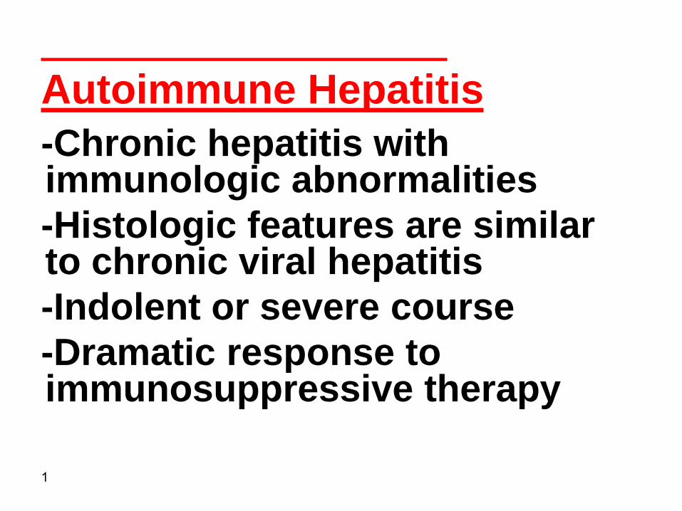
Autoimmune Hepatitis
-Chronic hepatitis with immunologic abnormalities
-Histologic features are similar to chronic viral hepatitis
-Indolent or severe course
-Dramatic response to immunosuppressive therapy

2
Features:
1-Female predominance (70%)
2-Negative serelogy for viral Ags.
3-↑serum Ig (>2.5 g/dl)
4-High titers of autoantibodies (80% of cases)
5-The presence of other autoimmune diseases as RA, thyroiditis, sjogern syndrome, UC in 60% of the cases

3
The type of autoantibodies 1-Antismooth muscle Abs
anti actin
anti troponin
anti tropomyosin
2-liver/kidney microsomal Abs
anti cytochrome P-450 components
anti UDP-glucuronosyl
transferases
3-Anti – soluble liver / pancreas antigen
4
Outcome
Mild to severe chronic hepatitis
Full remission is unusual
Risk of cirrhosis is 5% which is the main cause of death
5
Nonalcoholic Fatty Liver Disease
Types:
1.Steatosis ( Fatty liver)
2.Steatohepatitis
hepatocyte destruction
parenchymal inflammation
progressive pericellular fibrosis

6
Predisposing factors :
1-Type 2 DM
2-Obesity : body mass index
> 30 kg /m2 in caucasians
> 25 kg /m2 in Asians
3-Dyslipidemia ( ↑ TG, ↑LDL, ↓HDL)
7
Pathogenesis
.Metabolic syndrome
. Insulin resistance
. Obesity
. Dyslipidemia
8
Mechanism of fatty accumulation
1.Impaired oxidation of fatty acids
2.Increased synthesis & uptake of FFA
3.Decreased hepatic sec. of VLDL
. ↑TNF , IL6 , chemokine →liver inflammation & damage

9
Clinically
-NAFLD is the most common cause of
incidental ↑ in transaminases
-Most pts. are asymptomatic
-Non-specific symptoms
Fatigue, malaise, RUQ discomfort
-Severe symptoms
-Liver Bx is required for dx.
-NAFLD m.b a significant contributer to
cryptogenic cirrhosis

10
Hemochsomatosis
• Excessive accumalation of body iron (liver & pancreas)
• 1ry or 2ry (genetic or acquired )
• Genetic hemochromatosis ( 4 variants)
• The most common form is aut.recessive disease of adult onset caused by mutation in the HFE gene on chr.6

11
Causes of acquired hemosidrosis :
1-multiple transfusions
2-ineffective erythropoiesis (β-thalassemia )
3-increased iron intake (Bantu sidrosis )
4-chronic liver disease

12
Clinical Features:
1-Micronodular cirrhosis (all patients)
2-D.M ( 75 – 80%)
3-skin prigmentation 75-80%)
4-cardiomegaly (arrhythmias, cardiomyopathy)
5- joints disease
6- testicular atrophy
13
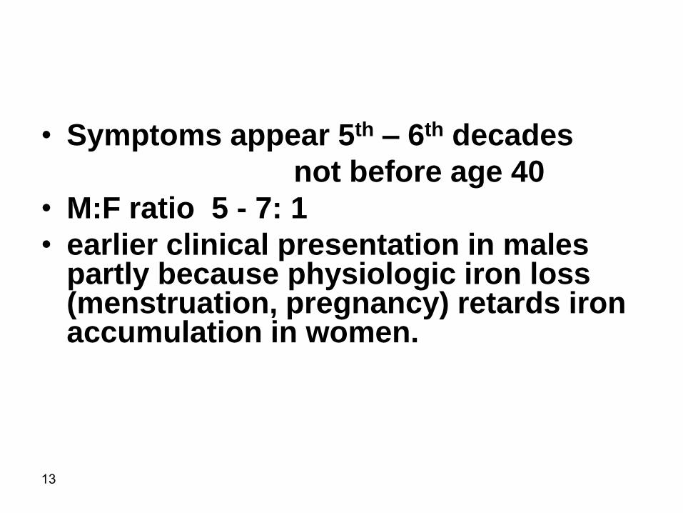
• Symptoms appear 5th – 6th decades
not before age 40
• M:F ratio 5 - 7: 1
• earlier clinical presentation in males partly because physiologic iron loss (menstruation, pregnancy) retards iron accumulation in women.
14
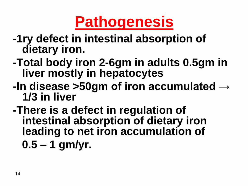
Pathogenesis -1ry defect in intestinal absorption of
dietary iron.
-Total body iron 2-6gm in adults 0.5gm in liver mostly in hepatocytes
-In disease >50gm of iron accumulated → 1/3 in liver
-There is a defect in regulation of intestinal absorption of dietary iron leading to net iron accumulation of
0.5 – 1 gm/yr.

15
• HFE gene regulates the level of hepcidin hormone synthesized in liver
• Hepicidin normally inhibits iron absorption.
• When hepcidin levels are reduced there is increased iron absorption.
• HFE gene deletion causes→ ↓Hepcidin levels→ iron overload

16
-Two mutations can occur in HFE gene:
1-Mutation at 845 nucleotide → tyrosine substitution for cystine at AA 282
( C282 Y )
2-aspartate substitution for histidine at AA 63 ( H63D)
10% of pts. have other gene mutations

17
-Carrier rate for C282Y is 1/70
-Homozygosity is 1/200
- 80% of pts. are homozygous for (C282Y) mutation & have the highest incidence of iron accumulation
-10% of pts. are either homozygous for H63D mutation or compound heterozygous for C282Y/H63D mutation
18
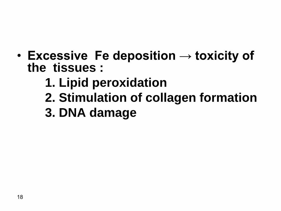
• Excessive Fe deposition → toxicity of the tissues :
1. Lipid peroxidation
2. Stimulation of collagen formation
3. DNA damage
19
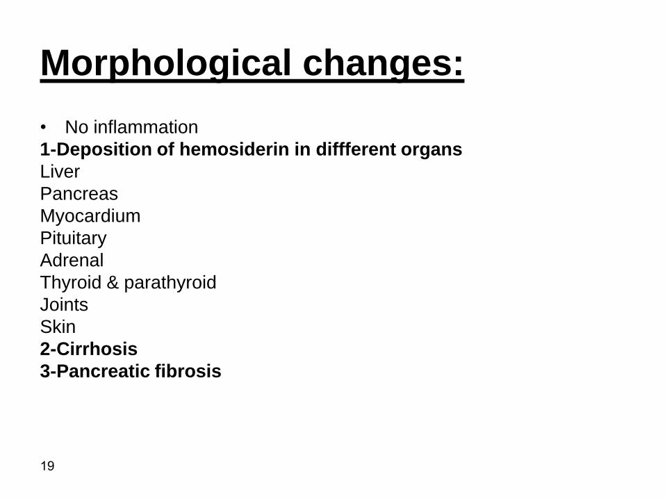
Morphological changes:
• No inflammation
1-Deposition of hemosiderin in diffferent organs
Liver
Pancreas
Myocardium
Pituitary
Adrenal
Thyroid & parathyroid
Joints
Skin
2-Cirrhosis
3-Pancreatic fibrosis

20
Hemosiderosis
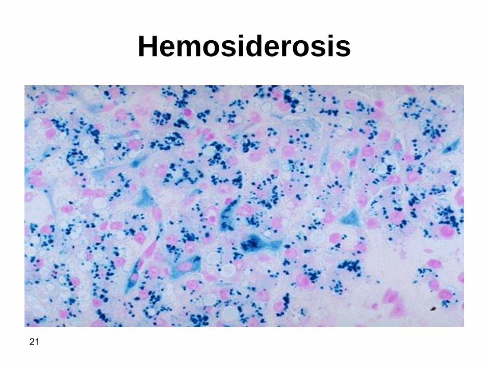
21
Hemosiderosis

22
4-Synovitis
5-Polyarthritis(pseudogout)
6-Pigmentation of liver
7-Fibrosis of pancreas & myocardium
8-Atrophy of testes

23
• Death may result from :
• 1-cirrhosis
• 2-hepatocellular carcinoma
• 3-cardiac disease.
• The risk of hepatocellular carcinoma
development in patients with
hemochromatosis is 200-fold higher than
in normal populations

24
Wilson Disease
-aut. Recessive disorder of Cu metabolism
-mutation in ATP7B gene on chr. 13 which encodes an ATPase metal ion transporter in Golgi region
-> 80 mutations
-Gene freq. 1:200
-Incidence is 1:30000
25
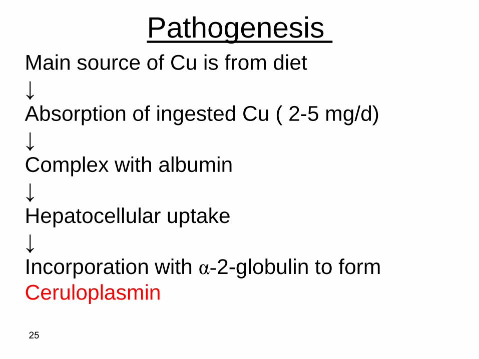
Pathogenesis
Main source of Cu is from diet
↓
Absorption of ingested Cu ( 2-5 mg/d)
↓
Complex with albumin
↓
Hepatocellular uptake
↓
Incorporation with α-2-globulin to form
Ceruloplasmin

26
↓
Sec. into plasma
(90 – 95% of plasma Cu)
↓
Hepatic uptake of ceruloplasmin
↓
Lysosomal degradation
↓
Secretion of free Cu into bile
27
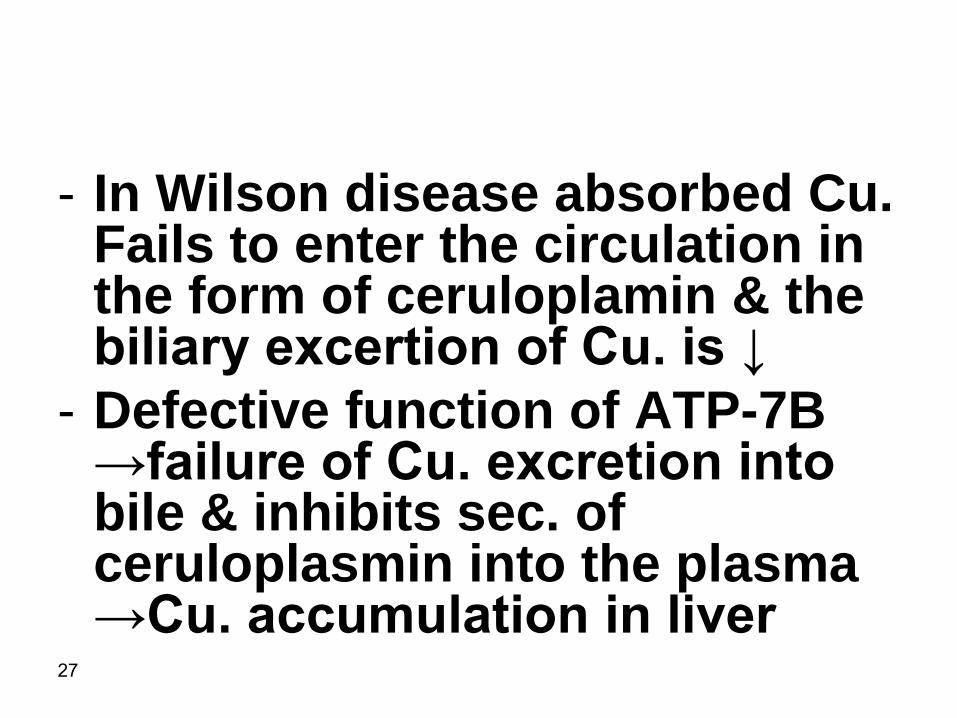
- In Wilson disease absorbed Cu. Fails to enter the circulation in the form of ceruloplamin & the biliary excertion of Cu. is ↓
- Defective function of ATP-7B →failure of Cu. excretion into bile & inhibits sec. of ceruloplasmin into the plasma →Cu. accumulation in liver

28
-↑Cu. Accumulation in the liver reults in:-
1-Production of free radicals
2-binding to sulfhydryl groups of cellular proteins
3-displacement of other metals in hepatic metalloenzymes
29
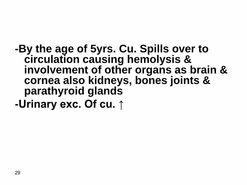
-By the age of 5yrs. Cu. Spills over to circulation causing hemolysis & involvement of other organs as brain & cornea also kidneys, bones joints & parathyroid glands
-Urinary exc. Of cu. ↑
30
Morphology
Liver
1-Fatty change
2-Acute hepatitis
3-chronic hepatitis
4-cirrhosis
5-massive hepatic necrosis
( rhodanine stain or orcein stain )
31
Brain:
Toxic injury to basal ganglia esp. the
putamen causing atrophy & cavitation
32
Eye:
kayser- Fleischer rings
green – brown depositis of Cu. in descemet membrane in the limbus of the cornea
(hepatolenticular degeneration)

33
• Clinically
-Presentation > 6 yrs of age
-Most common presentation is acute on top of chronic hepatitis
-Neuropsychiatric presentation can occur
behavioral changes
Frank psychosis
Parkinson disease- like syndrome

34
• DX
1- ↓ in serum ceruloplasmin level
2- ↑ in urinary exc. Of Cu.
3- ↑ hepatic content of copper
> 250 mg/gm dry wt.

35
α-1-Antitrypsin Defeciency
- Aut. Recessive disorder
- freq. 1:7000 in N. american white population
- α-1-antiryrpsin is a protease inhibtor as elastase, cathepsinG , proteinase 3 which are released from neutrophils at the site of inflammation.
-The gene pi. Is located on chr. 14.
-At least 75 forms of gene mutation are present
-The most common genotype is pi.MM present in 90% of individuals.
36
• PiZZ genotype→↓level of α-1-ntitrypsin
in blood (only 10% of normal) are at
high risk of developing clinical disease

37
Pathogenesis
-The mutant polypeptide (PiZ) is abnormally folded & polymerizes causing its retention in the ER of hepatocytes.
-Athoyugh all individual with Pizz genotype accumulate α-1-AT-Z protein only 10% of them develop clinical liver disease .
-This is due to lag in ER protein degradation pathway.

38
-The accumulated α-1-AT-Z is not toxic but the autophagocytic response stimulated within the hepatocytes appear to be the cause of liver injury by autophagocytosis of the mitochondria.
-8-10% of patients develop significant liver damage.

39
Morphology
• Intracytoplasmic globular inclusions in
hepatocytes which are acidophilic in
H&E sections.
• The inclusions are PAS+ve & diastase
resistant.
• Neonatal hepatitis cholestasis &
fibrosis
40
• Chronic hepatitis
• Cirrhosis
• Fatty change
• Mallory bodies

41
Clinical features
• Neonatal hepatitis with cholestatic
jaundice appears in 10 – 20% of
newborns with the disease .
• Attacks of hepatitis in adolescence
• Chronic hepatitis & cirrhosis
• HCC in 2- 3 % of Pizz adults

42
Antitrypsin Defeciency-1-α
Intracytoplasmic globular inclusions in hepatocytes (PAS stain)

43
Reye’s Syndrome
-Fatty change in liver &
encephalopathy.
-< 4 yr.
-3 – 5 d after viral illness.
-↑liver & abn. LFT.
-Vomiting lethargy.
-25% may go into coma.

44
• Death occurs from progressive neurologic
deterioration or liver failure.
• Survivors of more serious illness may be
left with permanent neurologic
impairments.

45
Pathogenesis
• The pathogenesis of Reye syndrome involves a generalized loss of mitochondrial function.
• Reye syndrome is now recognized as the prototype of a wide variety of conditions known as "mitochondrial hepatopathies."
• Reye syndrome has been associated with aspirin administration during viral illnesses, but there is no evidence that salicylates play a causal role in this disorder.

46
Morphology
• The key pathologic finding in the liver is microvesicular steatosis.
• Electron microscopy of hepatocellular mitochondria reveals pleomorphic enlargement and electron lucency of the matrices with disruption of cristae and loss of dense bodies.
• In the brain, cerebral edema is usually present.
47
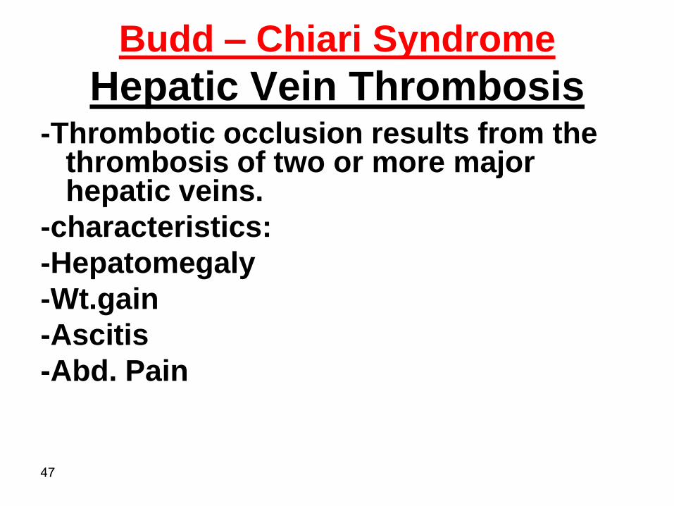
Budd – Chiari Syndrome
Hepatic Vein Thrombosis -Thrombotic occlusion results from the
thrombosis of two or more major hepatic veins.
-characteristics:
-Hepatomegaly
-Wt.gain
-Ascitis
-Abd. Pain

48
Causes:
1-PCV
2-Pregnancy
3-Postpartum
4-Oral contraceptive
5-PNH
7-Mechanical obstruction
8-Tumors as HCC
9-Idiopathic in 30% of the cases
-
49
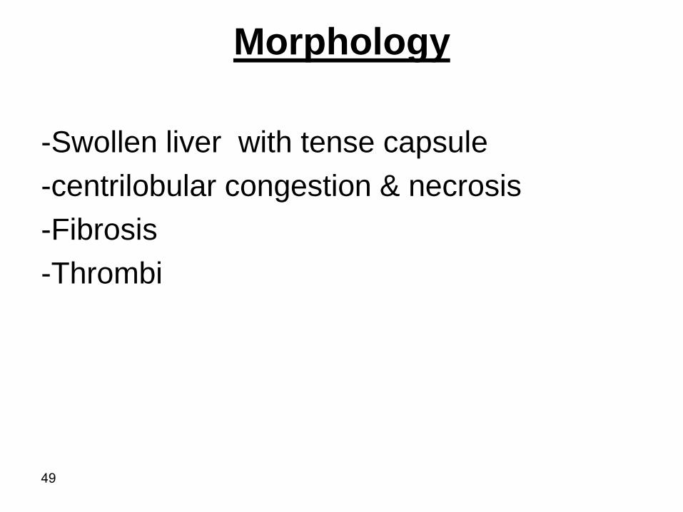
Morphology
-Swollen liver with tense capsule
-centrilobular congestion & necrosis
-Fibrosis
-Thrombi

50
Primary sclerosing cholangitis
-Inflammation , obliterative firosis & segmental dilation of the obstructed
intra hepatic & extra hepatic bile ducts.
-In PSC, UC coexists in 70% of patients.
-In patients of UC, 4% develop PSC.
-3-5th decades
-M: F 2:1

51
- asymptomatic pts.
- persistent ↑ serum alkaline phosphatase
- fatigue, pruritis, jaundice, wt loss, ascitis, bleeding, encephalopathy.
- antimitochondrial Abs < 10% of cases.
• Antinuclear cytoplasmic Abs (ANCA) in 80% of cases.

52
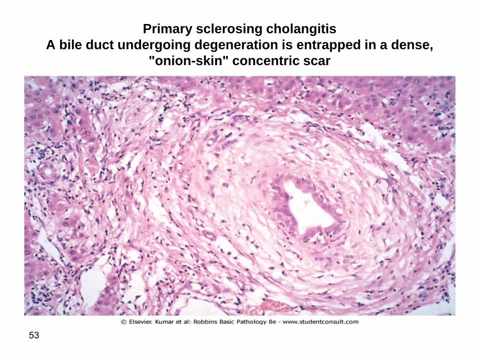
Morphology -Concentric periductal onion-skin fibrosis & lymphocytic infilrate
-Atrophy & obliteration of bile ducts
-Dilation of bile ducts inbetween areas of stricture
-Cholestasis & fibrosis
-Cirrhosis, cholangiocarcinoma ( 10 – 15%)
53
Primary sclerosing cholangitis
A bile duct undergoing degeneration is entrapped in a dense,
"onion-skin" concentric scar
54
Pathogenesis
-Exposure to gut derived toxins
-Immune attack
-Ischemia of biliary tree

55
biliary cirrhosis
• 1-primary
• 2-Secondary
• -Prolonged obst. To extrahepatic biliary tree
• Causes:
• 1-cholelithiasis
• 2-biliary atresia
• 3-malignancies
• 4-stricutres

56
Primary biliary Cirrhosis
-Chronic progressive & often fatal cholestatic liver disease
-Non-suppurative granulomatous destruction of medium-sized intrahepatic bile ducts, portal inflammation & scarring
57
-Age 20-80yrs ( peak 40-50yrs)
-F>M
-Insidious onset
-Pruritis, jaundice
-Cirrhosis over 2 or more decades

58
-↑Alkaline phosphatase & cholesterol
-Hyperbilirubinemia = hepatic decompansation
-Antimitochondrial Abs > 90%
Antimitochondrial pyruvate dehydrogenase
-Associated conditions: Sjogren synd. Scleroderma thyroiditis, RA, Raynauds phenomenon, MGN, celiac disease.

59
• Morphology • interlobular bile ducts are absent or severely destructed (florid
duct lesion)
• intra epithelial inflammation
• Granulomatous inflammation
• Bile ductular proliferation
• Cholestesis
• Necrosis of parenchyma
• Cirrhosis

60
Primary biliary cirrhosis. A portal tract is markedly expanded by an
infiltrate of lymphocytes and plasma cells. Note the granulomatous
reaction to a bile duct undergoing destruction (florid duct lesion(

61
Sinusoidal Obstruction Syndrome
( Veno-occlusive disease)
• Originally described in Jamaican drinkers of bush-tea containing pyrrolizidine alkaloids.
• Obstruction syndrome is caused by toxic injury to sinusoidal endothelium.
• Damaged endothelial cells slough off and create emboli that block blood flow.

62
• Endothelial damage is accompanied by passage of red blood cell into the space of Disse, proliferation of stellate cells, and fibrosis of terminal branches of the hepatic vein
• This occurs in the first 20-30 days after bone marrow transplantation
. Which is caused by:
1-Drugs as cyclophosphamide
2-Total body radiation
63
.Incidence -20% in recepients of allogeneic marrow
transplant
-Clinical presentation
Mild – severe
Death if does not resolve in 3 months
64
Liver tumors
• Most common benign tumor is
cavernous hemagioma
• Usually <2cm
• Subcapsular

65
Liver cell adenoma
• Young female
• Childbearing age who have used oral
contraceptive steroids.
• It may regress on discontinuance of
hormone use.

66
Hepatic adenoma
67
• Liver cell adenomas are significant for three reasons:
• (1) when they present as an intrahepatic mass, they may be mistaken for the more ominous hepatocellular carcinoma
• (2) subcapsular adenomas are at risk for rupture, particularly during pregnancy (under estrogenic stimulation), causing life-threatening intra-abdominal hemorrhage
• (3) although adenomas are not considered precursors of hepatocellular carcinoma, adenomas carrying β-catenin mutations carry a risk of developing into cancers.

68
Liver Nodules
Focal noudular hyperplasia
• Well demarcated hyperplastic hepatocytes with
central scar.
• Non-cirrhotic liver.
• Not neoplasm but nodular regeneration.
• Local vascular injury.
• Females of reproductive age.
• No risk of malignancy.
• 20% of cases have cavernous hemagnioma.

69
Macroregenerative Nodules
• Cirrhotic liver
• Larger than cirrhotic nodules
• No atypical features
• Reticulin is intact
• No malignant potential

70
Dysplastic nodules
• Larger than 1 mm
• Cirrhotic liver
• Atypical features, pleomorphism and crowding
• High proliferative activity
• High or low dysplasia
• Precancerous (monoclonal, +ve gene mutations
• Types:
1. Small – cell dysplastic nodules
2. Large – cell dysplastic nodules
71
Hepatocellular carcinoma
• 5.4% of all cancers
• Incidence:
<5/100000 population in N&S America
N& central Europe
Australia
15/100000 population in Mediterranean
36/100000 population in Korea, Taiwan
mozambique, china

72
• Blacks > white
• M:F ratio
3:1 in low incidence areas. >60yr
8:1 in high incidence areas. 20-40yr
73
Predisposing Factors
1. Hepatitis carrier state
vertical transmission increases the risk
200X
cirrhosis may be absent
young age group (20-40yr)
2. >85% of cases of HCC occur in countries
with high rates of chronic HBV infection
74
3-Cirrhosis
In western countries cirrhosis is
present in 85-90% of cases
>60yr
HCV & alcoholism
4. Aflatoxins
5. Hereditary tyrosinemia (in 40% of
cases)
6. Hereditary hemochromatosis
75
Pathogenesis
1. Repeated cycles of cell death &
regeneration
HBC, HCV, gene mutations, genomic
instability
2. Viral integration
HBV DNA intergration which leads to
clonal expansion of hepatocytes
3. HBV DNA intergration which leads to
genomic instability not limited to
integration site.

76
4. HBV
X-protein which leads to transactivation of
viral & cellular promoters,activation of
oncogenes and Inhibition of apoptosis
5. Aflatoxins ( fungus Aspirgillus flavus)
mutation of p53
6. Cirrhosis
HCV
Alcohol
Hemochromatosis
Tyrosinemia (40% of pts. Develop HCC
despite adequate dietary control)
77
Morphology
1. Hepatocellular carcinoma (HCC)
2. Cholangiocarcinoma (CC)
3. Mixed
• Unifocal
• Multfiocal
• Diffusely infiltrative

78
Hepatocellular carcinoma, unifocal, massive type. A large neoplasm with extensive areas
of necrosis has replaced most of the right hepatic lobe in this noncirrhotic liver. A satellite
tumor nodule is directly adjacent.

79
Hepatocellular carcinoma

80
• Vascular invasion is common in all types.
• Well ---- Anaplastic

81
• Fibrolamellar carcinoma
20-40 yr. M=F
No relation to HBV or cirrhosis
better prognosis
single hard scirrhous tumor
• Cholangiocarcinoma are desmoplastic

82
Fibrolamellar carcinoma .

83
metastasis
Vascular – lungs, bones, adrenals, brain,
in 50% of cholagiocarcinoma

84
• C/P
abd. Pain, malaise, wt. loss
increase α-feto protein in 60 – 75% of pts.
85
• α-feto protein increases also with:
1-yolk sac tumor
• 2- cirrhosis,
• 3-massive liver necrosis,
• 4-chronic hepatitis,
• 5-normal pregnancy,
• 6-fetal distress or death
• 7- fetal neural tube defect.

86
Prognosis
• Death within 7 -10 months
• Causes:
1-Cachexia
2-GI bleeding
3-Liver failure
4-Tumor rupture and hemorrhage

87
THE END