Embed Size (px)
Citation preview
LUND UNIVERSITY
PO Box 117221 00 Lund+46 46-222 00 00
Auto-antibodies and their association with clinical findings in women diagnosed withmicroscopic colitis.
Roth, Bodil; Gustafsson, Rita; Ohlsson, Bodil
Published in:PLoS ONE
DOI:10.1371/journal.pone.0066088
2013
Link to publication
Citation for published version (APA):Roth, B., Gustafsson, R., & Ohlsson, B. (2013). Auto-antibodies and their association with clinical findings inwomen diagnosed with microscopic colitis. PLoS ONE, 8(6), [e66088].https://doi.org/10.1371/journal.pone.0066088
General rightsUnless other specific re-use rights are stated the following general rights apply:Copyright and moral rights for the publications made accessible in the public portal are retained by the authorsand/or other copyright owners and it is a condition of accessing publications that users recognise and abide by thelegal requirements associated with these rights. • Users may download and print one copy of any publication from the public portal for the purpose of private studyor research. • You may not further distribute the material or use it for any profit-making activity or commercial gain • You may freely distribute the URL identifying the publication in the public portal
Read more about Creative commons licenses: https://creativecommons.org/licenses/Take down policyIf you believe that this document breaches copyright please contact us providing details, and we will removeaccess to the work immediately and investigate your claim.
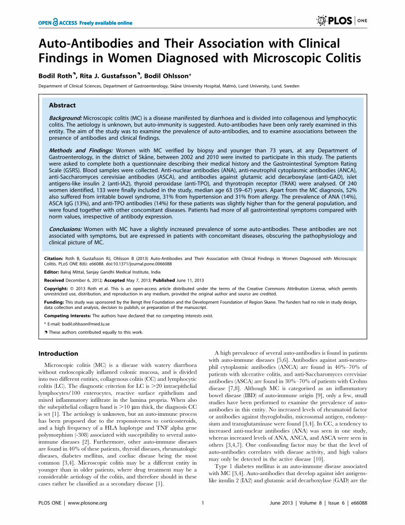
Auto-Antibodies and Their Association with ClinicalFindings in Women Diagnosed with Microscopic ColitisBodil Roth., Rita J. Gustafsson., Bodil Ohlsson*
Department of Clinical Sciences, Department of Gastroenterology, Skane University Hospital, Malmo, Lund University, Lund, Sweden
Abstract
Background: Microscopic colitis (MC) is a disease manifested by diarrhoea and is divided into collagenous and lymphocyticcolitis. The aetiology is unknown, but auto-immunity is suggested. Auto-antibodies have been only rarely examined in thisentity. The aim of the study was to examine the prevalence of auto-antibodies, and to examine associations between thepresence of antibodies and clinical findings.
Methods and Findings: Women with MC verified by biopsy and younger than 73 years, at any Department ofGastroenterology, in the district of Skane, between 2002 and 2010 were invited to participate in this study. The patientswere asked to complete both a questionnaire describing their medical history and the Gastrointestinal Symptom RatingScale (GSRS). Blood samples were collected. Anti-nuclear antibodies (ANA), anti-neutrophil cytoplasmic antibodies (ANCA),anti-Saccharomyces cerevisiae antibodies (ASCA), and antibodies against glutamic acid decarboxylase (anti-GAD), isletantigens-like insulin 2 (anti-IA2), thyroid peroxidase (anti-TPO), and thyrotropin receptor (TRAK) were analysed. Of 240women identified, 133 were finally included in the study, median age 63 (59–67) years. Apart from the MC diagnosis, 52%also suffered from irritable bowel syndrome, 31% from hypertension and 31% from allergy. The prevalence of ANA (14%),ASCA IgG (13%), and anti-TPO antibodies (14%) for these patients was slightly higher than for the general population, andwere found together with other concomitant diseases. Patients had more of all gastrointestinal symptoms compared withnorm values, irrespective of antibody expression.
Conclusions: Women with MC have a slightly increased prevalence of some auto-antibodies. These antibodies are notassociated with symptoms, but are expressed in patients with concomitant diseases, obscuring the pathophysiology andclinical picture of MC.
Citation: Roth B, Gustafsson RJ, Ohlsson B (2013) Auto-Antibodies and Their Association with Clinical Findings in Women Diagnosed with MicroscopicColitis. PLoS ONE 8(6): e66088. doi:10.1371/journal.pone.0066088
Editor: Balraj Mittal, Sanjay Gandhi Medical Institute, India
Received December 6, 2012; Accepted May 7, 2013; Published June 11, 2013
Copyright: � 2013 Roth et al. This is an open-access article distributed under the terms of the Creative Commons Attribution License, which permitsunrestricted use, distribution, and reproduction in any medium, provided the original author and source are credited.
Funding: This study was sponsored by the Bengt Ihre Foundation and the Development Foundation of Region Skane. The funders had no role in study design,data collection and analysis, decision to publish, or preparation of the manuscript.
Competing Interests: The authors have declared that no competing interests exist.
* E-mail: [email protected]
. These authors contributed equally to this work.
Introduction
Microscopic colitis (MC) is a disease with watery diarrhoea
without endoscopically inflamed colonic mucosa, and is divided
into two different entities, collagenous colitis (CC) and lymphocytic
colitis (LC). The diagnostic criterion for LC is .20 intraepithelial
lymphocytes/100 enterocytes, reactive surface epithelium and
mixed inflammatory infiltrate in the lamina propria. When also
the subepithelial collagen band is .10 mm thick, the diagnosis CC
is set [1]. The aetiology is unknown, but an auto-immune process
has been proposed due to the responsiveness to corticosteroids,
and a high frequency of a HLA haplotype and TNF alpha gene
polymorphism (-308) associated with susceptibility to several auto-
immune diseases [2]. Furthermore, other auto-immune diseases
are found in 40% of these patients, thyroid diseases, rheumatologic
diseases, diabetes mellitus, and coeliac disease being the most
common [3,4]. Microscopic colitis may be a different entity in
younger than in older patients, where drug treatment may be a
considerable aetiology of the colitis, and therefore should in these
cases rather be classified as a secondary disease [1].
A high prevalence of several auto-antibodies is found in patients
with auto-immune diseases [5,6]. Antibodies against anti-neutro-
phil cytoplasmic antibodies (ANCA) are found in 40%–70% of
patients with ulcerative colitis, and anti-Saccharomyces cerevisiae
antibodies (ASCA) are found in 30%–70% of patients with Crohns
disease [7,8]. Although MC is categorised as an inflammatory
bowel disease (IBD) of auto-immune origin [9], only a few, small
studies have been performed to examine the prevalence of auto-
antibodies in this entity. No increased levels of rheumatoid factor
or antibodies against thyroglobulin, microsomal antigen, endomy-
sium and transglutaminase were found [3,4]. In CC, a tendency to
increased anti-nuclear antibodies (ANA) was seen in one study,
whereas increased levels of ANA, ANCA, and ASCA were seen in
others [3,4,7]. One confounding factor may be that the level of
auto-antibodies correlates with disease activity, and high values
may only be detected in the active disease [10].
Type 1 diabetes mellitus is an auto-immune disease associated
with MC [3,4]. Auto-antibodies that develop against islet antigens-
like insulin 2 (IA2) and glutamic acid decarboxylase (GAD) are the
PLOS ONE | www.plosone.org 1 June 2013 | Volume 8 | Issue 6 | e66088
markers of the disease, and are present in 70%–80% of cases
[11,12]. In the majority of diabetes cases, immune reaction against
islet antigens and consequent formation of auto-antibodies begins
long before the disease is diagnosed clinically [13]. The prevalence
of these antibodies in MC has been only little examined.
The aim of this study was to further examine the prevalence of
auto-antibodies in a larger cohort of MC patients, and if present,
to examine the association between the presence of antibodies to
concomitant diseases and clinical findings.
Methods
Ethics StatementThe study protocol was approved by the Ethics Committee of
Lund University, and all participants gave their written, informed
consent when taking part in the study (LU 2009/565 and 2011/
209).
PatientsWomen who had been treated for MC at any outpatient clinic
of the Departments of Gastroenterology, throughout the district of
Skane, between 2002 and 2010, were identified by a search for the
ICD-10 classification for CC and LC (K52.8) in outpatient records
and the local register at the Department of Pathology, Skane
University Hospital, Malmo. About one-third of the total number
of the cases identified was not invited due to their being over 73
years of age, because they had many other concomitant diseases
and drug therapies, which could obscure the picture with several
cases of secondary MC [1]. Only the 240 patients (median 63
years, range 22–73 years) who had the diagnoses verified by
colonic biopsy, and were 73 years of age or younger, were invited
to participate in the present study. Altogether, 159 (median 63
years, range 22–73 years) of the 240 patients invited (66%) were
recruited to the study and 134 patients (56%) also agreed to
provide blood samples. One patient was excluded due to another
IBD diagnosis a few weeks after the inclusion, leaving 158 (66%)
and 133 patients (55%), respectively, to be included in the final
calculations and laboratory analyses. These patients represent the
majority of female cases of diagnosed MC in the southernmost
districts of Sweden, younger than 73 years of age. As MC and
auto-immunity are more frequent in women than in men [9,14],
and as the health-related quality of life and the experience of
symptoms differ between the genders [15], we chose to include
only women in the study.
Patient Recruitment and Study DesignBetween March and June 2011, invitations, including informa-
tion on the study and questionnaires about social and life style
factors, gastrointestinal symptoms, and medical condition and
medication were sent by mail to all 240 women. A reminding letter
was sent a month after the invitation letter to those who had not
answered. They were also invited to provide blood samples for
routine analyses according to standardised methods at the Division
of Clinical Chemistry, Department of Laboratory Medicine in
Skane, at their local hospital. Analyses for IgG auto-antibodies
were performed. ANA, ANCA, and ASCA were analysed at the
Division of Immunology, Department of Laboratory Medicine in
Skane, Lund, and analyses of anti-IA2, anti-GAD and anti-TPO
antibodies, and TRAK were performed at the Division of Clinical
Chemistry, Department of Laboratory Medicine in Skane,
Malmo.
Medical records were scrutinised, and age, gastrointestinal
symptoms, examinations, and treatments were recorded. Patients
were divided into two groups. One group included patients with at
least two episodes of watery diarrhoea; and/or those who were
dependent on long-term treatment of corticosteroids to maintain
remission; and/or those who had had two pathological intestinal
mucosa biopsies (MC1, n = 73). The other group included patients
who had concomitant coeliac disease [11], had undergone an
acute gastroenteritis shortly before the diagnostic colonoscopy [4],
had had only one episode of severe diarrhoea, or had had a
normal biopsy after the initial pathological intestinal biopsy, and
were in clinical remission (MC2, n = 60).
Immunological AnalysesHealthy blood donors served as controls for all immunological
analyses, except for TRAK, for which control values in healthy
controls are available by the same method [16]. Cut-off levels and
prevalence figures in the healthy population are the reference
values at the Department of Laboratory Medicine in Skane after
in-house examinations. Our reference values for anti-GAD and
anti-IA2 antibodies have been published previously [17].
ANA and ANCA were analysed by indirect immunofluores-
cence (IIF) [18,19]. The cut-off value for ANA, .14 IU/ml, is set
at the dilution where 5% of blood donors (50 men and 50 women)
showed a positive ANA. The assay is calibrated against the
international WHO reference for homogeneous ANA. Cytoplas-
mic (C)-and peri-nuclear (P)-ANCA have reference values of
.10 IU/ml. The cut-off is determined by the supplier (Euro
Immune AG, Lubeck, Germany) and of 50 healthy blood donors
tested, none showed a positive result. ANA and ANCA were
analysed in accordance with the International Organisation for
Standardisation (ISO)/International Electrotechnical Commission
(IEC) 17025 (General requirements for the competence for testing
and calibration laboratories).
ASCA is analysed by an automated ELISA method for which
the reference value is set to .10 IU/ml in accordance with the
manufacturer’s recommendation (Orgentec Diagnostika, AlegriaH,
Mainz, Germany), and both IgA and IgG levels are given. Of 50
healthy blood donors tested at our laboratory, 8% were positive
for both ASCA IgA and IgG, whereas other studies have found
0.6%–3.1% positive among healthy blood donors by the same
method [20].
Antibodies against IA2 and GAD were analysed with a
commercial ELISA kit (RSR Ltd, Cardiff, UK) according to the
manufacturers manual for which the cut-off values are defined as
.15 and .11 kIU/L, respectively [12]. In a cohort of 120 healthy
Swedish women, median age 32 years, range 19–44 years, none
was positive for anti-GAD antibodies and 31% were positive for
anti-IA2 antibodies [17].
Anti-TPO antibodies are analysed by a chemiluminiscence
enzyme immunological method (Roche Diagnostics Limited,
cobasH, West Sussex, UK) and TRAK by an RIA method
(Thermo B.R.A.H.M.S., TRAK Human RIA) [16]. Anti-TPO
antibodies are found in 7% of blood donors when the cut-off level
is set to .70 kIU/L [21]. The reference value for TRAK with a
sensitivity of 98.8% is set to .1.5 IU/L, and ,1% of 282 healthy
individuals of median age 45 years, range 20–73 years, were
positive [16].
QuestionnairesA self-administered questionnaire about marital status, educa-
tion, employment, smoking habits, wine consumption, physical
activity, medical conditions and medication was completed by the
patients. The patients were asked the questions: ‘‘ Have you ever
been treated for any of the following diseases, namely, hyperten-
sion, allergy, thyroid disorders, fracture, rheumatoid arthritis,
asthma, gastric ulcer, ovarial inflammation, herpes infection,
Auto-Antibodies in MC
PLOS ONE | www.plosone.org 2 June 2013 | Volume 8 | Issue 6 | e66088
endometriosis, Chlamydia Trachomatis, kidney stones, coeliac
disease, diabetes mellitus type 1 or 2, myocardial infarction,
intermittent claudication or stroke’’. A list of the drugs, vitamins
and minerals currently being taken was completed. The partici-
pants were also asked about heredity for cancer, stroke,
myocardial infarction, hypertension, diabetes mellitus, thyroid
disorders, asthma, rheumatoid arthritis, IBD or coeliac disease.
Gastrointestinal symptom rating scale (GSRS). The
GSRS is a Swedish, disease-specific and self-administered ques-
tionnaire, designed to evaluate perceived severity of gastrointes-
tinal symptoms during the previous week [22,23,24]. The
questionnaire includes 15 items and uses a 7-grade Likert scale.
This gives a total range value between 15 and 105 where the
highest score (seven) represents the most pronounced symptoms
and the lowest (one) no symptoms. The items are divided into five
dimensions representing Reflux Syndrome (two items), Abdominal
Pain Syndrome (three items), Constipation Syndrome (three
items), Indigestion Syndrome (four items) and Diarrhoea Syn-
drome (three items). The data are presented as the average of the
total score divided by the number of items. Norm values for the
healthy, gender-matched population are available [25].
Rome III criteria. The patients completed an abbreviated
version of the Rome III questionnaire, including only irritable
bowel syndrome (IBS) symptoms [26]. This questionnaire has been
translated and validated into the Swedish language (Magnus
Simren and Anna Ryden). Patients who fulfilled the criteria in the
Rome III questionnaire were classified as also suffering from IBS-
like symptoms.
Table 1. Prevalence of auto-antibodies in microscopic colitis.
Controls
Microscopiccolitis
Collagenouscolitis
Lymphocyticcolitis P- value
n=133 n=77 n=56
Hb (117–153 g/L) 132.269.3 132.269.5 132.269.0 0.987
Leucocytes
(3.5–8.86109/L) 6.962.0 7.161.9 6.862.1 0.430
S-Albumin
(36–48 g/L) 39.963.3 39.963.6 40.063.0 0.794
CRP (,3.1 mg/L) 2.0 (1.0–4.0) 4.0 (1.0–4.0) 1.6 (1.0–2.9) 0.014
P-Sodium 140.0 141.0 140.0 0.111
(137–145 mmol/L) (139.0–42.0) (139.0–142.0) (139.0–142.0)
P-Potassium
(3.5–4.4 mmol/L) 3.960.4 3.960.3 3.960.4 0.916
ANA
(#14 IU/ml)(n, %) (5)a 19 (14) 8 (10) 11 (20) 0.140
C-ANCA
(#10 IU/ml)(n, %) (0)b 1 (1) 1 (1) 0 (0) 1.000
P-ANCA
(#10 IU/ml) (n, %) (0)b 7 (5) 4 (5) 3 (5) 1.000
ASCA IgA
(#10 IU/ml) (n,%) (8)b 0 (0) 0 (0) 0 (0)
ASCA IgG
(#10 IU/ml)(n, %) (8)b 17 (13) 7 (9) 10 (18) 0.187
Anti-GAD
(#11 kIU/L) (n, %) (0)c 7 (5) 4 (5) 3 (5) 1.000
Anti-IA2
(#15 kIU/L) (n, %) (31)c 2 (2) 1 (1) 1 (2) 0.100
Anti-TPO
(#70 kIU/L) (n, %) (7)b 18 (14) 9 (12) 9 (16) 0.444
TRAK
(#1.5 IU/L)(n, %) (1)d 1 (1) 0 (0) 1 (2)
ANA= anti-nuclear antibodies, C-ANCA= cytoplasmic anti-neutrophil cytoplasmic antibodies, ASCA= anti-Saccharomyces cerevisiae antibodies, CRP =C-reactive protein,anti-IA2 = anti-islet antigens-like insulin 2, anti-GAD= anti-glutamic acid decarboxylase, Hb = haemoglobin, IgA = Immunglobulin A, IgG = Immunglobulin G, P = plasma,P-ANCA=peri-nuclear anti-neutrophil cytoplasmic antibodies, S = serum, anti-TPO= anti-thyroid peroxidase, TRAK= thyrotropin receptor antibodies. Reference values inbrackets. n (%) = number (percent) of positive antibodies. Controls consisted of: a=healthy blood donors, 50 men and 50 women, b= 50 healthy blood donors, c= 120healthy women, reference No 17, d= 282 healthy controls from reference No 16. Values are given as mean 6 standard deviation or median (interquartile range).Student t- test, Mann-Whitney U- test or Fishers exact test were used for comparisons between collagenous colitis and lymphocytic colitis. P,0.01 was consideredstatistically significant.doi:10.1371/journal.pone.0066088.t001
Auto-Antibodies in MC
PLOS ONE | www.plosone.org 3 June 2013 | Volume 8 | Issue 6 | e66088
Statistical AnalysesThe data were analysed using the statistical software package
SPSS for Windows� (Release 20.0; IBM). Values are given as
mean 6 standard deviation (SD) or median (interquartile range)
depending on distribution norm. Gastrointestinal symptoms are
given as the mean with a 95% confidence interval, because the
reference values in reference No 25 are given in this form. All
patients who expressed any form of measured auto-antibody were
classified as antibody positive, and calculations of the association
between antibodies and clinical features were performed both for
each antibody and for the total presence of antibodies. Differences
between groups were calculated by the 2-tailed Mann-Whitney U -
test or Student t- test. The Spearman correlation test was used to
examine correlations between groups. Fishers exact test was used
to examine differences between the presence of auto-antibodies in
diseases and heredity; differences in the prevalence of auto-
antibodies and diseases between CC and LC; and differences in
the prevalence of auto-antibodies between MC1 and MC2. Due to
several hypotheses tested, p,0.01 was considered statistically
significant.
Results
Patient CharacteristicsIn total, the 133 women with MC, who provided blood samples,
were included in the study (median age 63 (59–67) years, range
27–73 years). CC was diagnosed in 77 patients (58%) (median age
64 (59–68) years, range 31–73 years) and LC in 56 patients (42%)
(median age 63 (54–67) years, range 27–72 years). Of these 133,
69 patients (52%) also fulfilled the Rome III criteria for IBS, 40 of
the patients (52%) with CC and 29 of the patients (52%) with LC.
The duration of gastrointestinal symptoms at study inclusion was 8
(3–14) years. There was no correlation between duration of
symptoms and age (correlation coefficient =20.097, p = 0.286).
The body mass index was 24.8 (22.7–28.7) kg/m2. Of the patients,
25% were regular smokers, 8% smoked occasionally, 40% had
stopped smoking and 27% had never smoked. There was no
difference in smoking habits between CC and LC (p = 0.350).
Routine analyses were within the reference values used in the
laboratory in the vast majority of cases (Table 1).
Prevalence of Auto-antibodiesThe highest prevalence of antibodies was found of ANA, ASCA
IgG, and anti-TPO antibodies, where the prevalence was doubled
in the patients compared to the general population, with no
differences between CC and LC, or between MC1 and MC2
(Table 1 and Table 2). There was no difference between CC and
LC in the total presence of auto-antibodies (p = 0.375). The
median age in ANA-positive patients tended to be higher than in
ANA-negative patients (67 (62–71) years and 63 (56–67) years,
respectively, p = 0.013). In contrast, the median age tended to be
lower in ASCA-positive patients compared to ASCA-negative
patients (59 (46–66) years and 64 (59–68) years, respectively,
p = 0.031). The relatively high prevalence of anti-TPO antibodies
was not reflected by such a difference in age in patients who
expressed antibodies compared to those who did not (60 (47–66)
years and 64 (59–68) years, respectively, p = 0.097).
When examining the most prevalent concomitant diseases,
excluding sporadic diseases, ANA-positive patients also suffered
from hypertension (nine), allergy (seven), thyroid disorders (six),
rheumatoid arthritis (three), and coeliac disease (three). Patients
expressing ASCA IgG also suffered from hypertension (eight),
rheumatoid arthritis (four), asthma (three), coeliac disease (three),
and allergy (three). Anti-TPO antibodies were found together with
thyroid disorders in seven patients, with allergy in four and with
asthma and rheumatoid arthritis in three. Of the seven who
expressed P-ANCA, four suffered from allergy, and three from
thyroid disorders, rheumatoid arthritis, and hypertension, respec-
tively. Of the seven patients who expressed anti-GAD antibodies,
five also suffered from diabetes mellitus. One of the patients with
anti-IA2 antibodies also expressed anti-GAD antibodies, and
suffered from diabetes mellitus. Only eight of the patients with any
antibody had MC as a sole disease. The patient with the most
concomitant diseases suffered from seven of these, apart from MC
and IBS. There was no difference in the results of routine blood
Table 2. Prevalence of auto-antibodies in persistent (MC1)and transient microscopic colitis (MC2).
MC1 MC2 P-value
n=73 n=60
Age (years) 63 (57–67) 64 (59–68) 0.405
Hb (117–153 g/L) 132.368.8 132.169.9 0.916
Leucocytes
(3.5–8.86109/L) 7.062.3 6.961.5 0.906
S-Albumin (36248g/L)
40.163.5 39.763.1 0.494
CRP (,3.1 mg/L) 2.0 (1.024.0) 2.0 (1.023.5) 0.840
P-Sodium
(1372145 mmol/L) 141.0(139.02142.0)
140.0(139.02142.0)
0.253
P-Potassium
(3.524.4 mmol/L) 3.960.3 3.960.4 0.736
ANA (#14 IU/ml)(n, %)
12 (16.4) 7 (11.7) 0.619
C-ANCA
(#10 IU/ml)(n, %) 1 (1.4) 0 (0) 1.000
P-ANCA
(#10 IU/ml)(n, %) 5 (6.8) 2 (3.3) 0.463
ASCA IgA
(#10 IU/mL)(n, %) 0 (0) 2 (3.3)
ASCA IgG
(#10 IU/mL)(n, %) 11 (15.1) 6 (10) 0.602
Anti-GAD
(#11 kIU/L)(n, %) 5 (6.8) 2 (3.3) 0.460
Anti-IA2
(#15 kIU/L)(n, %) 0 (0) 2 (3.3) 0.100
Anti-TPO
(#70 kIU/L)(n, %) 9 (12.3) 9 (15.0) 0.799
TRAK (#1.5 IU/L)(n, %) 0 (0) 1 (1.7) 0.456
ANA= anti-nuclear antibodies, C-ANCA= cytoplasmic anti-neutrophilcytoplasmic antibodies, ASCA= anti-Saccharomyces cerevisiae antibodies,CRP = C-reactive protein, anti-IA2 = anti-islet antigens-like insulin 2, anti-GAD= anti-glutamic acid decarboxylase, Hb = haemoglobin,IgA = Immunglobulin A, IgG = Immunglobulin G, P =plasma, P-ANCA =peri-nuclear anti-neutrophil cytoplasmic antibodies, S = serum, anti-TPO= anti-thyroid peroxidase, TRAK = thyrotropin receptor antibodies. Reference values inbrackets. n (%) = number (percent) of patients with positive antibodies. Valuesare given as mean 6 standard deviation or median (interquartile range).Student t- test, Mann-Whitney U- test or Fishers exact test. P,0.01 wasconsidered statistically significant.doi:10.1371/journal.pone.0066088.t002
Auto-Antibodies in MC
PLOS ONE | www.plosone.org 4 June 2013 | Volume 8 | Issue 6 | e66088
analyses between patients with and those without auto-antibodies
(Table 3).
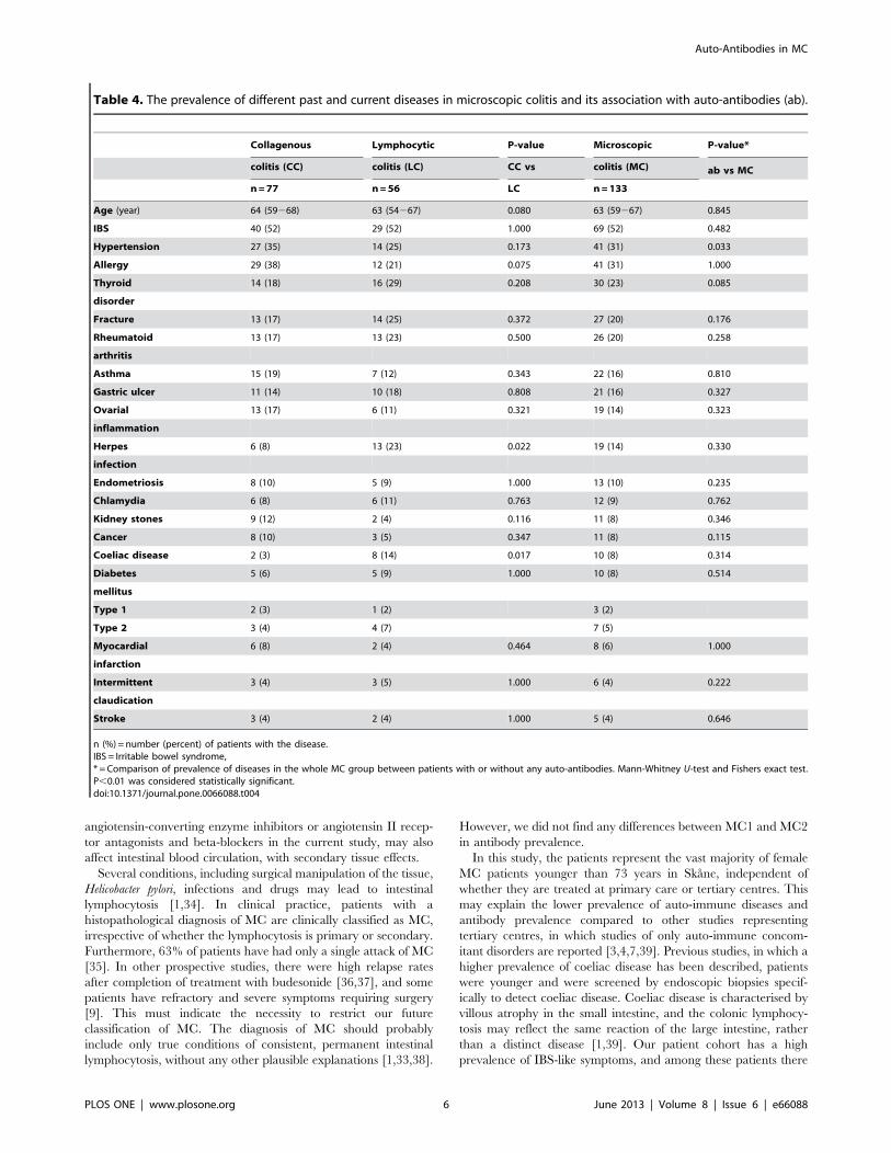
Prevalence of Other DiseasesThe distribution of present and past diseases is shown in Table 4.
More than half of the patients fulfilled the criteria both for MC
and IBS. Apart from IBS, only 26 of the patients (20%) reported
that they had had MC as the sole disease. No difference in the
prevalence of different diseases was found between the CC and LC
groups (Table 4). Three of the 10 patients with diabetes mellitus
had auto-immune type 1 diabetes, and the rest had type 2
diabetes. There was no association between the total presence of
antibodies and any of the diseases investigated in the statistical
calculations (Table 4), or between each antibody and disease (data
not shown). The patients were, at the time of the study, treated
with many drugs simultaneously, where corticosteroids (30%),
proton pump inhibitors (22%), angiotensin-converting enzyme
inhibitors or angiotensin II receptor antagonists (19%), thyroid
hormones (19%), anti-depressant drugs of the type selective
serotonin reuptake inhibitors (17%), statins (17%), and beta-
blockers (14%) were the most frequently used. Treatment with
corticosteroids tended to be more prevalent in MC1 (26) than in
MC2 (11) (p = 0.033).
There was no difference in heredity concerning cancer (60%)
(p = 0.648), hypertension (61%) (p = 0.208), myocardial infarction
(50%) (p = 0.031), stroke (36%) (p = 0.627), diabetes mellitus (32%)
(p = 0.217), rheumatoid arthritis (34%) (p = 0.665), thyroid disor-
ders (31%) (p = 0.870), asthma (29%) (p = 0.676), IBD (28%)
(p = 0.260) or coeliac disease (10%) (p = 1.000) between patients
with and those without antibodies.
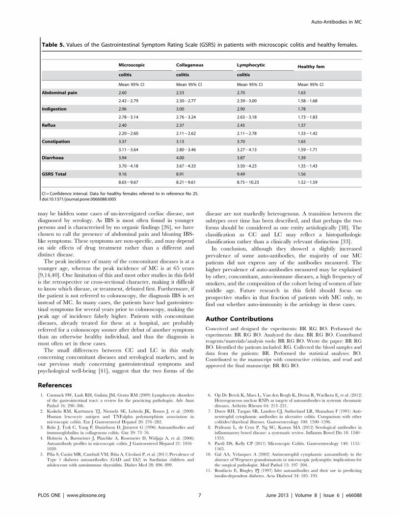
Prevalence of Gastrointestinal SymptomsPatients with MC had more gastrointestinal symptoms than
healthy females in all the dimensions Reflux Syndrome, Abdom-
inal Pain Syndrome, Constipation Syndrome, Indigestion Syn-
drome, and Diarrhoea Syndrome (Table 5). There was no
difference in symptoms between CC and LC (Table 5). There
was no difference in disease duration and degree of gastrointestinal
symptoms between patients with and those without any kind of
auto-antibodies (Table 3).
Discussion
The main finding of this cross-sectional study was that the
prevalence of common auto-antibodies such as ANA, P-ANCA,
ASCA IgG, and anti-GAD and anti-TPO antibodies is slightly
more prevalent in MC patients than in the general Swedish
population, but they are nonetheless present in only a minority of
cases. C-ANCA, ASCA IgA, anti-IA2 antibodies, and TRAK are
only occasionally found. There was no difference in gastrointes-
tinal symptoms between patients with and those without auto-
antibodies. The antibodies were present in patients who also
suffered from other concomitant diseases.
Auto-immunity has been suggested as a plausible aetiology of
MC, as an association with the same HLA genotypes in MC as
described in other known auto-immune diseases has been
described [2,9]. However, the prevalence of C-ANCA, ASCA
IgA, anti-IA2 antibodies, and TRAK was in the same range or
lower as in the general population, according to our laboratory
reference values of healthy blood donors, as has also been
described in previous studies [3,7,11,13,16,17]. Patients predis-
posed to auto-immunity usually express higher levels of several
antibodies in serum [5,6,7,8]. The prevalence of ANA, P-ANCA,
ASCA IgG, and anti-GAD and anti-TPO antibodies were slightly
increased compared to healthy controls, but not at all to the same
magnitude as in patients with Crohns disease, ulcerative colitis,
diabetes mellitus or thyroid disorders [7,8,12,27,28]. Furthermore,
reference values are set in relation to the general population,
including both genders and all ages. In this study of women of
upper middle age, the prevalence of auto-antibodies is higher than
in the general population [14]. World-wide, 12%–26% of healthy
women and 3%–14% of healthy men have anti-TPO antibodies
[28], and 6% of healthy women are considered positive for ANA
[29]. The increased prevalence of antibodies in our present study
can be explained by other concomitant diseases of auto-immune
character, e.g. rheumatoid arthritis, coeliac disease, thyroid
disorders and diabetes mellitus, and by our cohort being of upper
middle-aged women. As our older MC patients had many
concomitant diseases, not only of auto-immune origin, it is
difficult to estimate the true association with auto-immune
diseases. The associations observed could be due to secondary
effects on the digestive tract of the diseases, or their treatment, and
do not necessarily mean a common causal aetiology [1]. Smoking
per se is associated with a higher prevalence of ANA [30], and this
could explain the higher ANA positivity in our study with a high
frequency of former and current smokers. Thus, our finding in the
present study does not indicate that auto-immunity is the most
important cause of MC.
Previously described associations between MC and drug
treatment and smoking [31,32], and the higher age in the diseased
patients [9], suggest environmental and circulatory influences as
important aetiological factors for MC [33]. Intestinal ischemia
should also be considered, especially as smoking is a risk factor for
the disease [32], and the patients are older, with a high prevalence
of cardiovascular diseases [33]. The high prevalence of using
Table 3. Laboratory values and gastrointestinal symptoms inrelation to the presence of auto-antibodies.
Antibodies No antibodies P-value
n=56 n=77
Age (year) 62 (58268) 63 (58267) 0.845
Hb (1172153 g/L) 132.569.3 132.069.1 0.764
Leucocytes
(3.528.86109/L) 6.962.1 7.061.9 0.791
S-Albumin
(36248 g/L) 40.063.3 40.063.4 0.966
CRP (,3.1 mg/L) 2.0 (1.024.4) 2.0 (1.023.5) 0.056
P-Sodium
(1372145 mmol/L) 141.0 (139.02143.0) 140.0 (139.02142.0) 0.253
P-Potassium
(3.524.4 mmol/L) 3.960.3 3.960.4 0.907
Duration (years) 10.0 (5.8216.0) 6.0 (3.0212.0) 0.038
Abdominal pain 2.55 2.2822.83 2.63 2.3722.88 0.428
Indigestion 3.01 2.7623.26 2.92 2.6623.18 0.372
Reflux 2.50 2.1722.83 2.30 2.0422.56 0.195
Constipation 3.33 2.9223.74 3.40 3.0423.76 0.457
Diarrhoea 4.02 3.6824.35 3.90 3.5524.24 0.372
GSRS Total 8.92 8.0429.80 9.30 8.6929.92 0.299
Values are given as median (interquartile range) or mean with a 95% confidenceinterval. GSRS =Gatrointestinal Symptom Rating Scale. Mann-Whitney U-test orStudent t -test. P,0.01 was considered statistically significant.doi:10.1371/journal.pone.0066088.t003
Auto-Antibodies in MC
PLOS ONE | www.plosone.org 5 June 2013 | Volume 8 | Issue 6 | e66088
angiotensin-converting enzyme inhibitors or angiotensin II recep-
tor antagonists and beta-blockers in the current study, may also
affect intestinal blood circulation, with secondary tissue effects.
Several conditions, including surgical manipulation of the tissue,
Helicobacter pylori, infections and drugs may lead to intestinal
lymphocytosis [1,34]. In clinical practice, patients with a
histopathological diagnosis of MC are clinically classified as MC,
irrespective of whether the lymphocytosis is primary or secondary.
Furthermore, 63% of patients have had only a single attack of MC
[35]. In other prospective studies, there were high relapse rates
after completion of treatment with budesonide [36,37], and some
patients have refractory and severe symptoms requiring surgery
[9]. This must indicate the necessity to restrict our future
classification of MC. The diagnosis of MC should probably
include only true conditions of consistent, permanent intestinal
lymphocytosis, without any other plausible explanations [1,33,38].
However, we did not find any differences between MC1 and MC2
in antibody prevalence.
In this study, the patients represent the vast majority of female
MC patients younger than 73 years in Skane, independent of
whether they are treated at primary care or tertiary centres. This
may explain the lower prevalence of auto-immune diseases and
antibody prevalence compared to other studies representing
tertiary centres, in which studies of only auto-immune concom-
itant disorders are reported [3,4,7,39]. Previous studies, in which a
higher prevalence of coeliac disease has been described, patients
were younger and were screened by endoscopic biopsies specif-
ically to detect coeliac disease. Coeliac disease is characterised by
villous atrophy in the small intestine, and the colonic lymphocy-
tosis may reflect the same reaction of the large intestine, rather
than a distinct disease [1,39]. Our patient cohort has a high
prevalence of IBS-like symptoms, and among these patients there
Table 4. The prevalence of different past and current diseases in microscopic colitis and its association with auto-antibodies (ab).
Collagenous Lymphocytic P-value Microscopic P-value*
colitis (CC) colitis (LC) CC vs colitis (MC) ab vs MC
n=77 n=56 LC n=133
Age (year) 64 (59268) 63 (54267) 0.080 63 (59267) 0.845
IBS 40 (52) 29 (52) 1.000 69 (52) 0.482
Hypertension 27 (35) 14 (25) 0.173 41 (31) 0.033
Allergy 29 (38) 12 (21) 0.075 41 (31) 1.000
Thyroid 14 (18) 16 (29) 0.208 30 (23) 0.085
disorder
Fracture 13 (17) 14 (25) 0.372 27 (20) 0.176
Rheumatoid 13 (17) 13 (23) 0.500 26 (20) 0.258
arthritis
Asthma 15 (19) 7 (12) 0.343 22 (16) 0.810
Gastric ulcer 11 (14) 10 (18) 0.808 21 (16) 0.327
Ovarial 13 (17) 6 (11) 0.321 19 (14) 0.323
inflammation
Herpes 6 (8) 13 (23) 0.022 19 (14) 0.330
infection
Endometriosis 8 (10) 5 (9) 1.000 13 (10) 0.235
Chlamydia 6 (8) 6 (11) 0.763 12 (9) 0.762
Kidney stones 9 (12) 2 (4) 0.116 11 (8) 0.346
Cancer 8 (10) 3 (5) 0.347 11 (8) 0.115
Coeliac disease 2 (3) 8 (14) 0.017 10 (8) 0.314
Diabetes 5 (6) 5 (9) 1.000 10 (8) 0.514
mellitus
Type 1 2 (3) 1 (2) 3 (2)
Type 2 3 (4) 4 (7) 7 (5)
Myocardial 6 (8) 2 (4) 0.464 8 (6) 1.000
infarction
Intermittent 3 (4) 3 (5) 1.000 6 (4) 0.222
claudication
Stroke 3 (4) 2 (4) 1.000 5 (4) 0.646
n (%) = number (percent) of patients with the disease.IBS = Irritable bowel syndrome,* = Comparison of prevalence of diseases in the whole MC group between patients with or without any auto-antibodies. Mann-Whitney U-test and Fishers exact test.P,0.01 was considered statistically significant.doi:10.1371/journal.pone.0066088.t004
Auto-Antibodies in MC
PLOS ONE | www.plosone.org 6 June 2013 | Volume 8 | Issue 6 | e66088
may be hidden some cases of un-investigated coeliac disease, not
diagnosed by serology. As IBS is most often found in younger
persons and is characterised by no organic findings [26], we have
chosen to call the presence of abdominal pain and bloating IBS-
like symptoms. These symptoms are non-specific, and may depend
on side effects of drug treatment rather than a different and
distinct disease.
The peak incidence of many of the concomitant diseases is at a
younger age, whereas the peak incidence of MC is at 65 years
[9,14,40]. One limitation of this and most other studies in this field
is the retrospective or cross-sectional character, making it difficult
to know which disease, or treatment, debuted first. Furthermore, if
the patient is not referred to colonoscopy, the diagnosis IBS is set
instead of MC. In many cases, the patients have had gastrointes-
tinal symptoms for several years prior to colonoscopy, making the
peak age of incidence falsely higher. Patients with concomitant
diseases, already treated for these at a hospital, are probably
referred for a colonoscopy sooner after debut of another symptom
than an otherwise healthy individual, and thus the diagnosis is
most often set in these cases.
The small differences between CC and LC in this study
concerning concomitant diseases and serological markers, and in
our previous study concerning gastrointestinal symptoms and
psychological well-being [41], suggest that the two forms of the
disease are not markedly heterogenous. A transition between the
subtypes over time has been described, and that perhaps the two
forms should be considered as one entity aetiologically [38]. The
classification as CC and LC may reflect a histopathologic
classification rather than a clinically relevant distinction [33].
In conclusion, although they showed a slightly increased
prevalence of some auto-antibodies, the majority of our MC
patients did not express any of the antibodies measured. The
higher prevalence of auto-antibodies measured may be explained
by other, concomitant, auto-immune diseases, a high frequency of
smokers, and the composition of the cohort being of women of late
middle age. Future research in this field should focus on
prospective studies in that fraction of patients with MC only, to
find out whether auto-immunity is the aetiology in these cases.
Author Contributions
Conceived and designed the experiments: BR RG BO. Performed the
experiments: BR RG BO. Analyzed the data: BR RG BO. Contributed
reagents/materials/analysis tools: BR RG BO. Wrote the paper: BR RG
BO. Identified the patients included: RG. Collected the blood samples and
data from the patients: BR. Performed the statistical analyses: BO.
Contributed to the manuscript with constructive criticism, and read and
approved the final manuscript: BR RG BO.
References
1. Carmack SW, Lash RH, Gulizia JM, Genta RM (2009) Lymphocytic disorders
of the gastrointestinal tract: a review for the practicing pathologist. Adv Anat
Pathol 16: 290–306.
2. Koskela RM, Karttunen TJ, Niemela SE, Lehtola JK, Ilonen J, et al. (2008)
Human leucocyte antigen and TNFalpha polymorphism association in
microscopic colitis. Eur J Gastroenterol Hepatol 20: 276–282.
3. Bohr J, Tysk C, Yang P, Danielsson D, Jarnerot G (1996) Autoantibodies and
immnuoglobulins in collagenous colitis. Gut 39: 73–76.
4. Holstein A, Burmeister J, Plaschke A, Rosemeier D, Widjaja A, et al. (2006)
Autoantibody profiles in microscopic colitis. J Gastroenterol Hepatol 21: 1016–
1020.
5. Pilia S, Casini MR, Cambuli VM, Ibba A, Civolani P, et al. (2011) Prevalence of
Type 1 diabetes autoantibodies (GAD and IA2) in Sardinian children and
adolescents with autoimmune thyroiditis. Diabet Med 28: 896–899.
6. Op De Beeck K, Maes L, Van den Bergh K, Derua R, Waelkens E, et al. (2012)
Heterogeneous nuclear RNPs as targets of autoantibodies in systemic rheumatic
diseases. Arthritis Rheum 64: 213–221.
7. Duerr RH, Targan SR, Landers CJ, Sutherland LR, Shanahan F (1991) Anti-
neutrophil cytoplasmic antibodies in ulcerative colitis. Comparison with other
colitides/diarrheal illnesses. Gastroenterology 100: 1590–1596.
8. Prideaux L, de Cruz P, Ng SC, Kamm MA (2012) Serological antibodies in
inflammatory bowel disease: a systematic review. Inflamm Bowel Dis 18: 1340–
1355.
9. Pardi DS, Kelly CP (2011) Microscopic Colitis. Gastroenterology 140: 1155–
1165.
10. Gal AA, Velasquez A (2002) Antineutrophil cytoplasmic autoantibody in the
absence of Wegeners granulomatosis or microscopic polyangitis: implications for
the surgical pathologist. Mod Pathol 15: 197–204.
11. Bonifacio E, Bingley PJ (1997) Islet autoanibodies and their use in predicting
insulin-dependent diabetes. Acta Diabetol 34: 185–193.
Table 5. Values of the Gastrointestinal Symptom Rating Scale (GSRS) in patients with microscopic colitis and healthy females.
Microscopic Collagenous Lymphocytic Healthy fem
colitis colitis colitis
Mean 95% CI Mean 95% CI Mean 95% CI Mean 95% CI
Abdominal pain 2.60 2.53 2.70 1.63
2.4222.79 2.3022.77 2.3923.00 1.5821.68
Indigestion 2.96 3.00 2.90 1.78
2.7823.14 2.7623.24 2.6323.18 1.7321.83
Reflux 2.40 2.37 2.45 1.37
2.2022.60 2.1122.62 2.1122.78 1.3321.42
Constipation 3.37 3.13 3.70 1.65
3.1123.64 2.8023.46 3.2724.13 1.5921.71
Diarrhoea 3.94 4.00 3.87 1.39
3.7024.18 3.6724.33 3.5024.23 1.3521.43
GSRS Total 9.16 8.91 9.49 1.56
8.6529.67 8.2129.61 8.75210.23 1.5221.59
CI = Confidence interval. Data for healthy females referred to in reference No 25.doi:10.1371/journal.pone.0066088.t005
Auto-Antibodies in MC
PLOS ONE | www.plosone.org 7 June 2013 | Volume 8 | Issue 6 | e66088
12. Rahmati K, Lernmark A, Becker C, Foltyn-Zadura A, Larsson K, et al. (2008) A
comparison of serum and EDTA plasma in the measurement of glutamic aciddecarboxylase autoantibodies (GADA) and autoantibodies to islet antigen-2 (IA-
2A) using the RSR radioimmunoassay (RIA) and enzyme linked immunosorbent
assay (ELISA) kits. Clin Lab 54: 227–235.13. Tuomilehto J, Yliharsila H (1998) Antibodies as predictors of insulin-dependent
diabetes mellitus before the clinical onset. Nutrition 14: 403–405.14. Symmons DP (2002) Epidemiology of rheumatoid arthritis: determinants of
onset, persistence and outcome. Best Pract Res Clin Rheumatol 16: 707–722.
15. Simren M, Abrahamsson H, Svedlund J, Bjornsson ES (2001) Quality of life inpatients with irritable bowel syndrome seen in referral centers versus primary
care: the impact of gender and predominant bowel pattern. Scand J Gastroenterol36: 545–552.
16. Costagliola S, Morgenthaler NG, Hoermann R, Badenhoop K, Struck J, et al.(1999) Second generation assay for thyrotropin receptor antibodies has superior
diagnostic sensitivity for Graves’ disease. J Clin Endocrinol Metab 84: 90–97.
17. Persson A, Becker C, Hansson I, Nilsson A, Torn C (2010) Comparison ofMeasurements of Autoantibodies to Glutamic Acid Decarboxylase and Islet
Antigen-2 in Whole Blood Eluates from Dried Blood Spots Using the RSR-Enzyme Linked Immunosorbent Assay Kits and In-House Radioimmunoassays.
Exp Diabetes Res. Online June 3.
18. Westman KWA, Selga D, Bygren P, Segelmark M, Baslund B, et al. (1998)Clinical evaluation of a capture ELISA for detection of proteinase-3
antineutrophil cytoplasmic antibody. Technical Note Kidney Int 53: 1230–1236.19. Reichlin M (2006) Measurement and clinical significance of antinuclear
antibodies. Up to date Online ver 15.1.20. Mankaı A, Sakly W, Thabet Y, Achour A, Manoubi W, et al. (2013) Anti-
Saccharomyces cerevisiae antibodies in patients with systemic lupus erythematosus.
Rheumatol Int 33: 665–669.21. www.analysporten-labmedicin.skane.se.
22. Svedlund J, Sjodin I, Dotevall G (1988) GSRS – a clinical rating scale forgastrointestinal symptoms in patients with irritable bowel syndrome and peptic
ulcer disease. Dig Dis Sci 33: 129–134.
23. Dimenas E, Glise H, Hallerback B, Hernqvist H, Svedlund J, et al. (1993)Quality of life in patients with upper gastrointestinal symptoms An improved
evaluation of treatment regimens? Scand J Gastroenterol 28: 681–687.24. Dimenas E, Glise H, Hallerback B, Hernqvist H, Svedlund J, et al. (1995) Well-
being and gastrointestinal symptoms among patients referred to endoscopyowing to suspected duodenal ulcer. Scand J Gastroenterol 30: 1046–1052.
25. Dimenas E, Carlsson G, Glise H, Israelsson B, Wiklund I (1996) Relevance of
norm values as part of the documentation of quality of life instruments for use inupper gastrointestinal disease. Scand J Gastroenterol 31 Suppl 221: 8–13.
26. Drossman DA, Corrazziari E, Delvaux M, Spiller R, Talley NJ, et al. Rome III:The Functional Gastrointestinal Disorders. McLean, VA: Degnon Associates;
2006.
27. Weng J, Ekelund M, Lehto M, Li H, Ekberg G, et al. (2002) Screening for
MODY mutations, GAD antibodies, and Type 1 diabetes-associated HLA
genotypes in women with gestational diabetes mellitus. Diabetes Care 25: 68–71.
28. Prummel MF, Wiersinga WM (2005) Thyroid peroxidase autoantibodies in
euthyroid subjects. Best Pract Res Clin Endocrinol Metab 19: 1–15.
29. Matthiesen LS, Berg G, Ernerudh J, Skogh T (1999) A prospective study on the
occurrence of autoantibodies in low-risk pregnancies. Eur J Obstet Gynecol
Reprod Biol 83: 21–26.
30. Karabulut G, Kitapcioglu G, Inal V, Kalfa M, Yargucu F, et al. (2011) Cigarette
smoking in primary Sjogren’s syndrome: positive association only with ANA
positivity. Mod Rheumatol 21: 602–607.
31. Fernandez-Banares F, Eseve M, Espinos JC, Roinach M, Forne M, et al. (2007)
Drug consumption and the risk of microscopic colitis. Am J Gastroneterol 102:
324–330.
32. Yen EF, Pokhrel B, Du H, New S, Bianchi L, et al. (2012) Current and past
cigarette smoking significantly increase risk for microscopic colitis. Inflamm
Bowel Dis 18: 1835–1841.
33. Bjørnbak C, Engel PJ, Nielsen PL, Munck LK (2011) Microscopic colitis: clinical
findings, topography and persistence of histopathological subgroups. Aliment
Pharmacol Ther 34: 1225–1234.
34. Lee S, Ogilvie RT, Dupre M, Gao ZH (2009) Intravascular lymphocytosis in
acute appendicitis: potential mimicry of chronic lymphocytic leukaemia.
Histopathology 55: 660–664.
35. Olesen M, Eriksson S, Bohr J, Jarnerot G, Tysk C (2004) Lymphocytic colitis: a
retrospective clinical study of 199 Swedish patients. Gut 53: 536–541.
36. Miehlke S, Madisch A, Karimi D, Wonschik S, Kuhlisch E, et al. (2009)
Budesonide is effective in treating lymphocytic colitis: a randomized double-
blind placebo-controlled study. Gastroenterology 136: 2092–2100.
37. Bonderup OK, Hansen JB, Teglbjaerg PS, Christensen LA, Fallingborg JF
(2009) Long-term budesonide treatment of collagenous colitis: a randomised,
double-blind, placebo-controlled trial. Gut 58: 68–72.
38. Rasmussen MA, Munck LK (2012) Systematic review: are lymphocytic colitis
and collagenous colitis two subtypes of the same disease-microscopic colitis?
Aliment Pharmacol Ther 36: 79–90.
39. Stewart M, Andrews CN, Urbanski S, Beck PL, Storr M (2011) The association
of celiac disease and micrscopic colitis: a large population-based study. Aliment
Pharmacol Ther 33: 1340–1349.
40. Dahlquist GG, Nystrom L, Patterson CC; Swedish Childhood Diabetes Study
Group (2011) Diabetes Incidence in Sweden Study Group. Incidence of type 1
diabetes in Sweden among individuals aged 0–34 years, 1983–2007: an analysis
of time trends. Diabetes Care 34: 1754–1759.
41. Roth B, Ohlsson B (2013) Gastrointestinal symptoms and psychological well-
being in patients with microscopic colitis. Scand J Gastroenterol 48: 27–34.
Auto-Antibodies in MC
PLOS ONE | www.plosone.org 8 June 2013 | Volume 8 | Issue 6 | e66088