Embed Size (px)
Citation preview
1
Autism: The Centrality of Active Pathophysiology and the Shift from Static to Chronic Dynamic Encephalopathy
Martha R. Herbert, MD, PhD
Published in Autism: Oxidative stress, inflammation and immune abnormalities. Chauhan A,
Chauhan V, Brown T, eds., Fall, 2009, Taylor & Francis/CRC Press. DO NOT CIRCULATE THIS VERSION
Author contact information: TRANSCEND Research Program
Pediatric Neurology Massachusetts General Hospital
Harvard Medical School 149 13th St., Room 10.018
Charlestown, MA 02129 USA Phone: 617-724-5920
Fax: 617-812-6334 Email [email protected]
2
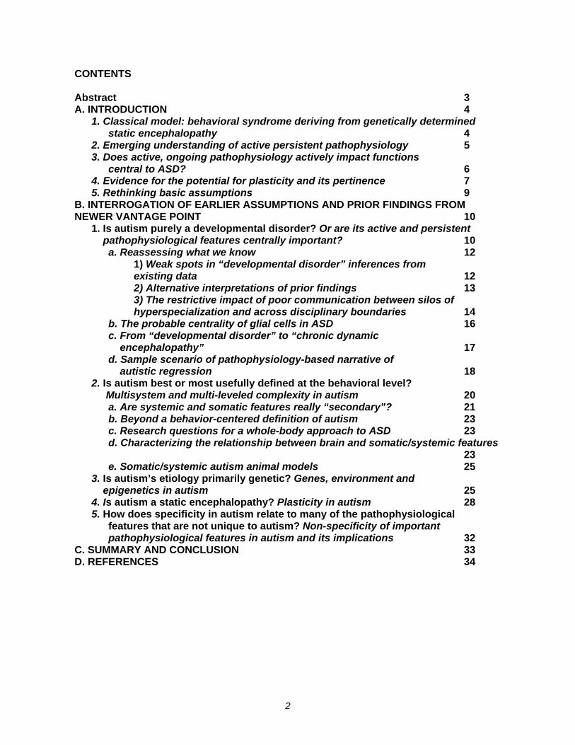
CONTENTS Abstract 3 A. INTRODUCTION 4
1. Classical model: behavioral syndrome deriving from genetically determined static encephalopathy 4
2. Emerging understanding of active persistent pathophysiology 5 3. Does active, ongoing pathophysiology actively impact functions
central to ASD? 6 4. Evidence for the potential for plasticity and its pertinence 7 5. Rethinking basic assumptions 9
B. INTERROGATION OF EARLIER ASSUMPTIONS AND PRIOR FINDINGS FROM NEWER VANTAGE POINT 10
1. Is autism purely a developmental disorder? Or are its active and persistent pathophysiological features centrally important? 10
a. Reassessing what we know 12 1) Weak spots in “developmental disorder” inferences from existing data 12 2) Alternative interpretations of prior findings 13 3) The restrictive impact of poor communication between silos of hyperspecialization and across disciplinary boundaries 14
b. The probable centrality of glial cells in ASD 16 c. From “developmental disorder” to “chronic dynamic encephalopathy” 17 d. Sample scenario of pathophysiology-based narrative of autistic regression 18
2. Is autism best or most usefully defined at the behavioral level? Multisystem and multi-leveled complexity in autism 20
a. Are systemic and somatic features really “secondary”? 21 b. Beyond a behavior-centered definition of autism 23 c. Research questions for a whole-body approach to ASD 23 d. Characterizing the relationship between brain and somatic/systemic features 23 e. Somatic/systemic autism animal models 25
3. Is autism’s etiology primarily genetic? Genes, environment and epigenetics in autism 25 4. Is autism a static encephalopathy? Plasticity in autism 28 5. How does specificity in autism relate to many of the pathophysiological
features that are not unique to autism? Non-specificity of important pathophysiological features in autism and its implications 32
C. SUMMARY AND CONCLUSION 33 D. REFERENCES 34
3
ABSTRACT
The purpose of this chapter is to reflect upon the implications of the identification of active pathophysiological processes in autism spectrum disorders (ASD), and to reflect back upon prior findings and formulations in the light of these recent discoveries. The chapter articulates challenges posed by these discoveries to deeply held assumptions about ASD. These assumptions are embodied in a classical model framing ASD as a problem of genes, brain and behavior – i.e., as a genetically determined developmental disorder of the brain whose main manifestation is behavioral alterations based upon an indelible static encephalopathy; this model would not have predicted the growing documentation of pathophysiological disturbances. The chapter describes an emerging pathophysiology-centered model of autism that can subsume genes, brain and behavior but also includes much more. Prior findings and models are re-evaluated to support the framing of ASD as 1) not only developmental but also a chronic condition based on active pathophysiology, 2) not only behavioral but also having somatic and systemic features that are not secondary but rather intrinsic consequences of underlying mechanisms, 3) not only genetic but also environmental, 4) not a static encephalopathy but a dynamic, recalcitrant encephalopathy, and 5) not a set of discrete behavioral features neatly mapping to specific genetic mechanisms but a set of emergent properties dynamically arising from pathophysiological systems whose parameters have been dramatically and interactively perturbed. It is argued that a research program based upon this approach will incorporate the strengths of the classical model, will encourage many more routes to investigations with practical and treatment applications, and may be a much more rapid path to providing much needed help to affected individuals and their families.
4
A. INTRODUCTION
While autism spectrum disorders (ASD) can involve exquisite gifts and unusual qualities of perception and thought, they can also involve a great deal of suffering, for the individual on the spectrum as well as family and community. On this account, a core question in autism work needs to be how to help the most people in the most effective ways as quickly as possible. The goal of making sense of autism and its mechanisms needs to be deeply harnessed to this core purpose. Our aim should be to relieve suffering at multiple levels – from aversive sensory overload, sleep disruption, recurrent infections and gastrointestinal troubles to overcoming obstacles to communication; to misunderstanding by non-autistic people of the experiences of people with autism; to aggression and self-injurious behavior; to the burden of allocating scarce resources to deliver therapies that may not be optimally designed, targeted or implemented; to acrimonious debate and fiscal drain. Last but hardly least, if any part of the impairment of optimal functioning in new cases of autism is not purely genetically determined, the suffering and severity that is therefore neither inevitable nor necessary should be prevented or ameliorated.
If we are to help most quickly and with the broadest and greatest effectiveness, then how do we do so, and how much can we really help? What can we realistically expect to accomplish? The answers we give to these questions are greatly conditioned by what we understand autism to be. The main goal of this chapter is explain and compare two models of autism which lead to greatly different expectations: a) a classical model of autism as a genetically determined developmental brain disorder and static encephalopathy, and b) an emerging model of autism centered around active systemic environmentally as well as genetically influenced pathophysiological processes beginning early in life and leading to an chronic persistent encephalopathy with dynamic features. This comparison will show that the emerging dynamic pathophysiological model includes the strengths of the classical static model but also takes account of emerging data that is incommensurable with the older formulations. It will also give support to the argument that the emerging more inclusive model offers more opportunities for constructive investigation and intervention.
1. Classical model: behavioral syndrome deriving from genetically determined static encephalopathy
Autism has until recently been considered to be a developmental disorder originating in faulty genes that skew early brain development and lead to a devastating and incurable static encephalopathy. Since this perspective frames autism as directly deriving from an indelibly fundamental alteration of brain structure and function, its adherents take the logical next step when they assume that there are fundamental profound limitations to the potential efficacy of any current therapies. An additional commonly held assumption of this classical viewpoint is that the core behavioral features of autism are specifically determined at the genetic and molecular level. From this vantage point, only extremely precise molecular or genetic interventions targeting some critical aberrant pathway have any chance of unlocking neural functioning, but these pathways have yet to be discovered and the molecules to target them are yet to be invented. Therefore, to identify targets and develop effective and safe interventions, an extensive, expensive and long-term research strategy is necessary in order even to begin to relieve suffering in any serious way.
5
The recent framing of autism as heterogeneous, or “autisms” (plural), modifies this model by suggesting that “autism” is really a collection of different “autisms,” each with its own mechanism and perhaps even its own gene(s). The research program derived from this framing would still look for distinctive mechanisms but now may implicitly propose multiple parallel searches for mechanisms. If this is not accompanied by seeking final common pathways that may bridge across these distinct “autisms” and provide routes of intervention that could be beneficial more broadly than to any one small subgroup, then the road ahead is even longer.
2. Emerging understanding of active persistent pathophysiology
Clearly some kind of atypical brain function must be going on in ASD in order for its atypical behaviors to be produced. The very high prevalence of sensory and sleep problems (Leekam et al. 2007; Tomchek and Dunn 2007) and the high rate of epilepsy in ASD (Canitano 2007) also support this. Critical questions for which we have enticing clues but no clearly worked out answers include: 1) what are the mechanisms underlying the altered brain function, and 2) what are the causes? Within the classical model, a common phrase heard is “genes-brain-behavior.” This suggests that genetic alteration of the brain causes autistic behavior, and it also implies that researching this specific chain of levels and their relationships is sufficient for understanding ASD.
The trouble with the “genes-brain-behavior” framework is that it promotes oversimplified thinking about the way genes alter brain and the way brain alters behavior. Even to use the three words in a string is a problem, because 1) genes themselves do not directly impact brain but shape other processes that alter brain, 2) these processes that alter brain are impacted by other things in addition to genes, 3) the combination of genes and these other processes alter not only brain but also the rest of the body including systemic molecular and cellular mechanisms that are not organ-specific, 4) there is not unidirectionality but bidirectionality – indeed web-line network interconnections – in that the consequences of all of these dynamical alterations can feed back and alter gene expression, and 5) the outputs of all of this complexity are not limited to behavior but also include phenomena at many other levels (Herbert 2002; Noble 2008). Therefore, to say “genes-brain-behavior” leaves unspecified many intermediary levels that need to be explicitly spelled out and investigated.
One formulation more inclusive than “genes-brain-behavior” is “input-pathophysiology-output” (Herbert 2005a). Input can include genes but also a range of environmental factors. Pathohysiology can include prenatal processes with early impacts on brain development that modulate fundamental features of brain, but it can also include processes and impacts at other time points that have other types of effects on both brain and body. Outputs can include behaviors but also medical illnesses and a host of other functions.
Critical findings in ASD at the level of active, ongoing pathophysiology that inspire the present volume would not have been predicted by the classical genes-brain-behavior model. Particularly of note are the phenomena of oxidative stress, mitochondrial dysfunction and inflammation that have been identified in a growing number of studies in a substantial number of individuals with ASD. Evidence of these processes has been identified in somatic tissue samples with measurement of alterations in a variety of substances including in membrane phospholipids(Bu et al. 2006; Chauhan et al. 2004; Vancassel et al. 2001), antioxidant enzymes and metabolites
6
in the glutathione synthesis pathway (Chauhan and Chauhan, 2006;James et al. 2006) documentation of both oxidative stress (Evans et al. 2008; Pardo et al. 2008; Vargas et al. 2006) and neuroinflammation (Li et al. 2009; Vargas et al. 2005) as well as rapid membrane turnover (Minshew et al. 1993) and altered energy metabolism (Chugani et al. 1999) in brain has also been produced. Much more is reviewed extensively in this volume.
These phenomena are active, ongoing pathophysiological abnormalities. While their chronic impacts can be stubbornly persistent, and while they can cause damage that is more stable, their primary mechanisms act on the time scale of hours and days or even less. They cannot be attributed to genetic errors or early insults in a simple or straightforward fashion, although those could contribute vulnerability or get these processes started. It needs to be emphasized that the identification of active, persistent disturbances of physiological functions, particularly in the brain, is a landmark in the history of autism science because it adds dimensions to the parameters we need to include in considering the condition, and also because it changes the temporality from a playing out of something that happened early on to a process that is continuingly active.
3. Does active, ongoing pathophysiology actively impact functions central to ASD?
Even granted the existence of active pathophysiological processes in ASD, a skeptic from the classical model vantage point might question whether they have any significant relevance to ASD. From the classical point of view, it would seem obvious that these sorts of influences could be little more than small bubbles on the surface of the genetically determined ocean of profound brain abnormality. To face this challenge, we need to determine whether the ASD phenotype or any of its components or contributors could be created or substantially aggravated by neural functioning alterations that are chronically and actively maintained.
From a pathophysiology-centered point of view, once the chronic persistence and active character of these processes is recognized, it is not so radical to suggest that perhaps these phenomena might affect synaptic and neural systems function. In the literature of neurobiology, there is plenty to suggest that oxidative stress and immune activity can be neuromodulatory. The immune system, energy metabolism and oxidative stress are abundantly documented as impacting the central nervous system and its function (Lowry et al. 2007; Lozovaya and Miller 2003; Mattson 2007; Mattson 2008; Mattson and Liu 2002; Miller, Maletic, and Raison 2009; Wrona 2006). These considerations may be particularly pertinent to the phenomenon of autistic regression, which generally occurs somewhere around the middle of the second year of life. Even if “regression” is preceded by a variety of subtle signs of dysfunction, it is occurring far beyond the most critical periods of brain development and deserves investigation as a new event and in particular, as a shift in functional/metabolic/neurodynamic state and not just as an inevitable playing out of early hard-wired brain alterations.
With chronic active pathophysiology identified systemically and in brain tissue from individuals with autism, with this active pathophysiology having potential neuromodulatory effects, and with functional changes such as regression needing mechanistic explanation, it becomes necessary to consider the possibility that the biological basis of the autism behavioral phenotype may not be determined “architecturally” once and for all in utero, but rather may be actively sustained, possibly
7
even caused or at least substantially aggravated by persistently active pathophysiology (Anderson, Hooker, and Herbert 2008; Zimmerman 2008).
We can imagine a number of possibilities: 1a) inputs (genes, environmental factors) create an indelible alteration in prenatal or early postnatal brain development; 1b) these indelible in utero impacts of genes and environmental factors are mediated by pathophysiological processes such as inflammation or oxidative stress; 2) some inputs (e.g. genes, teratogens, infections or immune responses to infections) increase vulnerability to other inputs that alter early prenatal or early postnatal brain development; 3) some inputs increase vulnerability to other inputs (e.g. excitotoxins) or pathophysiological states (e.g. immune triggers, oxidative stress) that alter neural function postnatally; and 4) chronic, persistent alteration in neural function (e.g. cumulative toxic body burden and/or chronic neuroinflammation having a persistent excitotoxic impact) can in turn lead to changes in brain tissue (e.g. mitochondrial damage cellular dysfunction cell death) which in turn may feed back to further affect function.
Once these additional dimensions beyond genetic determination of altered brain development join the parameters of concern, how do we assess what the type of influence and relative weight may be of each class of contributor? How far can this be pushed? For example, if excitotoxic modulation of synaptic function is chronic (i.e., from ongoing exposure or chronic inflammation) and/or persistent (i.e., with semi-permanent effects from even a transient exposure), can we consider whether it could contribute to a chronic encephalopathy? And could such a chronic encephalopathy potentially in some cases not simply modulate the autism but actually be the autism? Could genetic vulnerability and genetic impacts turn into autism (or more severe autism) with the onset of these pathophysiological processes? We obviously do not know the answer but this chapter reflects on the question.
Insofar as pathophysiological mechanisms can be affected by environmental input, it is also important to consider potential positive impacts. If there is a formative role for pathophysiology, this suggests that factors like diet, sleep quality, stress, exercise, autonomic arousal, environmental exposures and medications all could be having substantial short-term impacts on symptom severity and quality of life. It also suggests that such factors, which include both health-promoting and health-destroying variants, can have substantial effects over time on level of function and quality of life. On the scale of years, the "ongoing" nature of pathophysiological activity means that some interventions might be able to provide major long-term benefits as well.
4. Evidence for the potential for plasticity and its pertinence
To make a plausible argument that active, persistent pathophysiology might strongly modulate or even create core features of autism, there would need to be evidence of some kind of intra-individual variability in the phenotype that occurred in relationship to pathophysiologically pertinent processes. Such variability (e.g. symptom onset, marked worsening or marked improvement) would suggest that fluctuations in modulatory processes might have significant impact. As it happens, such evidence exists.
The idea that physiological modulation could contribute more than marginally is becoming less far-fetched in the light of published reports of short term marked
8
improvements in core features of autism. Investigators recently pursued suggestions from clinical case reports that behaviors and core capacities in autism may improve markedly in the setting of fever (Curran et al. 2007)—clinicians were fairly commonly hearing from parents that their affected children could relate better, make more eye contact and sometimes even talk transiently in the setting of fever—one mother poignantly described her experience during her child’s fever to the author of the present review as “visiting with my son.” A prospective study was thus performed utilizing the Aberrant Behavior Checklist to rate behavior changes; the study found that fewer aberrant behaviors were recorded for febrile patients on the subscales of irritability, hyperactivity, stereotypy, and inappropriate speech compared with control subjects in a fashion that was not associated with severity of illness. While lethargy scores were greater during fevers, and all improvements were transient, the behavioral improvements could not be attributed to the lethargy and the results instead suggested a genuine improvement in core functions. An earlier paper investigated 11 children with the history common in ASD of a period of often recurrent infection and antibiotic exposure followed by the development of chronic persistent diarrhea and then onset of autistic features, or “regression” (Sandler et al. 2000). This common phenomenon has spawned research demonstrating abnormal variants of clostridial bacteria in ASD (Finegold et al. 2002; Parracho et al. 2005; Song, Liu, and Finegold 2004) and animal models showing nervous system and behavioral impacts of propionic acid, a metabolic product of clostridia (MacFabe et al., 2007; Shultz et al. 2008a; Shultz et al. 2008b) which are part of a larger ferment of research on the influence of intestinal microecology (the “microbiome” on medical and psychiatric health (Alverdy and Chang 2008; Li et al. 2008; Nicholson, Holmes, and Wilson 2005).. This study investigated impact on behavior of oral vancomycin, which is a potent antibiotic normally given intravenously and minimally absorbed from the intestine but that devastates intestinal microorganisms. They noted significant short-term improvement using multiple pre- and post-therapy evaluations coded by a blinded clinical psychologist, with the transiency presumably due to re-growth of pathogenic intestinal microorganisms after cessation of antibiotic dosing. In both of these cases the improvement was notable, rapid in onset, and short in duration suggesting that the maladaptive physiological setpoint was insufficiently challenged by fever or transiently altered intestinal microbiota to shift to a different semi-stable state.
Some challenges to prior conceptions of developmental disorders have also emerged on the laboratory front. Symptom reversal has recently been reported in mouse models of developmental disorders—Fragile X syndrome (Hayashi et al. 2007), Rett Syndrome (Guy et al. 2007), and tuberous sclerosis (Ehninger et al. 2008), all considered genetic and incurable — through molecular intervention, including in older animals. This is striking because it forever undermines the basis for simply taking for granted that neurodevelopmental disorders are incurable or have only a narrow critical window after which intervention is pointless. At the other end of the lifecourse, the rapid transient reduction of Alzheimer’s disease symptoms within minutes of administration of perispinal etanercept suggests that chronic active and potentially reversible pathophysiology may also contribute to the encephalopathy in this devastatingly progressive disorder (Tobinick and Gross 2008a, 2008b).
With regard to the autism clinical papers discussed above, it is critical to note that fever does not create a permanent alteration of immunologic or neurobiological pathways, and oral vancomycin does not permanently alter intestinal flora, consistent with the changes not being persistent. But it is also critical to note that these supposedly lifelong core features of autism could be altered even in the short term, which itself is
9
inconsistent with a “static encephalopathy” model. All of this challenges us to think outside of the box of irretrievable brain damage in relation to the encephalopathy of ASD (and other conditions as well). The potential mechanisms that come to mind are in the domain of active, dynamic pathophysiology (including but hardly limited to altered gene expression) rather than genetic predetermination, as the genetic mechanisms causing an in utero disturbance of brain development would not explain such short-term fluctuations. In the Curran et al. (2007) paper on improvement with fever, the authors speculated that the phenomenon was driven by some mechanism related to immunologic and neurobiological pathways, intracellular signaling, and synaptic plasticity; in the Sandler et al. (2000) paper, the authors speculated that the oral antibiotic transiently suppressed an enteric microorganism and its production of a neurotoxin-like substance.
If such marked short term changes are possible, the idea that the encephalopathy in ASD is a dynamic (albeit recalcitrant) “state” rather than a wired-in static “trait” becomes conceivable, and the possibility of identifying the mechanism for and extending the duration of such changes can be framed as a worthwhile and important goal for research.
The implication of this is major: it means that we must consider the possibility that the functional impairments we observe in individuals with autism may be products not so much of innate “deficits” as of active (and obstinate) pathophysiological obstruction of capacities for which brain substrate is still at least partly present. Moreover, given that these processes are known to progressively assault and damage cellular integrity, and given the evidence suggesting progressive changes in brain tissue: (cellular changes (Bauman and Kemper 2005) and volume loss (Aylward et al. 2002)), the importance of finding ways to medically intervene to slow or stop this degeneration as early as possible comes into clear focus..
5. Rethinking basic assumptions
As our understanding of these new dimensions take shape, it starts to seem that the assumptions underlying the classical model of ASD need to be revisited. With these features in mind, it becomes possible and necessary to interrogate prior findings for fresh interpretations. The goal of this chapter is thus to spell out how emerging findings are revealing limitations in the assumptions of the classical view, and to outline some core features of a newer more inclusive view. These emerging findings are elucidating mechanisms suggesting that autism is more than a developmental disorder, that more than genes are etiologically contributory, and that the encephalopathy has dynamic features so that it is not strictly static.
We will develop the argument by posing the following questions, and explaining our rationale for the following responses:
Questions:
1. Is the category of “developmental disorder” adequate for autism?
2. Is autism best or most usefully defined at the behavioral level?
3. Is autism’s etiology primarily genetic?
10
4. Is autism best described as a static encephalopathy?
5. Is autism a unique and distinct syndrome?
Responses:
1. More than developmental: Autism is more inclusively framed as a chronic and also dynamical/semi-episodic multisystem condition that begins in utero or early in life during a period when developmental processes are greatly sensitive to perturbation and that continues through the lifecourse with persistent, ongoing, active and dynamic pathophysiology that may contribute critically to phenotypic features.
2. More than behavioral: Behavioral criteria alone do not encompass the multisystem features that are increasingly being appreciated in ASD, which are so common in affected individuals as to suggest that they may play central rather than secondary roles and/or reflect shared core underlying pathophysiological processes.
3. More than genes: Autism is likely to be the result of a complex interaction of multiple risk factors; neither genes nor environmental agents can a priori be assumed to be primary in their contributions, and the interaction of contributors persists beyond early development.
4. From static to active dynamic: Within-individual variability in severity of core features and emerging awareness of plasticity and improvement in autism, alongside of the relative intactness of neural structures in ASD, suggest that the encephalopathy in autism is recalcitrant but rooted strongly in dynamic processes, and that framing it as static is inaccurate.
5. From autism as a specific entity with specific genetic determination of each of its subcomponents to ASD as an emergent property of a neural and somatic system altered by physiological challenges during a sensitive period of early development. From a systems pathophysiology perspective, autism appears as a complex integration of continuously distributed abnormalities in multileveled features and has substantial physiological overlap with underlying pathophysiology in many other chronic diseases. It may be that we do not need to target features specific to autism but that therapies targeting underlying physiological features that are contributory but not unique to ASD could lead to altered emergent properties including altered behaviors.
B. INTERROGATION OF EARLIER ASSUMPTIONS AND PRIOR FINDINGS FROM NEWER VANTAGE POINT
1. Is autism purely a developmental disorder? Or are its active and persistent pathophysiological features centrally important?
The idea that ASD is a developmental disorder seems self-evident. ASD begins in early childhood, with abnormalities in responsiveness sometimes even evident at birth. Brain abnormalities have been documented at the neuropathological level consistent with changes occurring in utero. The high heritability and high recurrence rate
11
also support this framing. The characteristic clustering of behavioral features in the ASD phenotype suggests some kind of specific cause.
There are other ways of interpreting the above cluster of phenomena. These features of early onset, neuropathology changes suggestive of in utero onset, specific behavioral configuration and high heritability/high recurrence suggestive of genetic cause can be at least partly decoupled from the inferences with which they have been associated. Certainly important events occur at these early stages of development. The problem arises at the level of drawing implications from these observations about underlying mechanisms. If one assumes a priori that this is a “developmental disorder” in the neurobiological or neurogenetic sense, clinical and research observations may be given interpretations consistent with the implications of this assumption, while other potentially valid interpretations consistent with a more chronic model may be neglected.
The notion of a “developmental disorder” has a number of different connotations. From a developmental psychology point of view, it can connote simply that because function and capability change with development, a disorder in childhood will manifest differently at different ages. This is unquestionably true. However, there are other perspectives carrying more severe connotations. From a medical and neurobiological vantage point, “developmental disorder” commonly connotes at least the following four characteristics: 1) that there is a profound, if potentially subtle, alteration in the developmental trajectory of the brain, 2) that the ensuing developmental brain alterations are primary core targets of the etiological agent rather than incidental or secondary, 3) that these alterations directly cause the behavioral phenotype, and 4) that these brain features and the accompanying encephalopathy are indelibly unchangeable. This “developmental disorder” model is derived from observations in neurogenetic syndromes and syndromes of brain malformation where there are clearly observable and classifiable alterations in brain development based upon a fault in some neurochemical or regulatory process that leads to fairly predictable consequences in affected individuals.
While this framing of developmental disorders is most commonly associated with syndromes having genetic etiologies, the fields of developmental neurotoxicology and teratology have shown that exogenous substances can target early developmental processes and lead in a similar fashion to predictable malformations and developmental syndromes, such as fetal alcohol syndrome, fetal valproate syndrome, and fetal Minimata disease. Similar arguments are also being made about disorders of later onset such as schizophrenia (Arnold 2001; Opler and Susser 2005).
Given the widespread assumption that autism is not only a developmental disorder but a static encephalopathy, it appears that the stronger and more severe model outlined above has been applied in interpreting the presentation of ASD. But if we carefully examine the support for inferring the four characteristics connoted by the medical-neurobiological framing of “developmental disorder” listed above, it turns out that there are major gaps in our knowledge and more particularly in the evidence basis for the assumptions we have been making. We have certainly 1) identified a range of brain alterations that qualify as profound, often as subtle and sometimes pervasive, including changes in limbic system structures, cerebellum, white and gray matter volume, corpus callosum, subcortical gray matter structures and asymmetry. But we have not 2) shown for all of them that they are primary targets of an identifiable etiological agent rather than secondary consequences of a pathophysiological process, nor have we 3) conclusively demonstrated that they specifically cause the autism
12
behavioral phenotype (we have merely shown association and have not excluded the possibility that some of these changes may be downstream of something else that is driving the phenotype), and neither have we 4) proved that they are unchangeable, or that their unchangeability correlates with functional lack of plasticity—for all of these points our “knowledge” is at the level of plausible narrative, not empirical elucidation of mechanisms.
a. Reassessing what we know
In the light of emerging pathophysiological findings, it needs to be considered that the set of phenomena leading people to consider autism a “developmental disorder” – i.e. the brain changes, the early onset, the heritability and recurrence and the clustering of behavioral features – individually and together may have potential additional and/or alternative interpretations. Below are a series of considerations that cannot be encompassed within the “developmental disease” model as described above. Individually and together, they put autism in the “chronic active disease” category and pose challenging questions about what the interfaces may be between chronic processes that begin very early in life and alterations in development.
1) Weak spots in “developmental disorder” inferences from existing data
a) Brains of autistic individuals are for the most part remarkably normal looking. An MRI scan of the brain of most individuals with ASD would be interpreted by a clinical neuroradiologist as normal (and clinical abnormalities when identified are typically idiosyncratic, possibly incidental, and quite possibly secondary to some other process), and it is only by careful quantitative analysis that macroanatomical differences from brains of neurotypical subjects can be identified – it takes this level of intensive research-grade measurement because for the most part changes are too subtle to be identified by the unaided eye (Caviness et al. 1999). Indeed, some neuropathological researchers have held that since the brains lack major dysmorphology, they are unlikely to have suffered significant insult prior to the late gestational or early postnatal period (Ciaranello, VandenBerg, and Anders 1982; Coleman et al. 1985; Raymond, Bauman, and Kemper 1996). The observation has been made that there is a striking disconnection between the almost indiscernible white matter tract as well as general structural abnormalities and the dramatic functional impairments (Conturo et al. 2008).
b) Suggestions that neurodevelopmental disorders can be triggered by events during the fetal period are supported by a growing body of literature (Connors et al. 2008; Fatemi et al. 2002; Patterson 2002; Shi et al. 2003 ; Smith et al. 2007). There is a huge literature on developmental neurotoxicity (Slikker and Chang 1998) as well as developmental immune injury (Dietert and Dietert 2008; Hertz-Picciotto et al. 2008). However while these exposures can now be said to increase the potential for neurodevelopmental disorders, there is not support at this time for going further—i.e., such exposures have by no means been shown to sufficient on their own to cause postnatally emerging developmental disorders or ASD in particular. Nor have developmental disorders or ASD in particular have been shown to be necessarily or in all cases preceded by such events.
c) The model of autism derived from the connectivity literature related to connectivity impairments underlying impairments in complex processing (Just et al. 2004, 2007;
13
Muller 2007) is synchronic – i.e. it can be marshaled to explain the apparent selective impairment of complex processing in individuals with fully developed autism at a particular point in time. It is not a diachronic model – i.e., it does not help at all in explaining the phenomenon of the development of autism, and particular the phenomenon of regression into autism. We do not understand what changes so that a child who was producing behaviors closely consistent with normal developmental milestones either falters, plateaus, shifts tracks, or in some other way shifts to slow and/or alter development. If one assumes that autism is inborn, then it is possible to construct a narrative stating that the connectivity problem is innate or prenatal in origin, but doesn’t show itself until critical processes kick in (or fail to occur) postnatally at which point the innately altered wiring becomes a problem. An alternative narrative with a slightly later developmental timepoint is the idea that there “failure of pruning” of excess neural processes. We have no direct evidence to prove either narrative, and in fact imaging evidence as noted in point #4 below goes against the idea that there has been a pruning failure.
2) Alternative interpretations of prior findings
d) The brain findings to date contain many suggestions of prenatal events, but it must be remembered that explaining findings in a fully developed brain of a child past toddlerhood and particularly of an adult is an “archaeological” exercise in reading a developmental history from a snapshot—i.e. it is highly interpretive. At least some of the findings interpreted as supporting a prenatal onset have alternate possible interpretations. Moreover, given the scarcity of post-mortem brain specimens from people reliably diagnosed with ASD, most neuropathological observations have been noted in only a small number of cases and the observations have not always been replicated. Here are some examples where alternative explanations have been suggested.
• An observed tight packing of a larger number of smaller cells in limbic structures has been interpreted as indicating a mid-gestational event, but it is also becoming evident that limbic structures are especially sensitive to immune influences and could be altered in their cellular structure through other classes of events than the early developmental events initially considered—with these other classes of events conceivably occurring at somewhat later times (Buller and Day 2002; Buller, Hamlin, and Osborne 2005; Nyffeler et al. 2006).
• Minicolumnar alterations have been interpreted as occurring fairly early in gestation but arguments have been advanced for how they could occur later as well (Gustafsson 2004).
• Purkinje cells appear vulnerable but they may not necessarily be lost: while they do not pick up Nissl stain, they do appear when calbinden staining is used. Purkinje cells are highly vulnerable to excitotoxicity and their failure to be detected by Nissl stain may reflect chromatolysis or excitotoxic-induced alterations in their metabolism (Kern 2003).
• Brainstem and inferior olivary findings in ASD earlier interpreted as indicating prenatal disturbance of development have upon restudy been identified not only in ASD but also in control brain tissue (Thevarkunnel et al. 2004; Whitney et al.
14
2008), suggesting that interpretations regarding both developmental trajectory and specificity need to be rethought.
• Brain enlargement was early on attributed to a “failure of pruning” (i.e. a failure to eliminate the super-abundance of neurons produced early in brain development) but magnetic resonance spectroscopy (MRS) studies have shown reduced (DeVito et al. 2007; Endo et al. 2007; Friedman et al. 2003, 2006; Kleinhans et al. 2007) or unchanged (Vasconcelos et al. 2008; Zeegers et al. 2007) rather than increased n-acetylaspartate (NAA) not consistent with neuronal increase.
• Moreover, while NAA is often considered to be a measure of cell density, it can also be construed as a measures of cellular and even mitochondrial function, and its reduction may be an indicator not so much of neuronal loss as of neuronal dysfunction, particularly given the reversibility of NAA decrements in contralateral tissue following surgical resection of epileptic foci (Hugg et al. 1996; Pan et al. 2008; Serles et al. 2001).
• White matter enlargement has been identified in T1-weighted scans that offer only macroanatomic measures but no resolution at the scale of cellular changes; the distribution of this enlargement suggested an increase in short-cortico-cortical fiber density consistent with local hyperconnectivity and long-distance under-connectivity (Courchesne and Pierce 2005); although there was no neuropathological data on the tissue composition of this enlargement. But as results from diffusion tensor MRI imaging (pertinent to assessing white matter integrity) and MRS imaging (pertinent to measuring metabolites) are appearing, this inference is being contradicted by evidence suggesting that the expanded volume cannot be explained by increased fiber density and in fact may be due to altered water properties in the tissue more consistent with alternative pathophysiology such as neuroinflammation ( Hendry et al. 2006; Sundaram et al. 2008; Zimmerman 2008;).
The overall point here is not to argue that we have a clear-cut case in every respect for postnatal or pathophysiology/dynamical-influenced events in ASD brain development, but simply to say that there remains a fair amount of ambiguity in the limited data presently available to us.
3) The restrictive impact of poor communication between silos of hyperspecialization and across disciplinary boundaries
e) Functional imaging methods including fMRI (functional magnetic resonance imaging), EEG (electroencephalography), MEG magnetoencephalography), PET (positron emission tomography) and SPECT (single photon emission computed tomography) have shown alterations in regional interconnectivity by various methods (e.g. connectivity, coherence, covariation) (Herbert 2005b; Muller 2007; Herbert and Caviness 2006). Some investigators have inferred that this interconnectivity alteration might be linked to structural alterations in white matter. But demonstrating such a relationship would require coregistering functional data such as fMRI or MEG or PET with anatomical data such as diffusion tensor imaging or MRS, to see whether alterations in white matter integrity occur in a fashion that relates in any consistent manner with alterations in functional connectivity; to while such work is in progress, few results have been reported to date and so we actually have little
15
evidence based idea what tissue changes might be causing alterations in functional connectivity, EEG/MEG coherence or inter-regional covariation. The possibility has not been tested that an alteration of synaptic function secondary to the excitotoxic effects of chronic tissue pathophysiology could have systems impacts on patterning of neurodynamic activity that could contribute to altered functional connectivity. In fact, the investigators studying brain connectivity hardly even mention the emerging pathophysiology findings – the silo effect where groups of narrowly specialized investigators fail to cross-fertilize outside their own small circles and the cognitive dissonance effect are both apparently very strong, and the cross-fertilization between the levels of investigation is quite weak.
f) Several neuropathological investigations of brain tissue in ASD have found evidence of neuroinflammation and oxidative stress (Evans et al. 2008; Li et al. 2009; Sajdel-Sulkowska et al. 2008; Vargas et al. 2005, 2006). In addition, some neuropathological investigations into the nature of white matter enlargement are beginning to suggest that there is astroglial activation in the enlarged outer, radiate part of the white matter that is not present in the deeper, non-enlarged white matter, along with microglial activation that is present particularly in the cerebral cortex (Pardo et al. 2008). These early findings point toward fresh ways of making sense of both altered synaptic activity and brain hypoperfusion in ASD.
• Regarding synaptic function, an emerging field of literature relates to the active roles played by glial cells (astroglial, microglial and oligodendroglial cells) in signal transmission in the brain (Fields 2006; Fields 2008). These cells are being promoted in our understanding from handmaidens of neurons to active players in a much more complicated collaborative endeavor; the importance of these cells has prompted some to say that we should change the name of the field of study from “neurobiology” to neurogliobiology” (Peschanski 1991). Astroglial cells participate in a “tripartite synapse” (Halassa, Fellin, and Haydon 2007) as they wrap themselves around the ends of two synapsing neurons and neurochemically modulate the synaptic activity between these two cells. In two different specialized silos of the research world, it is known that i) immune-activated astroglial cells behave quite differently neurochemically, and ii) astroglial cells are exquisitely sensitive to toxicant exposures, which can also put them into an activated state. Apparently however, the impact of activation of glial cells on their function in the tripartite synapse has not yet been researched—i.e. the silos of glial-immune and glial-toxin specialists are still not communicating and synergizing with the silo of glial-synapse specialists. So some basic science that we would need to understand the functional impact of either white matter enlargement or glial activation simply has not been performed.
• Regarding brain hypoperfusion, it is also known that astroglial cells become larger when they are activated, and since they encircle small vessels, this enlargement can reduce capillary diameter by as much as 50% (Aschner et al. 1999); such a reduction is consistent with (though such consistency does not prove it is the cause of) measures of brain perfusion in ASD, where the several papers report perfusion reduced blood perfusion, albeit in different distributions (Herbert 2005b; Tuchman and Rapin 2006). Other possible pathophysiological contributors to hypoperfusion worth investigating include the modulation of vasoconstriction and platelet activation and aggregation by oxidative stress (Yao et al. 2006) and the impact of activation of microglia encircling cerebral
16
microvasculature (Vargas et al. 2005). Interestingly, the papers reporting hypoperfusion in ASD to date are almost entirely mute on the subject of the tissue biology or pathophysiology of this hypoperfusion, with the interpretations in the discussion sections of the papers focusing on the psychological significance of the localization of the hypoperfusion with an unstated assumption that this phenomenon is stable, static and persistent. Here the silo of pathophysiology specialists is not linked with the silo of psychology specialists.
The point of all of these examples is to give a taste of various ways that the introduction of pathophysiological variables can point to rather different interpretations of existing brain findings. It also serves to illustrate how much of what we think we know about the brain in autism is actually a morass of fragments of data being extrapolated to support inferences based on a priori assumptions. By showing that when we augment the conceptual input parameters to include chronic pathophysiology and not just genetics and brain development, we get as output a substantially different set of interpretations, I hope I have at least begun to demonstrate how tenuous are the established interpretations. On this basis, I would argue that we have no solid grounds for excluding or dismissing a research program based on a different set of assumptions than “developmental disorder of prenatal onset.” On the contrary, there are many reasons for arguing that it is very important that we pursue a research program based on these different assumptions, as well as communication and synergy across specialized silos, and do so aggressively.
b. The probable centrality of glial cells in ASD
The role of glial cell activation in brain dysfunction in autism needs more attention at the functional as well as at the neuropathological level (Coyle and Schwarcz 2000; Giaume et al. 2007). Glial cell activation can be set off by a myriad of triggers, and many of the downstream consequences are not specific to the initiating agents. While pathophysiologically oriented investigators have been greatly influenced by the identification of activated microglia and astroglia in brain tissue from individuals with autism (Vargas et al. 2005), adherents of the classical “developmental disorder” model often refuse even to discuss it, some arguing insufficient replication. Since the publication of the Vargas et al. (2005) paper which identified activated microglia and astroglia in all 11 of the brains studied, the group has collected at least another 9 brains, one from someone with Asperger’s syndrome, and all of these subsequent brains also showed this activation, including the one with Asperger’s syndrome, considered to be a milder condition (Zimmerman 2008). And now, as mentioned, another group has also identified central nervous system immune activation in ASD (Li et al. 2009).
Microglia comprise about 10% of the cells in the brain and perform important functions in both the resting and the activated state. They appear to release trophic factors during development, some of which have been measured as having different levels in infants who later develop autism (Nelson et al. 2001). When activated, microglia synthesize and secrete a range of pro-inflammatory cytokines (Hanisch 2002) several of which are neurotoxic, and in this activated state, they also promote astroglial overactivation and dysfunction, as well as edema (Orellana et al. 2009). Astroglia are multipurpose cells that not only support neurons but also perform metabolic and signaling functions (Aschner et al. 1999; Fields 2006; Halassa, Fellin, and Haydon 2007); astrocytes are highly networked into a syncitium through gap junctions (Theis et al. 2005) through which depolarization and calcium waves spread rapidly and interact
17
with neuronal activity; they are centrally involved in regulating neurovascular coupling (Koehler, Gebremedhin, and Harder 2006). It is increasingly appreciated that astrocytes are influenced by inflammation in a variety of disease states (Kielian 2008); activation of astroglial and microglial cells have a wide variety of effects that are arguably consistent with many observed features of ASD. Microglia activation is associated with vasogenic and cytotoxic edema associated with hypoperfusion; activated microglia release glutamate which induces astrocyte edema (Han et al. 2004; Liang et al. 2008). Microglial activation can occur rapidly in response to insults; when it persists, its neurotoxic impact progressively increases over time. Astroglial support of neuron chemistry and secretion of neuromodulators is altered in the activated state (Aschner, Sonnewald, and Tan 2002). Astroglia maintain the redox potential including through the production of glutathione, which they transfer to neurons. In their resting state astrocyte networks prevent glutamate excitotocity in the brain (Schousboe and Waagepetersen 2005). In the setting of acute inflammation these functions are compromised, leading to increased neuronal vulnerability (Orellana et al. 2009; Tilleux and Hermans 2007).. This might lead to a runaway self-reinforcing vicious cycle, with microglial activation releasing glutamate and activating astroglia, and the activation of astroglia reducing their ability to perform their multiple metabolic and signaling functions. In summary, the activation of these classes of glial cells leads to a series of pathophysiological phenomena that can be self-perpetuating and also progressively more excitotoxic and neurotoxic.
Given how insensitive existing in vivo imaging is to neuroinflammation and how few clinical measures collected to date pertain to these processes, we have no way of knowing how pervasive these processes are among people with ASD, how they interact with contributory genes, whether the above cascade of cellular and molecular changes is either sufficient or necessary to produce ASD, or whether genetic vulnerability is required. But all of the above raises the possibility that dysfunction in these cells could be central to ASD pathophysiology and functional impairment, prominently triggered by noxious environmental influences, and only subordinately related to genetic influence. These mechanisms suggest that there are substantial complexities beyond the boundaries implied by the assumption that ASD is simply a “developmental disorder.”
c. From “developmental disorder” to “chronic dynamic encephalopathy”
I would argue that an alternative to the “developmental disorder” model is that we are dealing at the core with an alteration of neural function. It would follow from this that the brain structural changes we observe might very well be to a significant extent a consequence of the underlying pathophysiology that alters function either in addition to or rather than being the structural basis for the functional alterations. In terms of this model, an alteration of cellular function would lead to gradual decrements; at some point, there would be a “tipping point” with a shift from quantity to quality leading to qualitative alterations in neurodynamics (i.e. interregional connectivity, patterns of oscillation and synchronization, etc). This shift would manifest itself at the brain systems level as “underconnectivity” and at the level of behavior as a “regression” process.
What is being offered here is a model of chronic dynamic encephalopathy. It is different from the classical model in the critical respect of being able to accommodate a number of features including the relative gross anatomical normality of most brains of people with ASD; and in particular the phenomenon of transient improvements that is increasingly being appreciated. It also can accommodate the highly common sensory and sleep problems and common epilepsy. It can accommodate somatic features. It
18
can even accommodate the high intra-individual variability in many individuals at the level of the intensity of their reactivity. And finally, it can accommodate autistic regression.
d. Sample scenario of pathophysiology-based narrative of autistic regression
Many scenarios have been advanced in the literature of how prenatal influences could set the stage for ASD; many of these are useful, but will not be repeated here.i We will instead present an example of a scenario that links existing data into a chronic pathophysiology-based narrative of autistic regression, since such a discussion is harder to find in the existing literature.
• In utero events (infection, toxicants, radiation, stress, maternal metabolic or immune factors), possibly but not necessarily in the setting of genetic vulnerabilities, have epigenetic effects that increase the responsivity of the organism to subsequent immune, metabolic and infectious stressors.
• The infant has a series of exposures or experiences that challenge the system at the points of vulnerability. These could include infections that the immune system cannot handle well, antibiotic exposure that disrupts gastrointestinal microecology and the immune and metabolic functions played by this complex intestinal microbiome, food allergens, toxic exposures and other stressors. These exposures to alter physiological function, and some of the alterations have neuromodulatory impact. Repeated exposures may lead to hypersensitivity and maladaptive responses at lower doses.
• Metabolic resiliency is cumulatively challenged: for example, every input that promotes the development of a pro-inflammatory cytokine profile and/or the depletion of glutathione and reduction of ability to buffer pro-oxidant stress and that is not followed by a recovery of a more normal cytokine profile, repletion of glutathione, etc. increases the infant’s vulnerability to subsequent challenging inputs. The weakening of metabolic resiliency may also be accompanied by subtle signs of impairment of higher cortical functions as well as by various medical symptoms.
• At some point, the ability of astrocytes to continue to maintain their local and syncitial support for neuronal function and their appropriate release of neuromodulatory “gliotransmitters” and glutathione is overcome by immune activation and toxic and redox challenges. At this point (which may have gradual or sudden onset), optimal neural systems connectivity can no longer be maintained. A functional consequence is the sharp curbing of the ability for engagement in activities requiring exquisitely timed and coordinated mental processing (such as the core behavioral domains of ASD – sensitivity to social nuance in communication, the ability to be flexible in the face of transitions). Mitochondrial function, a component of these physiological networks, is also challenged, which is a major problem given the enormous energy demands of brain activity; this further undermines neural systems integration and increases brain irritability and hypersensitivity. Cerebral microvascular regulation is altered. The system enters into a self-propelling pathogenic feedback loop that is difficult to interrupt and leads to a maladaptive “stable state.” The whole process has
i Table 2 in Herbert & Anderson, 2008 schematically lays out other possibilities.
19
many commonalities with mechanisms operative in neurodegenerative disorders (Standridge 2006).
• Impacts are widespread, including altered neural networks, altered perfusion patterns, neurotransmitter alterations and a pattern of potentially progressive inflammation and oxidative stress in the brain causing a chronic state of excitotoxicity, hyperreactivity, and increased excitation/inhibition ratio, with consequent electrophysiological disturbances causing disruption of sleep and sensory processing, motor tone and coordination decrements and increased onset of seizures with time and exposure to further stressors (e.g. pubertal hormonal shifts). With all of these system challenges and breakdowns of optimal neural systems activity and coordination, the child withdraws from the social universe and seeks a manageable sameness.
• Cellular function in other systems, e.g. gastrointestinal barrier function, is challenged by the same mechanisms that are challenging gap junction function and redox buffering in astrocyte syncitial networks, with resultant somatic symptoms as a consequence. This may be either due to problems with glial cells or analogs in extra-CNS sites like the GI tract (Ruhl 2005), or to related physiological vulnerabilities and cascades.
While the details of this scenario could be modified at various places along the way, and while many linkages have not been tested, the starting points and subsequent features for each step of the narrative are taken from existing literature. The point of presenting this sample narrative is to show that aberrant pathophysiology, with or perhaps even without genetic vulnerability, could lead to a systems shift in state that would cause altered brain function that could plausibly produce outputs including autistic behaviors.
Another very important point is that much of what has been identified in autism neuropathology and imaging could potentially be caused by rather than the cause of this cascade. Purkinje cell loss or dysfunction could be due to excitotoxicity (Blaylock and Strunecka 2009; Kern 2003; Yip, Soghomonian, and Blatt 2008). White matter enlargement could be due to inflammation (Dager et al. 2008; Hendry et al. 2006; Pardo et al. 2008). Limbic structure enlargement could also be due to inflammatory processes particularly given some evidence that these structures have greater immune sensitivity and vulnerability(Buller and Day 2002; Churchill et al. 2006; Kim et al. 2000). Altered connectivity could be due to an interaction of factors including reduced perfusion, gap junction closure, mitochondrial dysfunction and altered astrocyte metabolic activity as discussed above. Impairment in complex processing could be a result of the inability of a system whose cellular infrastructure is energetically and metabolically compromised to optimally coordinate information required to pull the components of complexity together in a timely and useful fashion (Anderson, Hooker, and Herbert 2008).
This chronic dynamic encephalopathy model could in particular accommodate the way that systemic alterations at the level of pathophysiology (e.g. oxidative stress, mitochondrial dysfunction, inflammation) could impact brain function not only on an ongoing basis but also in a fashion that is malleable – which is potentially consistent with reports of level of functioning that is dynamic – i.e. that changes with physiological alterations whether naturalistically or therapeutically induced (e.g. fever, metabolic treatment). That is, if cells in the CNS can be supported so that their degree of energetic
20
and metabolic compromise is reduced or eliminated and damage from chronic persistence of the pathological state is not too far advanced, the neurodynamic state of the system may be able to qualitatively shift and allow marked improvements in coordination and integrative function.
I think that this model needs to be built out into a detailed research program that in particular links cognitive neuroscience questions with pathophysiological considerations, and also includes a systematic re-interrogation of existing data in a fashion I could only begin to sketch here. Some further suggestions of what this could involve will appear in later sections below. This chronic dynamic encephalopathy model can incorporate developmental contributors to vulnerability but it can also accommodate the interaction of such risk factors with subsequent environmental triggers, something that the “developmental disorder” approach does less well.
If we sit in the “developmental disorder” model and assume that specific genetically based developmental mechanisms are in there messing up brain development but we just have not found them yet, we will intensively orient our research program to seeking these mechanisms and arguing for causal linkage of candidate mechanisms when we find them with core components of the behavioral phenotype before we have elucidated the intermediary mechanisms through all the levels that these candidate mechanisms must traverse to impact brain and behavior. The above arguments support a different approach, a pathophysiologically centered neurodynamic research program that incorporates etiological inputs and behavioral outputs but that focuses on core pathophysiological mechanisms and on their potential for dynamic change. The outcome can be cooperative and collaborative, since this approach does not lose the strengths of the classical model, but rather reincorporates those strengths into a more inclusive framework. More strongly, this dynamical model not only accommodates the observed metastability-variability-plasticity in ASD but also allows the investigation of intervention strategies that can be implemented in the short term with potential substantial reduction severity and suffering.
2. Is autism best or most usefully defined at the behavioral level? Multisystem and multi-leveled complexity in autism
Autism was initially identified by a psychiatrist (Kanner 1943); and with its prominent behavioral manifestations, it has been studied first as a psychiatric syndrome and for the last few decades with the accumulation of evidence of brain and nervous system abnormalities as a neuropsychiatric, neurodevelopmental syndrome (Tuchman and Rapin 2006). At the same time, there has long been a more whole-body physiological strand in autism research and treatment. Although several early scattered papers appeared describing measurable changes in somatic and systemic physiological features, these insights have not been integrated or assimilated into the dominant model of autism.
There are several reasons for this lack of integration of physiological and behavioral understanding. 1) Many of the physiological studies have been weak: small sample size, methodological problems, and inconsistency of results between studies have contributed to keeping these findings marginalized. 2) The immaturity of methods of investigation has limited the strength of such findings and hindered their ability to engender serious interest. 3) The behavioral definition of ASD has made it seem necessary or at least important to map physiological findings to specific behavioral
21
features of this syndrome in order to support their significance to the condition, but attempts to do this have produced weak results, probably because the systems pathophysiology is unlikely to lead to this kind of specific mechanism-to-behavior mapping. And 4) the heterogeneity of ASD is only recently being appreciated, so that most studies to date have not been designed to tease out distinctive subgroups. The problem of subgroups is particularly pertinent here: a pathophysiology-centered approach would emphasize that subgroups may be effectively distinguished at the physiological level; but at the same time, there is no guarantee of discerning any one specific measure at the metabolic or immune level that is present in the majority of a cohort. Thus physiological insights, particularly those that could be pertinent to such subgroups, have not been clearly identified.
In recent years, multisystem and systemic features of autism have been getting more attention, in part because of research (Herbert 2005a) but also because of the experiences of patients and the insistence of many such patients and their families that these are major issues and should not be ignored. Most commonly appreciated at this point are the gastrointestinal symptoms (such as chronic constipation, diarrhea, gastroesophageal reflux) (Afzal et al. 2003; Torrente et al. 2002; Valicenti-McDermott et al. 2006) and the immune abnormalities (such as recurrent infections and chronic allergies) (Ashwood and Van de Water 2004a, 2004b; Ashwood, Wills, and Van de Water 2006) both of which appear to have high prevalence in individuals with ASD and sometimes in their family members (Croen et al. 2005). Less widely known but supported by a growing body of literature are the underlying abnormalities in oxidative metabolism and sulfur metabolism already discussed above (Chauhan and Chauhan 2006; James et al. 2006). There are also various nervous system manifestations that are highly prevalent but that fall outside the triad of behaviors which define autism; these include sensory abnormalities (present in as many as 95% of individuals with autism) (Tomchek and Dunn 2007), sleep disturbances (Malow 2004; Malow et al. 2006), abnormal autonomic reactivity (Goodwin et al. 2006; Groden et al. 2005; Ming et al. 2004), epilepsy (Canitano 2007) and various motor and neuromuscular abnormalities. In parallel with these developments in the ASD literature, there are analogous developments in other neuropsychiatric fields where the interest is expanding beyond behaviors to include pathophysiology and systemic biomarkers (Schwarz and Bahn 2008).
a. Are systemic and somatic features really “secondary”?
From the vantage point of framing of autism as a genetically based neurodevelopmental syndrome, it is logical to assume that the brain problems come first, that developmentally rooted alterations in brain structure and function lead to the behaviors we observe and use to define autism, and that while we may find other features in large subsets of autistic individuals, they are secondary and not directly related to the core brain-based behavioral features. Even so, a growing number of people holding this classical point of view are acknowledging somatic/systemic features in ASD; how do they explain the frequent occurrence of these features?
Within the framework of a primarily genetic and developmental neurobiological model of ASD there are two main distinct but non-mutually exclusive explanations for this co-occurrence or “comorbidity” of somatic and neurological problems. One of them relates to the noxious impacts of physical discomfort: this is the idea that physical symptoms may create problem behaviors or reduce level of function; for example, pain
22
(e.g. from esophageal reflux or constipation) may contribute to aggression or self-injurious behavior, while sleep dysregulation may reduce attention and cognitive function (Bauman 2006). The second goes deeper and touches on cause: this is the important insight that genes may express in multiple systems, so that genes that impact the brain may also impact the gut or the immune system.
Both of these explanations seem substantially true and important. But they do not exhaust what needs to be said about the issue of so-called “comorbidities.” The pain argument takes for granted that the somatic features are secondary and not mechanistically related to brain alterations, while the “genes express in multiple systems” argument assumes that genes are the main effect and ignores environmental influences and gene-environment interactions (Rutter 2008). Neither explanation promotes reflection about other mechanisms of brain-body interaction that may be in play, either developmentally or chronically. Both explanations leave much unexplained.
What if the pathophysiology leading to pain is part of the same disturbance that is also altering brain function—either developmentally, chronically or both? And what if genes are contributory but not the main effect? Both of these are reasonable questions. If the answer to either is in any way positive, then the above two explanations for the comorbidity of brain and somatic/systemic features must be considered incomplete.
Because of the notion that autism is a genetically caused brain-based syndrome, the important clinical insight described above, that physical symptoms may aggravate behavioral problems or reduce levels of function, is often accompanied by an additional comment or implicit assumption: “but this has nothing to do with the core autism.” First of all, it needs to be asked, “How do you know it has nothing to do with the core autism? Where are the documented specific mechanisms proving that your framing of autism as not only specifically neurobiological but also nothing more than a genetically caused brain-based syndrome is actually the best framing? Do we have enough multidisciplinary systems biological phenomic research to prove that there are really cases of “pure autism” with absolutely no features other than the three core behaviors? Where are the systematic studies conducting sufficient appropriately sensitive measures in people with apparently non-systemic, non-somatic presentations to exclude all implicated dysregulated physiology? Can anyone point to a literature reporting systematic investigation and exclusion of the possibility that there may indeed be a relationship between brain and body features in affected individuals?”
In fact, from the vantage point of a pathophysiology-centered approach to autism, there are many reasons to expect that there is a vital linkage between body and brain, and strong reason to disagree with the idea that the somatic and systemic features are simply secondary to “the autism.” In truth, as mentioned in the introduction, there are many mechanisms by which brain and body may very well be related in autism (and in many other settings), and in particular, by which body may significantly influence brain, and there are many papers in the non-autism peer-reviewed literature showing immune-brain and gut-brain relationships via mechanisms that may very well also be operating in autism. We should not need to remind people that the notions that such mechanisms are irrelevant or of minimal effect because the brain is immune-privileged and/or the blood-brain barrier is fully protective are obsolete (Carson et al. 2006). Particularly pertinent are that both brain and body are known to be affected by oxidative stress, mitochondrial abnormalities and inflammation, mechanisms which growing evidence implicates in autism.
23
b. Beyond a behavior-centered definition of autism
Because systematic and phenomic studies of ASD are just beginning, it is premature to propose a rigorous definition of autism spectrum disorders that includes biological features. But it is time to treat the behavioral definition with a great deal of circumspection. With a high prevalence of a range of somatic/systemic features, the behavioral definition of autism can be appreciated as a starting point that gives some uniformity to subject characterization in research studies. But it should not be assumed that it is directly and specifically caused by the core underlying biology. This argument was made some years ago by Morton and Frith (1995), who diagrammed the complex pathways leading from genes (consistent with dominant genetic determinist biases they did not discuss environmental influences, but the argument about complex multileveled interacting cascades of influences would be the same for how any pathogenic factor leads to an impact on phenotype) to brain tissue changes to brain system changes to behaviors where the connections were much more likely to be to be circuitous and interactive than simple, straight and direct. In the meantime, systematic work needs to be done to tackle the question of somatic/systemic-brain-behavior relationships directly.
c. Research questions for a whole-body approach to ASD
Three of the core challenges facing a pathophysiology-centered approach to autism are
1) to develop study designs that have the capacity to concretely address and elucidate brain-body-systemic relationships in autism itself, and not merely by inference from other domains,
2) to develop research methods and identify measures optimally sensitive to the changes at the brain level that may be associated with changes at the somatic/systemic level in ASD, and
3) to develop treatment research programs that utilized these sensitive measures in whole-body, whole person treatment research and treatment efficacy tracking (Herbert 2007).
To achieve these goals, we need to work across silos of narrow specialization so that pathophysiology-centered studies incorporate brain function measures and cognitive neuroscience studies incorporate somatic and systemic measures. We also need a network of collaborating researchers and infrastructure to pool our data. All of this requires infrastructure capable of supporting these cross-silo integrative collaborations.
d. Characterizing the relationship between brain and somatic/systemic features
A number of key questions need to be addressed now that somatic/systemic features are on the table in ASD.
Are systemic features really secondary? To study this problem, we will need not only to look for the presence of systemic and somatic features in individuals with autism, but to assess what kinds of relationship these features may have to the brain. Is there any kind of correlation of somatic with brain features? Is there any covariation
24
of measures of somatic or systemic symptom severity with severity of behavior or neurocognitive impairment?
Does any kind of treatment of somatic systems measurably alter brain function?
In comparing biomedical treatments, is there a difference in measurable brain impact between treating somatic symptoms as compared with treating systemic/metabolic root causes? For practitioners holding the classical “developmental disorder” model, the goal is to relieve symptoms in order to achieve reduction of discomfort and improved function by virtue of absence of pain, sleeplessness, etc.; there is no goal of achieving change in the autistic encephalopathy itself. On the other hand, for practitioners with a pathophysiology orientation, targets further upstream would be sought, with the idea that correcting systemic pathophysiology would make possible reconfiguring of systems to healthier adaptation in body and brain together. To test whether it matters how far upstream treatments are targeted, outcomes could be compared between upstream and symptomatic approaches. For example, for diarrhea, stopping symptoms by medicating to reduce gut motility would not treat a mechanism that could drive both body and brain involvement, while treating an inflammatory process at an upstream point or removing an inflammatory trigger (such as by treating and eliminating a chronic infection) might have a more widespread effect; can this theoretical difference be demonstrated in practical studies?
What domains of brain structure or function might be most sensitive to pathophysiological disturbances and to modulating these disturbances therapeutically? What neurobiological dependent variables that can be measured non-invasively in a living individual (from coherence to sensory and motor to social and emotional, from auditory to language, and more) might be most useful to include in brain-body and treatment research in ASD? It would seem that if we are talking about chronic alterations of synaptic function, then measures sensitive to activity at the time scale of synaptic transmission, such as EEG and MEG which have millisecond temporal resolution, would probably be more sensitive than measures looking at brain activation in anatomical space, such as functional MRI, which has excellent spatial but poor temporal resolution.
What are the implications of tissue pathophysiology for cognitive neuroscience? Here are some questions that have hardly even been posed, let alone answered:
• Is there any correlation between particular pathophysiological features and particular behavioral features?
• Are language, communication and theory of mind impairments a manifestation of psychologically based lack of motivation or of a physiologically based inability to mobilize cellular activity to drive these functional systems? That is, are the core “impairments” we see in ASD the consequence of a “deficit” or of a pathophysiology-based heavily reinforced obstruction of a capacity that is potentially still at least partly present?
• What kinds of vulnerabilities are created by oxidative stress, neuroinflammation and immune dysfunction at the levels of neuronal and glial
25
functioning, synaptic functioning, and connectivity? Sensory processing and sleep? Is there any specificity or preferential impact?
Is autism a cluster of coexisting, comorbid distinct endophenotypic components? Or are there underlying mechanistic interconnections between apparently specific behavioral and somatic/systemic domains? This is a question that has received significant attention at the level of behavioral phenotype (Happe and Ronald 2008; Happe, Ronald, and Plomin 2006) but could also receive fruitful further attention with the inclusion of somatic and systemic features of autism. Investigation of pathophysiological mechanisms and their response to treatment, when accompanied by careful documentation of neurocognitive response, could help address whether change happens in modules or systemically, or somewhere in between.
Addressing the above research questions will lay the foundation for incorporating somatic/systemic features into phenotyping and defining autism spectrum disorders, and help us develop a coordinated research and clinical approach that integrates somatic and systemic features with brain, behavior and genetic factors.
e. Somatic/systemic autism animal models
Alongside human studies it is possible to utilize a somatic/systemic pathophysiological approach in constructing animal models. A particularly comprehensive approach to implementing this has been performed by MacFabe, using a propionic acid environmental stimulus. Propionate as mentioned in the introduction is a short chain fatty acid produced by clostridial bacteria, abnormal varieties of which have been documented in stool samples from children with ASD (Finegold et al. 2002, Parracho et al. 2005; Song, Liu, and Finegold 2004) Propionate is also used as a food preservative. MacFabe injected this substance into the ventricles of mice, and induced features spanning the levels of autism manifestations, ranging from stereotypies and social isolation behaviors through electrophysiological spiking to neuroinflammation and oxidative stress in brain tissue to upregulation of genes such as neurexin and neuregulin that have been identified as candidate genes by genetics researchers (MacFabe et al. 2007; Shultz et al. 2008a, 2008b). This model also includes reversibility, as the effects of the injection wear off over weeks; on the other hand it includes a kindling effect – repeated injections result in prolonged abnormalities and slower recovery.
The complex multi-level integrated model MacFabe has constructed could be repeated for a variety of other environmental stimuli, and would probably again show the unification of features across the range of complexity characterizing ASD. It could also be used as a treatment research platform, testing the multisystem effects and efficacy of treatment interventions.
3. Is autism’s etiology primarily genetic? Genes, environment and epigenetics in autism
There is nothing in the formal DSM-IV definition of autism relating to etiology except for one thing: to qualify for a diagnosis of childhood autism, the disturbance cannot be better accounted for by Rett’s Syndrome or Childhood Disintegrative Disorder. Beyond this, there is no exclusion for any specific genetic etiology or for that matter, for any biological etiology whatsoever. The disorder is defined simply by a constellation of behavioral symptoms. This clustering of behavioral symptoms into a diagnosis not
26
unique to ASD but is standard procedure in the DSM (Diagnostic and Statistical Manual) (American Psychiatric Association 94).
In the early literature, some papers noted that a high proportion of parents of individuals with autism had occupations that would expose them to potentially toxic chemicals (Katzman 79; Felicetti 1981; Rosenberger-Debiesse and Coleman 1986). But for decades, the emphasis in thinking about etiology has been on genetics. More recently, there has been increasing openness to considering environmental influences and gene - environment interactions (Campbell et al. 2006, 2008; D'Amelio et al. 2005; Newschaffer et al. 2007; Persico and Bourgeron 2006; Tsuang et al. 2004). By now, there are fewer who would maintain that autism is purely genetic, but still many who would expect that genetic influence is primary and greater, while environmental influence is lesser and of much smaller effect.
While a variety of genes have been implicated as associated with autism, no gene identified to date has both high impact and high prevalence. Even developmental or neurogenetic disorders associated with high rates of ASD, such as Fragile X or Tuberous Sclerosis, do not have anything near a 100% prevalence of ASD amongst affected individuals (Belmonte and Bourgeron 2006), suggesting that the genetic alterations underlying these conditions would be better construed as conferring high risk, rather than being called causal.
The basis for privileging genetics is largely inference from indirect evidence rather than a solid knowledge of which specific genes are implicated and in what ways they lead to what we call ASD, since such knowledge does not exist. As with discussion of prior points, the issue becomes examining whether the indirect evidence makes a strong case for a uniquely primary genetic contribution, or whether it is also consistent with a significant contribution from non-genetic factors.
A full discussion of these issues is beyond the scope of the present chapter, and good coverage of much of this is available elsewhere (Corrales and Herbert in press). The discussion here will focus on what is most pertinent in the setting of articulating a pathophysiology-centered approach to autism.
Two key pieces of indirect evidence for a strong or primary role for genetics are the high monozygotic twin concordance rates and the high sibling recurrence rates. However, a number of factors could contribute to at least somewhat altered interpretations of these numbers. First, high heritability is often overinterpreted as being exclusive of genetic influences, whereas in fact, high genetic contributions can co-exist with high environmental contributions – i.e. the total percentage can add up to much more than 100% (Rothman and Greenland 1998; Visscher, Hill, and Wray 2008). Second, and intriguingly, it has been found that when monozygotic twins are distinguished by whether they shared a placenta (were monochorionic) versus whether they each had their own (were dichorionic), the monozygotic monochorionic twins averaged 60% concordance for schizophrenia, whereas for the dichorionic monozygotic twins the concordance was only 10.7% (Davis, Phelps, and Bracha 1995). The investigators suggested that this implies an infection, probably viral; it could also be due to perfusion differential imposed between placentas; notably oxidative stress modulates vascular reactivity (Yao et al. 2006) and there is a particular sensitivity to this problem later in gestation during periods of rapid growth (McGinnis 2007). To date, a comparison of monochorionic with dichorionic monozygyotic twins has not been performed in ASD
27
research in spite of some effort, due to the obstacle of poor delivery room record keeping regarding placenta characteristics in US hospitals (Hallmayr 2008). Third, some recent as yet unpublished twin studies are showing high dizygotic concordance rates, one interpretation of which is that shared uterine environment is pertinent. Fetal impacts are being approached in a variety of ways in the ASD field by a number of investigators (Braunschweig et al. 2008; Connors et al. 2008; James et al. 2008; Smith et al. 2007) . Fourth, it is not clear at all how to quantitate the potential impact of epigenetics which could be an enormous confound, potentially reflecting environmental influences over several generations and not just in the current individual. Fifth, the identification of copy number variant genetic alterations in singleton children but not in their parents, as well as the increase in autism incidence with increasing paternal age, suggest that environmental influence could destabilize gene replication or mutate genes (Corrales and Herbert in press). Sixth, arguments that increases in ASD prevalence are due to greater awareness or earlier diagnosis and looser criteria are being challenged by recent empirical epidemiological studies suggesting a strong role for environment (Hertz-Picciotto and Delwiche 2009).
In addition, various pathophysiological abnormalities identified in ASD can result from environmental and not just (or perhaps even more than) genetic contributors. Some examples:
Some studies have identified mitochondrial abnormalities in a significant fraction of individuals with ASD (Correia et al. 2006; Filipek et al. 2004; Oliveira et al. 2005). This has raised the question of whether the abnormality in mitochondria is associated with disease entities (presumably genetically based and rare) or whether it is a dysfunction that is more common (Rossignol and Bradstreet 2008). Our appreciation of the complexity of mitochondrial disease and dysfunction has increased enormously in recent years. But it has been known for some time that mitochondria are exquisitely sensitive to dysfunction resulting from the impact of exogenous substances whether pharmaceutical or xenobiotic—thousands of which target various phases of mitochondrial metabolism (Wallace and Starkov 2000). Recent work by Holtzman et al. (2008) identified mitochondrial dysfunction, most commonly in Complex I of the electronic transport chain, in 12 out of 12 cell cultures of individuals with autism as compared with their unaffected siblings (Holtzman 2008). It is quite unlikely that all of these unrelated individuals carried the same mitochondrial genes; it is much more conceivable that Complex I as well as potentially other parts of mitochondrial metabolism are targets for a diverse array of influences and hence highly vulnerable.
The activation of microglia and astroglia is well known to be associated with a large range of environmental influences, including but not limited to infections, ultrafine particulate matter, heavy metals and other pollutants (Calderon-Garciduenas et al. 2008a, 2008b). The identification of significant numbers of these activated glial cells in the brains of individuals with autism suggests an environmental influence.
Many xenobiotics are known to impact aspects of synaptic development and activity (Slikker and Chang 98), although this is not often mentioned in discussions of “autism as a disorder of the synapse” (Zoghbi 2003). These impacts could interact with underlying genetic vulnerability and exert their effects at lower dosage or with more severe effects (Pessah and Lein 2008). The identification of genes associated with autism being regulated by neuronal activity (Morrow et al. 2008) raises the
28
question of how environmental (and particularly xenobiotic) modulation of this neuronal activity might interact with such genes to lead to amplification of impact on the phenotype.
In reviewing psychiatric gene-environment interaction literature, Rutter (2008) notes the apparent contradiction between high heritability and the miniscule effects of individual genes as assessed through molecular genetic investigation. He comments,
The G×E findings raise the possibility that the mistake has been to assume that all genetic effects are “main” effects independent of the environment. The truth may be that there is much more gene–environment interdependence than has been appreciated up until now (see Caspi and Moffitt 2006; Rutter 2007). Also, it is very striking that the G×E effects that have been found are of moderate size and by no means are as small as the main effects of single genes considered independently of the environment (Rutter 2008).
The chronic features of autism, the fact that environmental triggers are known more broadly to be associated with much of the pathophysiology identified in ASD (even if specific linkages have not yet been established in this context) as well as the other above arguments all point toward the need for a framework that includes environment as well as genes.
4. Is autism a static encephalopathy? Plasticity in autism
There is nothing in the formal definition of autism that specifies prognosis. A person simply has to meet behavioral criteria at a particular point of time. For a diagnosis of autism disorder, a child has to meet these criteria fully and before the age of 3. If some criteria are missing or if not all become evident before 3 years of age, there are variants of spectrum diagnoses available. But there is nothing to say that a person who meets these criteria at one point and no longer meets them later in life did not “really” meet them in the first place. Yet it is generally, though not universally, assumed that autism involves lifelong and irreversible impairment.
It also needs to be remembered that autism is not at all universally accompanied by mental retardation. The IQs of individuals with ASD range from mentally retarded to highly gifted. The alterations leading to the “autism” are not a function of intelligence.
The impact of any belief system is to focus attention toward information and questions consistent with belief, and to filter out perception of features not pertinent or contradictory to the belief system or conceptualization. In the field of ASD research and treatment, because improvement or loss of diagnosis has not been considered conceivable, most outcome studies in autism have not collected data pertinent to documenting and discriminating the details of such positive outcomes. In some questionnaires, a child who started as non-verbal and then becomes verbal will produce a score that goes down (implying worsening), presumably due to lack of anticipation of this outcome in the design of the measure. Reports of improvement have often been met with dismissive and even indignant assertions that the individual “could not have been really autistic in the first place,” generally without an acknowledgement that making such a statement goes beyond either the definition of autism (which doesn’t include prognosis) or the published outcomes evidence (which has not been sensitive to loss of diagnosis).
29
This dismissive attitude has not made recovery stories go away. A substantial number of anecdotal reports are circulating that describe transient improvement under conditions of stress, intense emotion, clear fluid diet in preparation for colonoscopy/endoscopy and after anesthesia. In addition, the internet and you-tube abound in narrative and video documentation and parent testimonials about recovered autistic children. But for decades, there has also been a small amount of academic documentation of improvement, loss of diagnosis and recovery. Early reports of improved outcome include the Case #1 in the 1943 paper by Kanner in which autism was first described and named. This individual appeared severely affected through childhood—his parents were in fact told that there was nothing to be done for him and were advised to let him live with a caring family elsewhere and get on with their lives. In late adolescence after a severe illness diagnosed as juvenile rheumatoid arthritis, for which gold salts treatment was administered, he experienced a remission not only of the arthritis but also of the autistic symptoms, and went on to earn a bachelor’s degree, live independently, hold down a job and travel widely (Kanner 1968,1971; Olmsted 2005). Early documentation of improvement and recovery also includes papers coauthored in 1967 by Rutter (Rutter, Greenfeld, and Lockyer, 1967, Rutter and Lockyer 1967), in 1974 by Gajzago and Prior (Gajzago and Prior 1974), in 1981 by DeMyer, Hingtgen and Jackson (DeMyer, Hingtgen, and Jackson 1981), and in 1987 by Lovaas (Lovaas 1987). Fein and colleagues have produced a further review of outcome studies and the notion of autism recovery (Helt et al. 2008). Recent reports of loss of diagnosis in children rigorously diagnosed with autism according to current diagnostic standards suggest that loss of diagnosis is likely to be accompanied by residual neurodevelopmental impairments such as attention deficit disorder or language impairment, and that good motor functioning is predictive of optimal outcome (Fein et al. 2005; Kelley et al. 2006; Sutera et al. 2007) In addition to recoveries, there are also the transient improvements (e.g. fever or oral antibiotic associated) and animal model reversals of developmental disorders already reviewed in the introduction.
The loss of diagnosis and the cases of transient improvement in core features – as well as the fairly common short term fluctuation between more lucid days and days of being more severely “zoned out” that parents can find maddening – raise intriguing questions about what kinds of underlying neurobiological basic features and changes could enable such variability and improvement to occur (Herbert and Anderson 2008). The transient improvements add further intrigue by suggesting that changes occurring over a very short time scale can lead to significant observable improvements in level of functioning, further honing the questions posed by these phenomena – what kinds of neurobiological mechanisms could be amenable to such rapid change?
An interesting potentially related phenomenon is an increasing recognition of an often substantial discrepancy between expressive and receptive language impairment. Many clinicians and parents are observing signs of receptive language abilities in some individuals with autism far in advance of their expressive language capabilities. Some such individuals test extremely well on IQ tests and can read and use keyboards to express themselves (sometimes showing great creativity and nuance), but not produce speech. Some attribute this discrepancy to oro-motor apraxia and others focus on sensory processing issues; increasing research attention is being paid to this phenomenon, which supports the importance of remembering that mental retardation is not coupled with ASD, and that individuals with ASD can have great potential. It again suggests that the brain changes causing the autism may not be about deficit but could be about difference or alteration or perhaps obstruction of a potentiality for which brain
30
capacity is present but whose utilization or expression is heavily obstructed in some fashion.
There are various further clinical findings and ASD-pertinent pathophysiological phenomena that suggest either the presence of plasticity or pathophysiological mechanisms that would be consistent with plasticity potential.
Certain metabolic disorders that are frequently accompanied by autism are amenable to treatment where resulting improvement in the metabolic conditions is sometimes accompanied by improvement in autistic features such as lessening of severity (Page 2000). Autistic symptoms are reduced in phenylketonuria (PKU) by a low phenylanlanine diet (Gillberg and Coleman 2000); in hyperuricosuric autism by a low purine diet with or without allopurinol (Coleman 2989; Gillberg and Coleman 2000; Page and Moseley 2002); in patients with low cerebrospinal fluid biopterin by biopterin supplementation (Fernell et al. 1997); in some hypocalcinuric autistic patients by calcium supplementation (Coleman 1989); in some patients with lactic acidemia by thiamine and/or ketogenic diet (Coleman 1989), in cerebral folate deficiency by folinic acid supplementation (Bauman 2006; Moretti et al. 2005), and in Smith-Lemli-Opitz syndrome by cholesterol treatments (Natowicz 2004). Some clinicians use vitamin cocktails to treat mitochondrial disease and report that when this is done with autistic children, some show significant improvement in function and reduction in severity of autistic-like behaviors (Gold and Cohen 2001).
Many individuals with ASD and other neurodevelopmental disorders are noted to have sub-epileptic electrophysiological disturbances on their EEGs. In addition, some children with autism experience improvement in core symptoms when treated with antiepileptic medications, even if they do not have epilepsy. This raises intriguing questions of the extent to which language impairment, emotional information processing, sensory disturbances and sleep problems may be on a continuous electrophysiological distribution with seizures and epilepsy. This may well be the level at which metabolic disturbances and neurophysiological disturbances interact to produce what is here being labeled “chronic dynamic encephalopathy.” This issue cannot be addressed by the current standard neurological evaluation for seizures, which does not include an evaluation for other nervous system functions governed by brain electrophysiological activity. While clinical evaluation of EEG studies is generally done by visual inspection only, contemporary computational analysis of electrophysiological tracings has great potential for identifying patterns of disturbance not apparent to the unaided eye, and their utilization in probing studies of the neurophysiology of sleep and sensory processing in ASD appear to be on a path to contributing insight beyond seizure diagnosis and expanding the clinical utility of electrophysiological assessment. This clinical research ferment is an example of a shift from a disease model (i.e. seizures vs. no seizures) to a functional pathophysiology model at the level of brain signaling (i.e., examination of the clinical impact of more subtle electrophysiological disturbances).
Findings from an MRS study documenting recovery of reduced n-acetylaspartate (NAA) discussed under question #1 in item #4 suggest both that cells are alive rather than missing and that function can potentially be restored if the irritant is removed. It also raises intriguing research questions, such as what the impact might be on cortical connectivity of cellular dysfunction sufficient to lower NAA, and whether both
31
of these measures might be expected to improve coordinately in improvement associated with pertinent pathophysiological change. This is a question that, to be entertained most appropriately, requires an integration of measures sensitive to pathophysiology with sophisticated cognitive neuroscience.
Administration of lipopolysaccharide (LPS), a bacterial cell membrane component, to wild-type mice and elicited an elevation of tumor necrosis factor (TNF)-alpha that subsided in the serum within 9 hours and in the liver within a week but persisted in the brain for 10 months (Qin et al. 2007); a variety of other pro-inflammatory brain factors also showed increased expression. This suggests that many triggers of brain inflammation, even if sporadic, can lead to a chronic inflammatory state. With any kind of re-exposure to triggers, this state can become unremitting and even self-propelling due to its long persistence. For all intents and purposes, this persistence will create the impression of a static trait, even though there is an underlying dynamic, active component.
The excitotoxic impact of glial activation, as well as its impact on gap junctions which in turn could substantially impact electrophysiology and have further downstream impacts on electrophysiologically mediated functions such as sleep, sensory processing and seizures, suggests that dysfunction in all of these downstream areas is dynamic rather than static.
The modulation of glial activation as well as the opening and closing of gap junctions modulated among other things by glial activation can occur on a short time scale commensurate with the time scale of the transient improvements reported in the Curran et al paper on improvement with fever and the Sandler et al paper on improvement with oral antibiotic discussed in the introduction.
Dietary depletion of tryptophan, which is a precursor of serotonin (very frequently documented as abnormal in ASD), has been shown to exacerbate autistic behaviors (McDougle et al. 1996); tryptophan can also be depleted by neuroinflammation which upregulates the tryptophan dependent synthesis of kynurenin, thereby depleting the tryptophan available for synthesis of brain serotonin (Maes et al. 1997).
A provocative recent theoretical paper hypothesizes that the strikingly improved behavior and enhanced communication manifested in some individuals with ASD during fever suggests the involvement of a pervasive neural system that can affect relatively rapid changes in the functional activity of widespread neural networks involved in the core features of ASD (Mehler and Purpura 2009). The authors specifically suggest that fever might transiently restore normal function to a dysregulated locus ceruleus-noradrenergic system (LC/NA). This system is capable of facilitating rapid and widespread neural network remodeling to behavioral adaptations to environmental challenges. The authors note that their hypothesis is in keeping with studies that have failed to find substantive neuropathological lesions in the cerebral cortex and other brain sites. Both their specific hypothesis about LC/NA, and the more general notion that a mechanism performing rapid regulation of functional remodeling is likely to be operative, are intriguing and will undoubtedly trigger further research on mechanisms of plasticity and interventions for enhancing it.
32
Each of these examples suggests in its own way that if some of the pathophysiology or dysfunction can be reduced, there is a potential for clinical improvement that would not be predicted in the classical “developmental disorder” framing of ASD. These insights come from a pathophysiology-centered approach to autism which, not being bound by the model of static encephalopathy, can orient us to a range of possible mechanisms that could contribute to transient and sustained improvement. Such an orientation is better suited than a central focus on genetic alterations of brain development for studying these changeable features that are more suggestive of dynamic than static encephalopathy. The possibility that we are not dealing with a developmentally based deficit but with potentially partly or fully functional domains whose dysfunction is not only or even necessarily structurally base but has a significant contribution from obstruction by pathophysiological dysfunction can be considered in this fresh framework; this can open a greater range of approaches to potentially helpful treatments. Moreover even if there is a developmentally based alteration of brain development, that in itself is not sufficient reason to exclude the possibilities of a) brain plasticity, and b) clinically significant improvement via amelioration of exacerbating contributors such as metabolic and energetic dysfunction.
Finally, investigating dynamic features of autism could contribute to our understanding of underlying mechanisms. Treatment of pathophysiological maladaptation (e.g. remediation of inflammation or oxidative stress) could be an interesting cognitive neuroscience probe. If these treatments have any efficacy in improving behavioral function, it would be most interesting to document the nature and distribution of the functional and structural neural systems impacts, and ask some important questions: Does functional improvement occur one domain at a time or in an across the board fashion? Might some treatments (e.g. treatment of disrupted gut microbiology) more specifically target repetitive behaviors, obsessions and stereotypies while other treatments (e.g. reduction of oxidative stress) have more general effects? The answers would tell us a lot about underlying brain mechanisms; but to get these answers, a real partnership in research and treatment between pathophysiology and cognitive neuroscience is necessary.
5. How does specificity in autism relate to many of the pathophysiological features that are not unique to autism? Non-specificity of important pathophysiological features in autism and its implications
Neither genetics nor environmental research has to date found any one factor that seems clearly and dominantly causal in ASD. On the other hand, some of the physiological (including metabolic) changes being identified relate to vulnerability to a multiplicity of agents and stressors. Methylation and transsulfuration pathways are vulnerable to a myriad of environmental agents; mitochondria are exquisitely vulnerable to an enormous number of pharmaceuticals and xenobiotics; the immune system shifts being identified are potentially both caused and perpetuated by a wide range of triggers. In addition, these features are not unique to ASD; very similar underlying physiological alterations are being identified in an impressive range of other contemporary chronic illnesses.
Meanwhile we know that there have been perpetrated an exponentially increasing number of technological innovations leading to an evolutionarily unprecedented increase in the range of new-to-nature exposures (Goldman and Koduru 2000; Grandjean and Landrigan 2006). In effect, a whole panoply of exposures and
33
stressors are converging on a smaller number of physiological pathways, overwhelming them and altering their systems dynamics. One output of this alteration arguably is autistic behaviors. .
Thus from a systems point of view, the “outputs” of the pathophysiological disturbances are “emergent properties” of the system rather than specifically determined by particular inputs, such as a particular gene or a particular toxin.
It is perhaps fortunate in an ironic kind of way that there appear to be final common pathways of physiological compromise. This means that interventions might not need to be so specific, and that support of the vulnerable physiological functions could serve to address harm from many inputs, break out of the gridlocked maladaptive state and allow a resetting of the system into a more adaptive homeostasis (Jones 2005; Rose 2001).
However, our methods for developing and evaluating interventions are based on a more determinist model, where we look for sensitivity and specificity of both biomarkers and targets for pharmacological intervention. From this determinist point of view, it would seem to be an odd notion to use a more generically oriented treatment (e.g. treatment of inflammation) to treat a much more specific problem. This presents an obstacle to the study of systems oriented treatment research.
As research accumulates to support the systems oriented active pathophysiological model of autism described above, it is hoped that methodologies will be developed and find acceptance that are suitable to the complexity and individuality of ASD pathophysiology, and to its amenability to a range of physiology-supportive interventions.
C. SUMMARY AND CONCLUSION
The complex dynamic pathophysiological features of ASD cannot be encompassed within a classical view that formulates the condition as a genetically determined brain-based static encephalopathy. Much information already exists suggesting a more inclusive model formulating ASD behaviors as one of a range of outputs of a set of active, persistent dynamic and interactive pathophysiological disturbances resulting from inputs that include environment as well as genes.
It is important to aggressively pursue approaches to research and treatment based upon this more inclusive model because there are strong reasons to believe that it will deliver palpable help sooner and will open the way to a greater range of constructive approaches. Cooperation and collaboration will allow the knowledge and skill sets of a range of specialists to come together in a synergistic approach to the multi-leveled challenges posed by autism (as also by other complex chronic conditions). In ASD, well-designed demonstrations of environmental influence, of physiological dimensions and of physiological interventions regarding whether and how they lead to demonstrable, measurable brain change are probably the most critical leverage points to address in helping to redirect the central force of our efforts toward the helping most effectively.
The identification of pathophysiological disturbances in ASD consistent with environmental contributors, alongside of an increase in prevalence of the condition, raise concerning questions not only about autism but about much broader features of our way
34
of life. The need for a transition to a more complex systems pathophysiological approach to autism is paralleled by the need for a transition to more integrative ecological approaches to healthy sustainable production that would reduce the destructive inputs that now appear to be overwhelming physiological (as well as social, psychological, ecological and biogeochemical) systems. That a dynamic pathophysiological is supported by phenomena consistent with plasticity and malleability is encouraging and should help speed the advance of our models and the delivery of much needed help to affected individuals, and also encourage us to look for critical leverage points in other challenged systems.
D. REFERENCES
Afzal, N., S. Murch, K. Thirrupathy, L. Berger, A. Fagbemi, and R. Heuschkel. 2003.
Constipation with acquired megarectum in children with autism. Pediatrics 112:939-942.
Alverdy, J. C., and E. B. Chang. 2008. The re-emerging role of the intestinal microflora in critical illness and inflammation: why the gut hypothesis of sepsis syndrome will not go away. J. Leukoc. Biol. 83:461-466.
American Psychiatric Association. 1994. Diagnostic and Statistical Manual of Mental Disorders. 4th Edtn.(DSM IV). Washington, DC: APA.
Anderson, M. P., B. S. Hooker, and M. R. Herbert. 2008. Bridging from Cells to Cognition in Autism Pathophysiology: Biological Pathways to Defective Brain Function and Plasticity. Special Issue on Autism Spectrum Disorders, Am. J. Biochem. Biotechnol. 4:167-176.
Arnold, S. E. 2001. Contributions of neuropathology to understanding schizophrenia in late life. Harv. Rev. Psychiatry 9:69-76.
Aschner, M., J. W. Allen, H. K. Kimelberg, R. M. LoPachin, and W. J. Streit. 1999. Glial cells in neurotoxicity development. Annu. Rev. Pharmacol. Toxicol. 39:151-173.
Aschner, M., U. Sonnewald, and K. H. Tan. 2002. Astrocyte modulation of neurotoxic injury. Brain Pathol. 12:475-481.
Ashwood, P., and J. Van de Water. 2004a. A review of autism and the immune response. Clin Dev Immunol 11:165-174.
Ashwood, P., and J. Van de Water. 2004b. Is autism an autoimmune disease? Autoimmun. Rev. 3:557-562.
Ashwood, P., S. Wills, and J. Van de Water. 2006. The immune response in autism: a new frontier for autism research. J. Leukoc. Biol. 80:1-15.
Aylward, E. H., N. J. Minshew, K. Field, B. F. Sparks, and N. Singh. 2002. Effects of age on brain volume and head circumference in autism. Neurology 59:175-183.
Bauman, M. L., and T. L. Kemper. 2005. Neuroanatomic observations of the brain in autism: a review and future directions. Int. J. Dev. Neurosci. 23:183-187.
Bauman, M. 2006. Beyond behavior--Biomedical diagnoses in autism spectrum disorders. Autism Advocate 45:27-29.
Belmonte, M. K., and T. Bourgeron. 2006. Fragile X syndrome and autism at the intersection of genetic and neural networks. Nat. Neurosci. 9:1221-1225.
Blaylock, R. L., and A. Strunecka. 2009. Immune-glutamatergic dysfunction as a central mechanism of the autism spectrum disorders. Curr. Med. Chem. 16:157-170.
Braunschweig, D., P. Ashwood, P. Krakowiak, I. Hertz-Picciotto, R. Hansen, L. A. Croen,
35
I. N. Pessah, and J. Van de Water. 2008. Autism: maternally derived antibodies specific for fetal brain proteins. Neurotoxicology 29:226-231.
Bu, B., P. Ashwood, D. Harvey, I. B. King, J. V. Water, and L. W. Jin. 2006. Fatty acid compositions of red blood cell phospholipids in children with autism. Prostaglandins Leukot. Essent. Fatty Acids. 74:215-221.
Buller, K. M., and T. A. Day. 2002. Systemic administration of interleukin-1beta activates select populations of central amygdala afferents. J. Comp. Neurol. 452:288-296.
Buller, K. M., A. S. Hamlin, and P. B. Osborne. 2005. Dissection of peripheral and central endogenous opioid modulation of systemic interleukin-1beta responses using c-fos expression in the rat brain. Neuropharmacology. 49:230-242.
Calderon-Garciduenas, L., A. Mora-Tiscareno, E. Ontiveros, G. Gomez-Garza, G. Barragan-Mejia, J. Broadway, S. Chapman, G. Valencia-Salazar, V. Jewells, R. R. Maronpot, C. Henriquez-Roldan, B. Perez-Guille , R. Torres-Jardon, L. Herrit, D. Brooks, N. Osnaya-Brizuela, M. E. Monroy, A. Gonzalez-Maciel, R. Reynoso-Robles, R. Villarreal-Calderon, A. C. Solt, and R. W. Engle. 2008a. Air pollution, cognitive deficits and brain abnormalities: a pilot study with children and dogs. Brain Cogn. 68:117-127.
Calderon-Garciduenas, L., A. C. Solt, C. Henriquez-Roldan, R. Torres-Jardon, B. Nuse, L. Herritt, R. Villarreal-Calderon, N. Osnaya, I. Stone, R. Garcia, D. M. Brooks, A. Gonzalez-Maciel, R. Reynoso-Robles, R. Delgado-Chavez, and W. Reed. 2008b. Long-term air pollution exposure is associated with neuroinflammation, an altered innate immune response, disruption of the blood-brain barrier, ultrafine particulate deposition, and accumulation of amyloid beta-42 and alpha-synuclein in children and young adults. Toxicol. Pathol. 36:289-310.
Campbell, D. B., C. Li, J. S. Sutcliffe, A. M. Persico, and P. Levitt. 2008. Genetic Evidence Implicating Multiple Genes in the MET Receptor Tyrosine Kinase Pathway in Autism Spectrum Disorder. Autism Res. 1:159-168.
Campbell, D. B., J. S. Sutcliffe, P. J. Ebert, R. Militerni, C. Bravaccio, S. Trillo, M. Elia, C. Schneider, R. Melmed, R. Sacco, A. M. Persico, and P. Levitt. 2006. A genetic variant that disrupts MET transcription is associated with autism. Proc. Natl. Acad. Sci. U.S.A. 103: 16834-16839.
Canitano, R. 2007. Epilepsy in autism spectrum disorders. Eur. Child Adolesc. Psychiatry 16:61-66.
Carson, M. J., J. M. Doose, B. Melchior, C. D. Schmid, and C. C. Ploix. 2006. CNS immune privilege: hiding in plain sight. Immunol. Rev. 213:48-65.
Caviness, V. S. Jr, N. T. Lange, N. Makris, M. R. Herbert, and D. N. Kennedy. 1999. MRI-based brain volumetrics: emergence of a developmental brain science. Brain Dev. 21:289-295.
Chauhan, A., and V. Chauhan. 2006. Oxidative stress in autism. Pathophysiology 13:171-181.
Chauhan, V., A. Chauhan, I. L. Cohen, W. T. Brown, and A. Sheikh. 2004. Alteration in amino-glycerophospholipids levels in the plasma of children with autism: a potential biochemical diagnostic marker. Life Scii.74:1635-1643.
Chugani, D. C., B. S. Sundram, M. Behen, M. L. Lee, and G. J. Moore. 1999. Evidence of altered energy metabolism in autistic children. Prog. Neuropsychopharmacol. Biol. Psychiatry 23:635-641.
Churchill, L., P. Taishi, M. Wang, J. Brandt, C. Cearley, A. Rehman, and J. M. Krueger. 2006. Brain distribution of cytokine mRNA induced by systemic administration of interleukin-1beta or tumor necrosis factor alpha. Brain Res. 1120:64-73.
Ciaranello, R. D., S. R. VandenBerg, and T. F. Anders. 1982. Intrinsic and extrinsic determinants of neuronal development: relation to infantile autism. J. Autism Dev.
36
Disord.12:115-145. Coleman, M. 1979. Studies of the Autistic syndromes. in Congenital and Acquired
Cognitive Disorders. ed. R Katzman, 265-275. New York: Raven Press. Coleman, N. 1989. Autism: Nondrug biological treatments. in Diagnosis and treatment of
autism. Ed. Gillberg C, 219-235. New York: Plenum Press. Coleman, P. D., J. Romano, L. Lapham, and W. Simon. 1985. Cell counts in cerebral
cortex of an autistic patient. J. Autism Dev. Disord. 15:245-255. Connors, S. L., P. Levitt, S. G. Matthews, T. A. Slotkin, M. V. Johnston, H. C. Kinney, W.
G. Johnson, R. M. Dailey, and A. W. Zimmerman. 2008. Fetal mechanisms in neurodevelopmental disorders. Pediatr. Neurol. 38:163-176.
Conturo, T. E., D. L. Williams, C. D. Smith, E. Gultepe, E. Akbudak, and N. J. Minshew. 2008. Neuronal fiber pathway abnormalities in autism: an initial MRI diffusion tensor tracking study of hippocampo-fusiform and amygdalo-fusiform pathways. J. Int. Neuropsychol. Soc. 14:933-946.
Corrales, M., and M. Herbert. 2009. Autism and environmental genomics: synergistic systems approaches to autism complexity. in Autism Spectrum Disorders. eds. D Amaral, G Dawson, and D Geschwind. New York: Oxford University Press.
Correia, C., A. M. Coutinho, L. Diogo, M. Grazina, C. Marques , T. Miguel, A. Ataide, J. Almeida, L. Borges, C. Oliveira , G. Oliveira, and A. M. Vicente. 2006. Brief report: High frequency of biochemical markers for mitochondrial dysfunction in autism: no association with the mitochondrial aspartate/glutamate carrier SLC25A12 gene. J. Autism Dev. Disord. 36:1137-1140.
Courchesne, E., and K. Pierce. 2005. Why the frontal cortex in autism might be talking only to itself: local over-connectivity but long-distance disconnection. Curr. Opin. Neurobiol .15:225-230.
Croen, L. A., J. K. Grether, C. K. Yoshida, R. Odouli, and J. Van de Water. 2005. Maternal autoimmune diseases, asthma and allergies, and childhood autism spectrum disorders: a case-control study. Arch. Pediatr. Adolesc. Med. 159:151-157.
Curran, L. K., C. J. Newschaffer, L. C. Lee, S. O. Crawford, M. V. Johnston, and A. W. Zimmerman. 2007. Behaviors associated with fever in children with autism spectrum disorders. Pediatrics 120:e1386-1392.
D'Amelio, M., I. Ricci, R. Sacco, X. Liu, L. D'Agruma, L. A. Muscarella, V. Guarnieri, R. Militerni, C. Bravaccio, M. Elia, C. Schneider, R. Melmed, S. Trillo, T. Pascucci, S. Puglisi-Allegra, K. L. Reichelt, F. Macciardi, J. J. Holden, and A. M. Persico. 2005. Paraoxonase gene variants are associated with autism in North America, but not in Italy: possible regional specificity in gene-environment interactions. Mol. Psychiatry 10: 1006-1016.
Dager, S. R., S. D. Friedman, H. Pegropoulos, and D. W. W. Shaw. 2008. Imaging evidence for pathological brain development in Autism Spectrum Disorders. in Autism: Current theories and evidence. ed. A. Zimmerman. Totowa, NJ: Humana Press.
Davis, J. O., J. A. Phelps, and H. S. Bracha. 1995. Prenatal development of monozygotic twins and concordance for schizophrenia. Schizophr. Bull. 21:357-366.
DeMyer, M. K., J. N. Hingtgen, and R. K. Jackson. 1981. Infantile autism reviewed: a decade of research. Schizophr. Bull. 7:388-451.
DeVito, T. J., D. J. Drost, R. W. Neufeld, N. Rajakumar, W. Pavlosky, P. Williamson, and R. Nicolson. 2007. Evidence for cortical dysfunction in autism: a proton magnetic resonance spectroscopic imaging study. Biol. Psychiatry 61:465-473.
Dietert, R. R., and J. M. Dietert. 2008. Potential for early-life immune insult including
37
developmental immunotoxicity in autism and autism spectrum disorders: focus on critical windows of immune vulnerability. J. Toxicol. Environ. Health B. Crit. Rev. 11:660-680.
Ehninger, D., S. Han, C. Shilyansky, Y. Zhou, W. Li, D. J. Kwiatkowski, V. Ramesh, and A. J. Silva. 2008. Reversal of learning deficits in a Tsc2+/- mouse model of tuberous sclerosis. Nat. Med. 14:843-848.
Endo, T., T. Shioiri, H. Kitamura, T. Kimura, S. Endo, N. Masuzawa, and T. Someya. 2007. Altered chemical metabolites in the amygdala-hippocampus region contribute to autistic symptoms of autism spectrum disorders. Biol. Psychiatry 62:1030-1037.
Evans, T. A., S. L. Siedlak, Lian Lu, X. Fu, Z. Wang, W. R. McGinnis, E. Fakhoury, R. J. Castellanio, S. L. Hazen, W. H. Walsh, A. T. Lewis, R. G. Salomon, M. A. Smith, and G. Zhu X. Perry. 2008. The Autistic Phenotype Exhibits a Remarkably Localized Modification of Brain Protein by Products of Free Radical-Induced Lipid Oxidation. Special Issue on Autism Spectrum Disorders, Am. J. Biochem. Biotechnol. 4:61-72.
Fatemi, S. H., J. Earle, R. Kanodia, D. Kist, E. S. Emamian , P. H. Patterson, L. Shi, and R. Sidwell. 2002. Prenatal viral infection leads to pyramidal cell atrophy and macrocephaly in adulthood: implications for genesis of autism and schizophrenia. Cell. Mol. Neurobiol. 22:25-33.
Fein, D., P. Dixon, J. Paul, and H. Levin. 2005. Pervasive Developmental Disorder Can Evolve Into ADHD: Case Illustrations. J. Autism Dev. Disord. 35:525-534.
Felicetti, T. 1981. Parents of autistic children: some notes on the chemical connection. Milieu Therapy 1:13-16.
Fernell, E., Y. Watanabe, I. Adolfsson, Y. Tani, M. Bergstrom, P. Hartvig, A. Lilja, A. L. von Knorring, C. Gillberg, and B. Langstrom. 1997. Possible effects of tetrahydrobiopterin treatment in six children with autism--clinical and positron emission tomography data: a pilot study. Dev. Med. Child Neurol. 39:313-318.
Fields, R. D. 2006. Advances in understanding neuron-glia interactions. Neuron Glia Biol. 2:23-26.
Fields, R. D. 2008. Oligodendrocytes changing the rules: action potentials in glia and oligodendrocytes controlling action potentials. Neuroscientist 14:540-543.
Filipek, P. A., J. Juranek, M. T. Nguyen, C. Cummings, and J. J. Gargus. 2004. Relative carnitine deficiency in autism. J. Autism Dev. Disord. 34:615-623.
Finegold, S. M., D. Molitoris, Y. Song, C. Liu, M. L. Vaisanen , E. Bolte, M. McTeague, R. Sandler, H. Wexler, E. M. Marlowe, M. D. Collins, P. A. Lawson, P. Summanen, M. Baysallar, T. J. Tomzynski, E. Read, E. Johnson, R. Rolfe, P. Nasir, H. Shah, D. A. Haake, P. Manning, and A. Kaul. 2002. Gastrointestinal microflora studies in late-onset autism. Clin. Infect. Dis. 35:S6-S16.
Friedman, S. D., D. W. Shaw, A. A. Artru, G. Dawson, H. Petropoulos, and S. R. Dager. 2006. Gray and white matter brain chemistry in young children with autism. Arch. Gen. Psychiatry. 63:786-794.
Friedman, S. D., D. W. Shaw, A. A. Artru, T. L. Richards, J. Gardner, G. Dawson, S. Posse, and S. R. Dager. 2003. Regional brain chemical alterations in young children with autism spectrum disorder. Neurology 60:100-107.
Gajzago, G., and M. Prior. 1974. Two cases of "recovery" in Kanner syndrome. Arch. Gen. Psychiatry. 31:264-268.
Giaume, C., F. Kirchhoff, C. Matute, A. Reichenbach, and A. Verkhratsky. 2007. Glia: the fulcrum of brain diseases. Cell Death Differ. 14:1324-1335.
Gillberg, C. Neuropsychiatric disorders. Curr Opin Neurol. 1998 Apr; 11(2):109-14.
38
Gillberg, Christopher, and M. Coleman. 2000. The Biology of the Autistic Syndromes (Clinics in Developmental Medicine).Christopher Gillberg, and Mary Coleman. Cambridge, UK: Cambridge University Press.
Gold, D. R., and B. H. Cohen. 2001. Treatment of mitochondrial cytopathies. Semin. Neurol. 21:309-325.
Goldman, L. R., and S. Koduru. 2000. Chemicals in the environment and developmental toxicity to children: a public health and policy perspective. Environ. Health Perspect. 108 Suppl 3:443-448.
Goodwin, M. S., J. Groden, W. F. Velicer, L. P. Lipsitt, M. G. Baron, S. G. Hofmann, and G Groden. 2006. Cardiovascular arousal in individuals with autism. Focus Autism Other Dev. Disabl. 21:100-123.
Grandjean, P., and P. J. Landrigan. 2006. Developmental neurotoxicity of industrial chemicals. Lancet 368:2167-2178.
Groden, June, Matthew S. Goodwin, M. Grace Baron, Gerald Groden, Wayne F. Velicer, Lewis P. Lipsitt, Stefan G. Hofmann, and Brett Plummer. 2005. Assessing Cardiovascular Responses to Stressors in Individuals With Autism Spectrum Disorders. Focus Autism Other Dev. Disabl. 20:244-252.
Gustafsson, L. 2004. Comment on "Disruption in the inhibitory architecture of the cell minicolumn: implications for autism". Neuroscientist 10:189-191.
Guy, J., J. Gan, J. Selfridge, S. Cobb, and A. Bird. 2007. Reversal of neurological defects in a mouse model of Rett syndrome. Science 315:1143-1147.
Halassa, M. M., T. Fellin, and P. G. Haydon. 2007. The tripartite synapse: roles for gliotransmission in health and disease. Trends Mol. Med. 13:54-63.
Hallmayr, J. 2008. Personal Communication. Han, B. C., S. B. Koh, E. Y. Lee, and Y. H. Seong. 2004. Regional difference of
glutamate-induced swelling in cultured rat brain astrocytes. Life Sci. 76:573-583. Hanisch, U. K. 2002. Microglia as a source and target of cytokines. Glia 40:140-155. Happe, F., and A. Ronald. 2008. The 'fractionable autism triad': a review of evidence
from behavioural, genetic, cognitive and neural research. Neuropsychol. Rev. 18:287-304.
Happe, F., A. Ronald, and R. Plomin. 2006. Time to give up on a single explanation for autism. Nat. Neurosci. 9:1218-1220.
Hayashi, M. L., B. S. Rao, J. S. Seo, H. S. Choi, B. M. Dolan, S. Y. Choi, S. Chattarji, and S. Tonegawa. 2007. Inhibition of p21-activated kinase rescues symptoms of fragile X syndrome in mice. Proc. Natl. Acad. Sci. U.S.A. 104:11489-11494.
Helt, M., E. Kelley, M. Kinsbourne, J. Pandey, H. Boorstein, M. Herbert, and D. Fein. 2008. Can children with Autism Recover? If so, How? Neuropsychol. Rev. 18: 339-366.
Hendry, J., T. DeVito, N. Gelman, M. Densmore, N. Rajakumar, W. Pavlosky, P. C. Williamson, P. M. Thompson, D. J. Drost, and R. Nicolson. 2006. White matter abnormalities in autism detected through transverse relaxation time imaging. Neuroimage 29:1049-1057.
Herbert, M. 2007. Transcending the gaps in autism research. Interview with Martha Herbert, MD by Frank Lampe and Suzanne Snyder. Altern. Ther. Health Med. 13:62-73 or http://www.alternative-therapies.com/at/web_pdfs/herbert_long.pdf .
Herbert, M. R. 2002. Genetics finding its place in larger living schemes. Critical Public Health 12:221-236.
Herbert, M. R. 2005a. Autism: A Brain disorder or a disorder that affects the brain? Clin. Neuropsychiatry 2:354-379.
Herbert, M. R. 2005b. Large brains in autism: the challenge of pervasive abnormality. Neuroscientist 11:417-440.
39
Herbert, M. R., and M. P. Anderson. 2008. An Expanding Spectrum of Autism Models: From Fixed Developmental Defects to Reversible Functional Impairments . Autism: Current Theories and Evidence. ed. A Zimmerman, Humana Press, pp. 429-463.
Herbert, M. R., and V. S. Caviness. 2006. Neuroanatomy and Imaging Studies. in Autism: A neurobiological disorder of early brain development. eds. R. Tuchman, and I. Rapin, Mac Keith Press, pp. 115-140.
Hertz-Picciotto, I., and L. Delwiche. 2009. The rise in autism and the role of age at diagnosis. Epidemiologh 20:84-90.
Hertz-Picciotto, I., H. Y. Park, M. Dostal, A. Kocan, T. Trnovec, and R. Sram. 2008. Prenatal exposures to persistent and non-persistent organic compounds and effects on immune system development. Basic Clin. Pharmacol. Toxicol. 102:146-154.
Holtzman, D. 2008. Autistic spectrum disorders and mitochondrial encephalopathies. Acta Paediatr. 97:859-860.
Hugg, J. W., R. I. Kuzniecky, F. G. Gilliam, R. B. Morawetz, R. E. Fraught, and H. P. Hetherington. 1996. Normalization of contralateral metabolic function following temporal lobectomy demonstrated by 1H magnetic resonance spectroscopic imaging. Ann. Neurol. 40:236-239.
James, S. J., S. Melnyk, S. Jernigan, M. A. Cleves, C. H. Halsted, D. H. Wong, P. Cutler, K. Bock, M. Boris, J. J. Bradstreet, S. M. Baker, and D. W. Gaylor. 2006. Metabolic endophenotype and related genotypes are associated with oxidative stress in children with autism. Am. J. Med. Genet. B Neuropsychiatr. Genet. 141:947-956.
James, S. J., S. Melnyk, S. Jernigan, A. Hubanks, S. Rose, and D. W. Gaylor. 2008. Abnormal transmethylation/transsulfuration metabolism and DNA hypomethylation among parents of children with autism. J. Autism Dev. Disord. 38:1966-1975.
Jones, D. S. 2005. Textbook of Functional Medicine. Gig Harbor, WA: Institute for Functional Medicine.
Just, M. A., V. L. Cherkassky, T. A. Keller, R. K. Kana, and N. J. Minshew. 2007. Functional and anatomical cortical underconnectivity in autism: evidence from an FMRI study of an executive function task and corpus callosum morphometry. Cereb. Cortex 17:951-961.
Just, M. A., V. L. Cherkassky, T. A. Keller, and N. J. Minshew. 2004. Cortical activation and synchronization during sentence comprehension in high-functioning autism: evidence of underconnectivity. Brain 127:1811-1821.
Kanner, L. 1943. Autistic disturbances of affective contact . Nervous Child 10:217-250. Kanner, L. 1968. Early infantile autism revisited. Psychiatry Dig. 29:17-28. Kanner, L. 1971. Follow-up study of eleven autistic children originally reported in 1943.
J. Autism Child Schizophr. 1:119-145. Kelley, E., J. J. Paul, D. Fein, and L. R. Naigles. 2006. Residual language deficits in
optimal outcome children with a history of autism. J. Autism Dev. Disord. 36:807-828.
Kern, J. K. 2003. Purkinje cell vulnerability and autism: a possible etiological connection. Brain Dev. 25:377-382.
Kielian, T. 2008. Glial connexins and gap junctions in CNS inflammation and disease. J Neurochem. 106:1000-1016.
Kim, W. G., R. P. Mohney, B. Wilson, G. H. Jeohn, B. Liu, and J. S. Hong. 2000. Regional difference in susceptibility to lipopolysaccharide-induced neurotoxicity in the rat brain: role of microglia. J. Neurosci. 20:6309-6316.
40
Kleinhans, N. M., B. C. Schweinsburg, D. N. Cohen, R. A. Muller, and E. Courchesne. 2007. N-acetyl aspartate in autism spectrum disorders: regional effects and relationship to fMRI activation. Brain Res. 1162:85-97.
Koehler, R. C., D. Gebremedhin, and D. R. Harder. 2006. Role of astrocytes in cerebrovascular regulation. J. Appl. Physiol. 100:307-317.
Leekam, S. R., C. Nieto, S. J. Libby, L. Wing, and J. Gould. 2007. Describing the sensory abnormalities of children and adults with autism. J. Autism Dev. Disord. 37:894-910.
Li, M., B. Wang, M. Zhang, M. Rantalainen, S. Wang, H. Zhou, Y. Zhang, J. Shen, X. Pang, M. Zhang, H. Wei, Y. Chen, H. Lu, J. Zuo, M. Su, Y. Qiu, W. Jia, C. Xiao, L. M. Smith, S. Yang, E. Holmes, H. Tang, G. Zhao, J. K. Nicholson, L. Li, and L. Zhao. 2008. Symbiotic gut microbes modulate human metabolic phenotypes. Proc. Natl. Acad. Sci. U.S.A. 105:2117-2122.
Li, X., A. Chauhan, A. M. Sheikh, S. Patil, V. Chauhan, X. M. Li, L. Ji, T. Brown, and M. Malik. 2009. Elevated immune response in the brain of autistic patients. J. Neuroimmunol. 207:111-116.
Liang, J., H. Takeuchi, Y. Doi, J. Kawanokuchi, Y. Sonobe, S. Jin, I. Yawata, H. Li, S. Yasuoka, T. Mizuno, and A. Suzumura. 2008. Excitatory amino acid transporter expression by astrocytes is neuroprotective against microglial excitotoxicity. Brain Res. 1210:11-19.
Lovaas, O. I. 1987. Behavioral treatment and normal educational and intellectual functioning in young autistic children. J. Consult. Clin. Psychol. 55:3-9.
Lowry, C. A., J. H. Hollis, A. de Vries, B. Pan, L. R. Brunet, J. R. Hunt, J. F. Paton, E. van Kampen, D. M. Knight, A. K. Evans, G. A. Rook, and S. L. Lightman. 2007. Identification of an immune-responsive mesolimbocortical serotonergic system: potential role in regulation of emotional behavior. Neuroscience 146:756-772.
Lozovaya, N., and A. D. Miller. 2003. Chemical neuroimmunology: health in a nutshell bidirectional communication between immune and stress (limbic-hypothalamic-pituitary-adrenal) systems. Chembiochem. 4:466-484.
MacFabe, D. F., D. P. Cain, K. Rodriguez-Capote, A. E. Franklin, J. E. Hoffman, F. Boon, A. R. Taylor, M. Kavaliers, and K. P. Ossenkopp. 2007. Neurobiological effects of intraventricular propionic acid in rats: possible role of short chain fatty acids on the pathogenesis and characteristics of autism spectrum disorders. Behav. Brain Res. 176:149-169.
Maes, M., R. Verkerk, E. Vandoolaeghe, F. Van Hunsel, H. Neels, A. Wauters, P. Demedts, and S. Scharpe. 1997. Serotonin-immune interactions in major depression: lower serum tryptophan as a marker of an immune-inflammatory response. Eur. Arch. Psychiatry Clin. Neurosci. 247:154-161.
Malow, B. A. 2004. Sleep disorders, epilepsy, and autism. Ment. Retard. Dev. Disabil. Res. Rev. 10:122-125.
Malow, B. A., M. L. Marzec, S. G. McGrew, L. Wang, L. M. Henderson, and W. L. Stone. 2006. Characterizing sleep in children with autism spectrum disorders: a multidimensional approach. Sleep 29:1563-1571.
Mattson, M. P. 2007. Mitochondrial regulation of neuronal plasticity. Neurochem. Res. 32:707-715.
Mattson, M. P. 2008. Glutamate and neurotrophic factors in neuronal plasticity and disease. Ann. N.Y. Acad. Sci. 1144:97-112.
Mattson, M. P., and D. Liu. 2002. Energetics and oxidative stress in synaptic plasticity and neurodegenerative disorders. Neuromolecular Med. 2:215-231.
McDougle, C. J., S. T. Naylor, D. J. Cohen, G. K. Aghajanian, G. R. Heninger, and L. H. Price. 1996. Effects of tryptophan depletion in drug-free adults with autistic
41
disorder. Arch. Gen. Psychiatry 53:993-1000. McGinnis, W. R. 2007. Could oxidative stress from psychosocial stress affect
neurodevelopment in autism? J. Autism Dev. Disord. 37:993-994. Mehler, M. F., and D. P. Purpura. 2009. Autism, fever, epigenetics and the locus
coeruleus. Brain Res. Rev. 59:388-392. Miller, A. H., V. Maletic, and C. L. Raison. 2009. Inflammation and Its Discontents: The
Role of Cytokines in the Pathophysiology of Major Depression. Biol. Psychiatry 2009, Jan 14 (Epub ahead of print).
Ming, X., P. O. Julu, J. Wark, F. Apartopoulos, and S. Hansen. 2004. Discordant mental and physical efforts in an autistic patient. Brain Dev. 26:519-524.
Minshew, N. J., G. Goldstein, S. M. Dombrowski, K. Panchalingam, and J. W. Pettegrew. 1993 . A preliminary 31P MRS study of autism: Evidence for undersynthesis and increased degradation of brain membranes. Biol.Psychiatry 33: 762-773.
Moretti, P., T. Sahoo, K. Hyland, T. Bottiglieri, S. Peters, D. del Gaudio, B. Roa, S. Curry, H. Zhu, R. H. Finnell, J. L. Neul, V. T. Ramaekers, N. Blau, C. A. Bacino, G. Miller, and F. Scaglia. 2005. Cerebral folate deficiency with developmental delay, autism, and response to folinic acid. Neurology 64:1088-1090.
Morrow, E. M., S. Y. Yoo, S. W. Flavell, T. K. Kim, Y. Lin, R. S. Hill, N. M. Mukaddes, S. Balkhy, G. Gascon, A. Hashmi, S. Al-Saad, J. Ware, R. M. Joseph, R. Greenblatt, D. Gleason, J. A. Ertelt, K. A. Apse, A. Bodell, J. N. Partlow, B. Barry, H. Yao, K. Markianos, R. J. Ferland, M. E. Greenberg, and C. A. Walsh. 2008. Identifying autism loci and genes by tracing recent shared ancestry. Science 321:218-223.
Morton, John, and Uta Frith. 1995. Causal modelling: a structural approach to developmental psychopathology. Manual of Developmental Psychopathology. eds. D. Cicchetti, and D. J. Cohen, New York: John Wiley, pp. 357-390.
Muller, R. A. 2007. The study of autism as a distributed disorder. Ment Retard Dev. Disabil. Res. Rev. 13:85-95.
Natowicz, M. 2004. Personal Communication . Nelson, K. B., J. K. Grether, L. A. Croen, J. M. Dambrosia, B. F. Dickens, L. L. Jelliffe, R.
L. Hansen, and T. M. Phillips. 2001. Neuropeptides and neurotrophins in neonatal blood of children with autism or mental retardation. Ann. Neurol .49:597-606.
Newschaffer, C. J., L. A. Croen, J. Daniels, E. Giarelli, J. K. Grether, S. E. Levy, D. S. Mandell, L. A. Miller, and J. Pinto-Martin. 2007. The Epidemiology of Autism Spectrum Disorders. Annu Rev Public Health 28: 235-256.
Nicholson, J. K., E. Holmes, and I. D. Wilson. 2005. Gut microorganisms, mammalian metabolism and personalized health care. Nat. Rev. Microbiol. 3:431-438.
Noble, D. 2008. The Music of Life. New York: Oxford University Press. Nyffeler, M., U. Meyer, B. K. Yee, J. Feldon, and I. Knuesel. 2006. Maternal immune
activation during pregnancy increases limbic GABAA receptor immunoreactivity in the adult offspring: implications for schizophrenia. Neuroscience 143:51-62.
Oliveira, G., L. Diogo, M. Grazina, P. Garcia, A. Ataide, C. Marques, T. Miguel, L. Borges, A. M. Vicente, and C. R. Oliveira. 2005. Mitochondrial dysfunction in autism spectrum disorders: a population-based study. Dev. Med. Child Neurol. 47:185-189.
Olmstead, D. 2005. The Age of Autism: Case 1 revisited. American Chronicle http://www.americanchronicle.com/articles/view/1872 .
Opler, M. G., and E. S. Susser. 2005. Fetal environment and schizophrenia. Environ. Health Perspect. 113:1239-1242.
42
Orellana, J. A., P. J. Saez, K. F. Shoji, K. A. Schalper, N. Palacios-Prado, V. Velarde, C. Giaume, M. V. Bennett, and J. C. Saez. 2009. Modulation of brain hemichannels and gap junction channels by pro-inflammatory agents and their possible role in neurodegeneration. Antioxid. Redox Signal. 11:369-399.
Page, T. 2000. Metabolic approaches to the treatment of autism spectrum disorders. J. Autism Dev. Disord. 30:463-469.
Page, T., and C. Moseley. 2002. Metabolic treatment of hyperuricosuric autism. Prog. Neuropsychopharmacol. Biol Psychiatry 26:397-400.
Pan, J. W., A. Williamson, I. Cavus, H. P. Hetherington, H. Zaveri, O. A. Petroff, and D. D. Spencer. 2008. Neurometabolism in human epilepsy. Epilepsia 49, Suppl 3:31-41.
Pardo, C. A., D. Wheeler, Vargas. D., N. Haughey, and A. Zimmerman. 2008. Abnormalities in cholesterol, ceramides and markers of oxidative stress are revealed by lipidomic analysis of brain tissues in autism. IMFAR: Poster #155.13.
Parracho, H. M., M. O. Bingham, G. R. Gibson, and A. L. McCartney. 2005. Differences between the gut microflora of children with autistic spectrum disorders and that of healthy children. J. Med. Microbiol. 54:987-991.
Patterson, P. H. 2002. Maternal infection: window on neuroimmune interactions in fetal brain development and mental illness. Curr. Opin. Neurobiol. 12:115-118.
Persico, A. M., and T. Bourgeron. 2006. Searching for ways out of the autism maze: genetic, epigenetic and environmental clues. Trends Neurosci 29:349-358.
Peschanski, M. 1991. Le temps venu de la "Neurogliobiologie". Médecine/Sciences 7:766-767.
Pessah, I. N., and P. J. Lein. 2008. Evidence for Environmental Susceptibility in Autism: What We Need to Know About Gene x Environment Interactions. in Autism.Current Theories and Models. ed. A Zimmerman, Humana Press, pp. 409-428.
Qin, L., X. Wu, M. L. Block, Y. Liu, G. R. Breese, J. S. Hong, D. J. Knapp, and F. T. Crews. 2007. Systemic LPS causes chronic neuroinflammation and progressive neurodegeneration. Glia 55:453-462.
Raymond, G. V., M. L. Bauman, and T. L. Kemper. 1996. Hippocampus in autism: a Golgi analysis. Acta Neuropathol. 91:117-119.
Rose, G. 2001. Sick individuals and sick populations. Int J Epidemiol 30:427-32; discussion 433-4.
Rosenberger-Debiesse, J., and M. Coleman. 1986. Preliminary evidence for multiple etiologies in autism. J. Autism Dev. Disord. 16:385-392.
Rossignol, D. A., and J. J. Bradstreet. 2008. Evidence of mitochondrial dysfunction in autism and implications for treatment. Special Issue on Autism Spectrum Disorders, Am. J. Biochem. Biotechnol. 4:208-217.
Rothman, K. J., and S. Greenland. 1998. Modern Epidemiology. Philadelphia: Lippincott Williams and Wilkins.
Ruhl, A. 2005. Glial cells in the gut. Neurogastroenterol. Motil. 17:777-790. Rutter, M. 2008. Biological implications of gene-environment interaction. J. Abnorm.
Child Psychol. 36:969-975. Rutter, M., D. Greenfeld, and L. Lockyer. 1967. A five to fifteen year follow-up study of
infantile psychosis. II. Social and behavioural outcome. Br. J. Psychiatry 113:1183-1199.
Rutter, M., and L. Lockyer. 1967. A five to fifteen year follow-up study of infantile psychosis. I. Description of sample. Br. J. Psychiatry 113:1169-1182.
Sajdel-Sulkowska, E. M., B. Lipinski, H. Windom, T. Audhya, and W. McGinnis. 2008. Oxidative stress in autism: Elevated Cerebellar 3-nitrotyrosine levels. Special
43
Issue on Autism Spectrum Disorders, Am. J. Biochem. Biotechnol. 4:73-84. Sandler, R. H., S. M. Finegold, E. R. Bolte, C. P. Buchanan, A. P. Maxwell, M. L.
Vaisanen, M. N. Nelson, and H. M. Wexler. 2000. Short-term benefit from oral vancomycin treatment of regressive-onset autism. J Child Neurol. 15:429-435.
Schousboe, A., and H. S. Waagepetersen. 2005. Role of astrocytes in glutamate homeostasis: implications for excitotoxicity. Neurotox. Res. 8:221-225.
Schwarz, E., and S. Bahn. 2008. The utility of biomarker discovery approaches for the detection of disease mechanisms in psychiatric disorders. Br. J. Pharmacol .153, Suppl 1:S133-136.
Serles, W., L. M. Li, S. B. Antel, F. Cendes, J. Gotman, A. Olivier, F. Andermann, F. Dubeau, and D. L. Arnold. 2001. Time course of postoperative recovery of N-acetyl-aspartate in temporal lobe epilepsy. Epilepsia 42:190-197.
Shi, L., S. H. Fatemi, R. W. Sidwell, and P. H. Patterson. 2003. Maternal influenza infection causes marked behavioral and pharmacological changes in the offspring. J. Neurosci. 23:297-302.
Shultz, S. R., D. F. Macfabe, S. Martin, J. Jackson, R. Taylor, F. Boon, K. P. Ossenkopp, and D. P. Cain. 2008a. Intracerebroventricular injections of the enteric bacterial metabolic product propionic acid impair cognition and sensorimotor ability in the Long-Evans rat: Further development of a rodent model of autism. Behav. Brain Res. 2008, Dec 30 (Epub ahead of print).
Shultz, S. R., D. F. MacFabe, K. P. Ossenkopp, S. Scratch, J. Whelan, R. Taylor, and D. P. Cain. 2008b. Intracerebroventricular injection of propionic acid, an enteric bacterial metabolic end-product, impairs social behavior in the rat: implications for an animal model of autism. Neuropharmacology54:901-911.
Slikker, W., and L. W. Chang. 1998. Handbook of Developmental Neurotoxicology. San Diego, CA: Academic Press.
Smith, S. E., J. Li, K. Garbett, K. Mirnics, and P. H. Patterson. 2007. Maternal immune activation alters fetal brain development through interleukin-6. J. Neurosci. 27:10695-106702.
Song, Y., C. Liu, and S. M. Finegold. 2004. Real-time PCR quantitation of clostridia in feces of autistic children. Appl. Environ. Microbiol. 70:6459-6465.
Standridge, J. B. 2006. Vicious cycles within the neuropathophysiologic mechanisms of Alzheimer's disease. Curr. Alzheimer Res. 3:95-108.
Sundaram, S. K., A. Kumar, M. I. Makki, M. E. Behen, H. T. Chugani, and D. C. Chugani. 2008. Diffusion Tensor Imaging of Frontal Lobe in Autism Spectrum Disorder. Cereb. Cortex 18: 2659-2665.
Sutera, S., J. Pandey, E. L. Esser, M. A. Rosenthal, L. B. Wilson, M. Barton, J. Green, S. Hodgson, D. L. Robins, T. Dumont-Mathieu, and D. Fein. 2007. Predictors of optimal outcome in toddlers diagnosed with autism spectrum disorders. J. Autism Dev. Disord. 37:98-107.
Theis, M., G. Sohl, J. Eiberger, and K. Willecke. 2005. Emerging complexities in identity and function of glial connexins. Trends Neurosci. 28:188-195.
Thevarkunnel, S., M. A. Martchek, T. L. Kemper, M. B. Bauman, and G. J. Blatt. 2004. A neuroanatomical study of the brainstem nuclei in autism . Society for Neuroscience Abstract 1028.14.
Tilleux, S., and E. Hermans. 2007. Neuroinflammation and regulation of glial glutamate uptake in neurological disorders. J. Neurosci. Res. 85:2059-2070.
Tobinick, E. L., and H. Gross. 2008a. Rapid cognitive improvement in Alzheimer's disease following perispinal etanercept administration. J. Neuroinflammation. 5:2.
Tobinick, E. L., and H. Gross. 2008b. Rapid improvement in verbal fluency and aphasia following perispinal etanercept in Alzheimer's disease. BMC Neurol. 8:27.

44
Tomchek, S. D., and W. Dunn. 2007. Sensory processing in children with and without autism: a comparative study using the short sensory profile. Am. J. Occup. Ther. 61:190-200.
Torrente, F., P. Ashwood, R. Day, N. Machado, R. I. Furlano, A. Anthony, S. E. Davies, A. J. Wakefield, M. A. Thomson, J. A. Walker-Smith, and S. H. Murch. 2002. Small intestinal enteropathy with epithelial IgG and complement deposition in children with regressive autism. Mol. Psychiatry 7:375-382, 334.
Tsuang, M. T., J. L. Bar, W. S. Stone, and S. V. Faraone. 2004. Gene-environment interactions in mental disorders. World Psychiatry 3:73-83.
Tuchman, R., and I. Rapin. 2006. Autism: A neurobiological disorder of early brain development. Mac Keith Press.
Valicenti-McDermott, M., K. McVicar, I. Rapin, B. K. Wershil, H. Cohen, and S. Shinnar. 2006. Frequency of gastrointestinal symptoms in children with autistic spectrum disorders and association with family history of autoimmune disease. J. Dev. Behav. Pediatr. 27:S128-136.
Vancassel, S., G. Durand, C. Barthelemy, B. Lejeune, J. Martineau, D. Guilloteau, C. Andres, and S. Chalon. 2001. Plasma fatty acid levels in autistic children. Prostaglandins Leukot. Essent. Fatty Acids 65:1-7.
Vargas, D. L., V. Bandaru, M. C. Zerrate, A. W. Zimmerman, N. Haughey, and C. A. Pardo. 2006. Oxidative stress in brain tissues from autistic patients: Increased concentration of isoprostanes. IMFAR: Poster PS2.6.
Vargas, D. L., C. Nascimbene, C. Krishnan, A. W. Zimmerman, and C. A. Pardo. 2005. Neuroglial activation and neuroinflammation in the brain of patients with autism. Ann. Neurol. 57:67-81.
Vasconcelos, M. M., A. R. Brito, R. C. Domingues, L. C. da Cruz Jr, E. L. Gasparetto, J. Werner Jr, and J. P. Goncalves. 2008. Proton magnetic resonance spectroscopy in school-aged autistic children. J. Neuroimaging 18:288-295.
Visscher, P. M., W. G. Hill, and N. R. Wray. 2008. Heritability in the genomics era--concepts and misconceptions. Nat. Rev. Genet. 9:255-266.
Wallace, K. B., and A. A. Starkov. 2000. Mitochondrial targets of drug toxicity. Annu. Rev. Pharmacol. Toxicol. 40:353-388.
Whitney, E. R., T. L. Kemper, M. L. Bauman, D. L. Rosene, and G. J. Blatt. 2008. Cerebellar Purkinje Cells are Reduced in a Subpopulation of Autistic Brains: A Stereological Experiment Using Calbindin-D28k. Cerebellum 7:406-416.
Wrona, D. 2006. Neural-immune interactions: an integrative view of the bidirectional relationship between the brain and immune systems. J. Neuroimmunol. 172:38-58.
Yao, Y., W. J. Walsh, W. R. McGinnis, and D. Pratico. 2006a. Altered vascular phenotype in autism: correlation with oxidative stress. Arch. Neurol. 63:1161-1164.
Yip, J., J. J. Soghomonian, and G. J. Blatt. 2008. Increased GAD67 mRNA expression in cerebellar interneurons in autism: implications for Purkinje cell dysfunction. J. Neurosci. Res. 86:525-530.
Zeegers, M., J. van der Grond, E. van Daalen, J. Buitelaar, and H. van Engeland. 2007. Proton magnetic resonance spectroscopy in developmentally delayed young boys with or without autism. J. Neural Transm. 114:289-295.
Zimmerman, A. 2008. Personal Communication. Zoghbi, H. Y. 2003. Postnatal neurodevelopmental disorders: meeting at the synapse?
Science 302:826-830.

45
Rutter, M. (2007). Gene–environment interdependence. Developmental Science, 10, 12–18. Caspi, A., & Moffitt, T. E. (2006). Gene–environment interactions in psychiatry: joining forces with neuroscience. Nature Reviews Neuroscience, 7, 583–590.
![Clinical Study Effectiveness of Methylcobalamin and ...downloads.hindawi.com/journals/aurt/2013/609705.pdfto autism pathophysiology[ , ]. ese ndings have particular clinical relevance](https://img.pdfslide.us/doc/110x75/5e2525faab9fe36f0f4dbba4/clinical-study-effectiveness-of-methylcobalamin-and-to-autism-pathophysiology.jpg)


![Effectiveness of Methylcobalamin and ... - Autism Is Medical...to autism pathophysiology [6,22]. *ese+ndings have particular clinical relevance since abnormalities in redox metabolism](https://img.pdfslide.us/doc/110x75/5e25264b723a9b62e97ababf/effectiveness-of-methylcobalamin-and-autism-is-to-autism-pathophysiology.jpg)