Embed Size (px)
Citation preview

Author(s)Rebecca L. Carrier, Maria Rupnick, Robert Langer, Frederick J. Schoen, Lisa E. Freed, and Gordana Vunjak-Novakovic
This article is available at IRis: http://iris.lib.neu.edu/chemical_eng_fac_pubs/14
175
TISSUE ENGINEERINGVolume 8, Number 2, 2002© Mary Ann Liebert, Inc.
Perfusion Improves Tissue Architecture of EngineeredCardiac Muscle
REBECCA L. CARRIER, Ph.D.,1 MARIA RUPNICK, M.D., Ph.D.,3
ROBERT LANGER, Sc.D.,1 FREDERICK J. SCHOEN, M.D., Ph.D.,4
LISA E. FREED, M.D., Ph.D.,2 and GORDANA VUNJAK-NOVAKOVIC, Ph.D.2
ABSTRACT
Cardiac muscle with a certain threshold thickness, uniformity of tissue architecture, andfunctionality would expand the therapeutic options currently available to patients with con-genital or acquired cardiac defects. Cardiac constructs cultured in well-mixed medium hadan approximately 100-mm-thick peripheral tissue-like region around a relatively cell-free in-terior, a structure consistent with the presence of concentration gradients within the tissue.We hypothesized that direct perfusion of cultured constructs can reduce diffusional distancesfor mass transport, improve control of oxygen, pH, nutrients and metabolites in the cell mi-croenvironment, and thereby increase the thickness and spatial uniformity of engineeredcardiac muscle. To test this hypothesis, constructs (9.5-mm-diameter, 2-mm-thick discs)based on neonatal rat cardiac myocytes and fibrous polyglycolic acid scaffolds were culturedeither directly perfused with medium or in control spinner flasks. Perfusion improved thespatial uniformity of cell distribution and enhanced the expression of cardiac-specific mark-ers, presumably due to the improved control of local microenvironmental conditions withinthe forming tissue. Medium perfusion could thus be utilized to better mimic the transportconditions within native cardiac muscle and enable in vitro engineering of cardiac constructswith clinically useful thicknesses.
INTRODUCTION
THE REGENERATION OF MYOCARDIUM remains a significant clinical problem, while cardiac transplantationremains limited by the shortage of donor tissue. Novel treatment options, currently under development,
include direct injection of stem cells into the scarred myocardium1–3 and tissue engineering of cardiac mus-cle.4–6 Cardiac muscle with a certain minimum thickness, uniformity of tissue architecture, and functional-ity would expand the therapeutic options currently available to patients with congenital or acquired cardiacdefects.
Functional cardiac muscle has been created using isolated neonatal heart cells in conjunction with either
1Department of Chemical Engineering and 2Division of Health Sciences and Technology, Massachusetts Institute ofTechnology, Cambridge, Massachusetts.
3Department of Medicine and 4Department of Pathology, Brigham and Women’s Hospital and Harvard MedicalSchool, Boston, Massachusetts.
polymer scaffolds and bioreactors,4,5,7–9 or collagen gel under tension,6,10,11 and studied in vitro and in vivo.However, in all cases, tissue formed only within an approximately 100-mm-thick surface layer, while theconstruct interior remained largely acellular. Most cells in the surface region displayed myocyte-specificmarkers.6,7,9 Constructs were contractile,6,7 generated tension in response to mechanical, electrical, andchemical stimuli,5,10,11 and sustained macroscopic signal propagation at rates of 60–400 beats per minute.5,9
Cardiac constructs based on fetal rat cardiac cells and either alginate gel12 or gelatine foam13 survived trans-plantation and vascularized, but no major improvement of the ventricular function was observed, presum-ably due to relatively low cellularity of engineered grafts.
The formation of only an outer layer of cardiac-like tissue was consistent with the presence of concen-tration gradients associated with diffusional mass transport between the construct surfaces and the innerphase. Cardiac myocytes are densely packed and highly metabolically active, therefore nutrients, particu-larly oxygen, are depleted within a relatively thin layer of viable tissue. Natural myocardium obviates thisdifficulty through a rich vasculature, with average capillary-to-capillary distances in rat heart of only 17–19mm, approximately the width of an individual cardiac myocyte.14–17
We hypothesized that perfusion can reduce diffusional gradients associated with mass transport over macro-scopic distances, improve control of local levels of pH and oxygen, and thereby increase the thickness andspatial uniformity of engineered cardiac muscle. In contrast to, for example, mixed flasks, where mass trans-port between the culture medium and construct surfaces is enhanced by convection but within the tissue con-struct occurs by molecular diffusion only, direct perfusion brings culture medium of a desired composition incontact with cells throughout the construct volume. This hypothesis was tested by culturing engineered con-structs based on neonatal rat heart cells and biodegradable polymer scaffolds either in cartridges with mediumperfusion through the developing tissue, or in control flasks with well mixed medium around the constructs.
MATERIALS AND METHODS
All experiments involving animals were performed according to a protocol approved by the Massachu-setts Institute of Technology Committee on Animal Care, which follows federal and state guidelines.
Medium
DMEM with 4.5 g/L glucose, 10% fetal bovine serum (FBS), 10 mM N-2-hydroxyethylpiperazine N9-2-ethanesulfonic acid (HEPES), 2 mM L-glutamine (all from Gibco, Grand Island, NY), and 100 units/mLpenicillin (Sigma, St. Louis, MO).
Cell isolation
Cardiac myocytes were isolated as previously described7 by digesting ventricles obtained from 1–2-day-old neonatal rats (Taconic, Germantown, NY). Ventricles were quartered, incubated overnight (4°C, 60 rpm)in a 1 mg/mL solution of trypsin (U.S. Biochemicals, Cleveland, OH) in Hank’s Balanced Salt Solution(HBSS) (Gibco, Grand Island, NY), washed in culture medium (4 min, 37°C, 60 rpm) and subjected to aseries of digestions (3 min, 37°C, 150 rpm) in 1 mg/mL collagenase (type II; Worthington, Freehold, NJ).The first digestate was discarded, and the cell suspensions from the subsequent four to six digestions werecentrifuged (1,000 rpm, 4 min), resuspended in 10 mL of HBSS each, pooled, and centrifuged (1,000 rpm,4 min). The resulting pellet was resuspended in medium and plated in T-75 flasks (8 3 106 cells/mL, 15mL/flask) for two 75-min periods to enrich the cell suspension with cardiac myocytes by allowing attach-ment of fibroblasts. Cells that remained unattached were used to seed polymer scaffolds. Approximately280 rat hearts from 28 different litters were used.
Cell seeding of polymer scaffolds
Discs 1.1-cm in diameter were cut from 2 mm thick nonwoven polyglycolic acid (PGA) mesh (bulk den-sity, 60 mg/cm3; fiber diameter, 13 mm) kindly provided by Smith & Nephew Group Research (York, U.K.).The scaffolds were sandwiched and sealed between two silicone gaskets (1.27/0.95-cm-diameter, 0.5-mm-
CARRIER ET AL.
176
thick; Millipore, Bedford, MA) using medical grade silicone adhesive (Dow Corning, Midland, MI), andsterilized by exposure to ethylene oxide. The exposed area of each scaffold had a diameter of 0.95 cm.Scaffolds were prewetted with medium, placed in 35-mm Petri dishes (one per well), and seeded by adding0.2 mL of cell suspension containing 24 3 106 cells. Cell–polymer construct was flipped, any extra cellsuspension not absorbed by the scaffold was reapplied, 7 mL of medium were added to each well, anddishes were mounted on an xyz gyrator (Shelton, Shelton, CT) set at 24 rpm in a 37°C humidified incuba-tor. After 2 h, unattached cells were reapplied to constructs. After 24 h, medium was replaced, and con-structs were flipped. After 48 h, constructs were transferred into perfusion cartridges (one per cartridge)and the perfusion was established as described below.
Perfusion culture
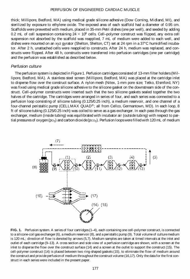
The perfusion system is depicted in Figure 1. Perfusion cartridges consisted of 13-mm filter holders (Mil-lipore, Bedford, MA). A stainless steel screen (Millipore, Bedford, MA) was placed at the cartridge inletto disperse flow over the construct surface. A nylon mesh (Nitex, 1-mm pore size; Tetko, Elsmford, NY)was fixed using medical grade silicone adhesive to the silicone gasket on the downstream side of the con-struct. Cell–polymer constructs were inserted such that the two silicone gaskets sealed together the twohalves of the cartridge. The cartridges were arranged in series of four, and each series was connected to aperfusion loop consisting of silicone tubing (0.125/0.25 inch), a medium reservoir, and one channel of afour-channel peristaltic pump (CELLMAX QUAD®; all from Cellco, Germantown, MD). In each loop, 8ft of silicone tubing (0.125/0.25 inch) was coiled to serve as a gas exchanger. In each pass through the gasexchanger, medium (inside tubing) was equilibrated with incubator air (outside tubing) with respect to par-tial pressures of oxygen (pO2) and carbon dioxide (pCO2). Perfusion loops were filled with 120 mL of medium
PERFUSION OF ENGINEERED CARDIAC MUSCLE
177
FIG. 1. Perfusion system. A series of four cartridges (1–4), each containing one cell-polymer construct, is connectedto a silicone-coil gas exchanger (6), a medium reservoir (8), and a peristaltic pump (9). Total volume of culture mediumis 120 mL; direction of flow is denoted by arrows (5,7). Medium samples are taken at timed intervals at the inlet andoutlet of each cartridge (9–13). A cross section and side view of a perfusion cartridge are shown, with a screen at theinlet to disperse the flow over the construct surface (14) and a screen at the outlet to support the construct (15). Thecell-polymer construct (14) is placed between two ring-shaped gaskets (15), to eliminate the flow of medium aroundthe construct and provide perfusion of medium throughout the construct volume (16,17). Only the data for the first con-struct in each series were included in the present paper.
and operated for 24 h. The series of cartridges was clamped off and drained, each cartridge was opened, acell–polymer construct transferred in, closed and refilled with medium, and the clamps were removed.
Series of cartridges (one construct per cartridge, four cartridges per series, four series per experiment)were perfused for 1 h at 0.2 mL/min, and then for 10 days at 3.24 6 0.12 mL/min (nominally 3 mL/min),or 0.98 6 0.02 mL/min (nominally 1 mL/min), or 0.62 6 0.00 mL/min (nominally 0.6 mL/min). Mediumsamples were taken using syringes (Becton Dickinson, Franklin Lakes, NJ) attached to three-way stopcocks(Baxter, Irvine, CA).
Spinner flask culture
Controls for comparing the effects of medium perfusion with the effects of diffusional transport were es-tablished for each experiment using spinner flasks (Bellco, Vineland, NJ). Cell–polymer constructs werethreaded onto 4-inch-long stainless steel wires (three to four per flask, with 1 disc apiece) embedded intothe stopper of a flask and cultured in 120 mL of medium stirred at 50 rpm as previously described.18 Gaswas exchanged via surface aeration of well mixed culture medium around the constructs. Medium sampleswere taken through the side arms using syringes. Medium was replaced at the same rate as in perfused car-tridges (50% or 60 mL every 2 days).
Analytical methods
Medium pH, pO2, and pCO2 were measured daily using a gas analyzer (model 1610; Instrumentation Lab-oratory, Lexington, MA) with an accuracy of 0.1% for pH and 2% for pO2 and pCO2.7 The rates of glucoseconsumption and lactate production were determined from medium concentrations measured using a glu-cose and L-lactate analyzer (model 2300 STAT Plus; YSI, Yellow Springs, OH).
Construct structure was analyzed using light and transmission electron microscopy. Samples taken forhistological analyses (n 5 2–4) were fixed for 10 min in 4% glutaraldehyde, embedded in paraffin, andcross-sectioned (5-mm-thick) through the center of the construct. Sections were stained with hematoxylinand eosin (H&E) for cells and immunohistochemically for cellular expression of sarcomeric a-actin.7 Sam-ples for TEM were fixed in Karnovsky’s reagent (0.1 M sodium cacodylate with 2% paraformaldehyde and2.5% glutaraldehyde, pH 5 7.4), postfixed in 2% osmium tetroxide, dehydrated in ethanol/propylene ox-ide, embedded in Poly/Bed812 (Polysciences, Warrington, PA), and sectioned (60-nm-thick). Sections werestained with lead citrate and uranyl acetate and examined with a transmission electron microscope (JEOL-100CX; JEOL, Peabody, MA).
DNA content was measured in homogenates of tissue constructs (n 5 4) using Hoescht 33258 dye (Poly-sciences, Warrington, PA) and a spectrofluorometer (Photon Technology International, South Brunswick,NJ) with calf thymus DNA (type 1, highly polymerized; Sigma, St. Louis, MO) as a standard.19 Proteincontents were determined from 100-ml samples of homogenates that were assayed as described in the mi-croplate protocol of the BioRad DC Protein Assay kit (BioRad, Hercules, CA).
Statistical analysis
Statistical analysis of data was performed by one-way analysis of variance (ANOVA) in conjunction withTukey’s test for multiple comparisons, using Systat 1.0 for Macintosh.
RESULTS
Cultivation conditions
For constructs cultured in cartridges, medium was perfused through constructs at flow rates of 0.6–3.0mL/min (corresponding to 0.8–4.2 cm3/cm2 ? min, or 140–700 mm/sec) and equilibrated in each pass throughgas exchanger, such that the inlet concentrations of oxygen and carbon dioxide were maintained at constantlevels (Fig. 1). For constructs cultured in spinner flasks, medium was in contact with the outer constructsurfaces and equilibrated with incubator air with surface aeration. As compared to flask cultures, medium
CARRIER ET AL.
178
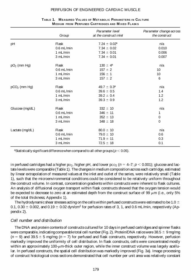
in perfused cartridges had a higher pO2, higher pH, and lower pCO2 (n 5 4–7; p , 0.001); glucose and lac-tate levels were comparable (Table 1). The changes in medium composition across each cartridge, estimatedby linear extrapolation of measured values at the inlet and outlet of the series, were relatively small (Table1), such that the microenvironmental conditions could be considered to be relatively uniform throughoutthe construct volume. In contrast, concentration gradients within constructs were inherent to flask cultures.An analysis of diffusional oxygen transport within flask constructs showed that the oxygen tension wouldbe expected to decrease to zero at an estimated depth from the construct surface of 99 mm (i.e., only 5%of the total thickness; Appendix 1).
The hydrodynamic shear stresses acting on the cells within perfused constructs were estimated to be 1.0 6
0.1, 0.30 6 0.002, and 0.19 6 0.02 dyn/cm2 for perfusion rates of 3, 1, and 0.6 mL/min, respectively (Ap-pendix 2).
Cell number and distribution
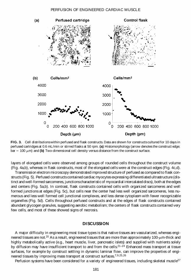
The DNA and protein contents of constructs cultured for 10 days in perfused cartridges and spinner flaskswere comparable, indicating comparable total cell number (Fig. 2). Protein/DNA ratios were 38.5 6 9 mg/mg(n 5 9) and 39.5 6 5 mg/mg (n 5 7) for perfused and flask constructs, respectively. However, perfusionmarkedly improved the uniformity of cell distribution. In flask constructs, cells were concentrated mostlywithin an approximately 100-mm-thick outer region, while the inner construct volume was largely acellu-lar. In perfused constructs, the spatial cell distribution was markedly improved (Fig. 3a). Image processingof construct histological cross sections demonstrated that cell number per unit area was relatively constant
PERFUSION OF ENGINEERED CARDIAC MUSCLE
179
TABLE 1. MEASURED VALUES OF METABOLIC PARAMETERS IN CULTURE
MEDIUM FROM PERFUSED CARTRIDGES AND MIXED FLASKS
Parameter level Parameter change acrossGroup at the construct inlet the construct
pH Flask 7.24 6 0.02a n/a0.6 mL/min 7.34 6 0.02 0.0101 mL/min 7.34 6 0.01 0.0063 mL/min 7.34 6 0.01 0.007
pO2 (mm Hg) Flask 130 6 4a n/a0.6 mL/min 157 6 2 101 mL/min 156 6 1 103 mL/min 157 6 2 6
pCO2 (mm Hg) Flask 49.7 6 0.3a n/a0.6 mL/min 39.6 6 0.5 1.41 mL/min 39.2 6 0.4 1.23 mL/min 39.3 6 0.9 1.2
Glucose (mg/dL) Flask 332 6 10 n/a0.6 mL/min 346 6 11 11 mL/min 352 6 13 03 mL/min 348 6 18 0
Lactate (mg/dL) Flask 80.0 6 10 n/a0.6 mL/min 79.0 6 10 0.61 mL/min 71.9 6 11 0.33 mL/min 72.5 6 18 0.1
aStatistically significant difference when compared to all other groups (p , 0.05).
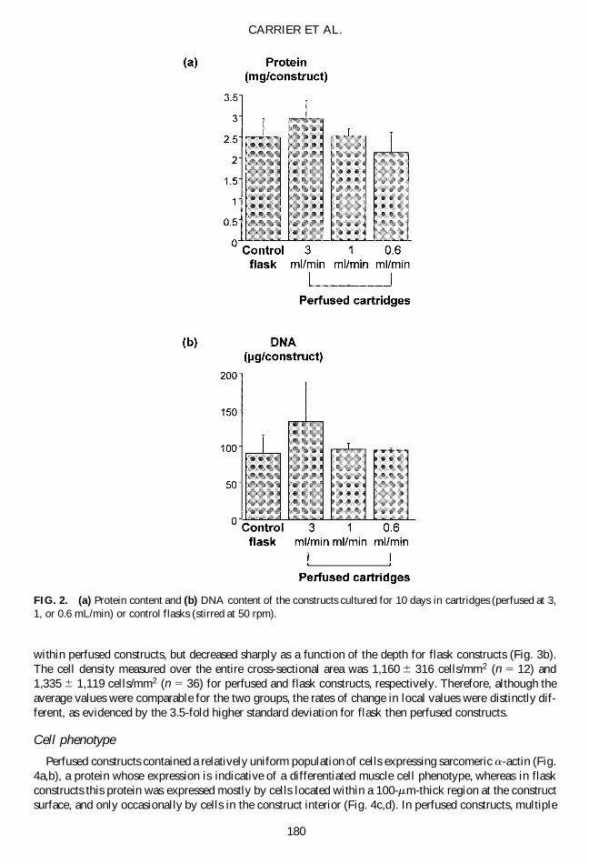
within perfused constructs, but decreased sharply as a function of the depth for flask constructs (Fig. 3b).The cell density measured over the entire cross-sectional area was 1,160 6 316 cells/mm2 (n 5 12) and1,335 6 1,119 cells/mm2 (n 5 36) for perfused and flask constructs, respectively. Therefore, although theaverage values were comparable for the two groups, the rates of change in local values were distinctly dif-ferent, as evidenced by the 3.5-fold higher standard deviation for flask then perfused constructs.
Cell phenotype
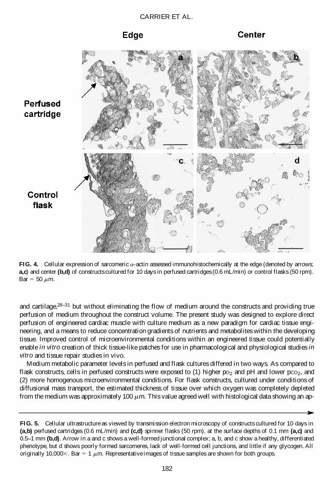
Perfused constructs contained a relatively uniform population of cells expressing sarcomeric a-actin (Fig.4a,b), a protein whose expression is indicative of a differentiated muscle cell phenotype, whereas in flaskconstructs this protein was expressed mostly by cells located within a 100-mm-thick region at the constructsurface, and only occasionally by cells in the construct interior (Fig. 4c,d). In perfused constructs, multiple
CARRIER ET AL.
180
FIG. 2. (a) Protein content and (b) DNA content of the constructs cultured for 10 days in cartridges (perfused at 3,1, or 0.6 mL/min) or control flasks (stirred at 50 rpm).
layers of elongated cells were observed among groups of rounded cells throughout the construct volume(Fig. 4a,b), whereas in flask constructs, most of the elongated cells were at the construct edges (Fig. 4c,d).
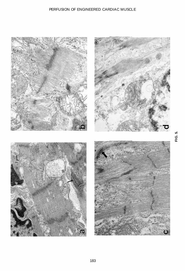
Transmission electron microscopy demonstrated improved structure of perfused as compared to flask con-structs (Fig. 5). Perfused constructs contained cardiac myocytes expressing differentiated ultrastructure (dis-tinct and well-formed sarcomeres, junctions characteristic of myocardial intercalated discs), both at the edgesand centers (Fig. 5a,b). In contrast, flask constructs contained cells with organized sarcomeres and wellformed junctions at edges (Fig. 5c), but cells near the center had less well organized sarcomeres, less nu-merous and less well formed cell junctional complexes, and less dense cytoplasm with fewer recognizableorganelles (Fig. 5d). Cells throughout perfused constructs and at the edges of flask constructs containedabundant glycogen granules, suggesting aerobic metabolism; the centers of flask constructs contained veryfew cells, and most of these showed signs of necrosis.
DISCUSSION
A major difficulty in engineering most tissue types is that native tissues are vascularized, whereas engi-neered tissues are not.20 As a result, engineered tissues that are more than approximately 100-mm-thick andhighly metabolically active (e.g., heart muscle, liver, pancreatic islets) and supplied with nutrients solelyby diffusion may have insufficient transport to and from the cells.21–24 Enhanced mass transport at tissuesurfaces, for example by construct settling in dynamic laminar flow, can improve the properties of engi-neered tissues by improving mass transport at construct surfaces.7,9,25,26
Perfusion systems have been considered for a variety of engineered tissues, including skeletal muscle27
PERFUSION OF ENGINEERED CARDIAC MUSCLE
181
FIG. 3. Cell distributions within perfused and flask constructs. Data are shown for constructs cultured for 10 days inperfused cartridges at 0.6 mL/min or stirred flasks at 50 rpm. (a) Histomorphology (arrow denotes the construct edge;bar 5 100 mm) and (b) Two-dimensional cell density versus distance from the construct surface.
and cartilage,28–31 but without eliminating the flow of medium around the constructs and providing trueperfusion of medium throughout the construct volume. The present study was designed to explore directperfusion of engineered cardiac muscle with culture medium as a new paradigm for cardiac tissue engi-neering, and a means to reduce concentration gradients of nutrients and metabolites within the developingtissue. Improved control of microenvironmental conditions within an engineered tissue could potentiallyenable in vitro creation of thick tissue-like patches for use in pharmacological and physiological studies invitro and tissue repair studies in vivo.
Medium metabolic parameter levels in perfused and flask cultures differed in two ways. As compared toflask constructs, cells in perfused constructs were exposed to (1) higher pO2 and pH and lower pCO2, and(2) more homogenous microenvironmental conditions. For flask constructs, cultured under conditions ofdiffusional mass transport, the estimated thickness of tissue over which oxygen was completely depletedfrom the medium was approximately 100 mm. This value agreed well with histological data showing an ap-
CARRIER ET AL.
182
FIG. 4. Cellular expression of sarcomeric a-actin assessed immunohistochemically at the edge (denoted by arrows;a,c) and center (b,d) of constructs cultured for 10 days in perfused cartridges (0.6 mL/min) or control flasks (50 rpm).Bar 5 50 mm.
FIG. 5. Cellular ultrastructure as viewed by transmission electron microscopy of constructs cultured for 10 days in(a,b) perfused cartridges (0.6 mL/min) and (c,d) spinner flasks (50 rpm), at the surface depths of 0.1 mm (a,c) and0.5–1 mm (b,d). Arrow in a and c shows a well-formed junctional complex; a, b, and c show a healthy, differentiatedphenotype, but d shows poorly formed sarcomeres, lack of well-formed cell junctions, and little if any glycogen. Alloriginally 10,0003. Bar 5 1 mm. Representative images of tissue samples are shown for both groups.
PERFUSION OF ENGINEERED CARDIAC MUSCLE
183
FIG
. 5.
proximately 100-mm-thick capsule of continuous tissue at the construct surface and a relatively cell-free in-terior (Fig. 3a).5,7,9 Moreover, 100 mm is also the approximate thickness of the zone of subendocardiumthat survives a myocardial infarct, owing to perfusion from blood in the left ventricular cavity.32
The control over the cell microenvironment was much better in perfused than in flask cultures, due to thedirect flow of medium throughout the construct rather than only at construct surfaces. Exposure to more ho-mogenous environmental conditions within perfused constructs was associated with significant improvementin the uniformity of the cell distribution across the entire construct thickness (Fig. 3). In spite of differencesin cell distribution, the total cell numbers were comparable in perfused and flask constructs (Fig. 2), presum-ably due to the same initial number of seeded cells. In flasks, the cell concentration was high at construct sur-faces, that is, in the region of high oxygen concentration, in contrast to perfused constructs where both thecells and oxygen were relatively uniformly distributed throughout the construct volume. Perfusion also en-hanced the expression of a differentiated cell phenotype within cardiac constructs, and resulted in a relativelyuniform population of cardiac myocytes showing cardiac-specific markers (e.g., expression of sarcomeric a-actin, sarcomeres, well formed cell junctions, elongated morphology) and evidence of aerobic metabolism(glycogen granules). In flask constructs, expression of these markers of differentiation and overall cell well-being were limited to a 100 mm thick region at construct surfaces (Fig. 4c,d).5,7,9
The fact that most cells within perfused cardiac constructs exhibited rounded phenotype may have beendue to shear. One response of various cell types to shear is rounding.33 In spite of the fact that the esti-mated shear forces within perfused constructs were relatively low (0.19–1.04 dyn/cm2; Appendix 2), slightlyhigher shear stresses (i.e., 1.6–3.3 dyn/cm2) applied in vitro over extended periods of time altered the mor-phology and decreased the viability of various mammalian cell types, including hybridoma and human em-bryonic kidney cells.34,35 The response to shear stress depends upon many factors, including cell type, ex-posure time, substrate, and flow dynamics.33,35 Cardiac cells are not exposed to direct shear forces in vivo,and the exchange of nutrients and metabolic wastes is carried out by diffusion between densely spaced cap-illaries and the cells.
The present study demonstrated that perfusion can markedly and significantly improve the uniformity ofthe engineered tissue. Recent in vivo studies of engineered cardiac grafts12,13 showed promising results withrespect to construct survival, vascularization and integration, but the functional improvement was either notobserved13 or was not significant.12 Future work is thus expected to be driven by the need to create thickergrafts with more uniform tissue architectures for controlled in vitro and in vivo studies. In particular, thereis a need to increase of the initial cell concentration, and to coculture the endothelial with cardiac musclecells in order to compartmentalize nutrient supply and waste removal in a more physiological way, and toenhance cell survival during in vitro cultivation and in vivo implantation.
To summarize, cultivation with direct perfusion of medium exposed engineered cardiac tissue to morehomogeneous conditions and maintained higher oxygen tension in the cell microenvironment, as comparedto spinner flasks. Perfused constructs had relatively uniform spatial distributions of cells expressing differ-entiated phenotype. Medium perfusion can thus be utilized to enhance mass transport to and from the cellsand allow the cultivation of engineered tissues .100 mm in thickness.
ACKNOWLEDGMENTS
We would like to thank J. Marler for providing the basis of the perfused bioreactor design, D. Sawyerfor his advice regarding cardiomyocyte isolation, H. Shing for carrying out TEM, A. Chan for assistancewith biochemical assays, and Sue Kangiser for her help with manuscript preparation. This work was sup-ported by the National Aeronautics and Space Administration (grant NCC8-174).
APPENDIX 1: CALCULATION OF OXYGEN GRADIENTS FOR CONSTRUCTSCULTURED IN FLASKS
A one-dimensional analysis of the diffusional oxygen transport through a flask-cultured construct wasused to estimate the depth, l, from the construct surface at which oxygen tension decreased to zero. The
CARRIER ET AL.
184
construct was modeled as a flat slab using the following simplifying assumptions: (1) constant rate of cel-lular oxygen consumption, Rv, (2) constant diffusivity of oxygen, D, (3) steady state conditions, (4) negli-gible convective flow through the construct, and (5) constant oxygen concentration, Co, at the construct sur-face, equal to the bulk medium oxygen concentration due to convective flow and mixing. Under theseconditions, the conservation equation for oxygen involves diffusion and consumption terms:
D * d2C/dx2 1 Rv 5 0 (1)
where x is the depth from the construct surface and x 5 0 refers to the construct surface. Equation 1 wassolved using the following boundary conditions:
Cx 5 0 5 Co (assumption 5) (2)
dC/dxx 5 l 5 0 (3)
to obtain the oxygen concentration profile:
C 5 2Rv/D * (2x2/2 1 l * x) 1 Co (4)
L was estimated from Equation 4 using the following parameter values:
Co 5 130 mm Hg (2.04 3 1027 moles/mL)
D 5 2.0 3 1025 cm2/sec
Rv 5 5.04 3 1026 mol O2/min/cm3, based on an oxygen consumption rate of 3.0 3 1028 mol/mg pro-tein/min,37 a cellular protein content of 8.4 3 1027 mg/cell,38 and a cell density of 2.0 3 108 cells/cm3.36
APPENDIX 2: CALCULATION OF THE SHEAR STRESS WITHIN PERFUSED CONSTRUCTS
Medium was perfused through cardiac constructs at flow rates of 0.6–3.0 mL/min (corresponding to0.8–4.2 cm3/cm2 min per construct cross-sectional area, or 140–700 mm/sec). Shear stress acting on cellswithin a perfused construct was estimated by equating the average energy dissipation with the drag forceper unit surface area of a polymer fiber coated with cells:39
tav 5 Fd/S (5)
where tav 5 the average shear stress on the cell surface, Fd 5 drag force, and S 5 surface area of polymerfiber. Drag force is equal to the pressure drop across a cell-polymer construct multiplied by its cross-sec-tional area, A, which can be calculated as:
A 5 V/L (6)
where L 5 the total length of perfused tissue in the direction of flow and V 5 the volume of the perfusedconstruct. By substitution, Equation 5 becomes:
tav 5 2DP/L * 1/(S/V) (7)
where DP 5 the pressure drop across the perfused tissue. The Reynold’s number (Re) within the constructwas estimated using:
PERFUSION OF ENGINEERED CARDIAC MUSCLE
185
Re 5 Uo * df * r/m (8)
Uo 5 Q/A (9)
where Uo is the superficial fluid velocity within the construct; df 5 diameter of polymer fibers coated withcells (df 5 23 mm assuming a 5-mm thick cell layer39 on 13-mm-diameter fibers); medium density of r 5
1.03 g/cm3;40 medium viscosity of m 5 0.7 cP (0.0007 Ns/m2) at 37°C39; the volumetric flow rate of mediumof Q 5 0.62–3.24 mL/min; and the construct cross sectional area A 5 0.712 cm2. Equation 8 was solvedto yield Re 5 0.005–0.026. Because Re , 10 at all conditions, the construct could be considered as an iso-topic porous medium, and Darcy’s Law could be applied41:
DP/L 5 m Uo/k (10)
where k 5 permeability, a property which depends on the size, concentration, and arrangement of the fibersin a fibrous medium. After substituting Equation 10 into Equation 7 and expressing S/V as a function ofthe construct void fraction, e, and fiber diameter, df, Equation 7 becomes:
tave 5 m * Uo/k * df/{4 * (1 2 e)} (11)
e was calculated from the volume fractions of cells and polymer fibers. The volume fraction of cells wasestimated using the measured DNA content of each construct, a measured value of 3.05 3 1025 mgDNA/cell, and a cell volume of 1.77 3 1029 cm3.42 The volume fraction of polymer fibers was previouslydetermined to be 3%.43 The resulting values of m for constructs perfused at 3, 1, and 0.6 mL/min were0.921, 0.929, and 0.934, respectively. The corresponding k values were estimated based the permeabilityof a random fibrous medium expressed as a function of fiber diameter and the volume fraction of the fibers,as previously suggested,41,44 to obtain 3.8, 4.2, and 4.8 for constructs perfused at 3, 1, and 0.6 mL/min, re-spectively. The average hydrodynamic shear tav was estimated from Equation 11 to be 1.04 6 0.11, 0.30 6
0.002, and 0.19 6 0.02 dyn/cm2 for the flowrate of 3, 1, and 0.6 mL/min, respectively.
REFERENCES
1. Soonpaa, M.H., Daud, A.I., Koh, G.Y., et al. Potential approaches for myocardial regeneration. Ann. N.Y. Acad.Sci. 752, 446, 1995.
2. Taylor, D.A., Atkins, B.Z., Hungspreugs, P., et al. Regenerating functional myocardium: improved performanceafter skeletal myoblast transplantation. Nat. Med. 4, 929, 1998.
3. Orlic, D., Kajstura, J., Chimenti, S., et al. Bone marrow cells regenerate infarcted myocardium. Nature 410, 701,2001.
4. Akins, R.E., Boyce, R.A., Madonna, M.L., et al. Cardiac organogenesis in vitro: reestablishment of three-dimen-sional tissue architecture by dissociated neonatal rat ventricular cells. Tissue Eng. 5, 103, 1999.
5. Bursac, N., Papadaki, M., Cohen, R.J., et al. Cardiac muscle tissue engineering: toward an in vitro model for elec-trophysiological studies. Am. J. Physiol. 277, H433, 1999.
6. Eschenhagen, T., Fink, C., Remmers, U., et al. Three-dimensional reconstitution of embryonic cardiomyocytes ina collagen matrix: a new heart model system. FASEB J. 11, 683, 1997.
7. Carrier, R.L., Papadaki, M., Rupnick, M., et al. Cardiac tissue engineering: cell seeding, cultivation parameters andtissue construct characterization. Biotechnol. Bioeng. 64, 580, 1999.
8. Freed, L.E., and Vunjak-Novakovic, G. Tissue culture bioreactors: chondrogenesis as a model system. In: Langer,R., and Chick, W., eds. Principles of Tissue Engineering. Austin: R.G. Landes, 1997, pp. 150–158.
9. Papadaki, M., Bursac, N., Langer, R., et al. Tissue engineering of functional cardiac muscle: molecular, structuraland electrophysiological studies. Am. J. Physiol. Heart Circ. Physiol. 280, H168, 2001.
10. Fink, C., Ergun, S., Kralisch, D., et al. Chronic stretch of engineered heart tissue induces hypertrophy and func-tional improvement. FASEB J. 14, 669, 2000.
11. Zimmermann, W.H., Schneiderbanger, K., Schubert, P., et al. Tissue engineering of a differentiated cardiac mus-cle construct. Circ. Res. 90, 223–230, 2002.
CARRIER ET AL.
186
12. Leor, J., Aboulafia-Etzion, S., Dar, A., et al. Bioengineered cardiac grafts: a new approach to repair the infarctedmyocardium: Circulation 102, III56, 2000.
13. Li, R.K., Jia, Z.Q., Weisel, R.D., et al. Survival and function of bioengineered cardiac grafts. Circulation 100, II63,1999.
14. Korecky, B., Hai, C.M., and Rakusan, K. Functional capillary density in normal and transplanted rat hearts. Can.J. Physiol. Pharm. 60, 23, 1982.
15. Rakusan, K., and Korecky, B. The effect of growth and aging on functional capillary supply of the rat heart. Growth46, 275, 1982.
16. Steinhausen, M., Tillmanns, H., and Thederan, H. Microcirculation of the epimyocardial layer of the heart. I. Amethod for in vivo observation of the microcirculation of superficial ventricular myocardium of the heart and cap-illary flow pattern under normal and hypoxic conditions. Pflugers Arch. Eur. J. Phy. 374, 57, 1978.
17. Zadeh, B.J., Gonzalez-Sanchez, A., Fischman, D.A., et al. Myosin heavy chain expression in embryonic cardiaccell culture. Dev. Biol. 115, 204, 1986.
18. Vunjak-Novakovic, G., Freed, L.E., Biron, R.J., et al. Effects of mixing on the composition and morphology oftissue-engineered cartilage. AIChE J. 42, 850, 1996.
19. Downs, T., and Wilfinger, M. Fluorometric quantification of DNA in cells and tissue. Anal. Biochem. 131, 538,1983.
20. Putnam, A.J., and Mooney, D.J. Tissue engineering using synthetic extracellular matrices. Nat. Med. 2, 824, 1996.21. Cima, L.G., and Langer, R. Engineering human tissue. Chem. Eng. Prog. June, 46, 1993.22. Colton, C.K. Implantable biohybrid artificial organs. Cell Transplant. 4, 415, 1995.23. Mooney, D.J., Mazzoni, C.L., Breuer, C., et al. Stabilized polyglycolic acid fiber-based tubes for tissue engineer-
ing. Biomaterials 17, 115, 1996.24. Peters, M.C., Isenberg, B.C., Rowley, J.A., et al. Release from alginate enhances the biological activity of vascu-
lar endothelial growth factor. J. Biomater. Sci. Polym. Ed. 9, 1267, 1998.25. Freed, L.E., and Vunjak-Novakovic, G. Tissue engineering bioreactors. In: Lanza, R.P., Langer, R., and Vacanti,
J., eds. Principles of Tissue Engineering. San Diego: Academic Press, 2000, p. 143–156.26. Vunjak-Novakovic, G., Martin, I., Obradovic, B., et al. Bioreactor cultivation conditions modulate the composi-
tion and mechanical properties of tissue engineered cartilage. J. Orthop. Res. 17, 130, 1999.27. Chromiak, J.A., Shansky, J., Perrone, C., et al. Bioreactor perfusion system for the long-term maintenance of tis-
sue engineered skeletal muscle organoids. In Vitro Cell. Dev. Biol. An. 34, 694, 1998.28. Dunkelman, N.S., Zimber, M.P., Lebaron, R.G., et al. Cartilage production by rabbit articular chondrocytes on
polyglycolic acid scaffolds in a closed bioreactor system. Biotechnol. Bioeng. 46, 299, 1995.29. Schreiber, R.E., Ilten-Kirby, B.M., Dunkelman, N.S., et al. Repair of osteochondral defects with allogeneic tissue-
engineered cartilage implants. Clin. Orthop. 367S, S382, 1999.30. Sittinger, M., Schultz, O., Keyszer, G., et al. Artificial tissue in perfusion culture. Int. J. Artif. Organs 20, 57, 1997.31. Wu, F., Dunkelman, N., Peterson, A., et al. Bioreactor development for tissue-engineered cartilage. Ann. N.Y.
Acad. Sci. 875, 405, 1999.32. Schoen, F.J. The heart. In: Cotran, R.S., Kumar, V., Collins, T., and Robbins, S.L., eds. Robbins Pathologic Ba-
sis of Disease. Philadelphia: W.B. Saunders, 1999, pp. 543–599.33. Kretzmer, G., and Schugerl, K. Response of mammalian cells to shear stress. Appl. Microbiol. Biotechnol. 34, 613,
1991.34. Smith, C.G., Greenfield, P.F., and Randerson, D. Shear sensitivity of three hybridoma cell lines in suspension cul-
ture. In: Spier, R.E., and Griffith, J.B., eds. Modern Approaches to Animal Cell Technology. Kent, UK: Butter-worth, 1987, pp. 316–327.
35. Stathopoulos, N.A., and Hellums, J.D. Shear stress effects on human embryonic kidney cells in vitro. Biotechnol.Bioeng. 27, 1021, 1985.
36. Piret, J.M., and Cooney, C.L. Model of oxygen transport limitations in hollow fiber bioreactors. Biotechnol. Bio-eng. 37, 80, 1991.
37. Yamada, T., Yang, J.J., Ricchiuti, N.V., and Seraydarian, M.W. Oxygen consumption of mammalian myocardial-cells in culture—measurements in beating cells attached to the substrate of the culture dish. Anal. Biochem. 145,302, 1985.
38. Qi, M., Ojamaa, K., Eleftheriades, E.G., Klein, I., and Samarel, A.M. Regulation of rat ventricular myosin heavychain expression by serum and contractile activity. Am. J. Physiol. 267, C520, 1994.
39. Perry, S.D., and Wang, D.I.C. Fiber bed reactor design for animal cell culture. Biotechnol. Bioeng. 34, 1, 1989.40. Freed, L.E., and Vunjak-Novakovic, G. Tissue engineering of cartilage. In: Bronzino, J.D., eds. Biomedical Engi-
neering Handbook. Boca Raton, FL: CRC Press, 1995, pp. 1788–1807.41. Jackson, G.W., and James, D.F. The permeability of fibrous porous media. Can. J. Chem. Eng. 64, 364, 1986.
PERFUSION OF ENGINEERED CARDIAC MUSCLE
187
42. Kira, Y., Nakaoka, T., Hashimoto, E., et al. Effect of long-term cyclic mechanical load on protein synthesis andmorphological changes in cultured myocardial cells from neonatal rat. Cardiovasc. Drugs Ther. 8, 251, 1994.
43. Freed, L.E., Vunjak-Novakovic, G., Biron, R., et al. Biodegradable polymer scaffolds for tissue engineering.Bio/Technology 12, 689, 1994.
44. Drummond, J.E., and Tahir, M.I. Laminar viscous flow through regular arrays of parallel solid cylinders. Int. J.Multiphas. Flow 10, 515, 1984.
Address reprint requests to:Gordana Vunjak-Novakovic, Ph.D.
Massachusetts Institute of Technology, E25-33045 Carleton St.
Cambridge, MA 02139
E-mail: [email protected]
CARRIER ET AL.
188