Embed Size (px)
Citation preview

This article appeared in a journal published by Elsevier. The attachedcopy is furnished to the author for internal non-commercial researchand education use, including for instruction at the authors institution
and sharing with colleagues.
Other uses, including reproduction and distribution, or selling orlicensing copies, or posting to personal, institutional or third party
websites are prohibited.
In most cases authors are permitted to post their version of thearticle (e.g. in Word or Tex form) to their personal website orinstitutional repository. Authors requiring further information
regarding Elsevier’s archiving and manuscript policies areencouraged to visit:
http://www.elsevier.com/copyright

Author's personal copy
Patterning network structure to spatially control cellular remodeling and stemcell fate within 3-dimensional hydrogels
Sudhir Khetan, Jason A. Burdick*
Department of Bioengineering, University of Pennsylvania, 210 S 33rd Street, Philadelphia, PA 19104, USA
a r t i c l e i n f o
Article history:Received 11 June 2010Accepted 6 July 2010Available online 31 July 2010
Keywords:HydrogelsStem cellsPatterningRemodelingHyaluronic acid
a b s t r a c t
The spatially directed 3-dimensional (3D) remodeling of synthetic materials may be useful to regionallycontrol cell behavior. In this work, we developed a process to synthesize hyaluronic acid hydrogels usingmultiple modes of crosslinking applied sequentially; a primary addition reaction to introduce proteasedegradable peptide crosslinks, then a UV light-induced secondary radical reaction (enabling spatialcontrol) to introduce non-degradable kinetic chains. These differential network structures eitherpermitted (primary crosslinking only, “�UV”) or inhibited (sequential crosslinking, “þUV”) cellularremodeling. This behavior was validated by controlling the outgrowth from chick aortic arches or thespreading of encapsulated mesenchymal stem cells (MSCs), where only �UV regions permitted archoutgrowth and MSC spreading. Additionally, network structures dictated adipogenic/osteogenic MSC fatedecisions, with spatial control, by controlling encapsulated MSC spreading. This manipulation ofmicroenvironmental cues may be valuable for advanced tissue engineering applications requiring thespatial control of cells in 3D.
� 2010 Elsevier Ltd. All rights reserved.
1. Introduction
The 3-dimensional (3D) interactions of cells and the extracel-lular matrix (ECM) comprise a dynamic regulatory systemresponsible for tissue morphogenesis during development, as wellas in response to injury [1,2]. However, much of the research inbiomaterial development for regenerative medicine applicationshas employed either cell seeding atop 2-dimensional (2D)substrates that do not adequately recapitulate the 3D nature ofnativemicroenvironments, or spatially uniform and staticmaterialsthat lack the heterogeneity that is found in vivo. As such, importantcausal relationships such as the dependence of stem cellmorphology [3] and differentiation [4] on 2D substrate elasticitymay not readily translate to 3D culture. Thus, the development of3D hydrogel systems, particularly with spatially controlled features,would be an important advance for investigating basic questions incell behavior and tissue development, as well as toward regener-ative medicine applications.
While considerable progress has been made in developinguniform ECM-mimetic 3D scaffolds capable of promoting hydrogelremodeling [5,6] and even stem cell differentiation [7], one
continuing challenge is the difficulty in translating patterningmethods well established for 2D or laminated substrates to 3D toenable the necessary spatial control over materials. The patterningof 3D hydrogel systems has been limited to only a few techniques,and most of these rely on pre-fabrication before the introduction ofcells due to cytocompatibility concerns [8,9]. Techniques have alsobeen developed where cellular adhesion is patterned in 3D usingclick chemistry reactions [10] or through the introduction ofa photodegradable linker [11] to alter cellular interactions tomaterials. These approaches use spatial control of adhesion to altercellular behavior, but rely on complex chemistry and are onlyapplicable to a few polymer systems.
To overcome these limitations, this report describes the devel-opment of a simple technique incorporating multiple modes ofcrosslinking, applied sequentially, to enable 3D spatially patternedremodeling of hydrogels. The technique is based on the basicunderstanding of how cells remodel certain crosslinks. Thesehydrogels support remodeling and infiltration of cells from ex vivotissues (e.g., chick aortic arches) or by cells that are encapsulateddirectly in the hydrogels (e.g., human mesenchymal stem cells,hMSCs). Hyaluronic acid (HA) was used as the primary structuralcomponent in the current work due to its biocompatibility, hydro-philicity, importance in vivo [12,13], and past use in 3D hydrogelsystems; [14e18]however, this approachcanbeeasilyapplied to anypolymeric material functionalized with compatible reactive groups.
* Corresponding author. Tel.: þ1 215 898 8537; fax: þ1 215 573 2071.E-mail address: [email protected] (J.A. Burdick).
Contents lists available at ScienceDirect
Biomaterials
journal homepage: www.elsevier .com/locate/biomateria ls
0142-9612/$ e see front matter � 2010 Elsevier Ltd. All rights reserved.doi:10.1016/j.biomaterials.2010.07.035
Biomaterials 31 (2010) 8228e8234

Author's personal copy
2. Materials and methods
All materials were purchased from SigmaeAldrich unless otherwise stated.
2.1. AHA synthesis
Acrylated hyaluronic acid (AHA) was synthesized via a two-step protocol:(1) Synthesis of the tetrabutylammonium salt of HA (HAeTBA) was performed byreacting sodium hyaluronate (64 kDa, Lifecore) with the highly acidic ion exchangeresin Dowex-100 and neutralization with 0.2M TBA-OH. (2) Coupling of acrylic acid(2.5 eq) and HAeTBA (1 eq, repeat unit) in the presence of dimethylamino pyridine(DMAP; 0.075 eq) and di-tert-butyl-dicarbonate (1.5 eq) in DMSO, followed bydialysis and lyophilization. Synthetic details (Fig. S1a), as well as the final structureand 1H NMR spectrum of AHA (Fig. S1b) are provided in Supplementary Information.
2.2. Peptides
The cell adhesive oligopeptide GCGYGRGDSPG (MW: 1025.1 Da; italics indicatecell adhesive domain) and MMP-degradable oligopeptide GCRDGPQGYIWGQDRCG(MW: 1754.0 Da; down arrow indicates site of proteolytic cleavage), both with >95%purity (per manufacturer HPLC analysis), were obtained from GenScript Corporation(Piscataway, NJ, USA).
2.3. Hydrogel formation
AHA was dissolved in sodium phosphate buffered saline (NaPBS buffer: 0.1 M
sodium phosphate, 0.3 M total osmolarity, pH 8.0) containing Irgacure 2959 (I2959,Ciba) photoinitiator (final concentration of 0.05 wt%). I2959 was chosen due to itsaqueous solubility and good cytocompatibility [19,20]. Sequentially crosslinked AHAhydrogels were synthesized using a two-step procedure of an addition reactionfollowed by radical polymerization. In the first step, stable “�UV” hydrogels weresynthesized via addition reactions between AHA macromers (first reacted with thecell adhesive peptide to a final concentration of 2 mM at room temperature for30min) andMMP-degradable peptides (acrylates react with thiols from cysteines onpeptides under these conditions) to consume 50% of the total available acrylategroups. Selected �UV gels were then exposed to 10 mW/cm2 365 nm ultravioletlight (Omnicure S1000 UV Spot Cure System, Exfo, Ontario, Canada) for 4 min(secondary crosslinking) through glass coverslips to produce uniform “þUV”hydrogels. Spatial patterning of the hydrogels was possible by substituting 10k DPIphotomasks (CAD/Art Services, Inc., Bandon, OR) for coverslips during the secondarystep. For studies involving photopatterned gels, 5 nM methacrylated rhodamine(MeRho) was mixed into the prepolymer solution for visualization ofþUV regions ofthe gels (methacrylates react extensively during the radical polymerization, butrelative to acrylates, coupling is too slow during the addition reaction).
2.4. Microscopy
For all studies, confocal laser scanningmicroscopy (CLSM) imageswere obtainedwith a Zeiss LSM 510Meta Confocal microscope (Carl Zeiss), using a DAPI/FITC/TRITCmulti-track configuration at 5�, 10�, and 20� objectives. All CLSM images areprojections of a 150 mm thick gel portion using a 15 mm slice thickness.
2.5. Hydrogel characterization
Samples were fabricated as described above and swelled to equilibrium in PBSfor 24 h. Photopatterned acellular gels containing MeRho were imaged at the topand bottom surfaces, and the color intensity distribution versus horizontal distancewas obtained using the “Plot Profile” tool in ImageJ (NIH). The elastic moduli ofuniform and photopatterned AHA hydrogels were quantified using atomic forcemicroscopy (AFM, Veeco Bioscope I). A silicon bead AFM tip with a spring constant of�0.06 N m�1 was used to obtain force curves for individual points on the gel surface(n ¼ 10 points chosen for each condition), from which a local elastic modulus wascalculated. For photopatterned gels, points were chosen at random locations fromthe interior of�UV orþUV regions of the gel. To characterize the real-time change inhydrogel mechanical properties with crosslinking, the shear elastic modulus wasmeasured via dynamic oscillatory shear rheometry at 1% strain and 1 Hz using anAR2000ex controlled stress rheometer (TA Instruments) with parallel plate geom-etry (20 mm diameter). Following the addition of crosslinker, the polymer solutionwas mixed and pipetted directly onto the bottom plate, and the top plate waslowered to contact the gelling solution with a 100 mm gap size. The evolution of theelastic stored modulus, G0 , and viscous loss modulus, G00 , was monitored with time.The uniform�UV hydrogel was allowed to undergo addition crosslinking for 15 minto a peak value of w0.8 kPa, at which point the gel was irradiated by 10 mW/cm2
365 nm UV light (Omnicure S2000 Spot Curing System), resulting in a rapid increasein the elastic modulus to a peak value ofw5.1 kPa. The gel point (i.e., the intersectionof G0 and G00) occurred almost immediately after mixing of the two components (i.e.,before themixturewas pipetted onto the rheometer stage) and thus does not appearon the displayed sweep.
To quantitatively assess degradation kinetics, uniform crosslinked �UV and þUVhydrogels were incubated in separatewells of a 24-well plate containing either 1.0mLPBS or 1.0 mL PBS with 40 nM human MMP-2 (R&D Systems) on an orbital shaker at37 �C. The solutions were refreshed every 48 h until complete degradation of �UVhydrogels occurred. The sample supernatants (frozen and stored at �20 �C aftercollection) were analyzed in triplicate via a modified uronic acid assay as previouslyreported [20]. For SEM analysis, uniform �/þ UV and 250 mm diameter dot photo-patterned hydrogels were incubated with PBS containing MMP-2 as above untilcomplete degradation of theuniform�UVhydrogels occurred. The sampleswere thenflash frozen in liquid N2, lyophilized, and imaged with a JEOL 7500 HR-SEM at 10 kV.
2.6. Chick aortic arch culture
Specific pathogen-free chick embryos (Charles River Labs) were sacrificed at day14 after fertilization, and dissection and extraction of aortic arch samples (w1 mm3
volume) were performed using aseptic techniques [21]. For encapsulation, archeswere suspended in the precursor solution prior to the first crosslinking step. AHAhydrogels with encapsulated arches were incubated in basal medium with supple-ments (EGM-2 media; Lonza) with media replacement every 2e3 days. Following 3weeks in culture, the gels were fixed with 10% formalin and stained with FITC-conjugated phalloidin using standard protocols.
2.7. hMSC cell culture
Human mesenchymal stem cells (hMSCs) were obtained from Lonza Corpora-tion (Walkersville, MD). For encapsulation studies, hMSCs were expanded in growthmedia (a-MEM, 10% FBS, 1% L-Glutamine & penicillinestreptomycin) and encapsu-lated at passage 3 in AHA hydrogels at a density of 5 � 106 cells mL�1 by suspensionin the precursor solution prior to the first crosslinking step. The constructs weremaintained in 2 mL growth media (for remodeling studies, Fig. 5) or a mixed adi-pogenic/osteogenic inductive media (for differentiation studies, Fig. 6) in a 24-wellplate and media was refreshed every 2e3 days until day 14. The adipogenic/oste-ogenic mixedmedia was made by combining commercially available osteogenic andadipogenic inductivemedia (R&D Systems) in a 1:1 ratio and supplementing with 1%(v/v) penicillinestreptomycin.
2.8. Live/dead staining
Following 14 days incubation in growth media, cell viability and morphology(for remodeling studies) were assessed using a live/dead staining kit (MolecularProbes).
2.9. Cellular aspect ratio measurements
Cellular aspect ratio is defined as the ratio of the maximum orthogonal length towidth of each cell. To quantify, three random CLSM images of live/dead stained gelsat 20� magnification were chosen from the interior of each gel; each image wasused for �20 measurements of randomly selected cells, or n � 60 for each sample.The measured aspect ratios were then sorted into bins and reported as histograms.
2.10. Oil red O and ALP quantification
Following 14 days of incubation in mixed adipogenic/osteogenic inductivemedia, production of oil red O and ALP by hMSCs encapsulated within uniform �UVand þUV hydrogels was assessed quantitatively. For oil red O, hydrogels werestained with the marker as described in Supplementary Information (see Fig. S5 forrepresentative stains). Quantification of the oil red O from the same samples wasthen performed as previously reported [22]. Briefly, oil red O was eluted from thestained hydrogels by incubation in 100% isopropanol for 30 min. Following manualdigestion of the gels and centrifugation to remove cellular and gel debris, thesupernatant absorbance containing the eluted oil red O was read at 500 nm. For ALP,hydrogels were manually digested in 250 mL CelLytic M cell lysis buffer, incubated at45 �C for 1 h, and centrifuged to remove cellular debris. ALP production was thenquantified by reacting 50 mL of the lysis buffer with an equal volume of substratesolution and measuring the absorbance at 405 nm. Quantitative values for oil red Oand ALP were normalized to total dsDNA content, which was determined using thePicoGreen assay as previously reported [23].
2.11. Immunocytochemistry
Following 14 days incubation in mixed adipogenic/osteogenic inductive media,uniform �UV and þUV and photopatterned (250 mm stripes) hydrogels with encap-sulated hMSCs were fixed overnight in 10% formalin. The gels were washed (PBScontaining 0.1% Tween 20 and 3% bovine serum albumin), permeabilized with 0.25%TritonX-100, blockedwith10%goat seruminPBS, and incubatedovernightat 4 �Cwithanti-human osteocalcin (1:10 dilution, mouse IgG) and anti-fatty acid binding protein(R&D Systems, 1:20 dilution, goat IgG) 1� Ab. Constructs were then washed andincubated for 2 h at room temperaturewithAlexa Fluor 488 anti-mouse (goat IgG) andAlexa Fluor 350 anti-goat (donkey IgG) 2� Ab for OC and FABP, respectively.
S. Khetan, J.A. Burdick / Biomaterials 31 (2010) 8228e8234 8229

Author's personal copy
2.12. Statistical analysis
Values are reported as mean � standard error of the mean (mechanics).Statistical differences between groups (p < 0.05) were determined using ANOVA inconjunction with Tukey’s post hoc test (JMP software).
3. Results and discussion
A two-step protocol was used to synthesize sequentially cross-linked hydrogels. In the primary crosslinking step, a uniform (i.e.,non-patterned) “�UV” hydrogel is formed using Michael-typereactivity between multi-acrylate HA macromers and bifunctional,proteolytically degradable peptides. Monofunctional, pendantRGDS-containing peptides are also added (prior to crosslinking) toincorporate cell adhesion. With these components (adhesion andproteolytic degradability), this hydrogel was expected to supportcellular remodeling [5,24]. The primary addition step is performedin the presence of a photoinitiator (at this point, non-reactive) anddesigned so that only a portion of total available acrylate groups areconsumed, making secondary free radical crosslinking possible. Inthe secondary step, �UV hydrogels are exposed to light to initiatefree radical photopolymerization of the remaining acrylate groups.The resulting “þUV” hydrogels were expected to prevent remod-eling due to the incorporation of non-degradable covalent cross-links from kinetic chain formation. Since mesh sizes in the þUVhydrogels are orders of magnitude smaller than typical cell diam-eters [24,25], secondary crosslinking was also predicted to preventcellular outgrowth from tissue and to confine encapsulated cells toa rounded morphology. This has been observed previously whencells are encapsulated in hydrogels using a purely radical poly-merization [26e28].
With this technique, it is possible to synthesize and characterizehydrogels patterned with �UV (permissive) and þUV (inhibitory)regions (Fig. 1). Since the secondary crosslinking is initiated bylight, spatially distinct zones of remodeling are created by applyinga photomask to the gel surface prior to the secondary crosslinking(Fig. 1a). To assess pattern resolution with depth, methacrylatedrhodamine (MeRho) was mixed into the prepolymer solution,which due to charge density and steric hindrance effects [29] onlycovalently incorporates into the network during the secondaryradical crosslinking. The pattern fidelity at the top and bottom
surfaces of a hydrogel photopatterned with 250 mm stripes wasconfirmed qualitatively by visualization using laser scanningconfocal microscopy (LSCM) (Fig. 1a), and quantitatively by plottingthe average color intensity versus horizontal distance across thepattern (Fig. 1b). These results demonstrate the retention of highresolution patterns throughout the hydrogel volume.
Mechanical testing of photopatterned hydrogels was thencarried out to determine if differences in gel structure predictablyinstructs differences in mechanics of �UV and þUV environments.Atomic force microscopy (AFM) was used to characterize the localelastic modulus of �UV and þUV regions of photopatternedhydrogels and for uniform gels (Fig. 2a). As expected, the increasedcrosslink density as a result of photopolymerization led to an in-crease in mechanical properties from 6.3� 0.7 kPa to 18.0� 1.5 kPain �UV and þUV uniform hydrogels, respectively, and from6.3 � 1.0 kPa to 15.2 � 2.4 kPa in �UV and þUV regions of photo-patterned gels, respectively. Further, the increase in elasticmodulus as a result of secondary crosslinking was observed inrheometry studies, which showed a rapid increase in shearmodulus with light exposure similar in fold to that observed withAFM testing (Fig. 2b). The mechanical properties in each environ-ment can be tuned for individual applications by adjusting themacromer concentration and ratio of addition to radical cross-linking, both of which were kept constant (3 wt% and 1:1, respec-tively) in this study.
To demonstrate that the incorporation of non-degradablecovalent kinetic chains restricts protease-mediated remodeling, weexposed hydrogels to exogenous MMP-2. When incubated in PBScontaining 40 nM MMP-2, uniform �UV hydrogels underwentcomplete degradation (i.e., 100% uronic acid release) within 12days, whereas very limited degradation (16.8 � 2.1% uronic acidrelease) was observed with uniform þUV hydrogels (Fig. 3a). Thesmall degree of mass loss in þUV hydrogels may be due to hydro-lysis of ester linkages in the crosslinks or a soluble fraction aftercrosslinking. To assess the degradation of patterned hydrogels, bothuniform �UV and þUV and patterned gels (250 mm dots) wereincubated in 40 nM MMP-2 and imaged using scanning electronmicroscopy (SEM) (Fig. 3b). As expected, incubation with MMP-2for 12 days led to complete degradation of �UV gels, but did notsignificantly affect the surface topography ofþUV regions, whereaspatterned gels degraded only in the �UV regions of the gel (i.e., the
Fig. 1. Photopatterning of AHA hydrogels. (a) Schematic of a sequentially crosslinked hydrogel photopatterned using a high resolution photomask. Laser scanning confocalmicroscopy (LSCM) images are shown from the top and bottom surfaces of a hydrogel photopatterned with 250 mm stripes. Light-initiated radical polymerization and kinetic chainformation only occurs in exposed (þUV) regions, whereas the unexposed (�UV) regions only consist of peptide crosslinks, resulting in spatially precise zones of crosslink type asillustrated schematically below the gel diagram. Scale bars ¼ 200 mm. (b) Quantification of photopattern fidelity at the top and bottom gel surfaces from images in (a) scannedperpendicularly with respect to the pattern.
S. Khetan, J.A. Burdick / Biomaterials 31 (2010) 8228e82348230

Author's personal copy
circular dot). Patterned degradation of the gels was furtherconfirmed by reacting macromers with thiolated FITC prior tocrosslinking and observing the progressive loss of fluorescencewithin �UV regions upon incubation with MMP-2 (Fig. S2). Theseresults support the hypothesis that the incorporation of non-degradable covalent kinetic chains from secondary crosslinkingrestricts the protease-mediated remodeling observed in �UVenvironments of uniform and patterned gels. Additionally, thedegradation behavior further illustrates the potential tunabilityafforded by these techniques, as the rate of remodeling can bematched to tissue engineering applications of interest by varyingboth the total crosslink concentration and the ratio of degradable tonon-degradable crosslinks.
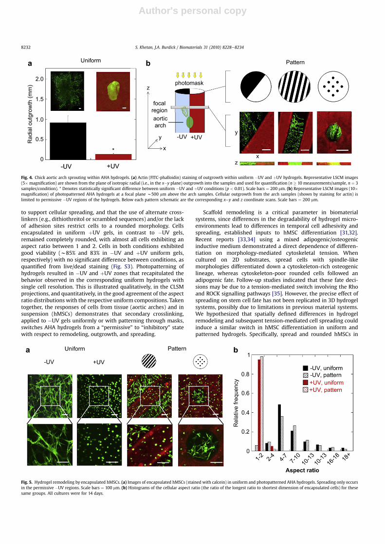
To assesswhether the degradation behavior of the gels translatesto directed cellular behavior, w1 mm3 chick aortic arch samples orhMSCs were mixed with MeRho into the macromer solution andencapsulated in both uniform and photopatterned gels. Aorticarches are useful as a model system for tissue growth, sinceoutgrowth can be readily imaged and quantified [21]. As illustratedin Fig. 4, the degree of cellular outgrowth from aortic arch sampleswas dependent on the hydrogel composition. Arches encapsulatedin uniform �UV hydrogels exhibited robust isotropic radial
outgrowth (0.92 mm � 0.18 mm) compared to largely restrictedoutgrowth in uniform þUV samples (0.14 mm� 0.02 mm) (Fig. 4a).When arch samples were encapsulated in gels synthesized withvarious patterns of light exposure and visualized using confocalmicroscopy (Fig. 4b), outgrowth as a result of gel remodeling wasonly observed into biodegradable�UV (i.e., black) versus restrictiveþUV (i.e., red from MeRho incorporation) regions of the gel. Therepresentative xey and z coordinate projections shown are froma focal region (150 mm thickness) of the gelw500 mmabove the archsample, highlighting the capacity of the patterned gel structures todirect cell migration over a length-scale comparable to the samplethickness (w1 mm).
The responses of encapsulated hMSCs to each hydrogelcomposition (Fig. 5) corroborated the arch findings and providefurther evidence of control of cellular remodeling using sequentialcrosslinking. As shown in representative CLSM projections from theinterior of the gels (Fig. 5a), cells encapsulated in uniform �UVhydrogels for 14 days locally degraded the matrix, spread andexhibited high aspect ratios (i.e., the ratio of the longest dimensionof the cell to the shortest orthogonal dimension) (Fig. 5b). We andothers have shown previously [5,30] that the inclusion of both sitesfor proteolytic degradation and for adhesion (e.g., RGD) is necessary
Fig. 2. Mechanical properties of photopatterned and uniform AHA hydrogels. (a) Elastic modulus of uniform �UV and þUV hydrogels, and �UV and þUV regions of photopatternedhydrogels measured by atomic force microscopy. * Denotes statistically significant difference (p < 0.05) between þUV and �UV conditions for uniform and patterned gels. (b) Thestorage (G0) and loss (G00) moduli of a uniform sequentially crosslinked AHA gel measured by parallel plate oscillatory shear rheometry, with secondary crosslinking performed after15 min of primary crosslinking.
Fig. 3. Enzymatic degradation of sequentially crosslinked AHA hydrogels by exogenous MMP-2. (a) Degradation kinetics of uniform �UV or þUV AHA hydrogels (50% theoreticalconsumption of acrylates during primary addition crosslinking) in the presence of 40 nM MMP-2. (b) Representative scanning electron microscopy (SEM) images of either uniform�UV or þUV hydrogels or hydrogels photopatterned using 250 mm diameter black dots at day 0 (i.e., no degradation) and 12 (following complete degradation of uniform �UVhydrogels) of incubation with 40 nM MMP-2. Scale bars ¼ 100 mm.
S. Khetan, J.A. Burdick / Biomaterials 31 (2010) 8228e8234 8231
Author's personal copy
to support cellular spreading, and that the use of alternate cross-linkers (e.g., dithiothreitol or scrambled sequences) and/or the lackof adhesion sites restrict cells to a rounded morphology. Cellsencapsulated in uniform þUV gels, in contrast to �UV gels,remained completely rounded, with almost all cells exhibiting anaspect ratio between 1 and 2. Cells in both conditions exhibitedgood viability (w85% and 83% in �UV and þUV uniform gels,respectively) with no significant difference between conditions, asquantified from live/dead staining (Fig. S3). Photopatterning ofhydrogels resulted in �UV and þUV zones that recapitulated thebehavior observed in the corresponding uniform hydrogels withsingle cell resolution. This is illustrated qualitatively, in the CLSMprojections, and quantitatively, in the good agreement of the aspectratio distributions with the respective uniform compositions. Takentogether, the responses of cells from tissue (aortic arches) and insuspension (hMSCs) demonstrates that secondary crosslinking,applied to �UV gels uniformly or with patterning through masks,switches AHA hydrogels from a “permissive” to “inhibitory” statewith respect to remodeling, outgrowth, and spreading.
Scaffold remodeling is a critical parameter in biomaterialsystems, since differences in the degradability of hydrogel micro-environments lead to differences in temporal cell adhesivity andspreading, established inputs to hMSC differentiation [31,32].Recent reports [33,34] using a mixed adipogenic/osteogenicinductive medium demonstrated a direct dependence of differen-tiation on morphology-mediated cytoskeletal tension. Whencultured on 2D substrates, spread cells with spindle-likemorphologies differentiated down a cytoskeleton-rich osteogeniclineage, whereas cytoskeleton-poor rounded cells followed anadipogenic fate. Follow-up studies indicated that these fate deci-sions may be due to a tension-mediated switch involving the Rhoand ROCK signalling pathways [35]. However, the precise effect ofspreading on stem cell fate has not been replicated in 3D hydrogelsystems, possibly due to limitations in previous material systems.We hypothesized that spatially defined differences in hydrogelremodeling and subsequent tension-mediated cell spreading couldinduce a similar switch in hMSC differentiation in uniform andpatterned hydrogels. Specifically, spread and rounded hMSCs in
Fig. 4. Chick aortic arch sprouting within AHA hydrogels. (a) Actin (FITC-phalloidin) staining of outgrowth within uniform �UV and þUV hydrogels. Representative LSCM images(5� magnification) are shown from the plane of isotropic radial (i.e., in the xey plane) outgrowth into the samples and used for quantification (n � 10 measurements/sample, n ¼ 3samples/condition). * Denotes statistically significant difference between uniform �UV and þUV conditions (p < 0.01). Scale bars ¼ 200 mm. (b) Representative LSCM images (10�magnification) of photopatterned AHA hydrogels at a focal plane w500 mm above the arch samples. Cellular outgrowth from the arch samples (shown by staining for actin) islimited to permissive �UV regions of the hydrogels. Below each pattern schematic are the corresponding xey and z coordinate scans. Scale bars ¼ 200 mm.
Fig. 5. Hydrogel remodeling by encapsulated hMSCs. (a) Images of encapsulated hMSCs (stained with calcein) in uniform and photopatterned AHA hydrogels. Spreading only occursin the permissive �UV regions. Scale bars ¼ 100 mm. (b) Histograms of the cellular aspect ratio (the ratio of the longest ratio to shortest dimension of encapsulated cells) for thesesame groups. All cultures were for 14 days.
S. Khetan, J.A. Burdick / Biomaterials 31 (2010) 8228e82348232

Author's personal copy
permissive �UV and inhibitory þUV environments were expectedto undergo primarily osteogenesis and adipogenesis, respectively,based simply on morphology. To test this hypothesis, low passagehMSCs were encapsulated in uniform �UV and þUV hydrogels andhydrogels photopatterned with a 250 mm stripe pattern. 14 daysfollowing incubation in 1:1 adipogenic/osteogenic media, hMSCdifferentiation was assessed by quantitative protein assays formarkers of osteogenesis (alkaline phosphatase, ALP) and adipo-genesis (oil red O), gene expression analysis, histological stainingfor ALP and oil red O, and immunostaining for osteocalcin (OC;osteogenic differentiation) and fatty acid binding protein (FABP;adipogenic differentiation). Our choice of differentiation markerswas based on literature demonstrating specificity of each marker toits respective lineage at 14 days following hMSC induction [36,37].
Consistent with our hypothesis and in good agreement with thedescribed previous reports, Fig. 6 illustrates a clear dependence ofhMSC differentiation within the respective AHA hydrogel micro-environments on cell morphology. Results of quantitative assays forALP and oil red O in normalized arbitrary units of activity are shownin Fig. 6a. ALP activity (osteogenic marker) in uniform �UVhydrogels (where cell spreading is permitted) was 2.1 � 0.3,compared to 1.0 � 0.1 within þUV gels (where cell spreading isrestricted). In contrast, oil red O activity (adipogenic marker) was1.7 � 0.3 in uniform þUV hydrogels compared to 1.0 � 0.2 within�UV gels. Representative xey coordinate projections from theinterior of uniform �UV and þUV gels stained for OC (osteogenicmarker) and FABP (adipogenic marker) are shown in Fig. 6b. Cellsencapsulated in �UV gels underwent primarily osteogenic differ-entiation, as illustrated by dense staining of OC in spindle-shapedcells, compared to sparse staining of comparatively rounded cells inthe þUV constructs. The opposite trend was observed for expres-sion of FABP; rounded cells in þUV stained predominantly for thisadipogenic marker relative to cells in �UV gels. Lineage specifica-tion of hMSCs in uniform �UV and þUV environments was furtherconfirmed via quantitative PCR and cytochemical staining(Fig. S4eS5). In photopatterned AHA hydrogels, multi-lineagehMSC differentiation based on the local gel structure wasconfirmed by immunostaining for the same targets (Fig. 6c). Cells in�UV andþUV regions were induced primarily down the osteogenicand adipogenic lineages, respectively.
Interestingly, the observed differentiation switch occurreddespite a mismatch in the mechanical properties traditionally asso-ciated with each lineage [4,38,39] (i.e., hMSCs in the higher elasticmodulusþUV environmentwere induced toward an adipogenic, not
osteogenic fate). Our results contrast, for example, those of a recentreport by Heubsch et al. [39] describing the dependence of MSCdifferentiation on the elastic modulus of physically crosslinked 3Dalginate scaffolds with a constant adhesive ligand concentration. Inthese studies, encapsulated hMSCs differentiated based on the gelelastic modulus (2.5e5 kPa and 11e30 kPa for adipogenesis andosteogenesis, respectively); however, the cells maintained a grosslyroundedmorphology in all conditions. The authors provide evidencethat lineage commitmentwasbasedon theabilityof thecells to reachthrough the surrounding mesh and utilize traction forces to re-organize and cluster adhesive ligands. The degree of cytoskeletaltension and adhesion ligand clustering was shown to correlatedirectly with the gel modulus, providing a mechanistic basis forosteogenic versus adipogenic differentiation in the stiffer versusmore compliant gels.
In our work, we hypothesize that kinetic chain formation in theþUV hydrogel environments, despite causing an increase in mechan-ical properties to a more typical osteogenic range (w15e18 kPa),functions as a non-degradable steric barrier, blocking the ability ofencapsulatedhMSCstospread. Incontrast, spreadhMSCs inpermissive�UV hydrogels undergo osteogenesis, potentially since their ability tospread allows them to apply greater tensions on the matrix, despiteinitial mechanical properties (w6 kPa) that are lower than the þUVsystem that supported adipogenesis. These differences illustrate that,relative to 2D systems, 3D substrate properties including elasticmodulus, adhesive ligand density, degradability and crosslink typemayact collectively onencapsulated cell fate, a dynamicprocess that isless amenable to generalized trends based upon any single property.
4. Conclusions
The sequential crosslinking and photopatterning techniquespresented here constitute a simple but powerful technique tocontrol hydrogel remodeling in 3D. The choice of material proper-ties (e.g., choice of polymer, type of functionalization, modificationefficiency) and experimental parameters (e.g., macromer weightpercentage and ratio of crosslinking types) are among the designparameters kept constant in the current work that can be adjustedto tune degradation and remodeling rates for individual applica-tions. The use of a light-initiated secondary crosslinking step mayalso enable more advanced processing capabilities, such as thesynthesis of a hydrogel with a gradient of crosslink type ratio (i.e.,by using a syringe pump to translate a photomask laterally abovethe gel during light exposure). Such techniques may be valuable for
Fig. 6. Spatially patterned hMSC lineage commitment in mixed adipogenic/osteogenic culture media. (a) Quantitative assays for markers of osteogenic (alkaline phosphatase) andadipogenic (oil red O) hMSC differentiation following 14 days of mixed media culture within uniform �UV and þUV hydrogels. * Denotes statistically significant differences(p < 0.05) between conditions. (b) Representative immunostaining images for fatty acid binding protein (blue; adipogenesis) and osteocalcin (green; osteogenesis) from the interiorof uniform �UV and þUV hydrogels, with channels shown separately for ease of viewing. Scale bars ¼ 100 mm. (c) Representative immunostaining images for the same targets fromthe interior of a hydrogel photopatterned with 250 mm stripes. Scale bar ¼ 100 mm.
S. Khetan, J.A. Burdick / Biomaterials 31 (2010) 8228e8234 8233

Author's personal copy
other basic cell-material interaction studies or advanced tissueengineering applications, including the engineering in vitro ofclinically relevant tissues with interfaces (e.g., osteochondraldefects) or anisotropic properties (e.g., vascular or nervous tissues).Importantly, this technology is applicable to a range of currentlyused polymeric biomaterials and incorporates spatial control that iscrucial in developing complex microenvironments.
Acknowledgements
This work was supported by funding from a Fellowship inScience and Engineering from the David and Lucile Packard Foun-dation (JAB) and a CAREER award (JAB) and Graduate ResearchFellowship (SK) from the National Science Foundation. The authorswould like to thank the following individuals for helpful discus-sions and experimental assistance: R. Marklein, J. Ifkovits,V. Ramanan, J. Katz, Dr. M. Charati, Dr. C. Shen and Dr. J. Miller.
Appendix. Supplementary information
Supplementary information associated with this article can befound in the on-line version, at doi:10.1016/j.biomaterials.2010.07.035.
Appendix
Figures with essential color discrimination. Figs. 1, 2, 4e6 in thisarticle are difficult to interpret in black and white. The fullcolor images can be found in the on-line version, at doi:10.1016/j.biomaterials.2010.07.035.
References
[1] Hynes R. Integrins: bidirectional, allosteric signalling machines. Cell 2002;110(6):673e87.
[2] Geiger B, Bershadsky A. Exploring the neighborhood: adhesion-coupled cellmechanosensors. Cell 2002;110(2):139e42.
[3] Marklein R, Burdick J. Spatially controlled hydrogel mechanics to modulatestem cell interactions. Soft Matter 2010;6(1):136e43.
[4] Engler A, Sen S, Sweeney H, Discher D. Matrix elasticity directs stem celllineage specification. Cell 2006;126(4):677e89.
[5] Lutolf M, Raeber G, Zisch A, Tirelli N, Hubbell J. Cell-responsive synthetichydrogels. Adv Mater 2003;15(11):888e92.
[6] Kim J, Kim I, Cho T, Lee K, Hwang S, Tae G, et al. Bone regeneration usinghyaluronic acid-based hydrogel with bone morphogenic protein-2 and humanmesenchymal stem cells. Biomaterials 2007;28(10):1830e7.
[7] Benoit D, Schwartz M, Durney A, Anseth K. Small functional groups forcontrolled differentiation of hydrogel-encapsulated human mesenchymalstem cells. Nat Mater 2008;7(10):816e23.
[8] Lee S, Moon J, West J. Three-dimensional micropatterning of bioactivehydrogels via two-photon laser scanning photolithography for guided 3D cellmigration. Biomaterials 2008;29(20):2962e8.
[9] Musoke-Zawedde P, Shoichet M. Anisotropic three-dimensional peptidechannels guide neurite outgrowth within a biodegradable hydrogel matrix.Biomed Mater 2006;1(3):162e9.
[10] DeForest C, Polizzotti B, Anseth K. Sequential click reactions for synthesizingand patterning three-dimensional cell microenvironments. Nat Mater 2009;8(8):659e64.
[11] Kloxin A, Kasko A, Salinas C, Anseth K. Photodegradable hydrogels fordynamic tuning of physical and chemical properties. Science 2009;324(5923):59e63.
[12] Yung S, Thomas G, Davies M. Induction of hyaluronan metabolism aftermechanical injury of human peritoneal mesothelial cells in vitro. Kidney Int2000;58(5):1953e62.
[13] Jiang D, Liang J, Noble P. Hyaluronan in tissue injury and repair. Annu Rev CellDev Biol 2007;23:435e61.
[14] Leach J, Bivens K, Collins C, Schmidt C. Development of photocrosslinkablehyaluronic acid-polyethylene glycol-peptide composite hydrogels for softtissue engineering. J Biomed Mater Res A 2004;70(1):74e82.
[15] Chung C, Burdick J. Influence of three-dimensional hyaluronic acid microen-vironments on mesenchymal stem cell chondrogenesis. Tissue Eng Part A2009;15(2):243e54.
[16] Sakai Y, Matsuyama Y, Takahashi K, Sato T, Hattori T, Nakashima S, et al. Newartificial nerve conduits made with photocrosslinked hyaluronic acid forperipheral nerve regeneration. Biomed Mater Eng 2007;17(3):191e7.
[17] Sahoo S, Chung C, Khetan S, Burdick J. Hydrolytically degradable hyaluronicacid hydrogels with controlled temporal structures. Biomacromolecules2008;9(4):1088e92.
[18] Ji Y, Ghosh K, Shu X, Li B, Sokolov J, Prestwich G, et al. Electrospun three-dimensional hyaluronic acid nanofibrous scaffolds. Biomaterials 2006;27(20):3782e92.
[19] Williams C, Malik A, Kim T, Manson P, Elisseeff J. Variable cytocompatibility ofsix cell lines with photoinitiators used for polymerizing hydrogels and cellencapsulation. Biomaterials 2005;26(11):1211e8.
[20] Burdick J, Chung C, Jia X, Randolph M, Langer R. Controlled degradation andmechanical behavior of photopolymerized hyaluronic acid networks.Biomacromolecules 2005;6(1):386e91.
[21] Miller J, Shen C, Legant W, Baranski J, Blakely B, Chen C. Bioactive hydrogelsmade from step-growth derived PEG-peptide macromers. Biomaterials2010;31(13):3736e43.
[22] Mauney J, Volloch V, Kaplan D. Matrix-mediated retention of adipogenicdifferentiation potential by human adult bone marrow-derived mesenchymalstem cells during ex vivo expansion. Biomaterials 2005;26(31):6167e75.
[23] Singer V, Jones L, Yue S, Haugland R. Characterization of PicoGreen reagentand development of a fluorescence-based solution assay for double-strandedDNA quantitation. Anal Biochem 1997;249(2):228e38.
[24] Kim J, Park Y, Tae G, Lee K, Hwang S, Kim I, et al. Synthesis and character-ization of matrix metalloprotease sensitive-low molecular weight hyaluronicacid based hydrogels. J Mater Sci Mater Med 2008;19(11):3311e8.
[25] Chung C, Mesa J, Miller G, Randolph M, Gill T, Burdick J. Effects of auricularchondrocyte expansion on neocartilage formation in photocrosslinkablehyaluronic acid networks. Tissue Eng 2006;12:2665e73.
[26] Chung C, Erickson I, Mauck R, Burdick J. Differential behavior of auricular andarticular chondrocytes in hyaluronic acid hydrogels. Tissue Eng Part A2008;14(7):1121e31.
[27] Burdick J, Anseth K. Photoencapsulation of osteoblasts in injectable RGD-modified PEG hydrogels for bone tissue engineering. Biomaterials 2002;23(22):4315e23.
[28] Lee H, Lee J, Chansakui T, Yu C, Elisseeff J, Yu S. Collagen mimetic peptide-conjugated photopolymerizable PEG hydrogel. Biomaterials 2006;27(30):5268e76.
[29] Osman R, Namboodiri K, Weinstein H, Rabinowitz J. Reactivities of acrylic andmethacrylic acids in a nucleophilic addition model of their biological activity.J Am Chem Soc 1988;110(6):1701e7.
[30] Khetan S, Katz J, Burdick J. Sequential crosslinking to control cellularspreading in 3-dimensional hydrogels. Soft Matter 2009;5:1601e6.
[31] Senechal H, Wahrmann J, Delain D, Macieira-Coelho A. Modulation of differ-entiation in vitro. II. Influence of cell spreading and surface events on myo-genesis. In Vitro 1984;20(9):692e8.
[32] Scintu F, Reali C, Pillai R, Badiali M, Sanna M, Argiolu F, et al. Differentiation ofhuman bone marrow stem cells into cells with a neural phenotype: diverseeffects of two specific treatments. BMC Neurosci 2006;7:14.
[33] Kilian K, Bugarija B, Lahn B, Mrksich M. Geometric cues for directing thedifferentiation of mesenchymal stem cells. Proc Natl Acad Sci U S A 2010;107(11):4872e7.
[34] Ruiz S, Chen C. Emergence of patterned stem cell differentiation withinmulticellular structures. Stem Cells 2008;26(11):2921e7.
[35] McBeath R, Pirone D, Nelson C, Bhadriraju K, Chen C. Cell shape, cytoskeletaltension, and RhoA regulate stem cell lineage commitment. Dev Cell 2004;6(4):483e95.
[36] Caballero M, Reed C, Madan G, Van Aalst J. Osteoinduction in umbilical cord-and palate periosteum-derived mesenchymal stem cells. Ann Plast Surg2010;64(5):605e9.
[37] Podechard N, Fardel O, Corolleur M, Bernard M, Lecureur V. Inhibition ofhuman mesenchymal stem cell-derived adipogenesis by the environmentalcontaminant benzo(a)pyrene. Toxicol In Vitro 2009;23(6):1139e44.
[38] Pek Y, Wan A, Ying J. The effect of matrix stiffness on mesenchymal stem celldifferentiation in a 3D thioxtropic gel. Biomaterials 2010;31:385e91.
[39] Huebsch N, Arany P, Mao A, Shvartsman D, Ali O, Bencherif S, et al. Harnessingtraction-mediated manipulation of the cell/matrix interface to control stem-cell fate. Nat Mater 2010;9:518e26.
S. Khetan, J.A. Burdick / Biomaterials 31 (2010) 8228e82348234