Embed Size (px)
Citation preview
This article appeared in a journal published by Elsevier. The attached
copy is furnished to the author for internal non-commercial research
and education use, including for instruction at the authors institution
and sharing with colleagues.
Other uses, including reproduction and distribution, or selling or
licensing copies, or posting to personal, institutional or third party
websites are prohibited.
In most cases authors are permitted to post their version of the
article (e.g. in Word or Tex form) to their personal website or
institutional repository. Authors requiring further information
regarding Elsevier’s archiving and manuscript policies are
encouraged to visit:
http://www.elsevier.com/copyright
Author's personal copy
Arg149 Is Involved in Switching the Low Affinity, OpenState of the Binding Protein AfProX into Its High Affinity,Closed State
Britta Tschapek1†, Marco Pittelkow2†, Linda Sohn-Bösser2,Gudrun Holtmann2, Sander H. J. Smits1, Holger Gohlke3,Erhard Bremer2 and Lutz Schmitt1!1Institute of Biochemistry, Heinrich Heine University Duesseldorf, Universitaetsstrasse 1,D-40225 Duesseldorf, Germany2Laboratory for Microbiology, Department of Biology, Philipps University Marburg,Karl-von-Frisch Strasse 8, D-35032 Marburg, Germany3Institute of Pharmaceutical and Medicinal Chemistry, Heinrich Heine University Duesseldorf,Universitaetsstrasse 1, D-40225 Duesseldorf, Germany
Received 8 April 2011;received in revised form21 May 2011;accepted 25 May 2011Available online2 June 2011
Edited by R. Huber
Keywords:ABC transporter;substrate binding protein;glycine betaine;substrate recognition;ligand binding site
The substrate binding protein AfProX from the Archaeoglobus fulgidus ProUATP binding cassette transporter is highly selective for the compatiblesolutes glycine betaine (GB) and proline betaine, which confer thermo-protection to this hyperthermophilic archaeon. A detailed mutationalanalysis of the substrate binding site revealed the contribution of individualamino acids for ligand binding. Replacement of Arg149 by an Ala residuedisplayed the largest impact on substrate binding. The structure of a mutantAfProX protein (substitution of Tyr111 with Ala) in complex with GB wassolved in the open liganded conformation to gain further insight into ligandbinding. In this crystal structure, GB is bound differently compared to theGB closed liganded structure of the wild-type AfProX protein. We foundthat a network of amino acid side chains communicates the presence of GBtoward Arg149, which increases ligand affinity and induces domain closureof AfProX. These results were corroborated by molecular dynamics studiesand support the view that Arg149 finalizes the high-affinity state of theAfProX substrate binding protein.
© 2011 Elsevier Ltd. All rights reserved.
Introduction
Microorganisms import a variety of chemicalcompounds from environmental sources through
high-affinity transport systems, many of whichbelong to the family of ATP binding cassette(ABC) transporters.1 These systems depend strictlyon a so-called substrate binding protein (SBP) thatcaptures the ligand with high affinity and subse-quently delivers it to its cognate transporter.2 TheSBP thereby determines the directionality of theoverall transport reaction, and its interaction withthe transmembrane domain of the transporterregulates the ATPase activity of the transportcomplex.2,3Structural studies of SBPs revealed a common fold
with a bilobal organization connected via a linkerregion.4,5 In the ligand-free, open conformation, the
*Corresponding author. E-mail address:[email protected].† B.T. and M.P. contributed equally to this work.Abbreviations used: ABC, ATP binding cassette; MD,
molecular dynamics; SBP, substrate binding protein; MBP,maltose binding protein; GB, glycine betaine; PB, prolinebetaine; PDB, Protein Data Bank.
doi:10.1016/j.jmb.2011.05.039 J. Mol. Biol. (2011) 411, 36–52
Contents lists available at www.sciencedirect.com
Journal of Molecular Biologyj ourna l homepage: ht tp : / /ees .e lsev ie r.com. jmb
0022-2836/$ - see front matter © 2011 Elsevier Ltd. All rights reserved.
Author's personal copy
two domains are separated from each other, therebyforming a deep solvent-exposed cleft, which harborsthe substrate binding site. Upon ligand binding,both domains move toward each other through ahinge-bending motion or rotation, which results inthe so-called closed liganded conformation. As aconsequence of this movement, residues originating
from both domains generate the ligand binding siteand trap the ligandwithin a deep cleft formed by thetwo lobes of the SBP.4 In the absence of a ligand,open and closed unliganded states of the SBP are inequilibrium, and the ligand solely shifts thisequilibrium toward the closed liganded state. Thissequence of events has been coined the “Venusflytrap mechanism”,6–8 which is supported bycrystal structures in the absence and presence of aligand9,10 and other biophysical techniques.4For the maltose binding protein (MBP),2 it has
been shown that both domains are dynamicallyfluctuating in the absence of the ligand.11 NMRspectroscopy revealed that the ligand-free form ofMBP consists of a predominantly open species (95%)and a minor species (5%) that corresponds to apartially closed state.12 The open form of MBPobserved by NMR is similar to the crystal structureof the open unliganded conformation.13 However,the partially closed species detected by NMR12 doesnot correspond to the ligand-bound, fully closedcrystal form. Instead, it represents an intermediate,partially closed conformation,14 suggesting that thesubstrate is required to reach the final, closedliganded conformation.Millet et al. have shown that every degree of
domain closure in the unliganded MBP requiresconformational deformation energy of approximate-ly 212 cal/mol. Since MBP closes by about 20°, thiswould lead to an energetic cost of !4 kcal/molwhen forming the closed unliganded conformationof MBP.15 This closed unliganded state, firstobserved by X-ray crystallography, is thereforehighly unfavorable and resembles a loaded spring.15Changing the stiffness of this region by introducingamino acids with bulky side chains changes thedegree of closure of MBP, which directly correlateswith maltose binding affinity.15,16 As a consequence,mutations in the hinge region likely alter theequilibrium between the open and partially closedstates of MBP.12 Upon maltose binding, a furthershift in this equilibrium occurs toward the fullyclosed liganded conformation, which becomesaccessible because maltose forms favorable interac-tions with the protein.Generally, the hinge region provides MBP with a
large degree of flexibility. This allows MBP to bind avariety of malto-oligosaccharides, some of whichlead to domain closure.13,17 Although this highlightsa distinct role of the substrate in domain closure, theexact nature of the substrate-induced closing mech-anism of MBPs and SBPs in general is unknown.Furthermore, SBPs bind their substrates with highaffinity,4 but it is not fully understood how thesubstrate is released to its cognate ABC importer.One scenario could be that interactions between theABC transporter and the SBP during the transportcycle lead to a modulation of the SBP's affinity to itssubstrate.3
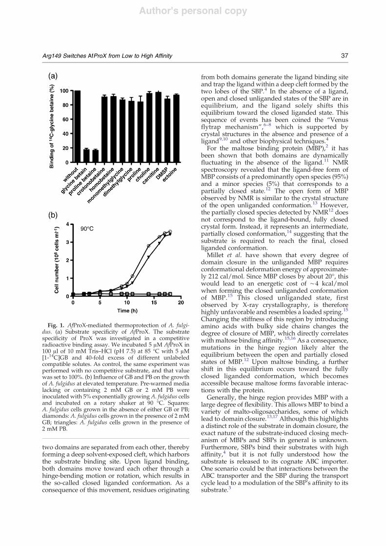
Fig. 1. AfProX-mediated thermoprotection of A. fulgi-dus. (a) Substrate specificity of AfProX. The substratespecificity of ProX was investigated in a competitiveradioactive binding assay. We incubated 5 !M AfProX in100 !l of 10 mM Tris–HCl (pH 7.5) at 85 °C with 5 !M[1-14C]GB and 40-fold excess of different unlabeledcompatible solutes. As control, the same experiment wasperformed with no competitive substrate, and that valuewas set to 100%. (b) Influence of GB and PB on the growthof A. fulgidus at elevated temperature. Pre-warmed medialacking or containing 2 mM GB or 2 mM PB wereinoculated with 5% exponentially growing A. fulgidus cellsand incubated on a rotary shaker at 90 °C. Squares:A. fulgidus cells grown in the absence of either GB or PB;diamonds: A. fulgidus cells grown in the presence of 2 mMGB; triangles: A. fulgidus cells grown in the presence of2 mM PB.
37Arg149 Switches AfProX from Low to High Affinity
Author's personal copy
In the thermophilic archaeon Archaeoglobus fulgi-dus, the ABC importer ProU has been annotated as aspecific uptake system for the compatible solutesglycine betaine (GB) and proline betaine (PB).18 Thecorresponding SBP (AfProX) has been crystallized indifferent conformations: a closed liganded confor-mation in complex with either GB or PB and in anopen unliganded conformation.19Here, we present the structure of AfProX in the
open liganded conformation together with a sys-tematic mutational analysis of the binding site of theAfProX protein and molecular dynamics (MD)simulations of liganded AfProX, using startingstructures of different conformational states. Basedon our results, residue Arg149 is crucial to switchAfProX from a low-affine open structure to a high-affine closed state. Our data imply that the “Venusflytrap” mechanism is composed of distinct molec-ular events to ensure the specific biological functionof SBPs because the presence of the substrate iscommunicated through a network of amino acidslocated in both domains, triggering domain closure.
Results
Thermoprotection of the hyperthermophilicarchaeon A. fulgidus by GB and PB
AfProX possesses striking substrate specificity:from all of the osmoprotectants tested, only GB andPB are substrates (Fig. 1a). Both GB and PB typicallyfunction as osmostress protectants in microorgan-isms. However, we observed that these compoundshad no osmoprotective effect for A. fulgidus (Sup-plementary Fig. 1). Therefore, the uptake of GB or PBmust serve another physiological role for thishyperthermophilic archaeon. Since GB has beenshown previously to serve as a thermoprotectant in
different bacteria,20 we explored a possible thermo-protective effect of GB and PB forA. fulgidus. NeitherGB nor PB had any effect on the growth ofA. fulgiduswhen it was cultivated at its optimal growthtemperature of 83 °C (Supplementary Fig. 1). Incontrast, both compounds exerted a strong thermo-protective effect for cells cultivated at the elevatedgrowth temperature of 90 °C. Without the additionof GB or PB to the growth medium, A. fulgidus wasunable to grow at this temperature (Fig. 1b). Such astriking thermoprotection of a hyperthermophilicarchaeon has never been observed before.
Mutational analysis of the ligand bindingsite in AfProX
AfProX has previously been crystallized withsubstrate at a resolution of 2.1 Å and 1.9 Å,respectively.19 The positively charged trimethylam-monium head group of GB and the dimethylammo-nium head group of PB are wedged into an aromaticcage that is formed by the main-chain carbonyl ofAsp109 and by four Tyr residues (Tyr63, Tyr111,Tyr119, and Tyr214). Here, cation–" interactions arekey determinants for coordinating the head group.The carboxylic tails of GB and PB protrude from thiscage and form two salt bridges and one hydrogenbond with Lys13, Thr66, and Arg149, respectively.19Binding of GB and PB to purified AfProX influencedthe spectroscopic properties of AfProX. Substratebinding of these compatible solutes leads to anincrease of the intrinsic tryptophan fluorescence. Weexploited this feature to determine the apparentaffinity constants (Kd) for both substrates. AfProXbinds GB and PB with high apparent affinity, 60±10 nM and 50±10 nM, respectively (Table 1). Thesedata represent high, but not unusual, apparentaffinities of SBPs5,21 of ABC transporters. To furthersupport our findings on the affinity ofAfProX for GBand PB, we also determined a similar bindingconstant of GB by isothermal calorimetry, revealinga Kd of 100±30 nM (Supplementary Fig. 2). It isimportant to note that, with both techniques, weonly observe the binding of the substrate, but wecannot differentiate between an initial binding of thesubstrate and subsequent domain closure.According to the structure of AfProX,19 it is
evident that the substrate binding pocket is builtup by four tyrosines (Tyr63, Tyr111, Tyr190, andTyr214) and Lys13, Thr66, and Arg149. To furtherunderstand the residues' roles in substrate binding,we mutated them to alanine, and we measured thesubstrate apparent affinity of the correspondingmutants (Table 1). Mutation of any of the fourtyrosines reduced the apparent affinity, rangingfrom 3.5±0.7 !M for Tyr214Ala to 149±17 !M forTyr63Ala, respectively (Table 1). Any one of thedouble Tyr-to-Ala mutants was not able to bind GBat all.
Table 1. Mutational analysis of the binding site of AfProX
Mutant Kd GB (!M) Kd PB (!M)
Wild type 0.06±0.01 0.05±0.01K13A 107±20 101±12Y63A 149±17 288±26T66A 1.8±0.2 18±1.4Y111A 76±4 148±28E145A 2.7±0.5 23.2±4F146A 6.6±0.6 4.0±0.5Y190A 67±9 19±5R149A 320±60 n.b.Y214A 3.5±0.7 3.5±0.7Double tyrosine mutations n.b. n.b.
The apparent affinities of wild-type AfProX and AfProX mutantsare summarized for GB and PB. No binding for any of the doublemutants was observed; hence, these mutants are not listed in thetable. n.b. indicates that no binding was observed under thisexperimental setup.
38 Arg149 Switches AfProX from Low to High Affinity
Author's personal copy
The Lys13Ala and Thr66Ala mutants also showed alower apparent affinity, 1.8±0.2 !M and 107±20 !M,respectively (Table 1). The largest decrease in apparentaffinity in the case of a single mutation was observedfor the Arg149Ala mutation. Here, the apparent Kddropped to 320±60 !M for GB, and no binding couldbe observed for PB (Table 1). This suggests a specificrole of Arg149 in the substrate binding to AfProX.
Crystal structure of Tyr111Ala AfProX
The Tyr mutations of the aromatic box had anunexpectedly large effect on the apparent affinity ofAfProX for GB. Therefore, we crystallized one ofthese mutants (Tyr111Ala) and solved its structurein the presence of GB at 2.0 Å. Tyr111Ala has anapparent affinity of 76±4 !M toward GB (Table 1).Crystals were grown as described in Material andMethods. A data set of the AfProX Tyr111Ala incomplex with GB was collected at beamline ID23
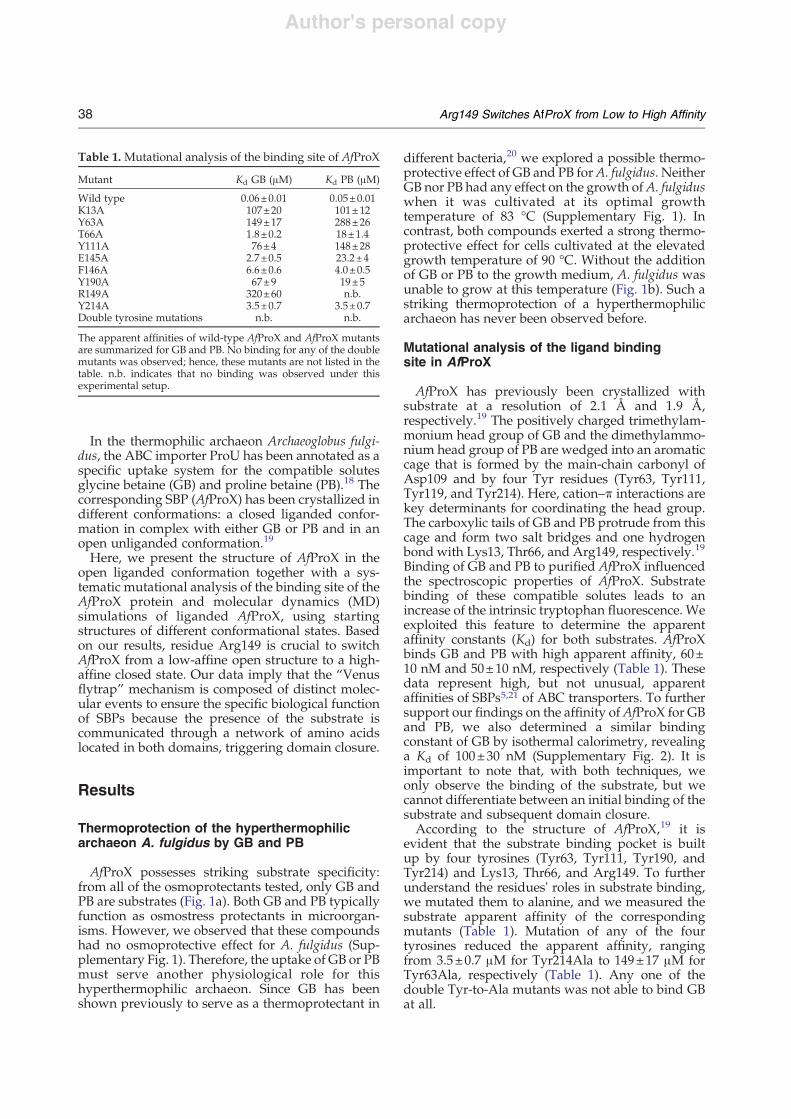
(European Synchrotron Radiation Facility, Greno-ble, France) and processed using XDS.22 Initialphases were obtained by molecular replacementusing the program Phaser,23 with the open unli-ganded AfProX structure as template [Protein DataBank (PDB) entry: 1SW5].19 The final structure wasrefined to a resolution of 2.0 Å. Data, refinementstatistics, and model content are summarized inTable 2.The structure of Tyr111Ala AfProX consists of two
domains, the ligand GB and a Hepes moleculecaptured from the crystallization solution. Due tothe binding of Hepes in close proximity, Asn16adopts an unusual conformation (as highlighted inthe Ramachandran plot). A comparison with theopen liganded structure and a closed ligandedstructure revealed that the structure of Tyr111AlaAfProX adopts an open liganded conformation, withthe hinge region positioned between residues 109–111 and residues 213–215 (Fig. 2). The RMSDbetween the two, open and closed liganded struc-tures of AfProX, was analyzed by the DynDomserver.26 The RMSD after the superimposition of allC# atoms of the Tyr111Ala structure with respect tothe closed liganded structure of AfProX (PDB code:1SW2) is 4.75 Å, whereas only minimal structuraldifferences were found with respect to the openunl iganded structure (PDB code: 1SW5)(RMSDb0.2 Å). In order to determine how muchdomain II moves with respect to domain I whengoing from the open to the closed structures, wesuperimposed residues 1–105 and 207–270 ofdomain I; subsequently, the RMSD was calculatedfor C# atoms of domain II only, which yields 13.3 Å.When reporting the MD simulation results below,the latter RMSD definition is used.A comparison between the open liganded structure
and the open unliganded structure revealed that onlyone loop (amino acids 142–153) differs (RMSD,1.6 Å). This loop contains Arg149 that is part of thesubstrate binding site in the closed liganded structureand will be referred to as “Arg loop”.
The substrate binding site of the AfProXTyr111Ala variant
In the AfProX Tyr111Ala mutant, the trimethylam-moniummoiety of GB is located at the same positionas in the closed liganded structure (Fig. 3). AlthoughGB lacks interactions with Tyr111, it is bound byTyr63, Tyr190, and Tyr214 of the aromatic cage andfurther stabilized by interactions with Thr66, Asp109,and the piperazinyl ring of the bound Hepes.Notably, when compared to the closed structure ofthe wild-type protein, the carboxyl tail of GB isrotated by almost 180° in the AfProX Tyr111Alamutant. Here, GB interacts through a water moleculewith Thr66 and through two water molecules withthe side chain of Tyr190. As the carboxyl group of GB
Table 2. Crystallographic parameters
Space group P1a, b, c (Å) 33.6, 36.9, 57.8#, $, % (°) 83.8, 80.5, 95.9
Data collection and processingWavelength (Å) 0.8726Resolution (Å) 20–2.0Mean redundancy 4.2 (4.3)Unique reflections 17,040Completeness (%) 92.6 (93.6)I/sigma 47.7 (29.0)Rsym
a 2.1 (4.3)
RefinementRF
b (%) 17.8Rfree
c (%) 24.4Overall B-factor from Wilson scaling (Å2) 20.9RMSD from idealBond lengths (Å) 0.023Bond angles (°) 0.956Average B-factors (Å2) 7.6Ramachandran plot (%)Most favored 93.4Allowed 6.2Disallowed 0.4Model contentMonomers per asymmetric unit 1Protein residues 6–275LigandsGB 1Hepes 1H2O 300
Crystal parameters and data collection statistics are derived fromXDS.24 Refinement statistics were obtained from REFMAC5.25Ramachandran analysis was performed using PROCHECK.Numbers in parentheses correspond to the highest-resolutionshell (2.1–2.0 Å).
a R sym is defined as Rsym =P
hklP
i j Ii hkl! " "hI hkl! "i j =Phkl
PI Ii hkl! ".
b RF is defined as RF =P
hkl jFobs j " jFcalc j =P
hkl jFobs j .c Rfree is calculated as RF but for 5% randomly chosenreflections that were omitted from all refinement steps.
39Arg149 Switches AfProX from Low to High Affinity
Author's personal copy
forms a neither direct nor water-mediated interactionwith the Hepes molecule. Therefore, the orientationof GB in the crystal structure of theAfProX Tyr111Alavariant should not be influenced.The binding sites in the closed liganded, open
liganded, and open unliganded structures of AfProXare closely related because Tyr63, Tyr214, Lys13,Thr66, and Asp109 are superimposable in all threestructures (Fig. 4a and b). However, the side chain ofTyr190 is flipped in the closed structure by 130°
when compared to both open structures. Interest-ingly, the position of the Tyr111 side chain in theclosed liganded structure is located at the position ofthe side chain of Tyr190 in the open unliganded andopen liganded structures (Fig. 4a and b). Thisimplies that, during the opening and closingmovements of AfProX, the binding site does notundergo a major conformational change; rather, it islargely preformed in the open unliganded structureand, thus, pre-dispositioned to capture GB. Still, the
Fig. 2. Overlay of Tyr111Ala AfProX with the closed liganded and the open unliganded structures of AfProX. (a)Overall structure of AfProX highlighted as cartoons. (b) Overlay of the Tyr111Ala AfProX structure with the closedliganded structure of AfProX (pink; PDB code: 1SW2). (c) Overlay of the Tyr111Ala AfProX structure with the openunliganded structure of AfProX (light green; PDB code: 1SW5). The overlays were calculated using LSQMAN.
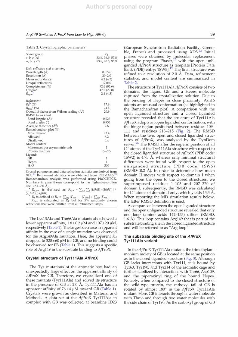
Fig. 3. The binding site of Tyr111Ala AfProX. (a) The GB binding site of Tyr111Ala AfProX is shown in sticks as isArg149 with its neighboring residues Asp145 and Glu151. Broken lines depict distances b3.5 Å between Arg149 andAsp145 or Glu151. Bound GB is highlighted in blue. (b) A simulated annealing omit map is shown for GB. For clarity, onlythe tyrosine residues of the binding site are shown. Both figures have slightly different orientations in order to show alsothe density of the tail of GB.
40 Arg149 Switches AfProX from Low to High Affinity
Author's personal copy
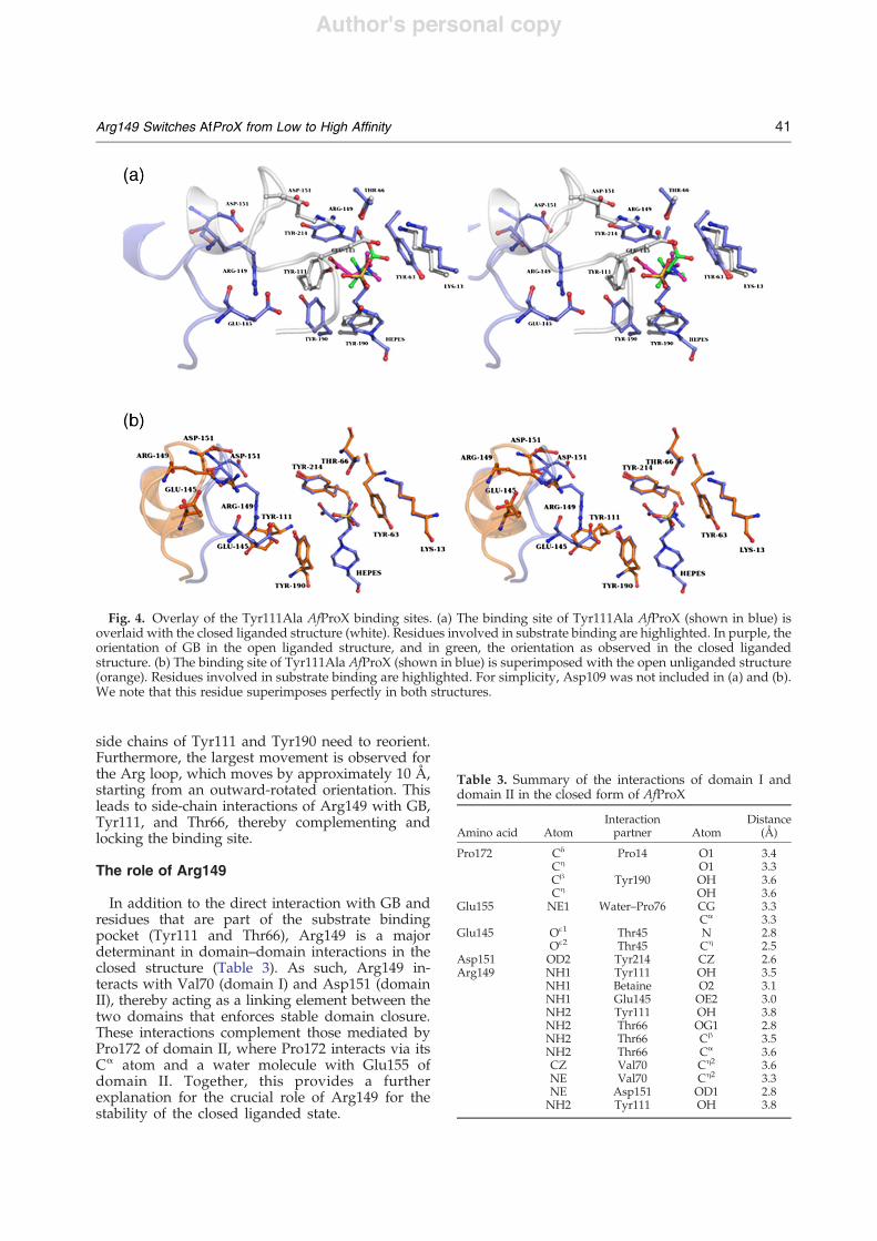
side chains of Tyr111 and Tyr190 need to reorient.Furthermore, the largest movement is observed forthe Arg loop, which moves by approximately 10 Å,starting from an outward-rotated orientation. Thisleads to side-chain interactions of Arg149 with GB,Tyr111, and Thr66, thereby complementing andlocking the binding site.
The role of Arg149
In addition to the direct interaction with GB andresidues that are part of the substrate bindingpocket (Tyr111 and Thr66), Arg149 is a majordeterminant in domain–domain interactions in theclosed structure (Table 3). As such, Arg149 in-teracts with Val70 (domain I) and Asp151 (domainII), thereby acting as a linking element between thetwo domains that enforces stable domain closure.These interactions complement those mediated byPro172 of domain II, where Pro172 interacts via itsC# atom and a water molecule with Glu155 ofdomain II. Together, this provides a furtherexplanation for the crucial role of Arg149 for thestability of the closed liganded state.
Fig. 4. Overlay of the Tyr111Ala AfProX binding sites. (a) The binding site of Tyr111Ala AfProX (shown in blue) isoverlaid with the closed liganded structure (white). Residues involved in substrate binding are highlighted. In purple, theorientation of GB in the open liganded structure, and in green, the orientation as observed in the closed ligandedstructure. (b) The binding site of Tyr111Ala AfProX (shown in blue) is superimposed with the open unliganded structure(orange). Residues involved in substrate binding are highlighted. For simplicity, Asp109 was not included in (a) and (b).We note that this residue superimposes perfectly in both structures.
Table 3. Summary of the interactions of domain I anddomain II in the closed form of AfProX
Amino acid AtomInteractionpartner Atom
Distance(Å)
Pro172 C& Pro14 O1 3.4C' O1 3.3C$ Tyr190 OH 3.6C' OH 3.6
Glu155 NE1 Water–Pro76 CG 3.3C# 3.3
Glu145 O!1 Thr45 N 2.8O!2 Thr45 C' 2.5
Asp151 OD2 Tyr214 CZ 2.6Arg149 NH1 Tyr111 OH 3.5
NH1 Betaine O2 3.1NH1 Glu145 OE2 3.0NH2 Tyr111 OH 3.8NH2 Thr66 OG1 2.8NH2 Thr66 C$ 3.5NH2 Thr66 C# 3.6CZ Val70 C'2 3.6NE Val70 C'2 3.3NE Asp151 OD1 2.8NH2 Tyr111 OH 3.8
41Arg149 Switches AfProX from Low to High Affinity
Author's personal copy
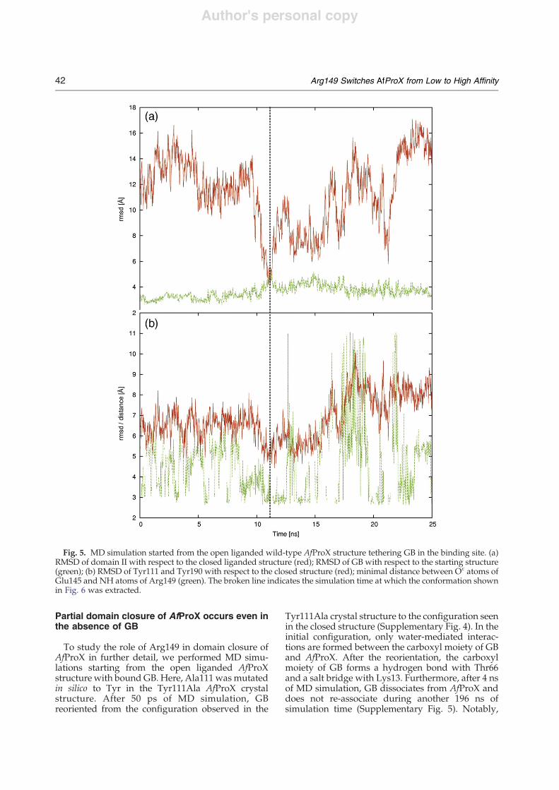
Partial domain closure of AfProX occurs even inthe absence of GB
To study the role of Arg149 in domain closure ofAfProX in further detail, we performed MD simu-lations starting from the open liganded AfProXstructure with bound GB. Here, Ala111 was mutatedin silico to Tyr in the Tyr111Ala AfProX crystalstructure. After 50 ps of MD simulation, GBreoriented from the configuration observed in the
Tyr111Ala crystal structure to the configuration seenin the closed structure (Supplementary Fig. 4). In theinitial configuration, only water-mediated interac-tions are formed between the carboxyl moiety of GBand AfProX. After the reorientation, the carboxylmoiety of GB forms a hydrogen bond with Thr66and a salt bridge with Lys13. Furthermore, after 4 nsof MD simulation, GB dissociates from AfProX anddoes not re-associate during another 196 ns ofsimulation time (Supplementary Fig. 5). Notably,
Fig. 5. MD simulation started from the open liganded wild-type AfProX structure tethering GB in the binding site. (a)RMSD of domain II with respect to the closed liganded structure (red); RMSD of GB with respect to the starting structure(green); (b) RMSD of Tyr111 and Tyr190 with respect to the closed structure (red); minimal distance between O! atoms ofGlu145 and NH atoms of Arg149 (green). The broken line indicates the simulation time at which the conformation shownin Fig. 6 was extracted.
42 Arg149 Switches AfProX from Low to High Affinity
Author's personal copy
after 50 ns of MD simulation, unliganded AfProXundergoes a drastic conformational change: domainII comes as close as 2 Å RMSD to the configurationfound in the closed AfProX structure (Supplemen-tary Fig. 5). During the remaining 150 ns of MDsimulation, domain II repeatedly opens again butalways returns to the configuration of the closedstructure. In summary, MD simulations that startedfrom the open AfProX structure reveal that atendency of domain closure exists even in theabsence of GB. Such motions are inline with the“Venus flytrap mechanism”4 and have been exper-imentally verified for MBP.12
Coupled structural reorganization in the bindingsite upon domain closure
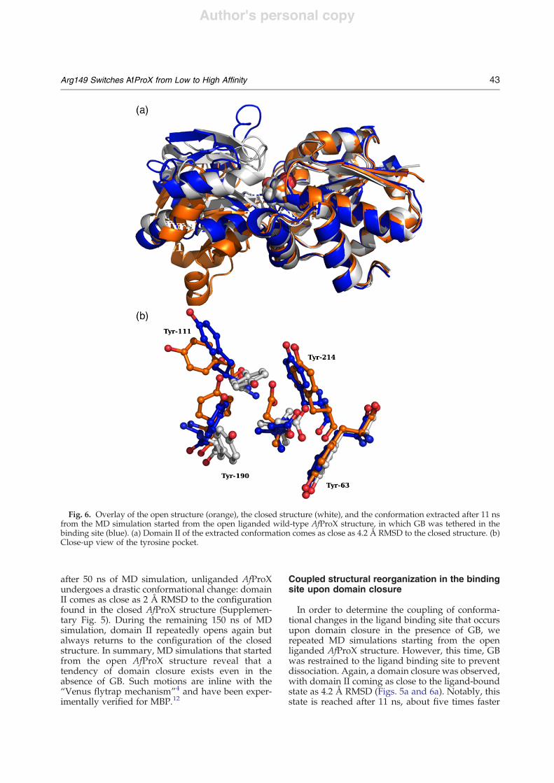
In order to determine the coupling of conforma-tional changes in the ligand binding site that occursupon domain closure in the presence of GB, werepeated MD simulations starting from the openliganded AfProX structure. However, this time, GBwas restrained to the ligand binding site to preventdissociation. Again, a domain closure was observed,with domain II coming as close to the ligand-boundstate as 4.2 Å RMSD (Figs. 5a and 6a). Notably, thisstate is reached after 11 ns, about five times faster
Fig. 6. Overlay of the open structure (orange), the closed structure (white), and the conformation extracted after 11 nsfrom the MD simulation started from the open liganded wild-type AfProX structure, in which GB was tethered in thebinding site (blue). (a) Domain II of the extracted conformation comes as close as 4.2 Å RMSD to the closed structure. (b)Close-up view of the tyrosine pocket.
43Arg149 Switches AfProX from Low to High Affinity
Author's personal copy
than in the absence of GB (Fig. 5a). Although singleevents that occur only once during a MD simulationmust be interpreted with caution, these resultsindicate that binding of GB facilitates conformation-al changes occurring during the transition from theopen state to the closed state. Furthermore, domainclosure is accompanied by a reorientation of GB (Fig.5a). In parallel, the Tyr cage that coordinates thebinding of the trimethylammonium moiety of GBvia cation–" interactions starts to adopt the confor-mation observed in the closed structure (Fig. 5b). Inthis process, Tyr111 shifts about halfway between
the conformations of the open and closed structures,whereas the conformation of Tyr190 is still close tothe one observed in the open structure (Fig. 6b). ForTyr111 to reach its final position, Tyr190 mustreorient. In addition, the minimal distance betweenany O! atom of Glu145 and any NH atom of Arg149fluctuates between 2.8 Å and !6 Å during the first8 ns of simulation time (Fig. 5b), indicating thetemporary formation of a salt bridge. This saltbridge consolidates during the next 2 ns, after whichit is almost permanently formed for another 2 ns.Upon domain opening after about 12 ns of
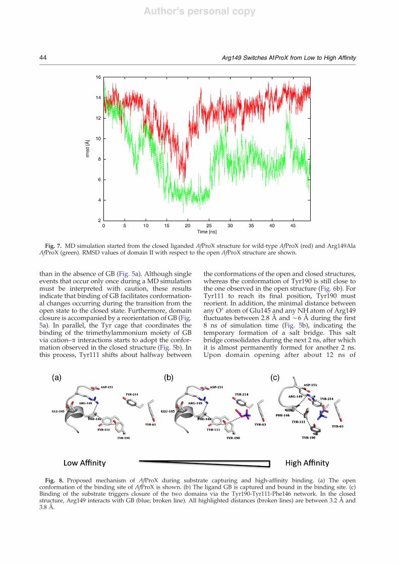
Fig. 7. MD simulation started from the closed liganded AfProX structure for wild-type AfProX (red) and Arg149AlaAfProX (green). RMSD values of domain II with respect to the open AfProX structure are shown.
Fig. 8. Proposed mechanism of AfProX during substrate capturing and high-affinity binding. (a) The openconformation of the binding site of AfProX is shown. (b) The ligand GB is captured and bound in the binding site. (c)Binding of the substrate triggers closure of the two domains via the Tyr190-Tyr111-Phe146 network. In the closedstructure, Arg149 interacts with GB (blue; broken line). All highlighted distances (broken lines) are between 3.2 Å and3.8 Å.
44 Arg149 Switches AfProX from Low to High Affinity
Author's personal copy
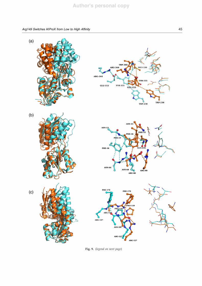
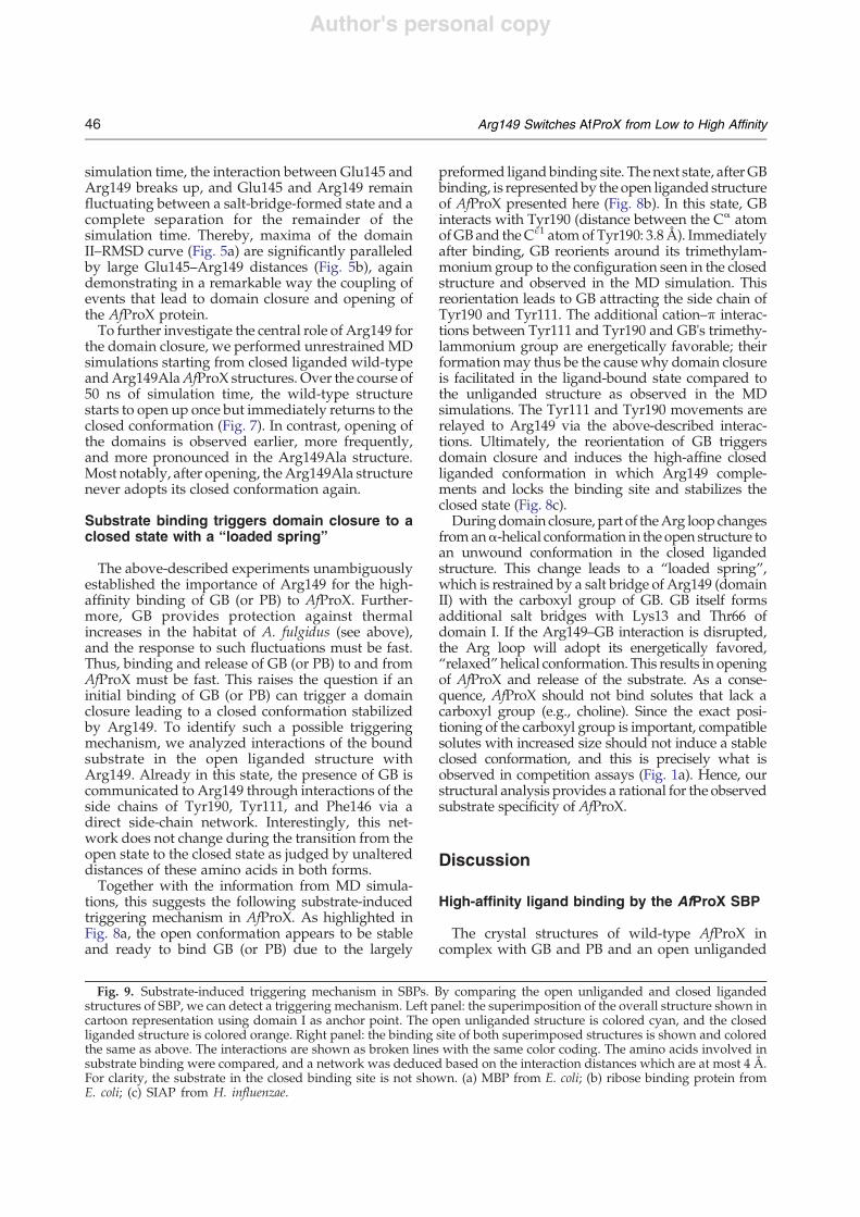
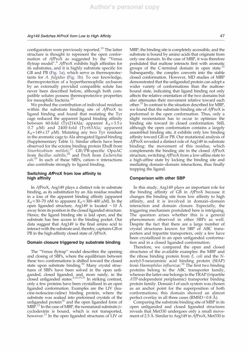
Fig. 9. (legend on next page)
45Arg149 Switches AfProX from Low to High Affinity
Author's personal copy
simulation time, the interaction between Glu145 andArg149 breaks up, and Glu145 and Arg149 remainfluctuating between a salt-bridge-formed state and acomplete separation for the remainder of thesimulation time. Thereby, maxima of the domainII–RMSD curve (Fig. 5a) are significantly paralleledby large Glu145–Arg149 distances (Fig. 5b), againdemonstrating in a remarkable way the coupling ofevents that lead to domain closure and opening ofthe AfProX protein.To further investigate the central role of Arg149 for
the domain closure, we performed unrestrained MDsimulations starting from closed liganded wild-typeandArg149AlaAfProX structures. Over the course of50 ns of simulation time, the wild-type structurestarts to open up once but immediately returns to theclosed conformation (Fig. 7). In contrast, opening ofthe domains is observed earlier, more frequently,and more pronounced in the Arg149Ala structure.Most notably, after opening, theArg149Ala structurenever adopts its closed conformation again.
Substrate binding triggers domain closure to aclosed state with a “loaded spring”
The above-described experiments unambiguouslyestablished the importance of Arg149 for the high-affinity binding of GB (or PB) to AfProX. Further-more, GB provides protection against thermalincreases in the habitat of A. fulgidus (see above),and the response to such fluctuations must be fast.Thus, binding and release of GB (or PB) to and fromAfProX must be fast. This raises the question if aninitial binding of GB (or PB) can trigger a domainclosure leading to a closed conformation stabilizedby Arg149. To identify such a possible triggeringmechanism, we analyzed interactions of the boundsubstrate in the open liganded structure withArg149. Already in this state, the presence of GB iscommunicated to Arg149 through interactions of theside chains of Tyr190, Tyr111, and Phe146 via adirect side-chain network. Interestingly, this net-work does not change during the transition from theopen state to the closed state as judged by unaltereddistances of these amino acids in both forms.Together with the information from MD simula-
tions, this suggests the following substrate-inducedtriggering mechanism in AfProX. As highlighted inFig. 8a, the open conformation appears to be stableand ready to bind GB (or PB) due to the largely
preformed ligandbinding site. The next state, afterGBbinding, is representedby the open liganded structureof AfProX presented here (Fig. 8b). In this state, GBinteracts with Tyr190 (distance between the C# atomofGBand theC!1 atomof Tyr190: 3.8Å). Immediatelyafter binding, GB reorients around its trimethylam-monium group to the configuration seen in the closedstructure and observed in the MD simulation. Thisreorientation leads to GB attracting the side chain ofTyr190 and Tyr111. The additional cation–" interac-tions between Tyr111 and Tyr190 and GB's trimethy-lammonium group are energetically favorable; theirformationmay thus be the cause why domain closureis facilitated in the ligand-bound state compared tothe unliganded structure as observed in the MDsimulations. The Tyr111 and Tyr190 movements arerelayed to Arg149 via the above-described interac-tions. Ultimately, the reorientation of GB triggersdomain closure and induces the high-affine closedliganded conformation in which Arg149 comple-ments and locks the binding site and stabilizes theclosed state (Fig. 8c).Duringdomain closure, part of theArg loop changes
froman#-helical conformation in the open structure toan unwound conformation in the closed ligandedstructure. This change leads to a “loaded spring”,which is restrained by a salt bridge of Arg149 (domainII) with the carboxyl group of GB. GB itself formsadditional salt bridges with Lys13 and Thr66 ofdomain I. If the Arg149–GB interaction is disrupted,the Arg loop will adopt its energetically favored,“relaxed”helical conformation. This results in openingof AfProX and release of the substrate. As a conse-quence, AfProX should not bind solutes that lack acarboxyl group (e.g., choline). Since the exact posi-tioning of the carboxyl group is important, compatiblesolutes with increased size should not induce a stableclosed conformation, and this is precisely what isobserved in competition assays (Fig. 1a). Hence, ourstructural analysis provides a rational for the observedsubstrate specificity of AfProX.
Discussion
High-affinity ligand binding by the AfProX SBP
The crystal structures of wild-type AfProX incomplex with GB and PB and an open unliganded
Fig. 9. Substrate-induced triggering mechanism in SBPs. By comparing the open unliganded and closed ligandedstructures of SBP, we can detect a triggering mechanism. Left panel: the superimposition of the overall structure shown incartoon representation using domain I as anchor point. The open unliganded structure is colored cyan, and the closedliganded structure is colored orange. Right panel: the binding site of both superimposed structures is shown and coloredthe same as above. The interactions are shown as broken lines with the same color coding. The amino acids involved insubstrate binding were compared, and a network was deduced based on the interaction distances which are at most 4 Å.For clarity, the substrate in the closed binding site is not shown. (a) MBP from E. coli; (b) ribose binding protein fromE. coli; (c) SIAP from H. influenzae.
46 Arg149 Switches AfProX from Low to High Affinity
Author's personal copy
configuration were previously reported.19 The latterstructure is thought to represent the open confor-mation of AfProX as suggested by the “Venusflytrap model”.4 AfProX exhibits high affinities forits substrates, and it is highly substrate specific forGB and PB (Fig. 1a), which serve as thermoprotec-tants for A. fulgidus (Fig. 1b). To our knowledge,thermoprotection of a hyperthermophilic archaeonby an externally provided compatible solute hasnever been described before, although both com-patible solutes possess thermoprotective propertiesfor mesophilic bacteria.20We probed the contribution of individual residues
within the substrate binding site of AfProX toligand binding and found that mutating the Tyrcage reduced the apparent ligand binding affinitybetween 60-fold (Tyr214Ala; apparent Kd=3.5±0.7 !M) and 2400-fold (Tyr63Ala; apparentKd=149±17 !M). Mutating any two Tyr residuesin the aromatic cage to Ala abrogated ligand binding(Supplementary Table 1). Similar effects have beenobserved for the ectoine binding protein EhuB fromSinorhizobium meliloti,27 GB/PB-specific OpuACfrom Bacillus subtilis,28 and ProX from Escherichiacoli.29 In each of these SBPs, cation–" interactionsalso contribute strongly to ligand binding.
Switching AfProX from low affinity tohigh affinity
In AfProX, Arg149 plays a distinct role in substratebinding, as its substitution by an Ala residue resultedin a loss of the apparent binding affinity (apparentKd#50–70 nM to apparent Kd#300–400 !M). In theopen liganded structure, Arg149 is located !10 Åaway from its position in the closed liganded structure.Hence, the ligand binding site is laid open, and thesubstrate has free access to the binding pocket. Ourdata suggest that Arg149 is the final amino acid tointeractwith the substrate and, thereby, capturesGBorPB in the high-affinity closed state of AfProX.
Domain closure triggered by substrate binding
The “Venus flytrap” model describes the openingand closing of SBPs, where the equilibrium betweenthese two conformations is shifted toward the closedstate upon substrate binding.30 Many crystal struc-tures of SBPs have been solved in the open unli-ganded, closed liganded, and, more rarely, in theclosed unliganded states.4,10,19,31 In striking contrast,only a few proteins have been crystallized in an openliganded conformation. Examples are the LIV (leu-cine-isoleucine-valine) binding protein, where thesubstrate was soaked into preformed crystals of theunliganded protein30 and the open liganded form ofMBP.13 In the case ofMBP, the nonnatural substrate$-cyclodextrin is bound, which is not transported,however.13 In the open liganded structures of LIV or
MBP, the binding site is completely accessible, and thesubstrate is bound by amino acids that originate fromonly one domain. In the case of MBP, it was thereforepostulated that maltose interacts first with aromaticgroups of the C-terminal domain in open state.32Subsequently, the complex converts into the stableclosed conformation. However, MD studies of MBPdemonstrated that the unliganded protein can adopt awider variety of conformations than the maltose-bound state, indicating that ligand binding not onlyaffects the relative orientation of the two domains butalso attenuates their movement relative toward eachother.33 In contrast to the situation described for MBP,we found that the substrate binding site of AfProX ispreformed in the open conformation. Thus, only aslight reorientation has to occur to optimize thebinding site toward its closed conformation. Still,although the open conformation contains a largelyassembled binding site, it exhibits only low bindingaffinity toward GB or PB. Our mutational analysis ofAfProX revealed a distinct role of Arg149 in substratebinding: the movement of this residue, whichcomplements the binding site in the closed AfProXstructure, switchingAfProX from a low-affine state toa high-affine state by locking the binding site andmediating domain–domain interactions, thus finallytrapping the ligand.
Comparison with other SBP
In this study, Arg149 plays an important role forthe binding affinity of GB in AfProX because itchanges the binding site from low affinity to highaffinity, and it is involved in domain–domaininteraction and domain closure. Especially, thetriggering mechanism postulated here is intriguing.The question arises whether this is a generalphenomenon observed in other SBPs as well.Despite the fact that there are a large number ofcrystal structures known for SBP of ABC trans-porters and tripartite transporters, only a few havebeen crystallized in an open unliganded conforma-tion and in a closed liganded conformation.Therefore, we compared the open and closed
structures of the available examples: the MBP andthe ribose binding protein from E. coli and the N-acetyl-5-neuraminic acid binding protein (SIAP)from Haemophilus influenzae.34 The first two bindingproteins belong to the ABC transporter family,whereas the latter one belongs to the TRAP (tripartiteATP-independent periplasmic) transporter bindingprotein family. Domain I of each system was chosenas an anchor point for the superposition of bothconformations; this domain showed an almostperfect overlay in all three cases (RMSDb0.8 Å).Comparing the substrate binding site of MBP in its
open unliganded and closed liganded structuresreveals that Met330 undergoes only a small move-ment of 2.5 Å. Similar to Arg149 inAfProX,Met330 is
47Arg149 Switches AfProX from Low to High Affinity
Author's personal copy
connected, via a network of side-chain interactions,to those amino acids that participate in substratebinding. This network forms through the followingamino acids: Met330-Tyr155-Glu153-Arg344. Fur-thermore, Tyr155 interacts with Trp230 and Phe156.The distances within this network do not differ in theopen and closed structures; however, the absolutepositions of these amino acids change by 6–10 Å,suggesting a rigid-body movement. That way, thesmall movement ofMet330 seems to trigger a pullingevent toward all amino acids involved in substratebinding in the second MBP domain. In addition,Met330 also interacts with Phe258, which is locatedat the beginning of a $-sheet. It has been shown thatthese $-sheets are important for stabilizing theclosed conformation as it contains Gly260, which ispart of a salt bridge only observed in the closedconformation of MBP33 (Fig. 9a).In ribose binding protein, the substrate is bound by
several amino acids originating from both domains.Here again, the superimposition of the open unli-ganded structure and the closed liganded structurerevealed a perfect overlay of domain I. In domain II,however, the amino acids undergo a significantmovement. Here, a network that starts at the sidechain of Gln235 and, in addition, contains Phe15,Phe16, Asn64, Arg90, and Asp89 was identified.Gln235 is also in contact with the substrate in theliganded structure, similar to Met330 in MBP and toTyr110 in AfProX, which undergoes a small confor-mational change of 1.5Åduring closing and is locateddirectly at the hinge between both domains. Thisexplains the small movement. In total, this highlightsthe importance of this single amino acid for ligandbinding, as seen for Met330 (MBP) and Arg149(AfProX), too (Fig. 9b).SIAP is the binding protein of the N-acetyl-5-
neuraminic acid TRAP transporter from H. influen-zae. Here, a specific role in ligand binding can beassigned to Arg127. Besides the interaction with thesubstrate in the closed conformation, a side-chaininteraction network is observed through interactionswith Pro149 and Asn187. Asn187 itself is in contactwith Phe170 and Arg147, which are the residues ofdomain II that build up the substrate binding site inSIAP. Here and inMBP and RBS, the distances in thenetwork do not change in the open and closed formsof the structures. In SIAP, Arg127 has been mutatedto Ala and Lys. In competition assays, the activity ofSIAP has altered for Arg127Ala and Arg127Lys.Furthermore, no binding of a substrate to theArg27Ala mutant could be observed anymore byisothermal calorimetry35 (Fig. 9c).
Substrate release by external modulation ofArg149 interactions
The Arg loop of the AfProX protein has an#-helical conformation in the open structure. In the
closed liganded structure, however, this helix isunwound. The energetic cost of unwinding isdiminished by multiple interactions of Arg149with domain II of AfProX and GB. Furthermore, inthe closed liganded structure, the side chain ofArg149 interacts with Glu145, whereas no suchinteraction occurs in the open structure. Finally, salt-bridge formation between Arg149 and Glu145 upondomain closure is also observed during the MDsimulations (see above). Interestingly, Glu145 isexposed at the membrane-facing side of AfProX.Taken together, our data suggest a model in
which a conformational change in the membranecomponents (ProW-1, ProW-2) and the ATPase(ProV) of the ProU ABC transporter from A.fulgidus might result in the manipulation or evenin the disruption of the Glu145–Arg149 salt bridge.This event is expected to weakened the interac-tions of Arg149 with GB or PB, which, in turn,would induce a reformation of the helical confor-mation of the Arg loop. Thereby, the binding siteof the AfProX protein would become accessible.Consequently, the affinity of the substrate bindingsite would change from low nanomolar to mediummicromolar range, inducing the release of GB/PBinto the substrate translocation pathway of theProU transporter for ATP-dependent import.In summary, Arg149 acts as a trigger to ensure a
fast and efficient ligand capture. A preformed ligandbinding site that only requires a single amino acid toswitch the system from a low- to a high-affine stateis a perfect solution to environmental restraints. Ourdata imply that distinct molecular events such as asingle amino acid switch are operational in theoverall “Venus flytrap” mechanism to ensure thespecific biological function of SBPs.
Materials and Methods
Heterologous expression and purification of AfProX
AfProX was heterologously expressed using the E. colistrain BL21 CodonPlus RIL strain containing the proX+
plasmid pHG26, a derivative of the pASKIBA6 expressionvector (IBA, Göttingen, Germany). A 10-l glass flaskcontaining 5 l of MMA supplemented with 150 !g ml"1 ofampicillin, 30 !g ml"1 chloramphenicol, 0.5% glucose, and0.2% casamino acids was inoculated to an OD578 of 0.1from an overnight culture. Cells were grown at 37 °C withvigorous stirring until the culture had reachedOD578=0.6–0.7. Expression was induced by the additionof anhydrotetracycline (final concentration, 0.2 !g ml"1).Cells were grown for an additional 2 h and weresubsequently harvested by centrifugation (10 min,3000g). To release the periplasmic proteins, we resus-pended the cell paste in 50 ml ice-cold buffer P [100 mMTris–HCl (pH 8), 500 mM sucrose, and 1 mM ethylene-diaminetetraacetic acid]. After a 30-min incubation on ice,the periplasmic protein extract was harvested by
48 Arg149 Switches AfProX from Low to High Affinity
Author's personal copy
centrifugation (15 min, 21,000g at 4 °C), followed by anultracentrifugation step (30 min, 120,000g at 4 °C) toremove insoluble material. The supernatant was thenloaded onto a 10-ml Strep-tactin column (IBA) equili-brated with buffer W [100 mM Tris–HCl (pH 8)]. Thecolumn was washed with 5 column volumes of buffer W,and bound proteins were released from the affinity resinby washing the column with buffer E [100 mM Tris–HCl(pH 8) and 2.5 mM desthiobiotin]. Since a portion of thepro-OmpA signal sequence was not proteolytically re-moved from the ProX protein by the E. coli cells, thepurified ProX protein was cleaved with factor Xa (1 !gfactor Xa per 200 !g ProX) for 16 h at room temperature ina buffer containing 100 mM Tris–HCl (pH 8), 100 mMNaCl, and 1mMCaCl2. This proteolytic cleavage removedthe OmpA signal sequence and the Strep-tag sequence,thereby resulting in AfProX containing no N-terminalextensions. To remove factor Xa from the solution, wediluted the protein solution 1:2 with deionized water andloaded them on a UnoQ6 column (Bio-Rad, Muenchen,Germany) equilibrated with 50 mM Tris–HCl (pH 8)(buffer A). The column was washed with 20 ml buffer A,and the protein was eluted with a linear NaCl gradient.AfProX was eluted at 250 mM NaCl. AfProX wassubsequently dialyzed overnight against 5 l of 10 mMTris–HCl (pH 7.5) at 4 °C and stored until further use at4 °C. In general, 2 mg of pureAfProX protein was obtainedper 1 l of overproducing cells.
Substrate binding of the purified AfProX protein
To study substrate binding properties of AfProX, weused the ammonium sulfate precipitation technique ofRicharme and Kepes.36 AfProX (5 !M) was incubated at85 °C (or at other temperatures as indicated) with 5 !M[1-14C]GB in a volume of 100 !l of 10 mM Tris–HCl(pH 7.5) for 5 min. AfProX was then precipitated by theaddition of 900 !l of an ice-cold saturated ammoniumsulfate solution. After a 5-min incubation on ice, themixture was sucked through cellulose filters (0.45-!mpore size; Schleicher and Schuell, Dassel, Germany), andthe radioactivity retained by the filters was determined byliquid scintillation counting. To determine the substratespecificity of AfProX, we incubated the protein at 85 °Cwith 5 !M radiolabeled GB and a 40-fold excess ofdifferent unlabeled compatible solutes, and we thenfurther treated the samples as described above.
Determination of the apparent binding affinity (Kd) ofAfProX using fluorescence spectroscopy
All spectra were obtained at room temperature with aVARIAN CARY Eclipse fluorometer (VARIAN, Darm-stadt, Germany). Emission scans were collected at anexcitation wavelength of 280 nm from 290 nm to 450 nm.Measurements were carried out in 1 ml of 10 mM Tris–HCl (pH 7.5). Five micrograms of AfProX protein wasadded to the buffer, and the solution was mixed. To titratethe protein, we added 5 !l substrate from a 40-!M GB orPB stock solution. After substrate addition, the samplewas mixed in the cuvette, and the change in thefluorescence was recorded. This step was repeated untila stable fluorescence signal was obtained. Assuming one
binding site per monomer in AfProX, the Kd for substratebinding was determined by nonlinear least-squares fittingof the data to the following equation: F=F0+(DF/2P0)[(Kd+P0+L0)" ((Kd+P0+L0)2"4L0P0)1/2], which correctfor the concentration of the receptor. F is the measuredfluorescence; F0 is the fluorescence of AfProX; DF is thechange in fluorescence at saturation; P0 and L0 are the totalconcentrations of protein and substrate. The change influorescence, however, gives no information about theopen or closed state of the protein. We can only observebinding of the substrate without any information of theconformation of AfProX.
Crystallization
AfProX crystals were obtained using the hanging-dropmethod at 293 K against a reservoir solution of 100 mMHepes (pH 7.0), 15–25% polyethylene glycol 6000, and 15–25% polyethylene glycol 8000. AfProX was incubated with10 mM GB prior to crystallization. Crystals grew slowlyand appeared after 1 month. They were flash frozen inliquid nitrogen withmother liquor supplemented with 20–25% ethylene glycerol as cryoprotectant.
Data collection, refinement, and structure analysis
AfProX crystals diffracted X-rays beyond 1.6 Å. How-ever, for refinement purposes, the data set was truncatedat a resolution of 2.0 Å. The data set was collected at theID23-EH2 beamline at the European Synchrotron Radia-tion Facility and processed with XDS.22 AfProX in theligand-free form (PDB code: 1SW5)19 was used as atemplate to obtain initial phases using Phaser.23 Thestructure was further refined using REFMAC525 andCoot.37 Data set and refinement statistics are listed inSupplementary Table 2. As analyzed with PROCHECK,38the Ramachandran plot of AfProX shows one residue inthe disallowed region. However, this residue interactswith the bound Hepes molecule, which explains the ratherunusual conformation. Figures of protein molecules wereprepared using PyMOL‡.
MD simulations
MD simulations were performed with the Amber 10suite of programs,39 together with the force field asdescribed by Cornell et al.,40 using modifications sug-gested by Simmerling et al.41 In total, four different MDsimulations were performed: (i) open liganded wild-typeAfProX. The starting structure was generated by mutatingAla111 in the crystal structure of Tyr111AlaAfProX to Tyr.The simulation length is 200 ns; (ii) open liganded wild-type AfProX. In contrast to (i), a distance restraint bymeans of a harmonic potential was applied between theammonium nitrogen of GB and the N of Asp109 ofAfProX. The simulation length is 25 ns; (iii) closedliganded wild-type AfProX. The starting structure wastaken from PDB code: 1SW4.19 The simulation length is50 ns; (iv) closed liganded Arg149Ala AfProX. The starting
‡www.pymol.org
49Arg149 Switches AfProX from Low to High Affinity
Author's personal copy
structure was generated from the closed liganded wild-type AfProX structure by mutating Arg149 to Ala. Thesimulation length is 50 ns.In all cases, the starting structure was placed into an
octahedral periodic box of TIP3P water molecules.42 Thedistance between the edges of the water box and theclosest atom of the protein was at least 11 Å, resulting in asystem of !43,000 atoms. The system was minimized by50 steps of steepest descent minimization followed by 450steps of conjugate gradient minimization. The particlemesh Ewald method43 was used to treat long-rangeelectrostatic interactions, and bond lengths involvingbonds to hydrogen atoms were constrained usingSHAKE.44 The time step for all MD simulations was 2 fs,with a direct-space nonbonded cutoff of 8 Å. Applyingharmonic restraints with force constants of 5 kcal mol"1Å"2 to all solute atoms, we carried out canonical ensemble(NVT)-MD for 50 ps, during which the system was heatedfrom 100 K to 300 K. Subsequent isothermal isobaricensemble (NPT)-MD was used for 150 ps to adjust thesolvent density. Finally, the force constants of theharmonic restraints on solute atom positions weregradually reduced to zero during 100 ps of NVT-MD.The following NVT-MD at 300 K with a time constant of10 ps for heat-bath coupling was used for analysis, withconformations extracted every 20 ps. Atomic charges forthe GB ligand were generated following the RESPprocedure;45 force field parameters for GB were takenfrom GAFF.46For the analysis of the trajectories, conformations were
superimposed with respect to the C# atoms of domain I.This resulted in an almost perfect overlay in all cases.RMSD values with respect to the open or closed startingstructures were then determined for C# atoms of domainII or for atoms of GB.
Accession number
Coordinates and structure factors have been depositedin the PDB with accession number 3MAM.
Acknowledgements
We thank the members of the laboratory of L.S.and E.B. for stimulating discussion. We are indebtedto Christoph Müller Dieckmann for excellent sup-port at the beamlines of the European MolecularBiology Laboratory Outstation Grenoble (France)and to Dr. Paul Tucker and Dr. Matthew Groves atthe BW7A beamline, European Molecular BiologyLaboratory Outstation Hamburg. Financial supportfor this study was provided by the Fonds derChemischen Industrie and the LOEWE program ofthe State of Hessen via the Centre for SyntheticMicrobiology (SynMicro; Marburg) to E.B. B.T. andL.S. gratefully acknowledge financial support by theEDICT EU program. H.G. is grateful to the“Zentrum fuer Informations- und Medientechnolo-
gie” at the Heinrich Heine University Duesseldorffor computational support.
Supplementary Data
Supplementary data associated with this articlecan be found, in the online version, at doi:10.1016/j.jmb.2011.05.039
References
1. Kempf, B. & Bremer, E. (1998). Uptake and synthesisof compatible solutes as microbial stress responses tohigh-osmolality environments. Arch. Microbiol. 170,319–330.
2. Davidson, A. L., Dassa, E., Orelle, C. & Chen, J. (2008).Structure, function, and evolution of bacterial ATP-binding cassette systems. Microbiol. Mol. Biol. Rev. 72,317–364; Table of Contents.
3. Cui, J., Qasim, S. & Davidson, A. L. (2010). Uncou-pling substrate transport from ATP hydrolysis in theEscherichia coli maltose transporter. J. Biol. Chem. 285,39986–39993.
4. Wilkinson, J. & Verschueren, K. H. G. (2003). Crystalstructures of periplasmic solute-binding proteins inABC transport complexes illuminate their function. InABC Proteins: From Bacteria toMan (Holland, I. B., Cole,S. P. C., Kuchler, K. & Higgins, C. F., eds), pp. 187–208,Academic Press (Elsevier Science), London, UK.
5. Berntsson, R. P., Smits, S. H., Schmitt, L., Slotboom,D. J. & Poolman, B. (2010). A structural classification ofsubstrate-binding proteins. FEBS Lett. 584, 2606–2617.
6. Quiocho, F. A. & Ledvina, P. S. (1996). Atomicstructure and specificity of bacterial periplasmicreceptors for active transport and chemotaxis: varia-tion of common themes. Mol. Microbiol. 20, 17–25.
7. Mao, B., Pear, M. R., McCammon, J. A. & Quiocho,F. A. (1982). Hinge-bending in L-arabinose-bindingprotein. The “Venus's-flytrap” model. J. Biol. Chem.257, 1131–1133.
8. Sack, J. S., Saper, M. A. & Quiocho, F. A. (1989).Periplasmic binding protein structure and function.J. Mol. Biol. 206, 171–191.
9. Oh, B. H., Pandit, J., Kang, C. H., Nikaido, K., Gokcen,S., Ames, G. F. & Kim, S. H. (1993). Three-dimensionalstructures of the periplasmic lysine/arginine/orni-thine-binding protein with and without a ligand.J. Biol. Chem. 268, 11348–11355.
10. Oswald, C., Smits, S. H., Höing, M., Sohn-Bösser, L.,Dupont, L., Le Rudulier, D. et al. (2008). Crystalstructures of the choline/acetylcholine substrate-binding protein ChoX from Sinorhizobium meliloti inthe liganded and unliganded-closed states. J. Biol.Chem. 283, 32848–32859.
11. Shilton, B. H. (2008). The dynamics of the MBP-MalFGK(2) interaction: a prototype for bindingprotein dependent ABC-transporter systems. Biochim.Biophys. Acta, 1778, 1772–1780.
12. Tang, C., Schwieters, C. D. & Clore, G. M. (2007).Open-to-closed transition in apo maltose-bindingprotein observed by paramagnetic NMR. Nature,449, 1078–1082.
50 Arg149 Switches AfProX from Low to High Affinity
Author's personal copy
13. Sharff, A. J., Rodseth, L. E. & Quiocho, F. A. (1993).Refined 1.8-Å structure reveals the mode of binding of$-cyclodextrin to the maltodextrin binding protein.Biochemistry, 32, 10553–10559.
14. Spurlino, J. C., Lu,G. Y.&Quiocho, F.A. (1991). The 2.3-Å resolution structure of the maltose- or maltodextrin-binding protein, a primary receptor of bacterial activetransport and chemotaxis. J. Biol. Chem. 266, 5202–5219.
15. Millet, O., Hudson, R. P. & Kay, L. E. (2003). Theenergetic cost of domain reorientation in maltose-binding protein as studied by NMR and fluorescencespectroscopy. Proc. Natl Acad. Sci. USA, 100,12700–12705.
16. Marvin, J. S. & Hellinga, H. W. (2001). Manipulationof ligand binding affinity by exploitation of confor-mational coupling. Nat. Struct. Biol. 8, 795–798.
17. Duan, X., Hall, J. A., Nikaido, H. & Quiocho, F. A.(2001). Crystal structures of the maltodextrin/maltose-binding protein complexed with reduced oligosaccha-rides: flexibility of tertiary structure and ligandbinding.J. Mol. Biol. 306, 1115–1126.
18. Klenk, H. P., Clayton, R. A., Tomb, J. F., White, O.,Nelson, K. E., Ketchum, K. A. et al. (1997). Thecomplete genome sequence of the hyperthermophilic,sulphate-reducing archaeon Archaeoglobus fulgidus.Nature, 390, 364–370.
19. Schiefner, A., Holtmann, G., Diederichs, K., Welte, W.& Bremer, E. (2004). Structural basis for the binding ofcompatible solutes by ProX from the hyperthermo-philic archaeon Archaeoglobus fulgidus. J. Biol. Chem.279, 48270–48281.
20. Holtmann, G. & Bremer, E. (2004). Thermoprotectionof Bacillus subtilis by exogenously provided glycinebetaine and structurally related compatible solutes:involvement of Opu transporters. J. Bacteriol. 186,1683–1693.
21. Vahedi-Faridi, A., Eckey, V., Scheffel, F., Alings, C.,Landmesser, H., Schneider, E. & Saenger, W. (2008).Crystal structures and mutational analysis of thearginine-, lysine-, histidine-binding protein ArtJ fromGeobacillus stearothermophilus. Implications for interac-tions of ArtJ with its cognate ATP-binding cassettetransporter, Art(MP)2. J. Mol. Biol. 375, 448–459.
22. Kabsch, W. (1993). Automatic processing of rotationdiffraction data from crystals of initially unknownsymmetry and cell constants. J. Appl. Crystallogr. 26,795–800.
23. McCoy, A. J., Grosse-Kunstleve, R. W., Adams, P. D.,Winn, M. D., Storoni, L. C. & Read, R. J. (2007). Phasercrystallographic software. J. Appl. Crystallogr. 40,658–674.
24. Otwinowski, Z. & Minor, W. (1997). Processing ofX-ray diffraction data collected in oscillation mode. InMethods in Enzymology (Carter, C. W. & Sweet, R. M.,eds), Methods in Enzymology, 276, pp. Academic Press,London, UK.
25. Murshudov, G., Vagin, A. A. & Dodson, E. J. (1997).Refinement of macromolecular structures by themaximum-likelihood method. Acta Crystallogr., Sect.D: Biol. Crystallogr. 53, 240–255.
26. Hayward, S. & Lee, R. A. (2002). Improvements in theanalysis of domain motions in proteins from confor-mational change: DynDom version 1.50. J. Mol.Graphics Modell. 21, 181–183.
27. Hanekop, N., Höing, M., Sohn-Bösser, L., Jebbar, M.,Schmitt, L. & Bremer, E. (2007). Crystal structure ofthe ligand-binding protein EhuB from Sinorhizobiummeliloti reveals substrate recognition of the compatiblesolutes ectoine and hydroxyectoine. J. Mol. Biol. 374,1237–1250.
28. Smits, S. H., Höing, M., Lecher, J., Jebbar, M., Schmitt,L. & Bremer, E. (2008). The compatible-solute-bindingprotein OpuAC from Bacillus subtilis: ligand binding,site-directed mutagenesis, and crystallographic stud-ies. J. Bacteriol. 190, 5663–5671.
29. Schiefner, A., Breed, J., Bösser, L., Kneip, S., Gade, J.,Holtmann, G. et al. (2004). Cation–pi interactions asdeterminants for binding of the compatible solutesglycine betaine and proline betaine by the periplasmicligand-binding protein ProX from Escherichia coli.J. Biol. Chem. 279, 5588–5596.
30. Sack, J. S., Saper, M. A. & Quiocho, F. A. (1989).Periplasmic binding protein structure and function.Refined X-ray structures of the leucine/isoleucine/valine-binding protein and its complex with leucine.J. Mol. Biol. 206, 171–191.
31. Oswald, C., Smits, S. H., Hoing, M., Bremer, E. &Schmitt, L. (2009). Structural analysis of the choline-binding protein ChoX in a semi-closed and ligand-freeconformation. Biol. Chem. 390, 1163–1170.
32. Diez, J., Diederichs, K., Greller, G., Horlacher, R.,Boos, W. &Welte, W. (2001). The crystal structure of aliganded trehalose/maltose-binding protein from thehyperthermophilic archaeon Thermococcus litoralis at1.85 Å. J. Mol. Biol. 305, 905–915.
33. Stockner, T., Vogel, H. J. & Tieleman, D. P. (2005). Asalt-bridge motif involved in ligand binding andlarge-scale domain motions of the maltose-bindingprotein. Biophys. J. 89, 3362–3371.
34. Muller, A., Severi, E., Mulligan, C., Watts, A. G., Kelly,D. J., Wilson, K. S. et al. (2006). Conservation ofstructure and mechanism in primary and secondarytransporters exemplified by SiaP, a sialic acid bindingvirulence factor from Haemophilus influenzae. J. Biol.Chem. 281, 22212–22222.
35. Johnston, J. W., Coussens, N. P., Allen, S., Houtman,J. C., Turner, K. H., Zaleski, A. et al. (2008). Charac-terization of the N-acetyl-5-neuraminic acid-bindingsite of the extracytoplasmic solute receptor (SiaP) ofnontypeable Haemophilus influenzae strain 2019. J. Biol.Chem. 283, 855–865.
36. Richarme, G. & Kepes, A. (1983). Study of bindingprotein–ligand interaction by ammonium sulfate-assisted adsorption on cellulose esters filters. Biochim.Biophys. Acta, 742, 16–24.
37. Emsley, P. & Cowtan, K. (2004). Coot: model-buildingtools for molecular graphics. Acta Crystallogr., Sect. D:Biol. Crystallogr. 60, 2126–2132.
38. Laskowski, R. A., MacArthur, M. W., Moss, D. S. &Thornton, J. M. (1993). PROCHECK: a program tocheck the stereochemical quality of protein structures.J. Appl. Crystallogr. 26, 283–291.
39. Case, D. A., Cheatham, T. E., 3rd, Darden, T., Gohlke,H., Luo, R., Merz, K. M., Jr et al. (2005). The Amberbiomolecular simulation programs. J. Comput. Chem.26, 1668–1688.
40. Cornell, W. D., Cieplak, P., Bayly, C. I., Gould, I. R.,Merz, K. M., Jr, Ferguson, D. M. et al. (1995). A second
51Arg149 Switches AfProX from Low to High Affinity
Author's personal copy
generation force field for the simulation of proteins,nucleic acids, and organic molecules. J. Am. Chem. Soc.117, 5179–5197.
41. Simmerling, C., Strockbine, B. & Roitberg, A. E.(2002). All-atom structure prediction and foldingsimulations of a stable protein. J. Am. Chem. Soc. 124,11258–11259.
42. Jorgensen, W. L., Chandrasekhar, J., Madura, J. &Klein, M. L. (1983). Comparison of simple potentialfunctions for simulating liquid water. J. Chem. Phys.79, 926–935.
43. Darden, T., York, D. & Pedersen, L. (1993). Particlemesh Ewald: An N·log(N) method for Ewald sums inlarge systems. J. Chem. Phys. 98, 10089–10092.
44. Ryckaert, J. P., Ciccotti, G. & Berendsen, H. J. C.(1977). Numerical integration of the cartesian equa-tions of motion of a system with constraints:molecular dynamics of n-alkanes. J. Comput. Phys.23, 327–341.
45. Bayly, C. I., Cieplak, P., Cornell, W. D. & Kollman,P. A. (1993). A well-behaved electrostatic potentialbased method using charge restraints for determiningatom-centered charges: the RESPmodel. J. Phys. Chem.97, 10269–10280.
46. Wang, J., Wolf, R. M., Caldwell, J. W., Kollman, P. A.& Case, D. A. (2004). Development and testing of ageneral amber force field. J. Comput. Chem. 25,1157–1174.
52 Arg149 Switches AfProX from Low to High Affinity


![Complete genome sequence of Archaeoglobus profundus · reservoirs [10], but no formal species description has been published, therefore this species ninth is excluded from comparisons](https://img.pdfslide.us/doc/110x75/5e594c745cb05e5ca404cb63/complete-genome-sequence-of-archaeoglobus-profundus-reservoirs-10-but-no-formal.jpg)