Embed Size (px)
Citation preview
This article appeared in a journal published by Elsevier. The attachedcopy is furnished to the author for internal non-commercial researchand education use, including for instruction at the authors institution
and sharing with colleagues.
Other uses, including reproduction and distribution, or selling orlicensing copies, or posting to personal, institutional or third party
websites are prohibited.
In most cases authors are permitted to post their version of thearticle (e.g. in Word or Tex form) to their personal website orinstitutional repository. Authors requiring further information
regarding Elsevier’s archiving and manuscript policies areencouraged to visit:
http://www.elsevier.com/copyright
Author's personal copy
History of Neurology
Figures and institutions of the neurological sciences in Parisfrom 1800 to 1950. Introduction and Part I: Neuroanatomy
Les figures et institutions des sciences neurologiques a Paris de 1800 a 1950.Introduction et partie I : neuroanatomie
F. Clarac a, J.-G. Barbara b,c, E. Broussolle d,*,e, J. Poirier f
a Laboratoire plasticite et physiopathologie de la motricite, CNRS, UMR 6196, universite Aix-Marseille II, 31, chemin Joseph-Aiguier,
13402 Marseille cedex 20, Franceb Laboratoire de neurobiologie des processus adaptatifs, CNRS, UMRS 7102, universite Pierre et Marie-Curie, case 14, 4, place Jussieu,
75252 Paris cedex 05, Francec Laboratoire SPHERE, CNRS UMR 7219, universite Denis-Diderot, case 7093, 5, rue Thomas-Mann, 75205 Paris cedex 13, FrancedService de neurologie C, hopital neurologique Pierre-Wertheimer, hospices civils de Lyon, universite Claude-Bernard Lyon-I, 59,
boulevard Pinel, 69500 Bron, FranceeCNRS UMR 5229, centre de neurosciences cognitives, 67, boulevard Pinel, 69675 Bron cedex, FrancefHopital Pitie-Salpetriere, 47-83, boulevard de l’Hopital, 75651 Paris cedex 13, France
r e v u e n e u r o l o g i q u e 1 6 8 ( 2 0 1 2 ) 2 – 1 4
i n f o a r t i c l e
Article history:
Received 2 May 2011
Received in revised form
29 July 2011
Accepted 8 August 2011
Published on line 16 January 2012
Keywords :
History
Paris
Neuroscience
Anatomy
Physiology
Neurology
Psychiatry
Psychology
Mots cles :
Histoire
Paris
Neurosciences
a b s t r a c t
We present a short historical review on the major institutions and figures that contributed to
make Paris a renowned centre of physiology and neurology during the XIXth and the first half
of the XXth centuries. We purposely chose to focus on the period 1800–1950, as 1800
corresponds to the development of brain science and 1950 marks the true beginning of
neuroscience. Our presentation is divided into four chapters, matching the main disciplines
which have progressed and contributed the most to the knowledge we have of the brain
sciences: anatomy, physiology, neurology, and psychiatry-psychology. The present article is
the first of four parts of this review, which includes an introduction followed by the chapter
on neuroanatomy and on anatomo-pathology, which includes biographical sketches of Felix
Vicq d’Azyr, Francois-Xavier Bichat, Franz Joseph Gall, Jean Cruveilhier, Jules Bernard Luys,
Paul Broca, Louis Ranvier, Andre-Victor Cornil, Albert Gombault, Jean Nageotte and Rene
Couteaux.
# 2011 Elsevier Masson SAS. All rights reserved.
r e s u m e
Nous presentons une revue generale historique breve sur les principales institutions et
personnalites ayant contribue a faire de Paris un centre renomme de physiologie et
de neurologie au cours du XIXe siecle et de la premiere partie du XX
e siecle. La raison du
choix de cette periode allant de 1800 a 1950 s’explique par le fait que 1800 marque les debuts
des sciences du cerveau et 1950 le reel developpement des neurosciences. Notre presenta-
tion est divisee en quatre chapitres, correspondant aux principales disciplines ayant
* Corresponding author.E-mail address : [email protected] (E. Broussolle).
Available online at
www.sciencedirect.com
0035-3787/$ – see front matter # 2011 Elsevier Masson SAS. All rights reserved.doi:10.1016/j.neurol.2011.08.013
Author's personal copy
1. Introduction
During the last decades, neuroscience research has experien-
ced a tremendous growth in many fields of interest, notably in
the anatomical, physiological, neurological or psychiatric
domains. However, most of today’s young scientists and
physicians do not have sufficient historical background of
these fascinating disciplines. Numerous authors from many
parts of the world contributed to the development of the
neurosciences and particularly neurology (Clarac and Boller,
2010; Clarac and Ternaux, 2008; Finger, 1994, 2000; Haymaker
and Schiller, 1953; Rancurel et al., 2004). As early as the XIXth
century, special credit should be given to German speaking
universities from Central Europe, institutions in Great Britain
and North America, French and Italian neurological schools,
and also medical universities from Belgium, the Netherlands,
Scandinavia, Southern Europe, Russia and subsequently from
South America, Asia and Africa during the XXth century.
We took the opportunity of the XVth annual meeting of the
international society for the history of the neurosciences, held
in Paris in June 2010, to prepare a historical work in order to
make a short presentation of the major institutions and figures
who contributed to make Paris a renowned centre of
physiology and neurology between the XIXth and the first
half of the XXth century. We purposely chose to focus on the
period 1800–1950, as 1800 corresponds to the actual beginning
of brain sciences and 1950 marks the rise of neuroscience.
The beginning of the XIXth century in France was
characterized by a remarkable improvement of the medical
and physiological knowledge. This was due to the influence of
several combined factors. First, the XVIIIth century enligh-
tenment and the mouvement encyclopedique (encyclopedic
movement), with an emerging scientific approach of philoso-
phy (positivism and materialism). Second, the French Revolu-
tion, with marked restructuring of the French hospital system
(establishment of internship/residentship and externship in
1802) and of the academic organization (re-foundation of the
three Ecoles de sante–Medical faculties–in 1794). Third, the
simultaneous presence of exceptional figures as Francois de
Fourcroy (1715–1809), Pierre-Jean Georges Cabanis (1757–1808)
and Philippe Pinel (1745–1826) to name but a few among the
pioneers.
In 1830, there were numerous hospitals in Paris, in which
5000 medical students were working and where more than
20,000 patients were treated. Three medical faculties were
developed at that time, in Paris, Montpellier and Strasbourg,
respectively. The other major cities such as Lyon, Marseille,
Bordeaux, Lille or Toulouse only had medical schools, which
did not become faculties of their own right before the end of
XIXth century and the beginning of the XXth century.
To put it shortly, the XIXth century in France, in what
regards the scientific domain, was deeply marked by two
major conflicting ideologies. As illustrated by Cabanis in his
Rapport du physique et du moral (Essay on body and mind)
published in 1802 in which he considered that ‘‘le cerveau
secrete la pensee comme le foie secrete la bile’’ (the brain secretes
the thought, just like the liver secretes bile) (Cabanis et al.,
1802), the materialists claimed that the human psyche was to
be understood uniquely with the help of physiology. They
adopted the positivism of Auguste Comte (1798–1857) and his
denial of the spiritualist psychology. Just like Emile Zola’s
(1840–1902) Docteur Pascal, they were convinced that the
advances of science would bring happiness to the world
(Zola, 1893). On the opposite, conservatives–who had the
support of the royalist side and religious authorities–followed
the ideas of Descartes and defended the body-mind dualism.
We have limited this presentation to Paris and its sur-
roundings because the French capital, unlike in other cities,
have long been the focal point of the country. After the reigns of
Louis XIV and of Napoleon, everything was centralized in Paris,
thus giving only a minor role to other cities. This explains why
many of the figures we are going to evoke in this article ‘‘went
up’’ to Paris, in search of a successful career. Our presentation
will be divided into four chapters, matching the four disciplines
which have progressed and contributed the most to the
knowledge we have of the brain sciences: anatomy, physiology,
neurology, and psychiatry-psychology.
2. Part I. Neuroanatomy
In the late XVIIIth century and all along the XIXth century,
anatomical and postmortem pathological studies did a
considerable growth in Europe. They were performed by
physicians who however were not specialized to any specific
organ. Nevertheless, a greater number of scientists oriented
their interest to the study of the brain and spinal cord. Besides
the macroscopic analysis, histological studies gained enor-
mous power in the second part of the XIXth century. This was
due to the improvement of microscopes and to the develop-
ment of histological fixation and staining techniques notably
the silver staining which made possible to visualize the
nervous cells and axons. Undoubtedly, this breakthrough can
be credited in a large part to German speaking universities.
Anatomical studies concerning the nervous system were
mainly dedicated during the XIXth century to the histology of
Anatomie
Physiologie
Neurologie
Psychiatrie
Psychologie
progresse et contribue le plus aux connaissances que nous avons sur les sciences du
cerveau : anatomie, physiologie, neurologie et psychiatrie-psychologie. Le present article
est la premiere des quatre parties de cette revue generale, qui inclut une introduction
generale puis le chapitre sur la neuroanatomie et sur l’anatomo-pathologie, avec les
biographies resumees de Felix Vicq d’Azyr, Francois-Xavier Bichat, Franz Joseph Gall, Jean
Cruveilhier, Jules Bernard Luys, Paul Broca, Louis Ranvier, Andre-Victor Cornil, Albert
Gombault, Jean Nageotte et Rene Couteaux.
# 2011 Elsevier Masson SAS. Tous droits reserves.
r e v u e n e u r o l o g i q u e 1 6 8 ( 2 0 1 2 ) 2 – 1 4 3
Author's personal copy
the nervous cells and to the cerebral cortex. We present below
some historical aspects, citing the giants, and indicating also
the contribution of the Paris school.
During the first half of the XIXth century, two French
anatomists Francois Leuret (1797–1851), psychiatrist and
student of Jean-Etienne Esquirol (1772–1840), and zoologist
Pierre Gratiolet (1815–1865) took major steps forwards in our
knowledge of the cortex. The fundamental data from their
work Anatomie comparee du systeme nerveux (Comparative
anatomy of the nervous system) allowed an accurate
localization of the different areas of the brain. Leuret defined
lobes and gyri as primary points of comparative anatomy on
which are based the psychic functions (Leuret, 1857a, 1857b).
Gratiolet complemented the results of his colleague by
describing five lobes (frontal, parietal, temporal, occipital
and insular) in each cerebral hemisphere, and the optic
radiations that allow the connexion between the thalamus
and the visual cortex (Gratiolet, 1861). In 1840, the French
psychiatrist Jules Gabriel Francois Baillarger (1809–1890)
brought to light six cortical layers, in turn transparent or
opaque, while working on thin sections of the cortex with a
microscope (Baillarger, 1840). This stratification has been
adopted and enhanced outside France by Robert Remak (1815–
1865) (Remak, 1838, 1841, 1847) and Albert von Kolliker (1817–
1905) in 1841 and 1850, respectively (Eulenburg et al., 1900;
Kolliker, 1850). These works on the brain led to intense
arguments between the localisationists, who claimed that
each area of the brain had its own function, and the unitarians
who thought that the whole brain achieved all the functions.
Concerning the nervous cells, they were first considered
separately with the cell bodies in the nervous centers and the
fibers in the nerves which were named neural tubes, Remak
tubes or ‘‘axis cylinders’’. The great step concerning these
researches was that made by Otto Deiters (1834–1863) when he
discovered the ‘‘laws governing the relations between cells
and fibers in central organs’’. Such a position must be replaced
in the more general context of the German cell theory after the
work by Theodor Schwann (1810–1882), Matthias Schleiden
(1804–1881), and the extension in the field of pathology by
Rudolf Ludwig Carl Virchow (1821–1902). The Berlin Virchow’s
Institute became a world famous research center for micro-
scopic studies. The term ‘‘motor cell’’ (‘‘motorischen Zellen’’)
was first used by histologists such as von Koelliker in his book,
Mikroskopische Anatomie, oder, Gewebelehre des Menschen. Follo-
wers of cell theory can be found in every nations referring to
the studies of the German school, as the French Jean-Martin
Charcot (1825–1893) Louis Ranvier (1835–1922) and Andre-
Victor Cornil (1837–1908). Indeed, with Louis Ranvier and its
great anatomical influence, anatomy and histology became
very potent disciplines in France. A real school was started in
Paris, and continued with Rene Couteaux (1909–1999), Jacques
Taxi and Andre Calas.
The complete description of the nervous cellular organisa-
tion was done in the second half of the XIXth century. The
nerve cell, which was termed the neuron in the 1890s by
Heinrich Wilhelm Gottfried von Waldeyer-Hartz (1836–1921)
after the work of Santiago Ramon y Cajal (1852–1934), Wilhelm
His (1831–1904) and August Henri Forel (1848–1931), appeared
to have the same essential characteristics as all the other cells
of the living beings despite its particularities.
Following this introductory paragraph on the scientific
context of the time, we will now present, without being
exhaustive - a biographical sketch of several scientists who
worked in Paris and greatly contributed to our knowledge of
brain anatomy and pathology from the late XVIIIth to the XXth
centuries: Felix Vicq d’Azyr, Francois-Xavier Bichat, Franz
Joseph Gall, Jean Cruveilhier, Jules Bernard Luys, Paul Broca,
Louis Ranvier, Andre-Victor Cornil, Albert Gombault, Jean
Nageotte and Rene Couteaux.
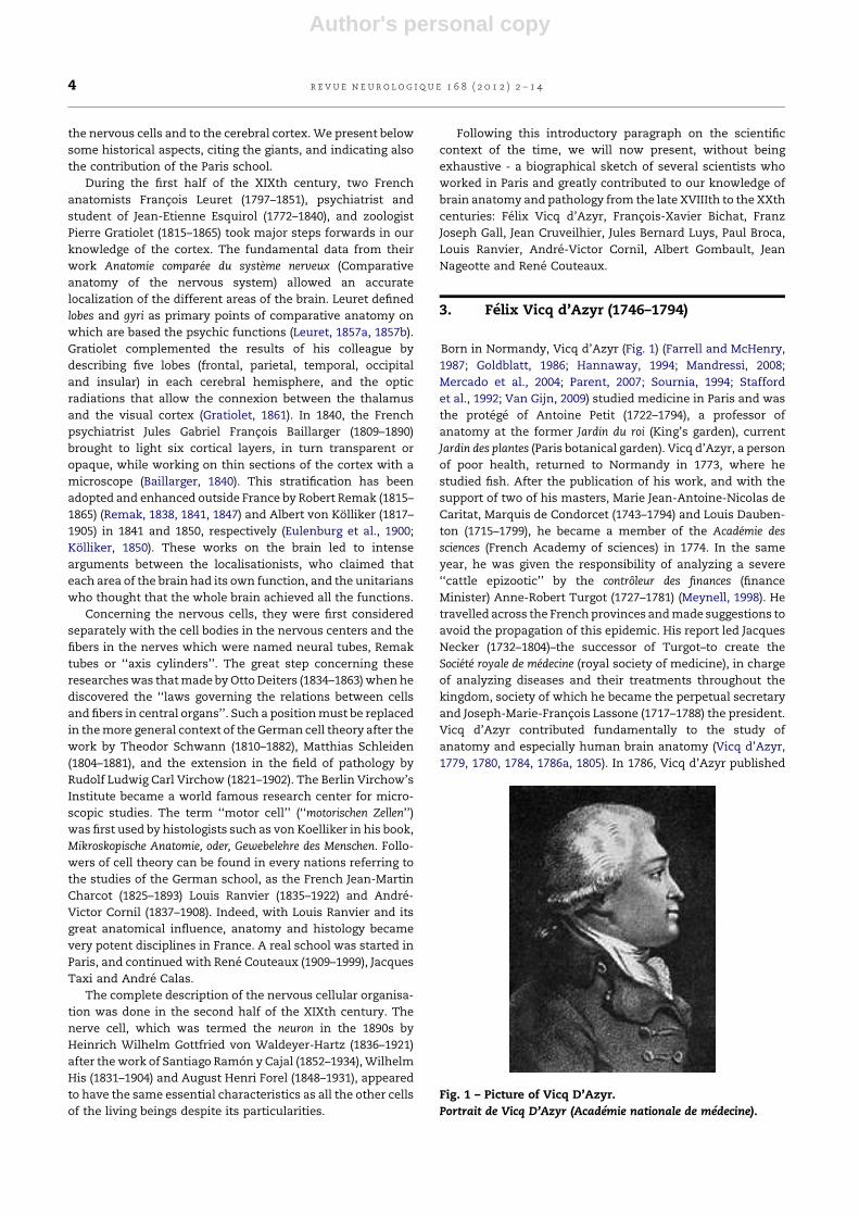
3. Felix Vicq d’Azyr (1746–1794)
Born in Normandy, Vicq d’Azyr (Fig. 1) (Farrell and McHenry,
1987; Goldblatt, 1986; Hannaway, 1994; Mandressi, 2008;
Mercado et al., 2004; Parent, 2007; Sournia, 1994; Stafford
et al., 1992; Van Gijn, 2009) studied medicine in Paris and was
the protege of Antoine Petit (1722–1794), a professor of
anatomy at the former Jardin du roi (King’s garden), current
Jardin des plantes (Paris botanical garden). Vicq d’Azyr, a person
of poor health, returned to Normandy in 1773, where he
studied fish. After the publication of his work, and with the
support of two of his masters, Marie Jean-Antoine-Nicolas de
Caritat, Marquis de Condorcet (1743–1794) and Louis Dauben-
ton (1715–1799), he became a member of the Academie des
sciences (French Academy of sciences) in 1774. In the same
year, he was given the responsibility of analyzing a severe
‘‘cattle epizootic’’ by the controleur des finances (finance
Minister) Anne-Robert Turgot (1727–1781) (Meynell, 1998). He
travelled across the French provinces and made suggestions to
avoid the propagation of this epidemic. His report led Jacques
Necker (1732–1804)–the successor of Turgot–to create the
Societe royale de medecine (royal society of medicine), in charge
of analyzing diseases and their treatments throughout the
kingdom, society of which he became the perpetual secretary
and Joseph-Marie-Francois Lassone (1717–1788) the president.
Vicq d’Azyr contributed fundamentally to the study of
anatomy and especially human brain anatomy (Vicq d’Azyr,
1779, 1780, 1784, 1786a, 1805). In 1786, Vicq d’Azyr published
Fig. 1 – Picture of Vicq D’Azyr.
Portrait de Vicq D’Azyr (Academie nationale de medecine).
r e v u e n e u r o l o g i q u e 1 6 8 ( 2 0 1 2 ) 2 – 1 44
Author's personal copy
his Traite d’anatomie comparee et de physiologie (Treatise on
comparative anatomy and physiology), a remarkable anatomy
and physiology treatise illustrated by sections of the human
brain of a quality and exactitude never attained before (Vicq
d’Azyr, 1786b, 1786c). He presented the deep brain structures
and was the first one to describe the locus coeruleus, the locus
niger and the mammillothalamic tract, also known as bundle
of Vicq d’Azyr. We owe him the concept ‘‘homology’’ in
biology. During the French Revolution, he presented a decisive
plan to reform the teaching of medicine in France, and
encouraged Francois de Fourcroy to have the three Ecoles de
sante (Medical faculties) re-created, in Paris, Montpellier and
Strasbourg voted by the convention in 1794 (Peumery, 2001).
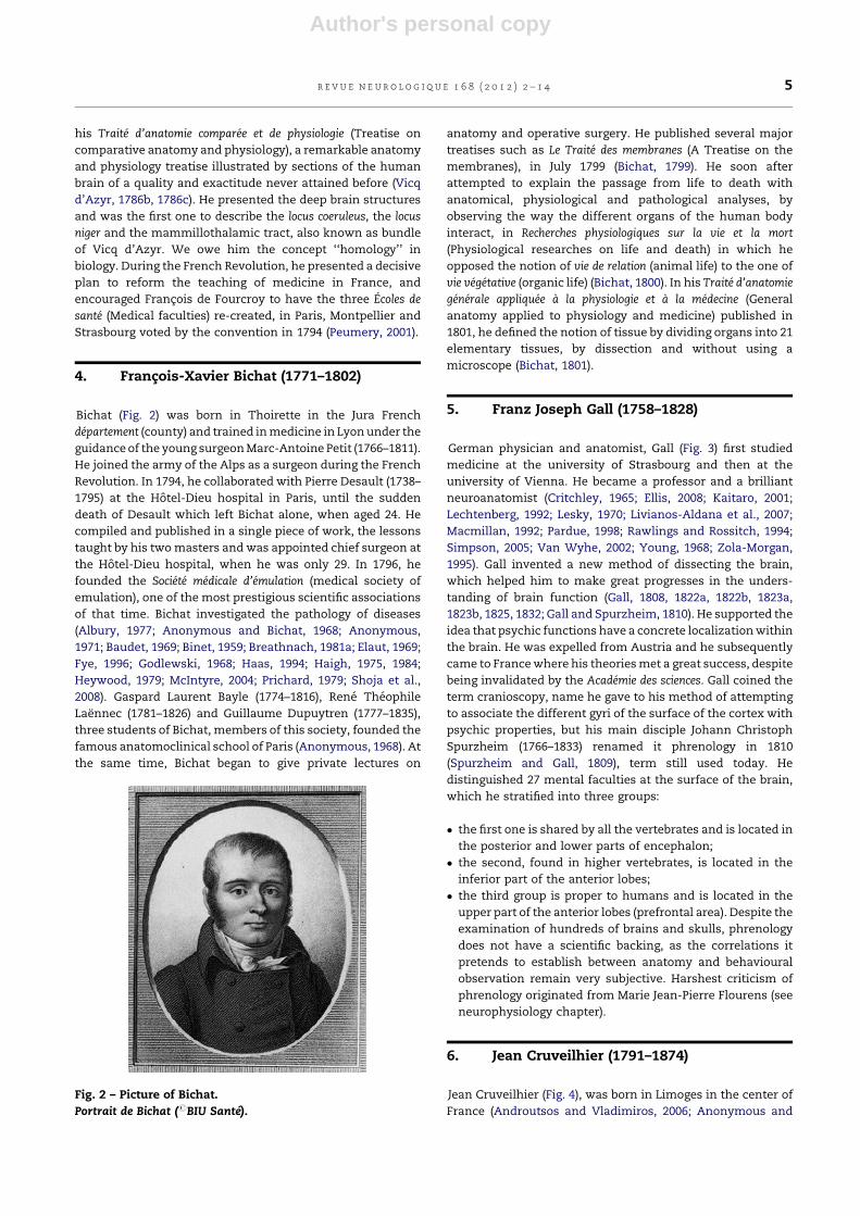
4. Francois-Xavier Bichat (1771–1802)
Bichat (Fig. 2) was born in Thoirette in the Jura French
departement (county) and trained in medicine in Lyon under the
guidance of the young surgeon Marc-Antoine Petit (1766–1811).
He joined the army of the Alps as a surgeon during the French
Revolution. In 1794, he collaborated with Pierre Desault (1738–
1795) at the Ho tel-Dieu hospital in Paris, until the sudden
death of Desault which left Bichat alone, when aged 24. He
compiled and published in a single piece of work, the lessons
taught by his two masters and was appointed chief surgeon at
the Ho tel-Dieu hospital, when he was only 29. In 1796, he
founded the Societe medicale d’emulation (medical society of
emulation), one of the most prestigious scientific associations
of that time. Bichat investigated the pathology of diseases
(Albury, 1977; Anonymous and Bichat, 1968; Anonymous,
1971; Baudet, 1969; Binet, 1959; Breathnach, 1981a; Elaut, 1969;
Fye, 1996; Godlewski, 1968; Haas, 1994; Haigh, 1975, 1984;
Heywood, 1979; McIntyre, 2004; Prichard, 1979; Shoja et al.,
2008). Gaspard Laurent Bayle (1774–1816), Rene Theophile
Laennec (1781–1826) and Guillaume Dupuytren (1777–1835),
three students of Bichat, members of this society, founded the
famous anatomoclinical school of Paris (Anonymous, 1968). At
the same time, Bichat began to give private lectures on
anatomy and operative surgery. He published several major
treatises such as Le Traite des membranes (A Treatise on the
membranes), in July 1799 (Bichat, 1799). He soon after
attempted to explain the passage from life to death with
anatomical, physiological and pathological analyses, by
observing the way the different organs of the human body
interact, in Recherches physiologiques sur la vie et la mort
(Physiological researches on life and death) in which he
opposed the notion of vie de relation (animal life) to the one of
vie vegetative (organic life) (Bichat, 1800). In his Traite d’anatomie
generale appliquee a la physiologie et a la medecine (General
anatomy applied to physiology and medicine) published in
1801, he defined the notion of tissue by dividing organs into 21
elementary tissues, by dissection and without using a
microscope (Bichat, 1801).
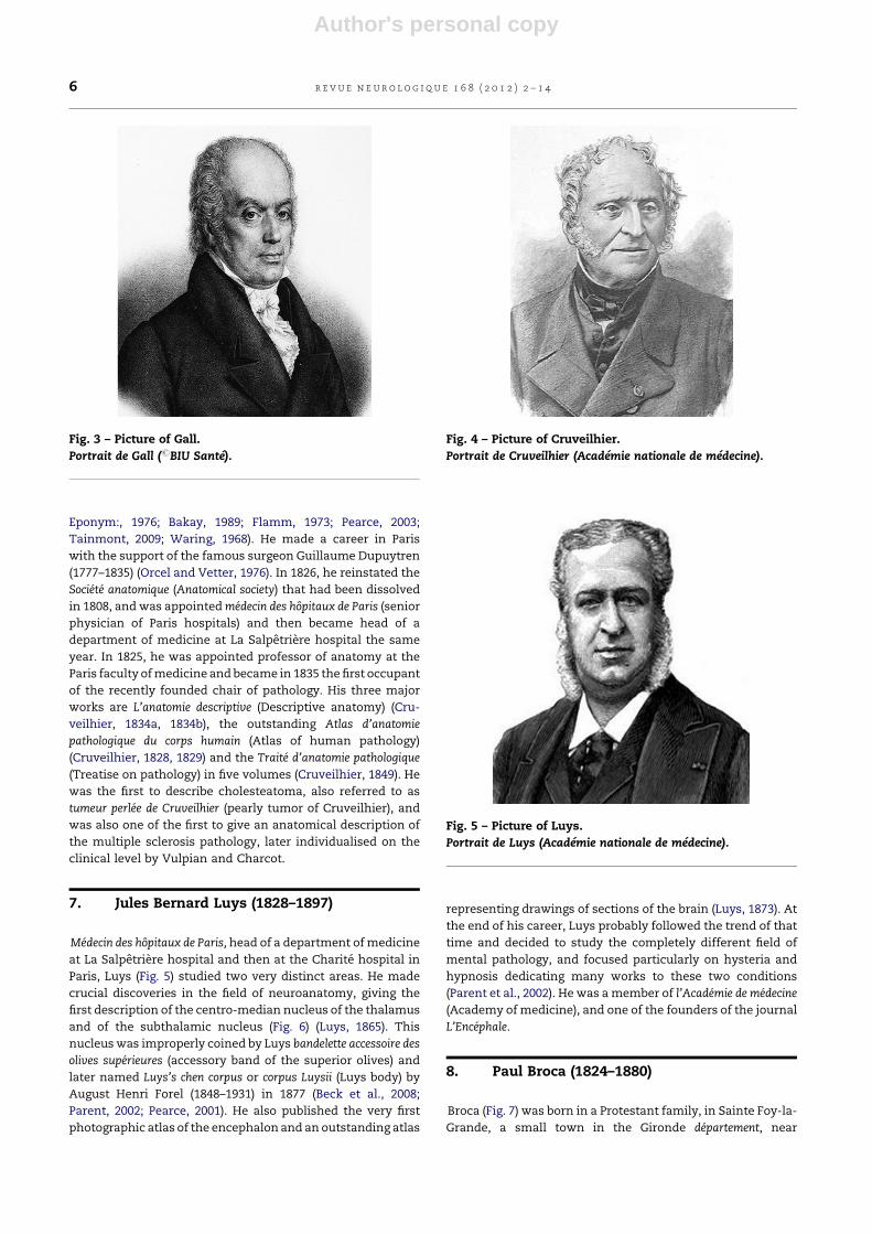
5. Franz Joseph Gall (1758–1828)
German physician and anatomist, Gall (Fig. 3) first studied
medicine at the university of Strasbourg and then at the
university of Vienna. He became a professor and a brilliant
neuroanatomist (Critchley, 1965; Ellis, 2008; Kaitaro, 2001;
Lechtenberg, 1992; Lesky, 1970; Livianos-Aldana et al., 2007;
Macmillan, 1992; Pardue, 1998; Rawlings and Rossitch, 1994;
Simpson, 2005; Van Wyhe, 2002; Young, 1968; Zola-Morgan,
1995). Gall invented a new method of dissecting the brain,
which helped him to make great progresses in the unders-
tanding of brain function (Gall, 1808, 1822a, 1822b, 1823a,
1823b, 1825, 1832; Gall and Spurzheim, 1810). He supported the
idea that psychic functions have a concrete localization within
the brain. He was expelled from Austria and he subsequently
came to France where his theories met a great success, despite
being invalidated by the Academie des sciences. Gall coined the
term cranioscopy, name he gave to his method of attempting
to associate the different gyri of the surface of the cortex with
psychic properties, but his main disciple Johann Christoph
Spurzheim (1766–1833) renamed it phrenology in 1810
(Spurzheim and Gall, 1809), term still used today. He
distinguished 27 mental faculties at the surface of the brain,
which he stratified into three groups:
� the first one is shared by all the vertebrates and is located in
the posterior and lower parts of encephalon;
� the second, found in higher vertebrates, is located in the
inferior part of the anterior lobes;
� the third group is proper to humans and is located in the
upper part of the anterior lobes (prefrontal area). Despite the
examination of hundreds of brains and skulls, phrenology
does not have a scientific backing, as the correlations it
pretends to establish between anatomy and behavioural
observation remain very subjective. Harshest criticism of
phrenology originated from Marie Jean-Pierre Flourens (see
neurophysiology chapter).
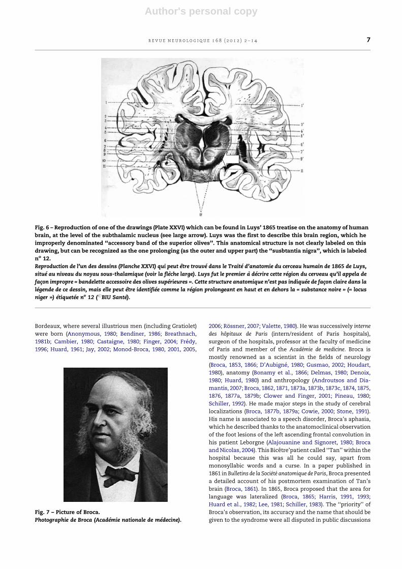
6. Jean Cruveilhier (1791–1874)
Jean Cruveilhier (Fig. 4), was born in Limoges in the center of
France (Androutsos and Vladimiros, 2006; Anonymous and
Fig. 2 – Picture of Bichat.
Portrait de Bichat (#BIU Sante).
r e v u e n e u r o l o g i q u e 1 6 8 ( 2 0 1 2 ) 2 – 1 4 5
Author's personal copy
Eponym:, 1976; Bakay, 1989; Flamm, 1973; Pearce, 2003;
Tainmont, 2009; Waring, 1968). He made a career in Paris
with the support of the famous surgeon Guillaume Dupuytren
(1777–1835) (Orcel and Vetter, 1976). In 1826, he reinstated the
Societe anatomique (Anatomical society) that had been dissolved
in 1808, and was appointed medecin des hopitaux de Paris (senior
physician of Paris hospitals) and then became head of a
department of medicine at La Salpetriere hospital the same
year. In 1825, he was appointed professor of anatomy at the
Paris faculty of medicine and became in 1835 the first occupant
of the recently founded chair of pathology. His three major
works are L’anatomie descriptive (Descriptive anatomy) (Cru-
veilhier, 1834a, 1834b), the outstanding Atlas d’anatomie
pathologique du corps humain (Atlas of human pathology)
(Cruveilhier, 1828, 1829) and the Traite d’anatomie pathologique
(Treatise on pathology) in five volumes (Cruveilhier, 1849). He
was the first to describe cholesteatoma, also referred to as
tumeur perlee de Cruveilhier (pearly tumor of Cruveilhier), and
was also one of the first to give an anatomical description of
the multiple sclerosis pathology, later individualised on the
clinical level by Vulpian and Charcot.
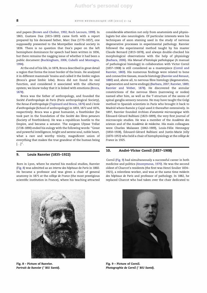
7. Jules Bernard Luys (1828–1897)
Medecin des hopitaux de Paris, head of a department of medicine
at La Salpetriere hospital and then at the Charite hospital in
Paris, Luys (Fig. 5) studied two very distinct areas. He made
crucial discoveries in the field of neuroanatomy, giving the
first description of the centro-median nucleus of the thalamus
and of the subthalamic nucleus (Fig. 6) (Luys, 1865). This
nucleus was improperly coined by Luys bandelette accessoire des
olives superieures (accessory band of the superior olives) and
later named Luys’s chen corpus or corpus Luysii (Luys body) by
August Henri Forel (1848–1931) in 1877 (Beck et al., 2008;
Parent, 2002; Pearce, 2001). He also published the very first
photographic atlas of the encephalon and an outstanding atlas
representing drawings of sections of the brain (Luys, 1873). At
the end of his career, Luys probably followed the trend of that
time and decided to study the completely different field of
mental pathology, and focused particularly on hysteria and
hypnosis dedicating many works to these two conditions
(Parent et al., 2002). He was a member of l’Academie de medecine
(Academy of medicine), and one of the founders of the journal
L’Encephale.
8. Paul Broca (1824–1880)
Broca (Fig. 7) was born in a Protestant family, in Sainte Foy-la-
Grande, a small town in the Gironde departement, near
Fig. 4 – Picture of Cruveilhier.
Portrait de Cruveilhier (Academie nationale de medecine).
Fig. 5 – Picture of Luys.
Portrait de Luys (Academie nationale de medecine).
Fig. 3 – Picture of Gall.
Portrait de Gall (#BIU Sante).
r e v u e n e u r o l o g i q u e 1 6 8 ( 2 0 1 2 ) 2 – 1 46
Author's personal copy
Bordeaux, where several illustrious men (including Gratiolet)
were born (Anonymous, 1980; Bendiner, 1986; Breathnach,
1981b; Cambier, 1980; Castaigne, 1980; Finger, 2004; Fredy,
1996; Huard, 1961; Jay, 2002; Monod-Broca, 1980, 2001, 2005,
2006; Rossner, 2007; Valette, 1980). He was successively interne
des hopitaux de Paris (intern/resident of Paris hospitals),
surgeon of the hospitals, professor at the faculty of medicine
of Paris and member of the Academie de medicine. Broca is
mostly renowned as a scientist in the fields of neurology
(Broca, 1853, 1866; D’Aubigne, 1980; Gusmao, 2002; Houdart,
1980), anatomy (Bonamy et al., 1866; Delmas, 1980; Denoix,
1980; Huard, 1980) and anthropology (Androutsos and Dia-
mantis, 2007; Broca, 1862, 1871, 1873a, 1873b, 1873c, 1874, 1875,
1876, 1877a, 1879b; Clower and Finger, 2001; Pineau, 1980;
Schiller, 1992). He made major steps in the study of cerebral
localizations (Broca, 1877b, 1879a; Cowie, 2000; Stone, 1991).
His name is associated to a speech disorder, Broca’s aphasia,
which he described thanks to the anatomoclinical observation
of the foot lesions of the left ascending frontal convolution in
his patient Leborgne (Alajouanine and Signoret, 1980; Broca
and Nicolas, 2004). This Bicetre’patient called ‘‘Tan’’ within the
hospital because this was all he could say, apart from
monosyllabic words and a curse. In a paper published in
1861 in Bulletins de la Societe anatomique de Paris, Broca presented
a detailed account of his postmortem examination of Tan’s
brain (Broca, 1861). In 1865, Broca proposed that the area for
language was lateralized (Broca, 1865; Harris, 1991, 1993;
Huard et al., 1982; Lee, 1981; Schiller, 1983). The ‘‘priority’’ of
Broca’s observation, its accuracy and the name that should be
given to the syndrome were all disputed in public discussions
Fig. 6 – Reproduction of one of the drawings (Plate XXVI) which can be found in Luys’ 1865 treatise on the anatomy of human
brain, at the level of the subthalamic nucleus (see large arrow). Luys was the first to describe this brain region, which he
improperly denominated ‘‘accessory band of the superior olives’’. This anatomical structure is not clearly labeled on this
drawing, but can be recognized as the one prolonging (as the outer and upper part) the ‘‘susbtantia nigra’’, which is labeled
no 12.
Reproduction de l’un des dessins (Planche XXVI) qui peut etre trouve dans le Traite d’anatomie du cerceau humain de 1865 de Luys,
situe au niveau du noyau sous-thalamique (voir la fleche large). Luys fut le premier a decrire cette region du cerveau qu’il appela de
facon impropre « bandelette accessoire des olives superieures ». Cette structure anatomique n’est pas indiquee de facon claire dans la
legende de ce dessin, mais elle peut etre identifiee comme la region prolongeant en haut et en dehors la « substance noire » (« locus
niger ») etiquetee no 12 (#BIU Sante).
Fig. 7 – Picture of Broca.
Photographie de Broca (Academie nationale de medecine).
r e v u e n e u r o l o g i q u e 1 6 8 ( 2 0 1 2 ) 2 – 1 4 7
Author's personal copy
and papers (Brown and Chobor, 1992; Roch Lecours, 1999). In
1863, Gustave Dax (1815–1893) came forth with a report
prepared by his deceased father, Marc Dax (1770–1837), one
supposedly presented to the Montpellier medical society in
1836. There is no question that Dax’s paper on the left
hemisphere dominance for speech had been written in 1836,
but there remains the nagging issue of whether it had been a
public document (Buckingham, 2006; Cubelli and Montagna,
1994).
At the end of his life, in 1878, Broca described in great detail
a region that forms the inner border of the brain. He analyzed
it in different mammals’ brains and called it the limbic region
(Broca’s great limbic lobe). Broca did not found its real
function, and considered it associated with the olfactory
system; we know today that it is linked with emotions (Broca,
1878).
Broca was the father of anthropology, and founded the
Societe d’anthropologie de Paris (Paris anthropological Society),
the Revue d’anthropologie (Topinard and Broca, 1876) and L’ecole
d’anthropologie (School of anthropology) in 1859, 1872 and 1876,
respectively. Broca was a great humanist, a freethinker (he
took part in the foundation of the Societe des libres penseurs
(Society of freethinkers). He was a republican hostile to the
Empire, and became a senator. The surgeon Ulysse Trelat
(1728–1890) ended his eulogy with the following words: ‘‘Great
and powerful intelligence, bright and serene soul, noble heart,
what a rare and worthy trinity, magnificent union of
everything that makes the true grandeur of the human being
[. . .]’’.
9. Louis Ranvier (1835–1922)
Born in Lyon, where he started his medical studies, Ranvier
(Fig. 8) was admitted as an interne des hopitaux de Paris in 1860.
He became a professor and was given a chair of general
anatomy in 1875 at the college de France (the most prestigious
scientific institution in France), where his teaching attracted
considerable attention not only from anatomists and physio-
logists but also neurologists. Of particular interests were his
techniques of axon staining used in the study of nervous
degenerative processes in experimental pathology. Ranvier
followed the experimental method taught by his master
Claude Bernard (1813–1878), and always double-checked his
morphological observations with the help of physiology
(Barbara, 2006). His Manuel d’histologie pathologique (A manual
of pathological histology) in collaboration with Victor Cornil
(1837–1908) is still considered as a masterpiece (Cornil and
Ranvier, 1869). His numerous findings were related to bone
and connective tissues, muscle histology (Ranvier and Renaut,
1880) and, above all, to nervous fibre histology (degeneration,
regeneration and nerve endings) (Barbara, 2007; Ranvier, 1889;
Ranvier and Weber, 1878). He discovered the annular
constrictions of the nervous fibers (narrowing or nodes)
named after him, as well as the T-structure of the axons of
spinal ganglia sensory neurons. He may have taught the Golgi
method to Spanish scientists in Paris who brought it back to
Madrid where Ramon y Cajal used it thereafter extensively. In
1897, Ranvier founded Archives d’anatomie microscopique with
Edouard-Gerard Balbiani (1823–1899), the very first journal of
microscopic studies. He was a member of the Academie des
sciences and of the Academie de medecine. His main colleagues
were Charles Malassez (1842–1909), Louis-Felix Henneguy
(1850–1928), Edouard-Gerard Balbiani and Justin-Marie Jolly
(1870–1953) who held a chair of histophysiology at the college de
France in 1925.
10. Andre-Victor Cornil (1837–1908)
Cornil (Fig. 9) had simultaneously a successful career in both
medicine and politics (Anonymous, 1970). He was the second
oldest of Charcot’s residents (the first was Henri Soulier 1834–
1921), a relentless worker, and was at the same time medecin
des hopitaux de Paris and professor of pathology. In 1882, he
replaced Charcot who had taken over the chair dedicated to
Fig. 8 – Picture of Ranvier.
Portrait de Ranvier (#BIU Sante).
Fig. 9 – Picture of Cornil.
Photographie de Cornil (#BIU Sante).
r e v u e n e u r o l o g i q u e 1 6 8 ( 2 0 1 2 ) 2 – 1 48
Author's personal copy
the study of the nervous system. Cornil was a clinician but
also, and mainly, a pathologist. He was passionate about
postmortem examinations and histopathology (Cornil, 1863,
1864, 1865, 1879; Cornil and Babes, 1885). He wrote many
papers and his Manuel d’histologie pathologique (A manual of
pathological histology), which he wrote in collaboration with
Louis Ranvier, is a remarkably clear condensation of all his
observations (Cornil and Ranvier, 1869). His collaboration with
Ranvier began with the small private histological laboratory
they founded on Rue Christine, and went on at the college de
France and at the chair of Pathological anatomy. On the
political level, Victor Cornil was a congressional representa-
tive of the Allier French departement (County), senator-mayor
of his small village Creuzier-le-neuf, regional councillor,
prefect from the 6th to the 24th of September 1870, and
vice-president then president of the regional council of Allier.
He was an anti-monarchist and presided over the republican
league in the 6th arrondissement of Paris during the Commune.
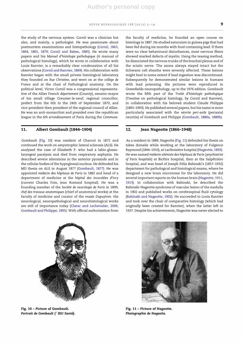
11. Albert Gombault (1844–1904)
Gombault (Fig. 10) was resident of Charcot in 1872 and
continued the work on amyotrophic lateral sclerosis (ALS). He
analysed the case of Elisabeth P. who had a labio-glosso-
laryngeal paralysis and died from respiratory asphyxia. He
described severe alterations in the anterior pyramids and in
the cellular bodies of the hypoglossal nucleus. He defended his
MD thesis on ALS in August 1877 (Gombault, 1877). He was
appointed medecin des hopitaux de Paris in 1882 and head of a
department of medicine at the hopital des incurables d’Ivry
(current Charles Foix, Jean Rostand hospital). He was a
founding member of the Societe de neurologie de Paris in 1899,
chef des travaux anatomiques (chief of anatomical works) at the
faculty of medicine and curator of the musee Dupuytren. His
neurological, neuropathological and neurohistological works
are still of importance today (Clarac and Lechevalier, 2006;
Gombault and Philippe, 1895). With official authorization from
the faculty of medicine, he founded an open course on
histology in 1887. He studied saturnism in guinea pigs that had
been fed during six months with food containing lead. If there
were no clear behavioural disturbances, most nervous fibers
showed marked defects of myelin. Using the teasing method,
he dissociated the nervous trunks of the brachial plexus and of
the sciatic nerve. The axons always stayed intact but the
Schwann cell sheaths were severely affected. These lesions
might heal to some extent if lead ingestion was discontinued.
Subsequently he demonstrated similar lesions in humans
with lead poisoning. His pictures were reproduced in
Greenfields neuropathology, up to the 1976 edition. Gombault
wrote the fifth part of the Traite d’histologie pathologique
(Treatise on pathological histology, by Cornil and Ranvier),
in collaboration with his beloved student Claude Philippe
(1865–1903). He published several papers, but his name is more
particularly associated with the nevrite peri-axile (periaxial
neuritis) of Gombault and Philippe (Gombault, 1880a, 1880b).
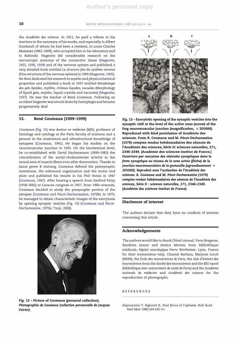
12. Jean Nageotte (1866–1948)
As a resident in 1889, Nageotte (Fig. 11) defended his thesis on
tabes dorsalis while working at the laboratory of Fulgence
Raymond (1844–1910), at Lariboisiere hospital (Nageotte, 1893).
He was named medecin alieniste des hopitaux de Paris ( psychiatrist
of Paris hospitals) at Bicetre hospital, then at the Salpetriere
hospital, and was head of Joseph Felix Babinski’s (1857–1932)
department for pathological and histological exams, where he
designed a new brain microtome for the laboratory. He did
several important reports on the human brain (Nageotte, 1911,
1913). In collaboration with Babinski, he described the
Babinski-Nageotte syndrome of vascular lesion of the medulla
in 1902 and published works on cerebrospinal fluid cytology
(Babinski and Nageotte, 1902). He succeeded to Louis Ranvier
and took over the chair of comparative histology (which had
originally been created for Ranvier), when the latter left in
1937. Despite his achievements, Nageotte was never elected to
Fig. 10 – Picture of Gombault.
Portrait de Gombault (#BIU Sante).
Fig. 11 – Picture of Nageotte.
Photographie de Nageotte.
r e v u e n e u r o l o g i q u e 1 6 8 ( 2 0 1 2 ) 2 – 1 4 9
Author's personal copy
the Academie des sciences. In 1911, he paid a tribute to his
teachers in the summary of his works, and especially to Albert
Gombault of whom he had been a resident, to Louis-Charles
Malassez (1842–1909), who accepted him in his laboratory and
to Babinski. Nageotte did considerable research on the
microscopic anatomy of the connective tissue (Nageotte,
1922, 1936, 1939) and of the nervous system and published a
very detailed book entitled La structure fine du systeme nerveux
(Fine structure of the nervous system) in 1905 (Nageotte, 1905).
He then dedicated his research to myelin and physicochemical
properties and published a book in 1937 entitled Morphologie
des gels lipoıdes, myeline, cristaux liquides, vacuoles (Morphology
of lipoid gels, myelin, liquid crystals and vacuoles) (Nageotte,
1937). He was the teacher of Rene Couteaux. Following an
accident Nageotte was struck down by hemiplegia and became
progressively deaf.
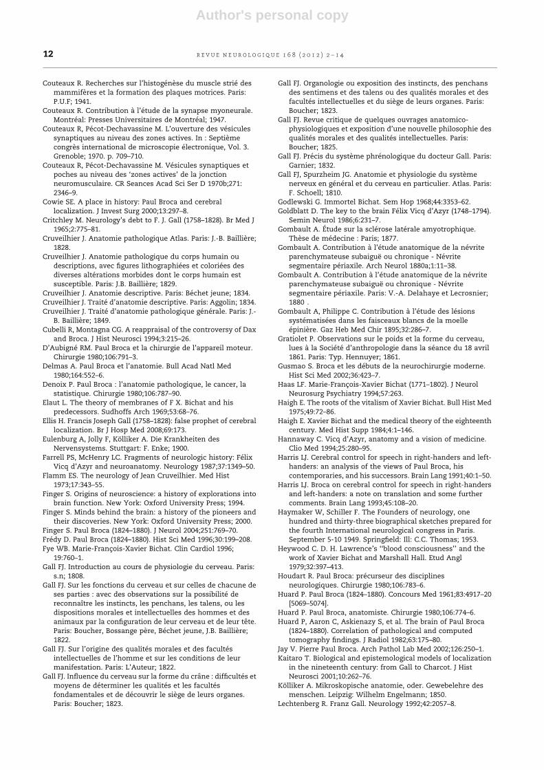
13. Rene Couteaux (1909–1999)
Couteaux (Fig. 12) was docteur en medecine (MD), professor of
histology and cytology at the Paris faculty of sciences and a
pioneer in the anatomical and ultrastructural knowledge of
synapses (Couteaux, 1941). He began his studies on the
neuromuscular junction in 1935. On the biochemical level,
he co-established with David Nachmanson (1899–1983) the
concentration of the acetyl-cholinesterase activity in the
neural area of muscle fibers even after denervation. Thanks to
Janus green B staining, Couteaux defined the postsynaptic
membrane, the subneural organization and the motor end
plate and published his results in his PhD thesis in 1947
(Couteaux, 1947). After hearing a speech from Sanford Palay
(1918–2002) at Caracas congress in 1957, from 1960 onwards,
Couteaux decided to study the presynaptic portion of the
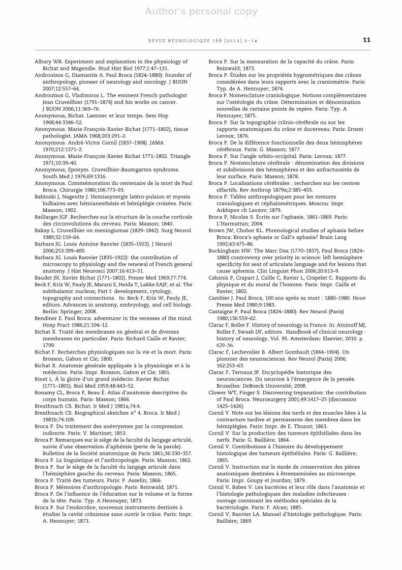
synapse (Couteaux and Pecot-Dechavassine, 1970b). In 1970,
he managed to obtain characteristic images of the exocytosis
by opening synaptic vesicles (Fig. 13) (Couteaux and Pecot-
Dechavassine, 1970a; Tsuji, 2006).
Disclosure of interest
The authors declare that they have no conflicts of interest
concerning this article.
Acknowledgements
The authors would like to thank Chloe Loiraud, Yann Bregeras,
Sandrine Jamen and Jessica Moreau from bibliotheque
medicale, hopital neurologique Pierre Wertheimer, Lyon, France
for their tremendous help, Chantal Barbara, Marjorie Lorch
(ISHN), the Ecole des neurosciences de Paris, the club d’histoire des
neurosciences from the Societe des neurosciences and the BIU sante
(bibliotheque inter universitaire de sante de Paris) and the Academie
nationale de medecine and Academie des sciences for the
reproduction of photographs.
r e f e r e n c e s
Alajouanine T, Signoret JL. Paul Broca et l’aphasie. Bull AcadNatl Med 1980;164:545–51.
Fig. 12 – Picture of Couteaux (personal collection).
Photographie de Couteaux (collection personnelle de Jacques
Poirier).
Fig. 13 – Exocytotic opening of the synaptic vesicles into the
synaptic cleft at the level of the active zone (arrow) of the
frog neuromuscular junction (magnification, T 205000).
Reproduced with kind permission of Academie des
sciences. From R. Couteaux and M. Pecot-Dechavassine
(1970) comptes rendus hebdomadaires des seances de
l’Academie des sciences, Serie D: sciences naturelles, 271,
2346–2349. (Academie des sciences–institut de France.)
Ouverture par exocytose des vesicules synaptiques dans la
fente synaptique au niveau de la zone active (fleche) de la
junction neuromusculaire de la grenouille (agrandissement T
205000). Reproduit avec l’autisation de l’Academie des
sciences. R. Couteaux and M. Pecot-Dechavassine (1970)
comptes rendus hebdomadaires des seances de l’Academie des
sciences, Serie D : sciences naturelles, 271, 2346–2349.
(Academie des sciences–institut de France).
r e v u e n e u r o l o g i q u e 1 6 8 ( 2 0 1 2 ) 2 – 1 410
Author's personal copy
Albury WR. Experiment and explanation in the physiology ofBichat and Magendie. Stud Hist Biol 1977;1:47–131.
Androutsos G, Diamantis A. Paul Broca (1824–1880): founder ofanthropology, pioneer of neurology and oncology. J BUON2007;12:557–64.
Androutsos G, Vladimiros L. The eminent French pathologistJean Cruveilhier (1791–1874) and his works on cancer.J BUON 2006;11:369–76.
Anonymous, Bichat. Laennec et leur temps. Sem Hop1968;44:3346–52.
Anonymous. Marie-Francois-Xavier-Bichat (1771–1802), tissuepathologist. JAMA 1968;203:291–2.
Anonymous. Andre-Victor Cornil (1837–1908). JAMA1970;212:1371–2.
Anonymous. Marie-Francois-Xavier Bichat 1771–1802. Triangle1971;10:39–40.
Anonymous, Eponym. Cruveilhier-Baumgarten syndrome.South Med J 1976;69:1316.
Anonymous. Commemoration du centenaire de la mort de PaulBroca. Chirurgie 1980;106:773–93.
Babinski J, Nageotte J. Hemiasynergie latero-pulsion et myosisbulbaires avec hemianesthesie et hemiplegie croisees. Paris:Masson; 1902.
Baillarger JGF. Recherches sur la structure de la couche corticaledes circonvolutions du cerveau. Paris: Masson; 1840.
Bakay L. Cruveilhier on meningiomas (1829–1842). Surg Neurol1989;32:159–64.
Barbara JG. Louis Antoine Ranvier (1835–1922). J Neurol2006;253:399–400.
Barbara JG. Louis Ranvier (1835–1922): the contribution ofmicroscopy to physiology and the renewal of French generalanatomy. J Hist Neurosci 2007;16:413–31.
Baudet JH. Xavier Bichat (1771–1802). Presse Med 1969;77:774.Beck F, Kriz W, Pauly JE, Marani E, Heida T, Lakke EAJF, et al. The
subthalamic nucleus; Part I: development, cytology,topography and connections. In: Beck F, Kriz W, Pauly JE,editors. Advances in anatomy, embryology, and cell biology.Berlin: Springer; 2008.
Bendiner E. Paul Broca: adventurer in the recesses of the mind.Hosp Pract 1986;21:104–12.
Bichat X. Traite des membranes en general et de diversesmembranes en particulier. Paris: Richard Caille et Ravier;1799.
Bichat F. Recherches physiologiques sur la vie et la mort. Paris:Brosson, Gabon et Cie; 1800.
Bichat X. Anatomie generale appliquee a la physiologie et a lamedecine. Paris: Impr. Brosson, Gabon et Cie; 1801.
Binet L. A la gloire d’un grand medecin: Xavier Bichat(1771–1801). Biol Med 1959;48:443–52.
Bonamy CL, Broca P, Beau E. Atlas d’anatomie descriptive ducorps humain. Paris: Masson; 1866.
Breathnach CS. Bichat. Ir Med J 1981a;74:4.Breathnach CS. Biographical sketches no 4. Broca. Ir Med J
1981b;74:109.Broca P. Du traitement des anevrysmes par la compression
indirecte. Paris: V. Martinet; 1853.Broca P. Remarques sur le siege de la faculte du langage articule,
suivie d’une observation d’aphemie (perte de la parole).Bulletins de la Societe anatomique de Paris 1861;36:330–357.
Broca P. La linguistique et l’anthropologie. Paris: Masson; 1862.Broca P. Sur le siege de la faculte du langage articule dans
l’hemisphere gauche du cerveau. Paris: Masson; 1865.Broca P. Traite des tumeurs. Paris: P. Asselin; 1866.Broca P. Memoires d’anthropologie. Paris: Reinwald; 1871.Broca P. De l’influence de l’education sur le volume et la forme
de la tete. Paris: Typ. A Hennuyer; 1873.Broca P. Sur l’endocra ne, nouveaux instruments destines a
etudier la cavite cranienne sans ouvrir le cra ne. Paris: Impr.A. Hennuyer; 1873.
Broca P. Sur la mensuration de la capacite du crane. Paris:Reinwald; 1873.
Broca P. Etudes sur les proprietes hygrometriques des cra nesconsiderees dans leurs rapports avec la craniometrie. Paris:Typ. de A. Hennuyer; 1874.
Broca P. Nomenclature craniologique. Notions complementairessur l’osteologie du cra ne. Determination et denominationnouvelles de certains points de repere. Paris: Typ. AHennuyer; 1875.
Broca P. Sur la topographie cra nio-cerebrale ou sur lesrapports anatomiques du cra ne et ducerveau. Paris: ErnestLeroux; 1876.
Broca P. De la difference fonctionnelle des deux hemispherescerebraux. Paris: G. Masson; 1877.
Broca P. Sur l’angle orbito-occipital. Paris: Leroux; 1877.Broca P. Nomenclature cerebrale : denomination des divisions
et subdivisions des hemispheres et des anfractuosites deleur surface. Paris: Masson; 1878.
Broca P. Localisations cerebrales : recherches sur les centresolfactifs. Rev Anthrop 1879a;2:385–455.
Broca P. Tables anthropologiques pour les mesurescraniologiques et cephalometriques. Moscou: Impr.Arkhipov ch Lavzov; 1879.
Broca P, Nicolas S. Ecrits sur l’aphasie, 1861–1869. Paris:L’Harmattan; 2004.
Brown JW, Chobor KL. Phrenological studies of aphasia beforeBroca: Broca’s aphasia or Gall’s aphasia? Brain Lang1992;43:475–86.
Buckingham HW. The Marc Dax (1770–1837), Paul Broca (1824–1880) controversy over priority in science: left hemispherespecificity for seat of articulate language and for lesions thatcause aphemia. Clin Linguist Phon 2006;20:613–9.
Cabanis P, Crapart J, Caille C, Ravier L, Crapelet C. Rapports duphysique et du moral de l’homme. Paris: Impr. Caille etRavier; 1802.
Cambier J. Paul Broca, 100 ans apres sa mort : 1880–1980. NouvPresse Med 1980;9:1983.
Castaigne P. Paul Broca (1824–1880). Rev Neurol (Paris)1980;136:559–62.
Clarac F, Boller F. History of neurology in France. In: Aminoff MJ,Boller F, Swaab DF, editors. Handbook of clinical neurology -history of neurology, Vol. 95. Amsterdam: Elsevier; 2010. p.
629–56.
Clarac F, Lechevalier B. Albert Gombault (1844–1904). Unpionnier des neurosciences. Rev Neurol (Paris) 2006;162:253–63.
Clarac F, Ternaux JP. Encyclopedie historique desneurosciences. Du neurone a l’emergence de la pensee.Bruxelles: DeBoeck Universite; 2008.
Clower WT, Finger S. Discovering trepanation: the contributionof Paul Broca. Neurosurgery 2001;49:1417–25 [discussion1425–1426].
Cornil V. Note sur les lesions des nerfs et des muscles liees a lacontracture tardive et permanente des membres dans leshemiplegies. Paris: Impr. de E. Thunot; 1863.
Cornil V. Sur la production des tumeurs epitheliales dans lesnerfs. Paris: G. Bailliere; 1864.
Cornil V. Contributions a l’histoire du developpementhistologique des tumeurs epitheliales. Paris: G. Bailliere;1865.
Cornil V. Instruction sur le mode de conservation des piecesanatomiques destinees a etreexaminees au microscope.Paris: Impr. Goupy et Jourdan; 1879.
Cornil V, Babes V. Les bacteries et leur ro le dans l’anatomie etl’histologie pathologiques des maladies infectieuses :ouvrage contenant les methodes speciales de labacteriologie. Paris: F. Alcan; 1885.
Cornil V, Ranvier LA. Manuel d’histologie pathologique. Paris:Bailliere; 1869.
r e v u e n e u r o l o g i q u e 1 6 8 ( 2 0 1 2 ) 2 – 1 4 11
Author's personal copy
Couteaux R. Recherches sur l’histogenese du muscle strie desmammiferes et la formation des plaques motrices. Paris:P.U.F; 1941.
Couteaux R. Contribution a l’etude de la synapse myoneurale.Montreal: Presses Universitaires de Montreal; 1947.
Couteaux R, Pecot-Dechavassine M. L’ouverture des vesiculessynaptiques au niveau des zones actives. In : Septiemecongres international de microscopie electronique, Vol. 3.Grenoble; 1970. p. 709–710.
Couteaux R, Pecot-Dechavassine M. Vesicules synaptiques etpoches au niveau des ‘zones actives’ de la jonctionneuromusculaire. CR Seances Acad Sci Ser D 1970b;271:2346–9.
Cowie SE. A place in history: Paul Broca and cerebrallocalization. J Invest Surg 2000;13:297–8.
Critchley M. Neurology’s debt to F. J. Gall (1758–1828). Br Med J1965;2:775–81.
Cruveilhier J. Anatomie pathologique Atlas. Paris: J.-B. Bailliere;1828.
Cruveilhier J. Anatomie pathologique du corps humain oudescriptions, avec figures lithographiees et coloriees desdiverses alterations morbides dont le corps humain estsusceptible. Paris: J.B. Bailliere; 1829.
Cruveilhier J. Anatomie descriptive. Paris: Bechet jeune; 1834.Cruveilhier J. Traite d’anatomie descriptive. Paris: Aggolin; 1834.Cruveilhier J. Traite d’anatomie pathologique generale. Paris: J.-
B. Bailliere; 1849.Cubelli R, Montagna CG. A reappraisal of the controversy of Dax
and Broca. J Hist Neurosci 1994;3:215–26.D’Aubigne RM. Paul Broca et la chirurgie de l’appareil moteur.
Chirurgie 1980;106:791–3.Delmas A. Paul Broca et l’anatomie. Bull Acad Natl Med
1980;164:552–6.Denoix P. Paul Broca : l’anatomie pathologique, le cancer, la
statistique. Chirurgie 1980;106:787–90.Elaut L. The theory of membranes of F X. Bichat and his
predecessors. Sudhoffs Arch 1969;53:68–76.Ellis H. Francis Joseph Gall (1758–1828): false prophet of cerebral
localization. Br J Hosp Med 2008;69:173.Eulenburg A, Jolly F, Kolliker A. Die Krankheiten des
Nervensystems. Stuttgart: F. Enke; 1900.Farrell PS, McHenry LC. Fragments of neurologic history: Felix
Vicq d’Azyr and neuroanatomy. Neurology 1987;37:1349–50.Flamm ES. The neurology of Jean Cruveilhier. Med Hist
1973;17:343–55.Finger S. Origins of neuroscience: a history of explorations into
brain function. New York: Oxford University Press; 1994.Finger S. Minds behind the brain: a history of the pioneers and
their discoveries. New York: Oxford University Press; 2000.Finger S. Paul Broca (1824–1880). J Neurol 2004;251:769–70.Fredy D. Paul Broca (1824–1880). Hist Sci Med 1996;30:199–208.Fye WB. Marie-Francois-Xavier Bichat. Clin Cardiol 1996;
19:760–1.Gall FJ. Introduction au cours de physiologie du cerveau. Paris:
s.n; 1808.Gall FJ. Sur les fonctions du cerveau et sur celles de chacune de
ses parties : avec des observations sur la possibilite dereconnaıtre les instincts, les penchans, les talens, ou lesdispositions morales et intellectuelles des hommes et desanimaux par la configuration de leur cerveau et de leur tete.Paris: Boucher, Bossange pere, Bechet jeune, J.B. Bailliere;1822.
Gall FJ. Sur l’origine des qualites morales et des facultesintellectuelles de l’homme et sur les conditions de leurmanifestation. Paris: L’Auteur; 1822.
Gall FJ. Influence du cerveau sur la forme du cra ne : difficultes etmoyens de determiner les qualites et les facultesfondamentales et de decouvrir le siege de leurs organes.Paris: Boucher; 1823.
Gall FJ. Organologie ou exposition des instincts, des penchansdes sentimens et des talens ou des qualites morales et desfacultes intellectuelles et du siege de leurs organes. Paris:Boucher; 1823.
Gall FJ. Revue critique de quelques ouvrages anatomico-physiologiques et exposition d’une nouvelle philosophie desqualites morales et des qualites intellectuelles. Paris:Boucher; 1825.
Gall FJ. Precis du systeme phrenologique du docteur Gall. Paris:Garnier; 1832.
Gall FJ, Spurzheim JG. Anatomie et physiologie du systemenerveux en general et du cerveau en particulier. Atlas. Paris:F. Schoell; 1810.
Godlewski G. Immortel Bichat. Sem Hop 1968;44:3353–62.Goldblatt D. The key to the brain Felix Vicq d’Azyr (1748–1794).
Semin Neurol 1986;6:231–7.Gombault A. Etude sur la sclerose laterale amyotrophique.
These de medecine : Paris; 1877.Gombault A. Contribution a l’etude anatomique de la nevrite
parenchymateuse subaigue ou chronique - Nevritesegmentaire periaxile. Arch Neurol 1880a;1:11–38.
Gombault A. Contribution a l’etude anatomique de la nevriteparenchymateuse subaigue ou chronique - Nevritesegmentaire periaxile. Paris: V.-A. Delahaye et Lecrosnier;1880 .
Gombault A, Philippe C. Contribution a l’etude des lesionssystematisees dans les faisceaux blancs de la moelleepiniere. Gaz Heb Med Chir 1895;32:286–7.
Gratiolet P. Observations sur le poids et la forme du cerveau,lues a la Societe d’anthropologie dans la seance du 18 avril1861. Paris: Typ. Hennuyer; 1861.
Gusmao S. Broca et les debuts de la neurochirurgie moderne.Hist Sci Med 2002;36:423–7.
Haas LF. Marie-Francois-Xavier Bichat (1771–1802). J NeurolNeurosurg Psychiatry 1994;57:263.
Haigh E. The roots of the vitalism of Xavier Bichat. Bull Hist Med1975;49:72–86.
Haigh E. Xavier Bichat and the medical theory of the eighteenthcentury. Med Hist Supp 1984;4:1–146.
Hannaway C. Vicq d’Azyr, anatomy and a vision of medicine.Clio Med 1994;25:280–95.
Harris LJ. Cerebral control for speech in right-handers and left-handers: an analysis of the views of Paul Broca, hiscontemporaries, and his successors. Brain Lang 1991;40:1–50.
Harris LJ. Broca on cerebral control for speech in right-handersand left-handers: a note on translation and some furthercomments. Brain Lang 1993;45:108–20.
Haymaker W, Schiller F. The Founders of neurology, onehundred and thirty-three biographical sketches prepared forthe fourth International neurological congress in Paris.September 5-10 1949. Springfield: Ill: C.C. Thomas; 1953.
Heywood C. D. H. Lawrence’s ‘‘blood consciousness’’ and thework of Xavier Bichat and Marshall Hall. Etud Angl1979;32:397–413.
Houdart R. Paul Broca: precurseur des disciplinesneurologiques. Chirurgie 1980;106:783–6.
Huard P. Paul Broca (1824–1880). Concours Med 1961;83:4917–20[5069–5074].
Huard P. Paul Broca, anatomiste. Chirurgie 1980;106:774–6.Huard P, Aaron C, Askienazy S, et al. The brain of Paul Broca
(1824–1880). Correlation of pathological and computedtomography findings. J Radiol 1982;63:175–80.
Jay V. Pierre Paul Broca. Arch Pathol Lab Med 2002;126:250–1.Kaitaro T. Biological and epistemological models of localization
in the nineteenth century: from Gall to Charcot. J HistNeurosci 2001;10:262–76.
Kolliker A. Mikroskopische anatomie, oder. Gewebelehre desmenschen. Leipzig: Wilhelm Engelmann; 1850.
Lechtenberg R. Franz Gall. Neurology 1992;42:2057–8.
r e v u e n e u r o l o g i q u e 1 6 8 ( 2 0 1 2 ) 2 – 1 412
Author's personal copy
Lee DA. Paul Broca and the history of aphasia: Roland P. MackayAward Essay, 1980. Neurology 1981;31:600–2.
Lesky E. Structure and function in Gall. Bull Hist Med1970;44:297–314.
Leuret F. Anatomie comparee du systeme nerveux consideredans ses rapports avec l’intelligence.Tome premiercomprenant la description de l’encephale et de la moellerachidienne. Paris: J.B. Bailliere et fils; 1857.
Leuret F. Anatomie comparee du systeme nerveux consideredans ses rapports avec l’intelligence. Tome secondcomprenant l’anatomie du cerveau de l’homme et dessinges. Paris: J.B. Bailliere et fils; 1857.
Livianos-Aldana L, Rojo-Moreno L, Sierra-Sanmiguel P. F. J. Galland the phrenological movement. Am J Psychiatry2007;164:414.
Luys J. Recherches sur le systeme nerveux cerebro-spinal : sastructure, ses fonctions et ses maladies. Paris: J.B. Bailliere etfils; 1865.
Luys J. Iconographie photographique des centres nerveux. Paris:J.B. Balliere; 1873.
Macmillan M. Inhibition and the control of behavior. From Gallto Freud via Phineas Gage and the frontal lobes. Brain Cogn1992;19:72–104.
Mandressi R. The past, education and science. Felix Vicq d’Azyrand the history of medicine in the 18th century. Med Secoli2008;20:183–212.
McIntyre N. Xavier Bichat (1771–1802). J Med Biogr 2004;12:184.Mercado R, Santos-Franco J, Ortiz-Velazquez I, Gomez-Llata S.
Vascular anatomy of the foramen of Vicq d’Azyr: amicrosurgical perspective. Minim Invasive Neurosurg2004;4:102–6.
Meynell E. Vicq d’Azyr and a cattle plague. J R Soc Med1998;91:105–6.
Monod-Broca P. Paul Broca (1824–1880). Le chirurgien, l’homme.Bull Acad Natl Med 1980;164:536–44.
Monod-Broca P. Paul Broca: 1824–1880. Ann Chir 2001;126:801–7.
Monod-Broca P. Paul Broca un geant du XIXe siecle. Paris:Vuibert; 2005.
Monod-Broca P. L’autre Paul Broca. Rev Prat 2006;56:923–5.Nageotte J. Tabes et paralysie generale. These de Medecine :
Paris; 1893.Nageotte J. La structure fine du systeme nerveux. Paris: Maloine;
1905.Nageotte J. Notice sur les travaux scientifiques. Paris: s.n; 1911.Nageotte J. La carte de l’ecorce cerebrale. Paris: Felix Alcan; 1913.Nageotte J. L’organisation de la matiere dans ses rapports avec
la vie : etudes d’anatomie generale et de morphologieexperimentale sur le tissu conjonctif et le nerf : avec 152figures dans le texte et 4 planches contenant 16 figures dont14 microphotographies autochromes. Paris: Felix Alcan; 1922.
Nageotte J. Structures a surfaces hydrophobes structuresvacuolaires. Paris: Hermann; 1936.
Nageotte J. Morphologie des gels lipoıdes. Myeline. Cristauxliquides. Vacuoles. Paris: Hermann; 1937.
Nageotte J. Sur l’emploi des greffes de tissu conjonctif mortdans la chirurgie reparatrice (Tendon et Nerf). Paris: Masson;1939.
Orcel L, Vetter T. Dupuytren, Cruveilhier and the anatomicalsociety. Arch Anat Cytol Pathol 1976;24:167–79.
Pardue ML. Joseph Gall- pioneering nuclear biology. Trends CellBiol 1998;8:208–10.
Parent A. Jules Bernard Luys and the subthalamic nucleus. MovDisord 2002;17:181–5.
Parent A. Felix Vicq d’Azyr: anatomy, medicine and revolution.Can J Neurol Sci 2007;34:30–7.
Parent A, Parent M, Leroux-Hugon V. Jules Bernard Luys: asingular figure of 19th century neurology. Can J Neurol Sci2002;29:282–8.
Pearce JM. The subthalamic nucleus and Jules Bernard Luys(1828–1897). J Neurol Neurosurg Psychiatry 2001;71:783.
Pearce JM. Cruveilhier and acoustic neuroma. J NeurolNeurosurg Psychiatry 2003;74:1015.
Peumery JJ. Vicq d’Azyr et la Revolution Francaise. Hist Sci Med2001;35:263–70.
Pineau H. Paul Broca et l’anthropologie. Bul Acad Natl Med1980;164:557–62.
Prichard R. Selected items from the history of pathology:Marie-Francois-Xavier Bichat (1771–1802). Am J Pathol1979;96:256.
Rancurel G, Delattre JY, Evrard P, et al. Les noms en neurologie.Le Raincy: Editions Congres relation; 2004.
Ranvier LA. Traite technique d’histologie. Paris: Librairie F. Savy;1889.
Ranvier LA, Renaut JL. Lecons d’anatomie generale sur lesysteme musculaire recueillies par M J. Renaut. Paris:Bureaux du Progres medical; 1880.
Ranvier LA, Weber E. Lecons sur l’histologie du systemenerveux. Paris: Savy; 1878.
Rawlings CE, Rossitch E. Franz Josef Gall and his contribution toneuroanatomy with emphasis on the brain stem. SurgNeurol 1994;42:272–5.
Remak R. Observationes anatomicae et microscopicae desystematis nervosi structura: Accedunt duae tabulae aeriincisae. Berolini: Sumtibus et formis Reimerianis; 1838.
Remak R. Bericht uber die Leistungen im Gebiele der Physiologieim Jahre 1841. Berlin: s.n; 1841.
Remak R. Ueber ein selbstandiges Darmnervensystem. Berlin: G.Reimer; 1847.
Roch Lecours A. Aphasie : querelles. Rev Neurol (Paris)1999;150:833–47.
Rossner S. Paul Pierre Broca (1824–1880). Obes Rev 2007;8:277.Schiller F. Paul Broca and the history of aphasia. Neurology
1983;33:667.Schiller F. Paul Broca, founder of French anthropology explorer
of the brain. New York: Oxford University Press; 1992.Shoja MM, Tubbs RS, Loukas M, Shokouhi G, Ardalan MR. Marie-
Francois-Xavier Bichat (1771–1802) and his contributions tothe foundations of pathological anatomy and modernmedicine. Ann Anat 2008;190:413–20.
Simpson D. Phrenology and the neurosciences: contributions ofF. J. Gall and J.G. Spurzheim. ANZ J Surg 2005;75:475–82.
Sournia JC. Felix Vicq d’Azyr, inventeur de l’Academie demedecine (1748–1794). Bull Acad Natl Med 1994;178:1237–43[discussion 1243–1244].
Spurzheim JG, Gall FJ. Recherches sur le systeme nerveux engeneral et sur celui du cerveau en particulier, memoirepresente a l’Institut de France, le 14 mars 1808 suivid’observations sur le rapport qui a ete fait a cette compagniepar ses commissaires. Paris: F. Schoell; 1809.
Stafford BM, Klein E, Haskins K, Liebman E, Teslow T. Depthstudies: illustrated anatomies from Vesalius to Vicq d’Azyr.Caduceus 1992;8:39–48.
Stone JL. Paul Broca and the first craniotomy based on cerebrallocalization. J Neurosurg 1991;75:154–9.
Tainmont J. A historical vignette (14). The anatomoclinicalmethod applied to ENT at the time of Jean Cruveilhier. B-ENT2009;5:129–36.
Topinard P, Broca P. L’anthropologie. Paris: C. Reinwald; 1876.Tsuji S. Rene Couteaux (1909–1999) and the morphological
identification of synapses. Biol Cell 2006;98:503–9.Valette G. Allocution prononcee lors de la seance au centenaire
de la mort de Paul Broca (1824–1880). Bull Acad Natl Med1980;164:535.
Van Gijn J. Felix Vicq d’Azyr (1748–1794). J Neurol 2009;256:1384–5.
Van Wyhe J. The authority of human nature: the Schadellehre ofFranz Joseph Gall. Br J Hist Sci 2002;35:17–42.
r e v u e n e u r o l o g i q u e 1 6 8 ( 2 0 1 2 ) 2 – 1 4 13
Author's personal copy
Vicq d’Azyr F. Observations anatomiques. In: Memoires del’Academie royale des sciences. Paris: Impr. Royale; 1779[700–703].
Vicq d’Azyr F. Memoire sur la description des nerfs de la secondeet troisieme paire cervicale. In: Memoires de l’Academieroyale des sciences. Paris: Impr. Royale; 1780 [21–40].
Vicq d’Azyr F. Second memoire contenant des observations surplusieurs regions du cerveau disseque par sa base, et surl’origine des nerfs. In: Memoires de l’Academie royale dessciences. Paris: Impr. Royale; 1784 [543–566].
Vicq d’Azyr F. Suite des recherches sur la structure du cerveau.Quatrieme memoire. Sur la structure du cerveau desanimaux compare avec celui de l’homme. In: Memoires del’Academie royale des sciences. Paris: Impr. Royale; 1786[468–504].
Vicq d’Azyr F. Traite d’anatomie et de physiologie avec desplanches coloriees representant au naturel les divers
organes de l’homme et des animaux, Vol. I. Paris: F. A. Didotl’aine; 1786.
Vicq d’Azyr F. Traite d’anatomie et de physiologie avec desplanches coloriees representant au naturel les diversorganes de l’homme et des animaux, Vol. II. Paris: F. A. Didotl’aine; 1786 [planche].
Vicq d’Azyr F. ¨uvres completes. Textes recueillis par JacquesLouis Moreau de la Sarthe. Paris: Duprat Duverger; 1805.
Waring JI. William Middleton Michel in Paris, 1842–1846. Avignette of Cruveilhier. J Hist Med Allied Sci 1968;23:349–55.
Young RM. The functions of the brain: Gall to Ferrier (1808–1886). Isis 1968;59:25–68.
Zola E. Le Docteur Pascal. Paris: Charpentier et E. Fasquelle; 1893.Zola-Morgan S. Localization of brain function: the legacy of
Franz Joseph Gall (1758–1828). Annu Rev Neurosci1995;18:359–83.
r e v u e n e u r o l o g i q u e 1 6 8 ( 2 0 1 2 ) 2 – 1 414


![BYOD @ Université Paris Descartes - cip.dauphine.frcip.dauphine.fr/.../images/Presentation_CIP_BYOD_sbadeau.pdf · • “[BYOD is] a term used to refer to the trend of bringing](https://img.pdfslide.us/doc/110x75/5c1378f509d3f2e3218c986c/byod-universite-paris-descartes-cip-byod-is-a-term-used-to-refer.jpg)