Embed Size (px)
Citation preview
This article appeared in a journal published by Elsevier. The attachedcopy is furnished to the author for internal non-commercial researchand education use, including for instruction at the authors institution
and sharing with colleagues.
Other uses, including reproduction and distribution, or selling orlicensing copies, or posting to personal, institutional or third party
websites are prohibited.
In most cases authors are permitted to post their version of thearticle (e.g. in Word or Tex form) to their personal website orinstitutional repository. Authors requiring further information
regarding Elsevier’s archiving and manuscript policies areencouraged to visit:
http://www.elsevier.com/copyright
Author's personal copy
European Journal of
PROTISTOLOGYEuropean Journal of Protistology 44 (2008) 241–253
Ultrastructure and molecular phylogeny of Stephanopogon minuta:An enigmatic microeukaryote from marine interstitial environments
Naoji Yubuki, Brian S. Leander�
Canadian Institute for Advanced Research, Program in Integrated Microbial Biodiversity, The Departments of Botany and Zoology,
University of British Columbia, 6270 University Boulevard, Vancouver, BC, Canada V6 T 1Z4
Received 24 November 2007; received in revised form 15 December 2007; accepted 21 December 2007
Abstract
Although Stephanopogon was described as a putative ciliate more than a century ago, its phylogenetic positionwithin eukaryotes has remained unclear because of an unusual combination of morphological characteristics (e.g. ahighly multiflagellated cell with discoidal mitochondrial cristae). Attempts to classify Stephanopogon have includedplacement with the Ciliophora, the Euglenozoa, the Heterolobosea and the Rhizaria. Most systematists have chosen,instead, to conservatively classify Stephanopogon as incertae sedis within eukaryotes. Despite the obvious utility ofmolecular phylogenetic data in resolving this issue, DNA sequences from Stephanopogon have yet to be published.Accordingly, we characterized the molecular phylogeny and ultrastructure of Stephanopogon minuta, a species weisolated from marine sediments in southern British Columbia, Canada. Our results showed that S. minuta sharesseveral features with heteroloboseans, such as discoidal mitochondrial cristae, a heterolobosean-specific (17_1 helix)insertion in the small subunit ribosomal RNA gene (SSU rDNA) and the lack of canonical Golgi bodies. Molecularphylogenetic analyses of SSU rDNA demonstrated that S. minuta branches strongly within the Heterolobosea andspecifically between two different tetraflagellated lineages, both named ‘Percolomonas cosmopolitus.’ Severalultrastructural features shared by S. minuta and P. cosmopolitus reinforced the molecular phylogenetic data andconfirmed that Stephanopogon is a highly divergent multiflagellated heterolobosean that represents an outstandingexample of convergent evolution with benthic eukaryovorous ciliates (Alveolata).r 2008 Elsevier GmbH. All rights reserved.
Keywords: Discoidal mitochondrial cristae; Heterolobosea; Percolomonas cosmopolitus; Vahlkampfiidae; Stephanopogon
Introduction
The diversity of eukaryotes consists mainly ofmicrobial lineages that can be confidently classified intoone of several well-circumscribed ‘supergroups’. Somesystematists have advocated six major groups ofeukaryotes: Opisthokonta (e.g. animals, fungi), Amoe-bozoa (e.g. dictyostelids and myxomycetes), Plantae
(e.g. green algae, land plants and red algae), Chromal-veolata (e.g. stramenopiles and alveolates), Rhizaria(e.g. cercozoans, foraminiferans and radiozoans) andExcavata (e.g. heteroroboseans, euglenozoans, paraba-salids, Carpediemonas) (Adl et al. 2005; Keeling et al.2005; Parfrey et al. 2006; Simpson and Roger 2004).Other systematists, however, remain skeptical of thesegroupings and take a more conservative approach thatinvolves the recognition of many more ‘sisterless’supergroups (Patterson 1999). Nonetheless, there aremany lineages of eukaryotes that are difficult to classify
ARTICLE IN PRESS
www.elsevier.de/ejop
0932-4739/$ - see front matter r 2008 Elsevier GmbH. All rights reserved.
doi:10.1016/j.ejop.2007.12.001
�Corresponding author. Fax: +1604 822 6089.
E-mail address: [email protected] (B.S. Leander).
Author's personal copy
because they are either poorly understood or possessenigmatic combinations of ultrastructural features. Theclassification presented by Adl et al. (2005), for instance,listed 199 genera that were lumped together as incertaesedis within the Eukaryota. Stephanopogon is amongthese genera and is especially interesting because theseorganisms are morphologically complex predators thathave converged in many characters normally associatedwith eukaryovorous ciliates (Alveolata).
Stephanopogon possesses several longitudinal rows offlagella ( ¼ cilia) and was originally described as amember of the Ciliophora by Entz (1884). Severalauthors subsequently interpreted this genus to be aplesiomorphic ciliate because all members of the grouplacked nuclear dimorphism (i.e. macro- and micronu-clei) (Corliss 1979; Lwoff 1923, 1936; Raikov 1969). Thepresence of isomorphic nuclei in Stephanopogon wasconsidered by some authors to be indicative of theancestral condition of ciliates (Corliss 1979; Raikov1969). Lipscomb and Corliss (1982) and Patterson andBrugerolle (1988) studied the ultrastructural features ofStephanopogon apogon in anticipation that they mightbe able to reconstruct early stages in the evolution ofciliates. These authors discovered instead that Stepha-
nopogon lacks basic ciliate features such as corticalalveoli and recognizable infraciliature. Moreover,S. apogon possessed mitochondria with discoidal cristaerather than tubular cristae and lacked conspicuous(canonical) Golgi bodies. Patterson and Brugerolle(1988) suggested that S. apogon was most likely relatedto other eukaryotes with discoidal cristae, such as theEuglenozoa.
These ultrastructural studies led to a great deal ofuncertainty about the evolutionary history and taxon-omy of Stephanopogon; and, over the past 20 years,neither molecular data nor additional ultrastructuraldata have been published on these enigmatic predators.This is due, in large part, to the difficulty of isolatingthese organisms from natural samples. Nonetheless,some systematists have used cladistic analyses ofmorphological character states to infer that Stephano-
pogon should be classified with the Euglenozoa, mainlybecause both groups possess discoidal mitochondrialcristae (Corliss 1984; Lipscomb 1991). Cavalier-Smith(1991, 1993a, b, 2003), by contrast, used the sameevidence and the lack of conspicuous Golgi bodies tointuitively classify Stephanopogon as a member of theHeterolobosea (or Percolozoa). Cavalier-Smith (2002)also temporarily moved Stephanopogon into the Cerco-zoa (Rhizaria) based on the presence of Spongomonas-like ‘sheet structures’ attached to the proximal end ofeach basal body (Hibberd 1983). Hayward and Ryland(1990) and Lei et al. (1999) continued to treatStephanopogon as an unusual member of the Ciliophora.Most modern systematists, however, have taken a moreconservative approach and tentatively classify Stepha-
nopogon as lacking any clear sister-lineage of eukaryotes(Adl et al. 2005; Alongi 1991; Al-Qassab et al. 2002;Hausmann et al. 2003; Larsen and Patterson 1990;Patterson 1999; Patterson and Zolffel 1991; Patterson etal. 2002b; Simpson 1997).
The confusing taxonomic history of Stephanopogon
indicates that molecular phylogenetic evidence will beextremely helpful in resolving how this lineage relates toother eukaryotes. After successfully isolating Stephano-
pogon minuta from marine sediments in British Colum-bia, we were able to investigate the molecularphylogenetic position and novel ultrastructural featuresof these enigmatic predators.
Material and methods
Isolation and cultivation
S. minuta was collected and manually isolated frommarine sediments at low tide in Boundary Bay, BritishColumbia, Canada (1231030W, 491010N) on May 17, 2007.We were able to establish and temporarily maintain a low-abundance culture of S. minuta with a standard f/2medium prepared with seawater. A small amount ofsediment was inoculated into the medium and cultured at22 1C. A single S. minuta cell was isolated by micropipet-ting from the enrichment culture. The low-abundanceculture of S. minuta was established with a small pennatediatom (Nitzschia sp.) as a food source and maintained atthe same conditions as the enrichment culture. All of themolecular and ultrastructural data in this study wasobtained from a culture containing only two eukaryotes,namely S. minuta and the diatom, Nitzschia sp. Thisapproach eliminated any chance of contamination fromany other source of aquatic flagellates.
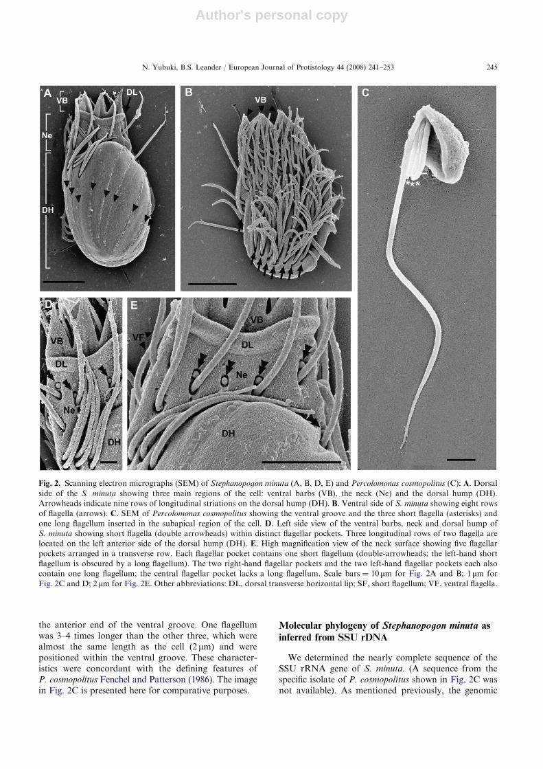
The image of Percolomonas cosmopolitus shown inthis study was taken from organisms collected from thesediment in Tokyo Bay, Japan on May 28, 2004.
Light and electron microscopy
Light microscopy was performed using a ZeissAxioplan 2 imaging microscope and a Leica DC500digital chilled CCD camera. Light micrographs weretaken of cells fixed in seawater mixed with an equalvolume of 2% glutaraldehyde (GA).
For scanning electron microscopy (SEM), cells ofS. minuta were mixed with an equal volume of fixativecontaining 2% GA in 0.1M sodium cacodylate buffer(SCB) (pH 7.2) and mounted on cover glasses coatedwith poly-L-lysine at room temperature for 1 h. Thecover glasses were rinsed with 0.1M SCB and fixed in1% OsO4 for 30min. The fixed cells were then rinsedwith 0.1M SCB and dehydrated with a graded ethanol
ARTICLE IN PRESSN. Yubuki, B.S. Leander / European Journal of Protistology 44 (2008) 241–253242
Author's personal copy
series from 30% to absolute ethanol. Samples werecritical point dried with CO2 using a Tousimis CriticalPoint Dryer. Samples were then coated with gold using aCressington 208HR high Resolution Sputter Coater,and observed with a Hitachi S-4700 field emissionscanning electron microscope.
For SEM of P. cosmopolitus, cells were mixed with anequal volume of fixative, containing 2.5% GA in 0.1MSCB (pH 7.2), and were mounted on cover glasses coatedby poly-L-lysine at room temperature for 2h. The coverglasses were rinsed with 0.2M SCB and fixed in 1% OsO4
for 30min. These were then rinsed with 0.2M SCB anddehydrated with a graded ethanol series from 30% toabsolute ethanol. Ethanol was replaced by dehydrated t-butanol before samples were freeze-dried with a freeze drierVFD-21S (Shinku-Device, Japan). Samples were coatedwith platinum/palladium with an E102 ion-sputter coater(Hitachi, Japan), and observed with a JSM-6330F fieldemission scanning electron microscope (JEOL, Japan).
For transmission electron microscopy (TEM), cellsuspensions were mixed with 4% (v/v) GA in 0.2M SCB(pH 7.2) at room temperature for 1 h. Cells wereaggregated into a pellet by centrifugation at 1000g for5min and then rinsed with 0.2M SCB (pH 7.2). Thepellet of cells was embedded in 2% agarose (Type IX:ultra-low gelling temperature). The specimens were thenfixed in 1% (w/v) osmium tetroxide in 0.2M SCB (pH7.2) at room temperature for 1 h followed by dehydra-tion through an ethanol series, and substitution withacetone. The specimens were embedded in resin (Epon812). Ultrathin sections were stained with 2% (w/v)uranyl acetate and lead citrate (Reynolds 1963), andobserved using a Hitachi H7600 electron microscope.
DNA extraction, PCR amplification, alignment and
phylogenetic analysis
Thirty individual cells of S. minuta were isolated fromthe two-eukaryote culture and washed twice in sterilizedf/2 medium. Genomic DNA was extracted from theseisolated cells using MasterPure Complete DNA andRNA purification Kit (Epicentre, WI, USA). Thepolymerase chain reaction (PCR) was performed usinga total volume of 25 ml and the PuRe Taq Ready-To-GoPCR beads kit (GE Healthcare, Buckinghamshire, UK).Nearly the entire SSU rRNA gene was amplifiedfrom genomic DNA using eukaryotic universal primers(PF1: 50-GCGCTACCTGGTTGATCCTGCCAGT-30
and R4: 50-GATCCTTCTGCAGGTTCACCTAC-30).The PCR protocol had an initial denaturation stage at95 1C for 2min; 35 cycles involving 94 1C for 45 s(denaturation), 55 1C for 45 s (annealing), and 72 1C for1.5min (extension); and final extension at 72 1C for5min. The amplified DNA fragments were purified fromagarose gels using UltraClean 15 DNA Purification Kit
(MO Bio, CA, USA), and then cloned into the TOPOTA Cloning Kit (Invitrogen, CA, USA). The S. minuta
sequence was deposited in DDBJ/EMBL/GenBankunder the accession number AB365646.
The SSU rRNA sequence of S. minuta was manuallyaligned with taxa representing all of the major groups ofeukaryotes, forming a 39-taxon alignment with 1016unambiguously aligned positions. PhyML (Guindon andGascuel 2003) was used to analyze this alignment withmaximum-likelihood (ML) using a general-time reversible(GTR) model of base substitutions (Rodriguez et al. 1990)incorporating invariable sites and a discrete gammadistribution (eight categories) (GTR+I+G model).Model parameters were estimated from the originaldataset. ML bootstrap analysis (100 replicates) wasconducted with the same settings described above.
In order to more comprehensively evaluate thephylogenetic position of S. minuta within the Hetero-lobosea, we analyzed an 18-taxon alignment of mostlyheterolobosean sequences and 1039 unambiguouslyaligned positions. The ML phylogenetic analyses de-scribed above were repeated on this data set.
Both the 39- and 18-taxon data sets were also analyzedwith Bayesian methods using the MrBayes program(Huelsenbeck and Ronquist 2001). The program was setto operate with a gamma distribution and four Monte–Carlo–Markov chains (MCMC) starting from a randomtree. A total of 2,000,000 generations were calculated withtrees sampled every 50 generations and with a prior burn-in of 100,000 generations (2000 sampled trees werediscarded). A majority rule consensus tree was constructedfrom 38,000 post-burn-in trees. Posterior probabilitiescorrespond to the frequency at which a given node wasfound in the post-burn-in trees.
Results
General morphology and ultrastructure of
Stephanopogon minuta
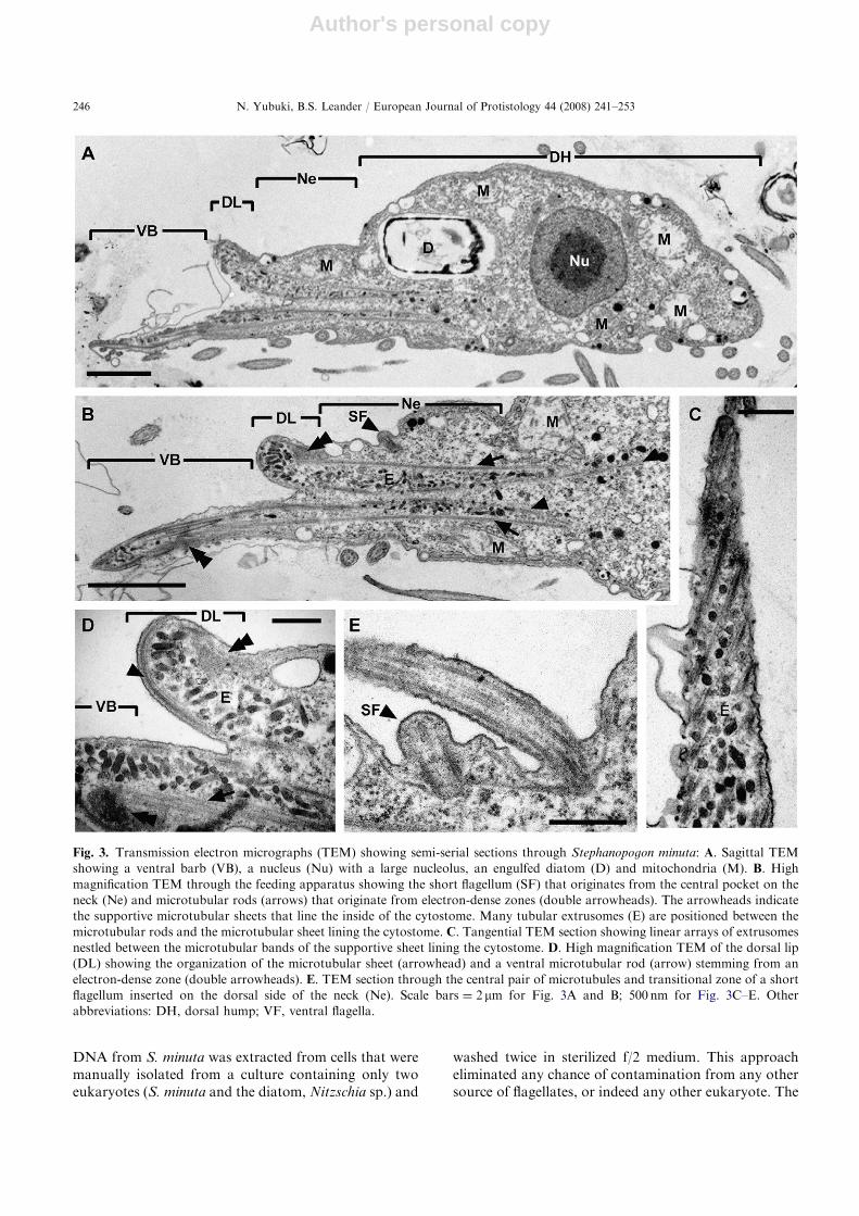
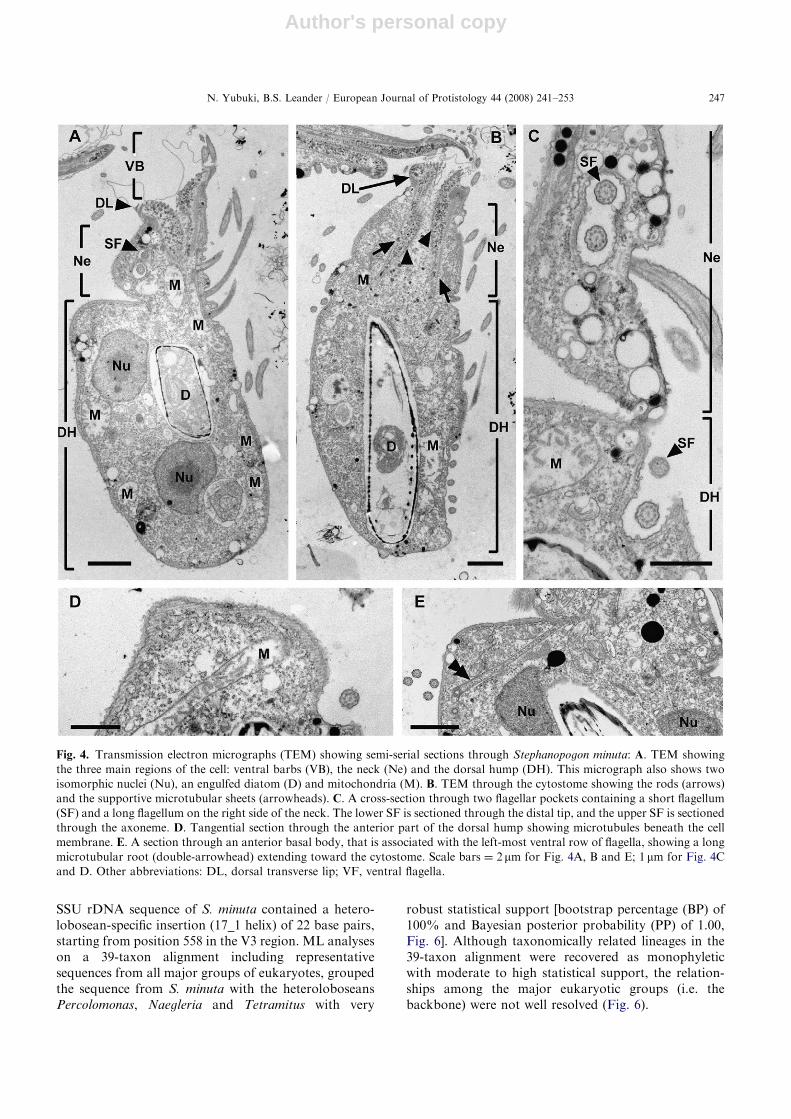
S. minuta was 26.5 mm (22.5–30 mm) long and 12.3 mm(10.3–16.5 mm) wide. The cell was vase-shape and curvedtoward the right when observed from the dorsal side(Fig. 1). The cell was divided into three main regions,from anterior to posterior: ventral barbs (VB), a neck(Ne) and a dorsal hump (DH) (Figs 2A, D–E, 3A, B and4A–C). S. minuta was a eukaryovore that preyed ondiatoms in our culture (Figs 1C, 3A and 4A, B) using apronounced feeding apparatus formed by three barbsand a dorsal transverse lip (DL) (Figs 1 and 2A). TheVBs and the dorsal lip defined a slit-shaped cytostome(Figs 2A, E and 3A, B). The cells were capable ofcrawling forwards and backwards using an array offlagella on the ventral surface (Figs 2B and 5A) and
ARTICLE IN PRESSN. Yubuki, B.S. Leander / European Journal of Protistology 44 (2008) 241–253 243
Author's personal copy
tended to turn to the right (clockwise) along thesubstrate. The cells were also able to swim in the watercolumn in a backwards direction when the culture vesselwas disturbed. Cysts attached to the substrate of theculture vessel were common (data not shown), and S.
minuta reproduced by asexual division within the restingcysts. Other possible stages in the life cycle (e.g. sexualreproduction) were not evident.
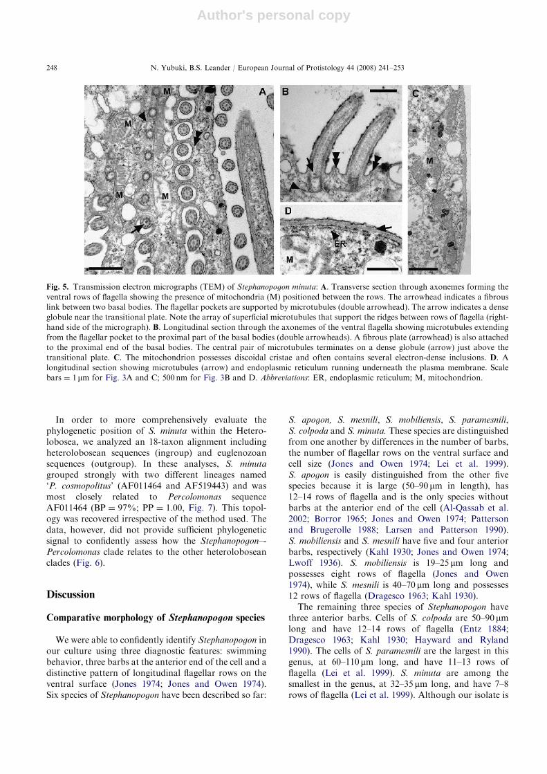
Two isomorphic nuclei, each with a central nucleolus,were present within the DH (Figs 3A, 4A and E).Endoplasmic reticulum (ER) was positioned beneath alayer of superficial microtubules (Fig. 5D). The mito-chondria of S. minuta contained discoidal cristae andoften contained several densely stained inclusions withinthe matrix (Fig. 5C). Mitochondria were distributedthroughout the cell (Figs 3A, B, 4A, B) and wereparticularly abundant between the ventral rows offlagella (Fig. 5A). The mitochondria were not sur-rounded by ER (Fig. 4B), and canonical Golgi bodieswere not observed. The feeding apparatus was sup-ported by a pair of microtubular bundles, or rods, thatextended posteriorly from electron dense zones presentwithin the dorsal lip and at the base of the VBs(Figs 3A–D and 4B). Moreover, the inside of thecytostome was lined by a supportive microtubular sheetthat extended anteriorly before turning posteriorly alongthe curve formed by the dorsal lip (Fig. 3D). Anaccumulation of tubular extrusomes was positionedbetween the microtubular rods and were arranged inlinear rows between the microtubules of the supportivesheet (Figs 3A–D and 4B).
Organization of the flagella in Stephanopogonminuta
Five flagellar pockets were arranged in a transverse rowon the neck just behind the dorsal lip (Fig. 2A and E). Onelong and one short flagellum emerged from four of the fivepockets; the central pocket lacked a long flagellum and
only contained a short flagellum (Figs 2A, D, E, 3B, E and4C). In addition, both a long and a short flagellum insertedat the right-hand side of the neck (Figs 2E and 4C), andonly one long flagellum inserted at the left-hand side of theneck (Fig. 2A, D, E). The left-hand side of the DHcontained three pairs (or clusters) of long flagella, however,some cells possessed three flagella in the right-hand cluster(Fig. 2A and D). Therefore, a total of 12–13 long flagellawere visible on the dorsal surface of S. minuta (Fig. 2A).The surface of the DH also contained 8–9 faint long-itudinal striations that were each about 2–3mm wide(Fig. 2A). The cell membrane of the DH was supported bymicrotubules (Figs 4D and 5D). The ventral surface ofS. minuta was concave and contained eight longitudinalgrooves, each supporting 4–22 flagella (Fig. 2B). Micro-tubules supported the ridges that were positioned betweenthe flagellar grooves on the ventral side of the cells(Fig. 5A). The total number of flagella on the ventral sidewas variable and ranged from 100 to 132. Therefore, thetotal number of flagella on each cell of S. minuta rangedfrom 112 to 144.
The basal bodies were characteristically short atapproximately 200 nm in length (Figs 3E and 5B). Theproximal end of each basal body was associated with afibrous sheet and microtubules that supported the wallof the flagellar depression (Figs 3E and 5A, B). Theanterior-most basal body in each longitudinal row ofventral flagella had a different configuration of micro-tubules that extended toward the cytostome region(Fig. 4E). The central pair of axonemal microtubulesterminated at their proximal ends on an electron denseglobule positioned above the transitional plate (Fig. 5A,B). Flagellar hairs (mastigonemes) were absent.
General morphology of Percolomonas
P. cosmopolitus was semi-spherical in shape andpossessed a conspicuous ventral groove (Fig. 2C). Fourflagella emerged from the subapical region of the cell, at
ARTICLE IN PRESS
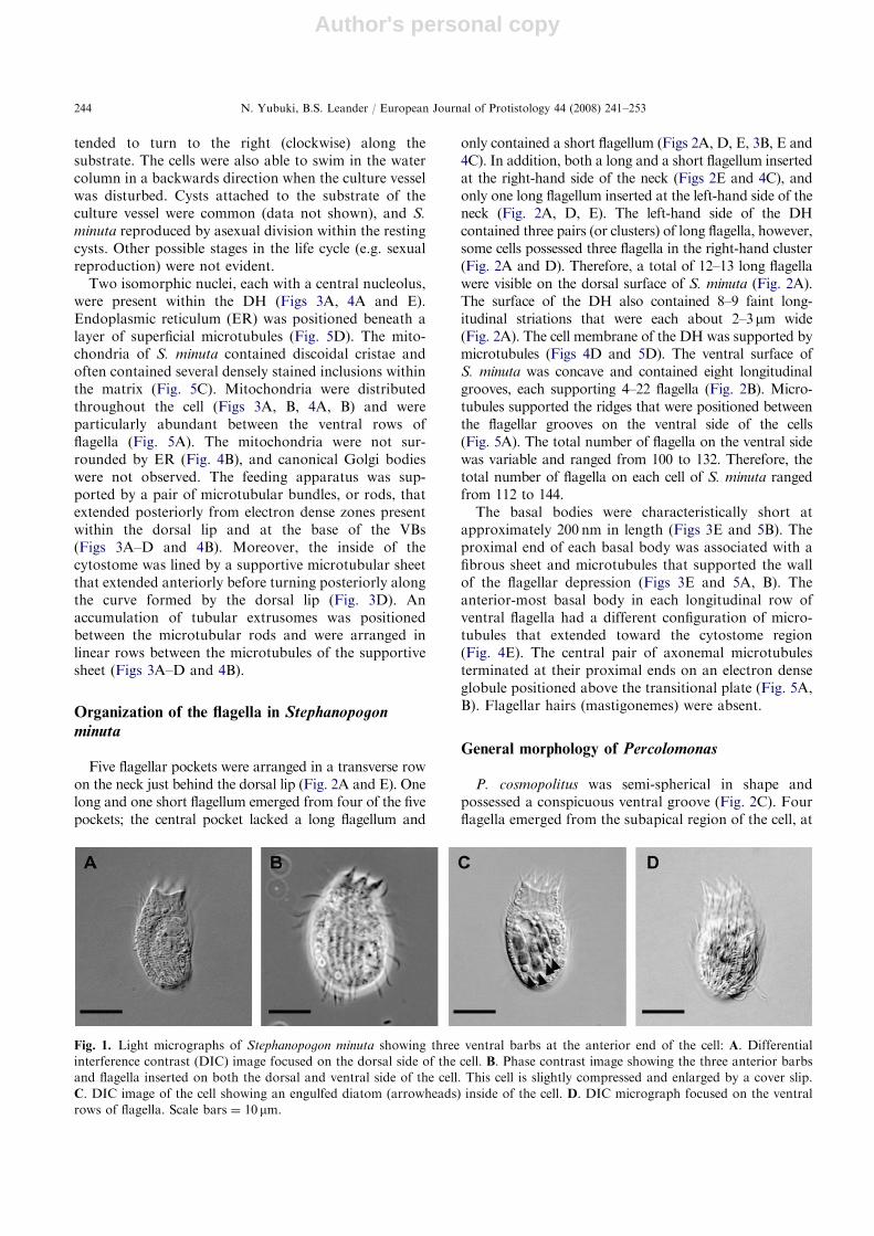
Fig. 1. Light micrographs of Stephanopogon minuta showing three ventral barbs at the anterior end of the cell: A. Differential
interference contrast (DIC) image focused on the dorsal side of the cell. B. Phase contrast image showing the three anterior barbs
and flagella inserted on both the dorsal and ventral side of the cell. This cell is slightly compressed and enlarged by a cover slip.
C. DIC image of the cell showing an engulfed diatom (arrowheads) inside of the cell. D. DIC micrograph focused on the ventral
rows of flagella. Scale bars ¼ 10 mm.
N. Yubuki, B.S. Leander / European Journal of Protistology 44 (2008) 241–253244
Author's personal copy
the anterior end of the ventral groove. One flagellumwas 3–4 times longer than the other three, which werealmost the same length as the cell (2 mm) and werepositioned within the ventral groove. These character-istics were concordant with the defining features ofP. cosmopolitus Fenchel and Patterson (1986). The imagein Fig. 2C is presented here for comparative purposes.
Molecular phylogeny of Stephanopogon minuta as
inferred from SSU rDNA
We determined the nearly complete sequence of theSSU rRNA gene of S. minuta. (A sequence from thespecific isolate of P. cosmopolitus shown in Fig. 2C wasnot available). As mentioned previously, the genomic
ARTICLE IN PRESS
Fig. 2. Scanning electron micrographs (SEM) of Stephanopogon minuta (A, B, D, E) and Percolomonas cosmopolitus (C): A. Dorsal
side of the S. minuta showing three main regions of the cell: ventral barbs (VB), the neck (Ne) and the dorsal hump (DH).
Arrowheads indicate nine rows of longitudinal striations on the dorsal hump (DH). B. Ventral side of S. minuta showing eight rows
of flagella (arrows). C. SEM of Percolomonas cosmopolitus showing the ventral groove and the three short flagella (asterisks) and
one long flagellum inserted in the subapical region of the cell. D. Left side view of the ventral barbs, neck and dorsal hump of
S. minuta showing short flagella (double arrowheads) within distinct flagellar pockets. Three longitudinal rows of two flagella are
located on the left anterior side of the dorsal hump (DH). E. High magnification view of the neck surface showing five flagellar
pockets arranged in a transverse row. Each flagellar pocket contains one short flagellum (double-arrowheads; the left-hand short
flagellum is obscured by a long flagellum). The two right-hand flagellar pockets and the two left-hand flagellar pockets each also
contain one long flagellum; the central flagellar pocket lacks a long flagellum. Scale bars ¼ 10mm for Fig. 2A and B; 1mm for
Fig. 2C and D; 2mm for Fig. 2E. Other abbreviations: DL, dorsal transverse horizontal lip; SF, short flagellum; VF, ventral flagella.
N. Yubuki, B.S. Leander / European Journal of Protistology 44 (2008) 241–253 245
Author's personal copy
DNA from S. minuta was extracted from cells that weremanually isolated from a culture containing only twoeukaryotes (S. minuta and the diatom, Nitzschia sp.) and
washed twice in sterilized f/2 medium. This approacheliminated any chance of contamination from any othersource of flagellates, or indeed any other eukaryote. The
ARTICLE IN PRESS
Fig. 3. Transmission electron micrographs (TEM) showing semi-serial sections through Stephanopogon minuta: A. Sagittal TEM
showing a ventral barb (VB), a nucleus (Nu) with a large nucleolus, an engulfed diatom (D) and mitochondria (M). B. High
magnification TEM through the feeding apparatus showing the short flagellum (SF) that originates from the central pocket on the
neck (Ne) and microtubular rods (arrows) that originate from electron-dense zones (double arrowheads). The arrowheads indicate
the supportive microtubular sheets that line the inside of the cytostome. Many tubular extrusomes (E) are positioned between the
microtubular rods and the microtubular sheet lining the cytostome. C. Tangential TEM section showing linear arrays of extrusomes
nestled between the microtubular bands of the supportive sheet lining the cytostome. D. High magnification TEM of the dorsal lip
(DL) showing the organization of the microtubular sheet (arrowhead) and a ventral microtubular rod (arrow) stemming from an
electron-dense zone (double arrowheads). E. TEM section through the central pair of microtubules and transitional zone of a short
flagellum inserted on the dorsal side of the neck (Ne). Scale bars ¼ 2mm for Fig. 3A and B; 500 nm for Fig. 3C–E. Other
abbreviations: DH, dorsal hump; VF, ventral flagella.
N. Yubuki, B.S. Leander / European Journal of Protistology 44 (2008) 241–253246
Author's personal copy
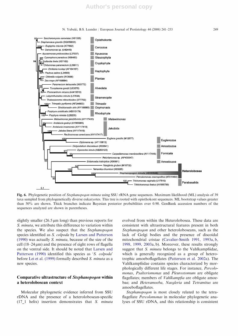
SSU rDNA sequence of S. minuta contained a hetero-lobosean-specific insertion (17_1 helix) of 22 base pairs,starting from position 558 in the V3 region. ML analyseson a 39-taxon alignment including representativesequences from all major groups of eukaryotes, groupedthe sequence from S. minuta with the heteroloboseansPercolomonas, Naegleria and Tetramitus with very
robust statistical support [bootstrap percentage (BP) of100% and Bayesian posterior probability (PP) of 1.00,Fig. 6]. Although taxonomically related lineages in the39-taxon alignment were recovered as monophyleticwith moderate to high statistical support, the relation-ships among the major eukaryotic groups (i.e. thebackbone) were not well resolved (Fig. 6).
ARTICLE IN PRESS
Fig. 4. Transmission electron micrographs (TEM) showing semi-serial sections through Stephanopogon minuta: A. TEM showing
the three main regions of the cell: ventral barbs (VB), the neck (Ne) and the dorsal hump (DH). This micrograph also shows two
isomorphic nuclei (Nu), an engulfed diatom (D) and mitochondria (M). B. TEM through the cytostome showing the rods (arrows)
and the supportive microtubular sheets (arrowheads). C. A cross-section through two flagellar pockets containing a short flagellum
(SF) and a long flagellum on the right side of the neck. The lower SF is sectioned through the distal tip, and the upper SF is sectioned
through the axoneme. D. Tangential section through the anterior part of the dorsal hump showing microtubules beneath the cell
membrane. E. A section through an anterior basal body, that is associated with the left-most ventral row of flagella, showing a long
microtubular root (double-arrowhead) extending toward the cytostome. Scale bars ¼ 2mm for Fig. 4A, B and E; 1 mm for Fig. 4C
and D. Other abbreviations: DL, dorsal transverse lip; VF, ventral flagella.
N. Yubuki, B.S. Leander / European Journal of Protistology 44 (2008) 241–253 247
Author's personal copy
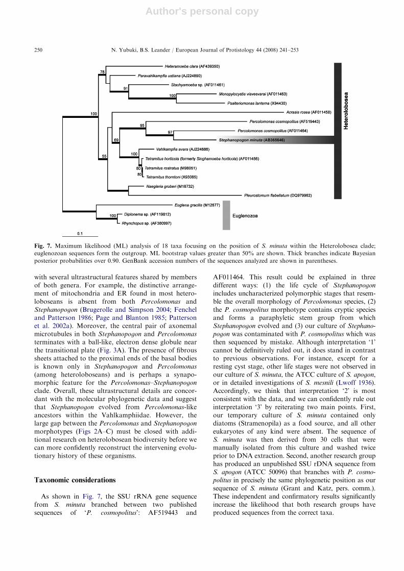
In order to more comprehensively evaluate thephylogenetic position of S. minuta within the Hetero-lobosea, we analyzed an 18-taxon alignment includingheterolobosean sequences (ingroup) and euglenozoansequences (outgroup). In these analyses, S. minuta
grouped strongly with two different lineages named‘P. cosmopolitus’ (AF011464 and AF519443) and wasmost closely related to Percolomonas sequenceAF011464 (BP ¼ 97%; PP ¼ 1.00, Fig. 7). This topol-ogy was recovered irrespective of the method used. Thedata, however, did not provide sufficient phylogeneticsignal to confidently assess how the Stephanopogon–-Percolomonas clade relates to the other heteroloboseanclades (Fig. 6).
Discussion
Comparative morphology of Stephanopogon species
We were able to confidently identify Stephanopogon inour culture using three diagnostic features: swimmingbehavior, three barbs at the anterior end of the cell and adistinctive pattern of longitudinal flagellar rows on theventral surface (Jones 1974; Jones and Owen 1974).Six species of Stephanopogon have been described so far:
S. apogon, S. mesnili, S. mobiliensis, S. paramesnili,S. colpoda and S. minuta. These species are distinguishedfrom one another by differences in the number of barbs,the number of flagellar rows on the ventral surface andcell size (Jones and Owen 1974; Lei et al. 1999).S. apogon is easily distinguished from the other fivespecies because it is large (50–90 mm in length), has12–14 rows of flagella and is the only species withoutbarbs at the anterior end of the cell (Al-Qassab et al.2002; Borror 1965; Jones and Owen 1974; Pattersonand Brugerolle 1988; Larsen and Patterson 1990).S. mobiliensis and S. mesnili have five and four anteriorbarbs, respectively (Kahl 1930; Jones and Owen 1974;Lwoff 1936). S. mobiliensis is 19–25 mm long andpossesses eight rows of flagella (Jones and Owen1974), while S. mesnili is 40–70 mm long and possesses12 rows of flagella (Dragesco 1963; Kahl 1930).
The remaining three species of Stephanopogon havethree anterior barbs. Cells of S. colpoda are 50–90 mmlong and have 12–14 rows of flagella (Entz 1884;Dragesco 1963; Kahl 1930; Hayward and Ryland1990). The cells of S. paramesnili are the largest in thisgenus, at 60–110 mm long, and have 11–13 rows offlagella (Lei et al. 1999). S. minuta are among thesmallest in the genus, at 32–35 mm long, and have 7–8rows of flagella (Lei et al. 1999). Although our isolate is
ARTICLE IN PRESS
Fig. 5. Transmission electron micrographs (TEM) of Stephanopogon minuta: A. Transverse section through axonemes forming the
ventral rows of flagella showing the presence of mitochondria (M) positioned between the rows. The arrowhead indicates a fibrous
link between two basal bodies. The flagellar pockets are supported by microtubules (double arrowhead). The arrow indicates a dense
globule near the transitional plate. Note the array of superficial microtubules that support the ridges between rows of flagella (right-
hand side of the micrograph). B. Longitudinal section through the axonemes of the ventral flagella showing microtubules extending
from the flagellar pocket to the proximal part of the basal bodies (double arrowheads). A fibrous plate (arrowhead) is also attached
to the proximal end of the basal bodies. The central pair of microtubules terminates on a dense globule (arrow) just above the
transitional plate. C. The mitochondrion possesses discoidal cristae and often contains several electron-dense inclusions. D. A
longitudinal section showing microtubules (arrow) and endoplasmic reticulum running underneath the plasma membrane. Scale
bars ¼ 1mm for Fig. 3A and C; 500 nm for Fig. 3B and D. Abbreviations: ER, endoplasmic reticulum; M, mitochondrion.
N. Yubuki, B.S. Leander / European Journal of Protistology 44 (2008) 241–253248
Author's personal copy
slightly smaller (26.5 mm long) than previous reports forS. minuta, we attribute this difference to variation withinthe species. We also suspect that the Stephanopogon
species identified as S. colpoda by Larsen and Patterson(1990) was actually S. minuta, because of the size of thecell (18–24 mm) and the presence of eight rows of flagellaon the ventral side. It should be noted that Larsen andPatterson (1990) identified this species as ‘S. colpoda’before Lei et al. (1999) formally described S. minuta as anew species.
Comparative ultrastructure of Stephanopogon within
a heterolobosean context
Molecular phylogenetic evidence inferred from SSUrDNA and the presence of a heterolobosean-specific(17_1 helix) insertion demonstrates that S. minuta
evolved from within the Heterolobosea. These data areconsistent with ultrastructural features present in bothStephanopogon and other heteroloboseans, such as thelack of Golgi bodies and the presence of discoidalmitochondrial cristae (Cavalier-Smith 1991, 1993a, b,1998, 1999, 2003a, b). Moreover, these results stronglysuggest that S. minuta belongs to the Vahlkampfiidae,which is generally recognized as a group of hetero-trophic amoeboflagellates (Patterson et al. 2002a). TheVahlkampfiidae contains species characterized by mor-phologically different life stages. For instance, Percolo-
monas, Psalteriomonas and Pleurostomum are obligateflagellates; members of Vahlkampfia are obligate amoe-bae; and Heteramoeba, Naegleria and Tetramitus areamoeboflagellates.
Stephanopogon is most closely related to the tetra-flagellate Percolomonas in molecular phylogenetic ana-lyses of SSU rDNA, and this relationship is consistent
ARTICLE IN PRESS
Fig. 6. Phylogenetic position of Stephanopogon minuta using SSU rRNA gene sequences. Maximum likelihood (ML) analysis of 39
taxa sampled from phylogenetically diverse eukaryotes. This tree is rooted with opisthokont sequences. ML bootstrap values greater
than 50% are shown. Thick branches indicate Bayesian posterior probabilities over 0.90. GenBank accession numbers of the
sequences analyzed are shown in parentheses.
N. Yubuki, B.S. Leander / European Journal of Protistology 44 (2008) 241–253 249
Author's personal copy
with several ultrastructural features shared by membersof both genera. For example, the distinctive arrange-ment of mitochondria and ER found in most hetero-loboseans is absent from both Percolomonas andStephanopogon (Brugerolle and Simpson 2004; Fencheland Patterson 1986; Page and Blanton 1985; Pattersonet al. 2002a). Moreover, the central pair of axonemalmicrotubules in both Stephanopogon and Percolomonas
terminates with a ball-like, electron dense globule nearthe transitional plate (Fig. 3A). The presence of fibroussheets attached to the proximal ends of the basal bodiesis known only in Stephanopogon and Percolomonas
(among heteroloboseans) and is perhaps a synapo-morphic feature for the Percolomonas–Stephanopogon
clade. Overall, these ultrastructural details are concor-dant with the molecular phylogenetic data and suggestthat Stephanopogon evolved from Percolomonas-likeancestors within the Vahlkamphiidae. However, thelarge gap between the Percolomonas and Stephanopogon
morphotypes (Figs 2A–C) must be closed with addi-tional research on heterolobosean biodiversity before wecan more confidently reconstruct the intervening evolu-tionary history of these organisms.
Taxonomic considerations
As shown in Fig. 7, the SSU rRNA gene sequencefrom S. minuta branched between two publishedsequences of ‘P. cosmopolitus’: AF519443 and
AF011464. This result could be explained in threedifferent ways: (1) the life cycle of Stephanopogon
includes uncharacterized polymorphic stages that resem-ble the overall morphology of Percolomonas species, (2)the P. cosmopolitus morphotype contains cryptic speciesand forms a paraphyletic stem group from whichStephanopogon evolved and (3) our culture of Stephano-
pogon was contaminated with P. cosmopolitus which wasthen sequenced by mistake. Although interpretation ‘1’cannot be definitively ruled out, it does stand in contrastto previous observations. For instance, except for aresting cyst stage, other life stages were not observed inour culture of S. minuta, the ATCC culture of S. apogon,or in detailed investigations of S. mesnili (Lwoff 1936).Accordingly, we think that interpretation ‘2’ is mostconsistent with the data, and we can confidently rule outinterpretation ‘3’ by reiterating two main points. First,our temporary culture of S. minuta contained onlydiatoms (Stramenopila) as a food source, and all othereukaryotes of any kind were absent. The sequence ofS. minuta was then derived from 30 cells that weremanually isolated from this culture and washed twiceprior to DNA extraction. Second, another research grouphas produced an unpublished SSU rDNA sequence fromS. apogon (ATCC 50096) that branches with P. cosmo-
politus in precisely the same phylogenetic position as oursequence of S. minuta (Grant and Katz, pers. comm.).These independent and confirmatory results significantlyincrease the likelihood that both research groups haveproduced sequences from the correct taxa.
ARTICLE IN PRESS
Fig. 7. Maximum likelihood (ML) analysis of 18 taxa focusing on the position of S. minuta within the Heterolobosea clade;
euglenozoan sequences form the outgroup. ML bootstrap values greater than 50% are shown. Thick branches indicate Bayesian
posterior probabilities over 0.90. GenBank accession numbers of the sequences analyzed are shown in parentheses.
N. Yubuki, B.S. Leander / European Journal of Protistology 44 (2008) 241–253250
Author's personal copy
Therefore, because the two sequences from P. cosmo-
politus do not form a clade exclusive of Stephanopogon,they almost certainly represent cryptic species. The longbranch-lengths that separate the two sequences ofP. cosmopolitus also indicate that they represent twovery different lineages. Because it would not be reason-able or informative to re-classify Stephanopogon withinthe genus Percolomonas, the name of one of the twoP. cosmopolitus sequences should eventually be changed.Resolution of this taxonomic issue, however, requiresfurther molecular phylogenetic and ultrastructuralstudies of both Stephanopogon species and Percolomonas
species, especially the organism from which sequenceAF011464 was derived.
Convergent evolution of Stephanopogon and the
multiflagellated state
Stephanopogon was originally described as a memberof the Ciliophora, because, superficially, members ofboth groups look very similar to one another, behavesimilarly and tend to occupy the same habitats (Entz1884). Other microeukaryotes with many flagella ar-ranged in distinct rows, like opalinids, were also initiallyclassified within the Ciliophora for essentially the samereasons (Metcalf 1923, 1940). Opalinids, however, areendocommensals that live within the hindguts ofamphibians and squamates. Molecular phylogeneticstudies and detailed utrastructural data have demon-strated that opalinids have evolved from within thestramenopiles and are closely related to biflagellatesknown as proteromonads (Cavalier-Smith 1997; Patter-son 1985, 1989; Silberman et al. 1996). Although thegeneral appearance of the opalinid flagellar apparatusdiffers from that of most stramenopiles, both groupsshare some key flagellar features, such as a transitionalhelix located just above the flagellar transition zone(Patterson 1985, 1989).
It should also be emphasized that multiflagellatedorganisms have also been described in several othereukaryotic ‘supergroups’. For instance, the multiflagel-lated amoeba Multicilia has been shown to be a memberof the Amoebozoa (Mikrjukov and Mylnikov 1998;Nikolaev et al. 2006); hypermastigotes and devescov-inids are members of the Parabasalia (Brugerolle andMuller 2000; Carpenter and Keeling 2007); and themultiflagellated (and multicellular) life history stages ofporiferans and metazoans (e.g. planulae, trochophores,Trichoplax and dicyemids) are members of the Opistho-konta. The distance between the phylogenetic positionsof all of these lineages demonstrates that a propulsiveapparatus consisting of distinct rows of flagella acrossthe cell surface evolved several times independentlyacross the tree of eukaryotes. In this paper, we haveshown that Stephanopogon is the only known lineage
within the Heterolobosea to have converged in overallmorphology with ciliates, opalinids and the otherlineages mentioned above. Moreover, the dinuclear(isomorphic) condition in S. minuta has also convergedwith the dinuclear (heteromorphic) condition found inmost ciliates. Nonetheless, adaptive explanations for thedistinctive organization of the flagella on S. minuta (e.g.the short flagella on the neck and the three rows offlagella on the DH) remain enigmatic and awaitexperimental studies focused on behavioral and func-tional analyses.
Acknowledgments
This work was supported by grants from the TulaFoundation (Centre for Microbial Diversity and Evolu-tion), the National Science and Engineering ResearchCouncil of Canada (NSERC 283091-04) and theCanadian Institute for Advanced Research, Programin Integrated Microbial Biodiversity. We would like tothank Jessica Grant and Dr. Laura Katz (Smith College,USA) for sharing their unpublished molecular phyloge-netic analyses of S. apogon (ATCC50096), and Dr. S.Agatha (University of Salzburg, Austria) for help inacquiring relevant literature. N.Y. would also like toexpress thanks to Dr. Isao Inouye (University ofTsukuba, Japan) for supervising the SEM work onP. cosmopolitus.
References
Adl, S.M., Simpson, A.G.B., Farmer, M.A., Andersen, R.A.,
Anderson, O.R., Barta, J.R., Bowser, S.S., Brugerolle, G.,
Fensome, R.A., Fredericq, S., James, T.Y., Karpov, S.,
Kugrens, P., Krug, J., Lane, C.E., Lewis, L.A., Lodge, J.,
Lynn, D.H., Mann, D.G., McCourt, R.M., Mendoza, L.,
Moestrup, Ø., Mozley-Standridge, S.E., Nerad, T.A.,
Shearer, C.A., Smirnov, A.V., Spiegel, F.W., Taylor,
M.F., 2005. The new higher level classification of eukar-
yotes with emphasis on the taxonomy of protists.
J. Eukaryot. Microbiol. 52, 399–459.
Alongi, D.M., 1991. Flagellates of benthic communities:
characteristics and methods of study. In: Patterson, D.J.,
Larsen, J. (Eds.), The Biology of Free-living Heterotrophic
Flagellates. Clarendon Press, Oxford, pp. 57–75.
Al-Qassab, S., Lee, W.J., Murray, S., Simpson, A.G.B.,
Patterson, D.J., 2002. Flagellates from stromatolites and
surrounding sediments in Shark Bay, Western Australia.
Acta Protozool. 41, 91–144.
Borror, A.C., 1965. New and little-known tidal marsh ciliates.
Trans. Am. Microsc. Soc. 84, 550–565.
Brugerolle, G., Muller, M., 2000. Amitochondriate flagellates.
In: Leadbeater, B.S.C., Green, J.C. (Eds.), The Flagellates.
Unity, Diversity and Evolution. Taylor & Francis, London,
New York, pp. 166–189.
ARTICLE IN PRESSN. Yubuki, B.S. Leander / European Journal of Protistology 44 (2008) 241–253 251
Author's personal copy
Brugerolle, G., Simpson, A.G.B., 2004. The flagellar apparatus
of heteroloboseans. J. Eukaryot. Microbiol. 51, 96–107.
Carpenter, K.J., Keeling, P.J., 2007. Morphology and
phylogenetic position of Eucomonympha imla (Parabasalia:
Hypermastiga). J. Eukaryot. Microbiol. 54, 325–332.
Cavalier-Smith, T., 1991. Cell diversification in heterotrophic
flagellates. In: Patterson, D.J., Larsen, J. (Eds.), The
Biology of Free-living Heterotrophic Flagellates. Claren-
don Press, Oxford, pp. 113–131.
Cavalier-Smith, T., 1993a. Kingdom protozoa and its 18
phyla. Microbiol. Rev. 57, 953–994.
Cavalier-Smith, T., 1993b. Percolozoa and the symbiotic
origin of the metakaryotic cell. In: Ishikawa, H., Ishida,
M., Sato, S. (Eds.), Endocytobiology V. University Press,
Tubingen, pp. 399–406.
Cavalier-Smith, T., 1997. Sagenista and Bigyra, two phyla of
heterotrophic heterokont chromists. Arch. Protistenkd.
148, 253–267.
Cavalier-Smith, T., 2002. The phagotrophic origin of eukar-
yotes and phylogenetic classification of Protozoa. Int. J.
Syst. Evol. Microbiol. 52, 297–354.
Cavalier-Smith, T., 2003. The excavate protozoan phyla
Metamonada Grasse emend (Anaeromonadea, Parabasa-
lia, Carpediemonas, Eopharyngia) and Loukozoa emend
(Jakobea, Malawimonas): their evolutionary affinities and
new higher taxa. Int. J. Syst. Evol. Microbiol. 53,
1741–1758.
Corliss, J.O., 1979. The Ciliated Protozoa. Characterization,
Classification and Guide to the Literature. Pergamon Press,
Oxford.
Corliss, J.O., 1984. The kingdom Protista and its 45 phyla.
Biosystems 17, 87–126.
Dragesco, J., 1963. Complements a la connaissance des cilie
mesopsammiques de Roscoff. I. Holotriches. Cah. Biol.
Mar. 4, 91–119.
Entz, G., 1884. Uber Infusorien des Golfes von Neapel. Mitt.
Zoologisch. Station zu Neapel 5, 289–444.
Fenchel, T., Patterson, D.J., 1986. Percolomonas cosmopolitus
(Ruinen) n. gen., a new type of filter feeding flagellate from
marine plankton. J. Mar. Biol. Assoc. UK 66, 465–482.
Guindon, S., Gascuel, O., 2003. Simple, fast, and accurate
algorithm to estimate large phylogenies by maximum
likelihood. Syst. Biol. 52, 696–704.
Hausmann, K., Hulsmann, N., Radek, R., 2003. Protistology,
third ed. E. Schweizerbart’sche Verlagsbuchhandlung,
Stuttgart.
Hayward, P.J., Ryland, J.S., 1990. The Marine Fauna of the
British Isles and North-West Europe, vol. 1. Clarendon
Press, Oxford.
Hibberd, D.J., 1983. Ultrastracture of the colonial colourless
zooflagellates Phalansterium digitatum Stein (Phalansteriida
ord. nov.) and Spongomonas ucella Stein (Spongomonadida
ord. nov.). Protistologica 19, 523–535.
Huelsenbeck, J.P., Ronquist, F., 2001. MrBayes: Bayesian
inference of phylogenetic trees. Bioinformatics 17, 754–755.
Jones, E.E., 1974. The protozoa of Mobile Bay, Alabama.
University of South Alabama Monographs, vol. 1,
pp. 1–113.
Jones, E.E., Owen, G., 1974. New species of protozoa from
Mobile Bay, Alabama. J. Mar. Sci. Alabama 2, 41–56.
Kahl, A., 1930. Wimpertiere oder Ciliata (Infsoria). In: Dahl,
F. (Ed.), Die Tierwelt Deutschlands. Gustav Fischer, Jena,
Pts. 18, 21, 25, 30.
Keeling, P.J., Burger, G., Durnford, D.G., Lang, B.F., Lee,
R.W., Pearlman, R.E., Roger, A.J., Gray, M.W., 2005. The
tree of eukaryotes. Trends Ecol. Evol. 20, 670–676.
Larsen, J., Patterson, D.J., 1990. Some flagellates (Protista)
from tropical marine sediments. J. Nat. Hist. 24, 801–937.
Lei, Y., Xu, K., Song, W., 1999. Two new species,
Stephanopogon minuta n. sp. and S. paramesnili n. sp. from
mariculture waters. J. Fish. Sci. China 6, 25–28.
Lipscomb, D.L., 1991. Broad classifications: the kingdoms and
the protozoa. In: second edKreier, J.P., Baker, J.R. (Eds.),
Parasitic Protozoa, vol. 1. Academic Press, San Diego,
pp. 81–136.
Lipscomb, D.L., Corliss, J.O., 1982. Stephanopogon, a
phylogenetically important ‘‘ciliate,’’ shown by ultrastruc-
tural studies to be a flagellate. Science 215, 303–304.
Lwoff, A., 1923. Sur un infusoire cilie homocaryote a vie libre.
C. R. Acad. Sci. 177, 910–912.
Lwoff, A., 1936. Le cycle nucleaire de Stephanopogon mesnili
Lw. (Cilie Homocaryote). Arch. Zool. Exp. Gen. 78,
117–132.
Metcalf, M.M., 1923. The opalinid ciliate infusorians. Bull. US
Natl. Mus. 120, 1–484.
Metcalf, M.M., 1940. Further studies on the opalinid ciliate
infusorians and their hosts. Proc. US Natl. Mus. 87,
465–634.
Mikrjukov, K.A., Mylnikov, A.P., 1998. The fine structure of
a carnivorous multiflagellar protist, Multicilia marina
Cienkoski, 1881 (Flagellata incetae sedis). Eur. J. Protistol.
34, 392–401.
Nikolaev, S.I., Berney, C., Petrov, N.B., Mylnikov, A.P.,
Fahrni, J.F., Pawlowski, J., 2006. Phylogenetic position of
Multicilia marina and the evolution of Amoebozoa. Int. J.
Syst. Evol. Microbiol. 56, 1449–1458.
Page, F.C., Blanton, R.L., 1985. The Heterolobosea (Sarco-
dina: Rhizopoda), a new class uniting the Schizopyrenida
and the Acrasidae (Acrasida). Protistologica 21, 121–132.
Parfrey, L.W., Barbero, E., Lasser, E., Dunthorn, M.,
Bhattacharya, D., Patterson, D.J., Katz, L.A., 2006.
Evaluating support for the current classification of eukar-
yotic diversity. PLoS Genet. 2, 2062–2073.
Patterson, D.J., 1985. The fine structure of Opalina ranarum
(family Opalinidae): Opalinid phylogeny and classification.
Protistologica 21, 413–428.
Patterson, D.J., 1989. Stramenopiles: chromophytes from a
protistan perspective. In: Green, J.C., Leadbeater, B.S.C.,
Diver, W.L. (Eds.), The Chromophyte Algae: Problems and
Perspective. Clarendon Press, Oxford, pp. 357–379.
Patterson, D.J., 1999. The diversity of eukaryotes. Am. Nat.
154, S96–S124.
Patterson, D.J., Brugerolle, G., 1988. The ultrastructural
identity of Stephanopogon apogon and the relatedness of
the genus to other kinds of protists. Eur. J. Protistol. 23,
279–290.
Patterson, D.J., Zolffel, M., 1991. Heterotorophic flagellate of
uncertain taxonomic position. In: Patterson, D.J., Larsen,
J. (Eds.), The Biology of Free-living Heterotrophic
Flagellates. Clarendon Press, Oxford, pp. 127–175.
ARTICLE IN PRESSN. Yubuki, B.S. Leander / European Journal of Protistology 44 (2008) 241–253252
Author's personal copy
Patterson, D.J., Rogerson, A., Vørs, N., 2002a. Class
Heterolobosea. In: Lee, J.J., Leedale, G.F., Bradbury, P.
(Eds.), An Illustrated Guide to the Protozoa, second ed.
Allen Press, Lawrence, pp. 1104–1111.
Patterson, D.J., Vørs, N., Simpson, A.G.B., O’Kelly, C.,
2002b. Residual free-living and predatory heterotrophic
flagellates. In: Lee, J.J., Leedale, G.F., Bradbury, P. (Eds.),
An Illustrated Guide to the Protozoa, second ed. Allen
Press, Lawrence, pp. 1302–1328.
Raikov, I.B., 1969. The macronucleus of ciliates. In: Chen,
T.T. (Ed.), Research in Protozoology, vol. 3. Pergamon
Press, Oxford, pp. 1–128.
Reynolds, E.S., 1963. The use of lead citrate at high pH as an
electron-opaque stain in electron microscopy. J. Cell Biol.
17, 208–212.
Rodriguez, F., Oliver, J.L., Marin, A., Medina, J.R., 1990. The
general stochastic model of nucleotide substitution.
J. Theor. Biol. 142, 485–501.
Silberman, J.D., Sogin, M.L., Leipe, D.D., Clark, C.G., 1996.
Human parasite finds taxonomic home. Nature 380, 398.
Simpson, A.G.B., 1997. The identity and composition of the
Euglenozoa. Arch. Protistenkd. 148, 318–328.
Simpson, A.G.B., Roger, A.J., 2004. The real ‘kingdoms’ of
eukaryotes. Curr. Biol. 14, R693–R696.
ARTICLE IN PRESSN. Yubuki, B.S. Leander / European Journal of Protistology 44 (2008) 241–253 253