Embed Size (px)
Citation preview
Voluntary and Involuntary Attention Affect Face DiscriminationDifferently
Michael Esterman1,2, William Prinzmetal2, Joe DeGutis2, Ayelet Landau2, Eliot Hazeltine3,Timothy Verstynen4, and Lynn Robertson2,5
1Department of Psychological and Brain Sciences, Johns Hopkins University, Baltimore, MD 21218
2Department of Psychology, University of California, Berkeley CA 94720
3Department of Psychology, University of Iowa, Iowa City, IA 52242
4Keck Center for Integrative Neuroscience, University of California, San Francisco, CA 94143
5Veterans Administration, Martinez, CA 94553
AbstractDo voluntary (endogenous) and involuntary (exogenous) attention have the same perceptualconsequences? Here we used fMRI to examine activity in the fusiform face area (FFA-a region inventral visual cortex responsive to faces) and frontal-parietal areas (dorsal regions involved in spatialattention) under voluntary and involuntary spatial cueing conditions. The trial and stimulusparameters were identical for both cueing conditions. However, the cue predicted the location of anupcoming target face in the voluntary condition but was nonpredictive in the involuntary condition.The predictable cue-condition led to increased activity in the FFA compared to the nonpredictablecue-condition. These results show that voluntary attention leads to more activity in areas of the brainassociated with face processing than involuntary attention, and they are consistent with differentialbehavioral effects of attention on recognition-related processes.
KeywordsFFA; exogenous attention; endogenous attention; fMRI; spatial cueing
Visual attention can be attracted to a location by a sudden onset but it can also be voluntarilymoved to a location in anticipation of an upcoming target (Posner, 1980; Posner, Nissen, &Ogden, 1978; Posner, Snyder, & Davidson, 1980). These two forms of spatially orientingattention are also called exogenous and endogenous attention, respectively (Posner, 1978). Itis generally proposed that this distinction refers to differences in the control of spatial attention.For example, evidence suggests that involuntary attention is automatic and transient, whereasvoluntary attention can be sustained (Nakayama & Mackeben, 1989). It is often assumed thatthe two forms of attention enhance perceptual processing in the same way and are controlledby the same neural mechanisms (see Gazzaniga, Ivry, & Mangun, 1998).
Send Correspondence to: Michael Esterman, Department of Psychological and Brain Sciences, Johns Hopkins University, 3400 N. CharlesSt., Baltimore, MD 21218-2686, Email: [email protected], Phone: 510 642-6266.Publisher's Disclaimer: This is a PDF file of an unedited manuscript that has been accepted for publication. As a service to our customerswe are providing this early version of the manuscript. The manuscript will undergo copyediting, typesetting, and review of the resultingproof before it is published in its final citable form. Please note that during the production process errors may be discovered which couldaffect the content, and all legal disclaimers that apply to the journal pertain.
NIH Public AccessAuthor ManuscriptNeuropsychologia. Author manuscript; available in PMC 2009 January 1.
Published in final edited form as:Neuropsychologia. 2008 ; 46(4): 1032–1040.
NIH
-PA Author Manuscript
NIH
-PA Author Manuscript
NIH
-PA Author Manuscript
Recent behavioral data challenge this assumption (Prinzmetal, McCool, & Park, 2005;Prinzmetal, Park, & Garette, 2005). Using a spatial cueing paradigm, they found that bothvoluntary and involuntary attention affected reaction time similarly. Participants were fasterwhen the target appeared in the cued location (“valid” trials) than in an uncued location(“invalid” trials), as would be expected if both types of attention affected processing in thesame way. However, when accuracy was the dependent variable, participants were moreaccurate when the target appeared at the cued location than at the uncued location only undervoluntary attention conditions (Prinzmetal, McCool et al., 2005; Prinzmetal, Park et al.,2005). Furthermore, increasing perceptual difficulty of the target had differential effects onvoluntary and involuntary attention (Prinzmetal, Zvinyatskovskiy, & Dilem, 2004). Together,these studies suggest these two types of spatial attention have different perceptualconsequences.
Here we examine the neural consequences of the two modes of spatial attention in a facediscrimination task and focused primarily on activity in the fusiform face area (FFA) of theventral processing stream (Allison et al., 1994; Kanwisher, McDermott, & Chun, 1997; Puce,Allison, Gore, & McCarthy, 1995; Sergent, Ohta, & MacDonald, 1992) under voluntary andinvoluntary conditions. It is well established that the FFA responds to faces more whenattention is directed to faces as compared to places or other object categories (Serences,Schwarzbach, Courtney, Golay, & Yantis, 2004; O’Craven, Downing, & Kanwisher, 1999;Wojciulik, Kanwisher, & Driver, 1998). In other words, voluntary object-based attentionincreases FFA activity. It has also been shown that there is increased FFA activity when spatialattention is directed to a location containing a task-irrelevant face (Downing, Liu, & Kanwisher,2001). However, to our knowledge, voluntary and involuntary spatial attention effects on FFAresponses have not been investigated or compared directly under similar experimentalconditions. In the present study, we combine spatial cueing methods with a face discriminationtask employing predictable and unpredictable peripheral cueing conditions to determinewhether the neural response in the FFA differs under voluntary and involuntary attentionconditions. To manipulate whether voluntary or involuntary attention is brought to bear, theprobability of a target face appearing at a cued location is varied, while all other aspects of thetask are kept constant.
Several studies have used fMRI to compare voluntary and involuntary attention (Kim et al.,1999; Kincade, Abrams, Astafiev, Shulman, & Corbetta, 2005; Mayer, Dorflinger, Rao, &Seidenberg, 2004; Peelen, Heslenfeld, & Theeuwes, 2004; Rosen et al., 1999). Three of thesestudies found little or no differences between voluntary and involuntary conditions (Kim et al.,1999; Peelen et al., 2004; Rosen et al., 1999). Two studies found differences in parietal-frontalnetworks that probably mediate the control of attention (Kincade et al., 2005; Mayer et al.,2004). However, none of these studies focused on differences in ventral areas responsible forobject recognition. Thus, these studies do not differentiate between potentially distinct neuralconsequences of the two forms of attention. In the present paper, we exploit the well-establishedproperties of the FFA to examine the consequences of attention in a cortical area related to facerecognition.
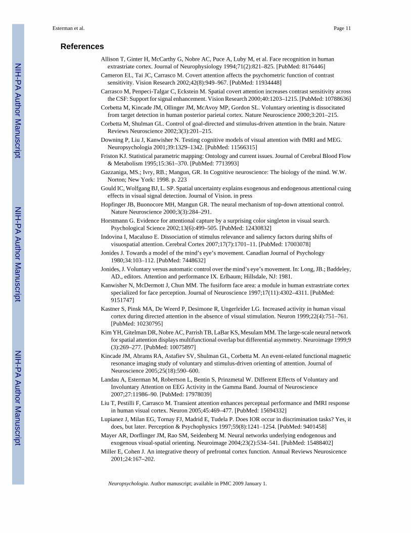
Importantly, the previous studies compared peripheral cues in involuntary conditions to centralcues in voluntary conditions, often with different timing parameters. Under thesecircumstances, different sensory input between the two conditions cannot be ruled out as thecause of the differences. In contrast, the trial events for the two attention conditions wereidentical in the present study (Figure 1). Every trial began with a fixation point and twoperipheral achromatic rectangles. A spatial cue was presented by having one of the rectanglesturn red. At cue offset, a target face briefly appeared in either the cued location (valid trial) orin the uncued location (invalid trial). In the involuntary condition, the cue was not predictiveof target location (Jonides, 1980,1981): The target appeared at the cued location on 50% of the
Esterman et al. Page 2
Neuropsychologia. Author manuscript; available in PMC 2009 January 1.
NIH
-PA Author Manuscript
NIH
-PA Author Manuscript
NIH
-PA Author Manuscript
trials and at the uncued location on 50% of the trials. In the voluntary condition the cue waspredictive of target location: The target appeared at the cued location on 75% of the trials andat the uncued location on 25% of the trials.
The predictive cue summons voluntary attention in that the participant is instructed to use thatcue to anticipate the location of the target. The nonpredictive cue manipulates involuntaryattention in that participants are told to ignore these cues as they do not carry information abouttarget location. In this way predictive and nonpredictive cue conditions are used as operationalvariables for voluntary and involuntary attention. Note that using a peripheral cue means thatour predictive session has an involuntary component as well. Furthermore, the nature of thetask determines that upon target appearance, the participants are required to voluntarily attendto the target location regardless of cue condition.
A critical feature of our design is that the stimulus displays and timing were identical forvoluntary and involuntary attention conditions. Thus, the temporal parameters had to becarefully selected. A short period of time between the cue and target favors a larger involuntaryattention effect, whereas a long period favors a larger voluntary attention effect (Posner, Cohen,& Rafal, 1982; Warner, Juola, & Koshino, 1990). Fortunately, with a discrimination task (asopposed to detection task), the time between the cue and target can be as long as 400 ms withoutdiminishing the effect of involuntary attention (Lupianez, Milan, Tornay, Madrid, & Tudela,1997). On the basis of pilot work with these stimuli, we chose a cue-stimulus interval thatwould give us approximately equivalent voluntary and involuntary attention effects on reactiontime (RT).
To preview our results, we find that voluntary attention increases FFA responses when the faceappears at the cued location as compared to when it appears at the uncued location. However,this difference is absent with involuntary attention. These findings support the hypothesis thatattentional mechanisms involved in voluntary and involuntary attention can have differentconsequences on ventral, recognition-related cortical functions.
There has been considerable controversy in the literature about whether involuntary attentionaffects the perceptual representation. As mentioned above, Prinzmetal and his colleagues havefound that voluntary attention, but not involuntary attention, influenced accuracy ondiscrimination tasks (Prinzmetal, McCool et al., 2005; Prinzmetal, Park et al., 2005). On theother hand, Carrasco and her colleagues have found several cases where “exogenous” attentionaffects accuracy. For example, Cameron, Tai, and Carrasco (2002) and Carrasco, Penpeci-Talgar, and Eckstein (2000) compared a 100% predictive cue with a neutral cue and foundhigher accuracy with the 100% predictive cue. They interpreted the results as reflectingexogenous attention because they used peripheral cues and relatively short SOAs (<120 ms)1. Thus there are cases where involuntary attention may affect accuracy and other where isdoes not. Because of this, we also conducted a behavioral experiment, the goal of which wasto determine which pattern of accuracy we would obtain when the same stimuli used in thefMRI experiment were made difficult to discriminate. We found that involuntary attention didnot affect accuracy using faces that were difficult to discriminate with otherwise the identicalparameters used in the fMRI experiment. Conversely, voluntary attention did improvediscrimination performance. Later we will suggest one possible explanation of why someresearchers find effects of involuntary attention on accuracy and others do not.
1Warner et al. (1990) found that the SOA where voluntary attention began to become effective depended on the level of practice of theobserver. The more practice that observers had, the shorter the critical SOA for voluntary attention, as short as 50 ms. Thus the resultsof Carrasco et al. (2000, 2002) may have reflected voluntary attention.
Esterman et al. Page 3
Neuropsychologia. Author manuscript; available in PMC 2009 January 1.
NIH
-PA Author Manuscript
NIH
-PA Author Manuscript
NIH
-PA Author Manuscript
Materials and MethodsParticipants
Ten healthy undergraduate and graduate students at UC Berkeley participated in the fMRIstudy. Another 32 healthy undergraduate students at UC Berkeley participated in the behavioralstudy. All had normal or corrected-to-normal vision and ages ranging from 18-30. All the fMRIparticipants were right handed. All provided informed consent as approved by the UC BerkeleyIRB committee.
Behavioral procedureThe target stimuli were created from digital photographs of two males who were similar inappearance (see Figure 1). One of the 2 faces appeared as a target on every trial. Before thetarget appeared, two boxes were present, and one turned red for 300 ms, directly followed bythe face target either in the cued location (“valid” trials) or in the uncued location (“invalid”trials). Participants were instructed to decide which of the two faces was present on each trial.For the predictive cueing condition, 75% of trials were valid, and 25% were invalid, andparticipants were told that the face usually appeared at the cued location, so it was beneficialto attend to that location. For the nonpredictive cueing condition, 50% of trials were valid and50% were invalid, and participants were told that the cue was irrelevant, so they should ignoreit. Faces were presented for 150 ms. Eye movements were monitored, and trials on which theyoccurred were eliminated from analysis2.
For the imaging experiment and the preceding training session, we chose to conduct a versionthat emphasized RT over accuracy. In this version, the faces were only slightly morphed, sothat discrimination was over 90%. Participants were urged to respond as quickly as possible.We used the easier RT version in the scanner because we wanted to control for extraneousneural responses due to differences in guessing, certainty, or accuracy, and focus only ondifferences in attention. For the behavioral study, faces were morphed such that participantswere only 75-80% correct, and accuracy was stressed rather than RT.
Simulated scanner procedureScanning sessions were preceded by a practice session in a simulated scanner the day before.In the simulated scanner, participants laid on their back with mirror glasses that enabled themto see the projected screen presentation. Eye movements were monitored with feedback usingan Applied Science Laboratory (ASL) eye tracking system. This session allowed participantsto practice the task and learn to maintain fixation. By the final block of practice, eye movementswere less than 2%, and discrimination accuracy was better than 90% for all participants. Duringthe simulated scanner session, participants performed the same number of blocks/trials theywould receive in the scanner (see fMRI procedure below). The order of voluntary andinvoluntary blocks was counterbalanced between participants.
fMRI procedureParticipants were scanned using a 4 Tesla Varian Scanner at UC Berkeley. Functional imageswere acquired using a gradient echoplanar sequence (TR = 2000 ms, TE = 28 ms, matrix size= 64 × 64, FOV = 22.4 cm, 3.5 × 3.5 × 5.5 mm voxel size) sensitive to BOLD contrast. Eachfunctional volume consisted of 18 × 5 mm thick axial slices with 0.5 mm gaps between eachslice, providing whole brain coverage. Images were projected onto a custom screen mountedat the participant’s chest level and viewed via an angled mirror placed inside the head coil.Responses were made by the right hand using a hand-held fiber optic button box.
2This paradigm was similar to Prinzmetal, McCool, et al. (2005) Experiments 9-11, but used a different cue-target interval (SOA).
Esterman et al. Page 4
Neuropsychologia. Author manuscript; available in PMC 2009 January 1.
NIH
-PA Author Manuscript
NIH
-PA Author Manuscript
NIH
-PA Author Manuscript
Cueing Task—Each participant performed 6 blocks of the cueing task: 3 consecutive blockswith a predictive cue, and 3 consecutive blocks with a nonpredictive cue. Predictive/nonpredictive order was counterbalanced. Before each block, participants were reminded ofthe cue predictability (i.e., whether to ignore the cue or not) and to maintain fixation. Eachblock consisted of 64 trials. ITI was jittered: 25% were 8 sec, 25% were 6 sec, and 50% were4 sec.
FFA Localizer—Between the predictive and nonpredictive blocks of the main experiment,each participant performed a FFA localizer task with a standard procedure (Kanwisher et al.,1997), previously used for ROI definition to explore attention effects of lateralized faces (e.g.Wojciulik et al., 1998). The faces in the localizer task were presented at fixation and weredifferent in identity and size (larger) than the faces used in the subsequent cueing task. Theywere shown 16-second blocks consisting of either faces, scenes, or fixation. In each block, 20images were presented for 500 ms each with 300 ms ISI. Participants were instructed to pressthe response pad when the current image was the same as the image immediately preceding it(on average, one response was required for each block of images). There were 7 blocks of eachtype, and the scan lasted 5 minutes and 20 seconds. Face selective regions were derived fromcontrasting face and scene blocks in this one-back task.
fMRI Data AnalysisInitial data preparation included image reconstruction, motion correction using a six-parameter, rigid-body, least-squares alignment, and spatial smoothing (8-mm FWHMGaussian kernel). SPM2 was used for all processing and analyses (Wellcome Institute ofCognitive Neurology, London, UK).
FFA Localizer—For each participant, the FFA was defined as the peak 15 voxels whichincluded the maximum peak voxel in the middle fusiform gyrus with greater BOLD signal forfaces compared to scenes. This yielded a right and left FFA for 6 participants, a right FFA onlyfor 2 participants, and a left FFA only for 2 participants. For participants with both left andright FFA, twice as much data were available for the ROI analyses.
ROI analysis-native space FFA—BOLD signal corresponding to each trial type wasassessed using a finite impulse response (FIR) model. The signal change in BOLD responsefrom baseline (fixation pattern alone) for each time point corresponding to the first 16 time-interpolated TRs (16 seconds) was estimated for each voxel and condition. This technique doesnot assume a canonical shape of the hemodynamic response function. The response functionfor each FFA was the average of all voxels in the ROI. Planned comparisons of peak BOLDresponses were done on the signal collapsed across 4-6 seconds. The ANOVA was conductedwith activation from 1-7 seconds, which corresponded to the time period in which all BOLDresponses were greater than zero. Note that these response functions are a result of BOLDresponses to both cue and target, since a single trial is modeled as a single event. However,this was the case for both voluntary and involuntary attention conditions. All trials wereincluded in the analyses, included errors, which occurred infrequently (4% of trials), and didnot differ between the conditions (see Results).
Whole brain group analysis—In addition to the ROI analysis, a whole brain analysis wasconducted. Estimates of task-related effects were obtained using a general linear model whichtakes into account the intrinsic variance-covariance structure of the time-series (Friston,1995). Separate parameter estimates were modeled for each condition. These models weregenerated for each participant in native space. For a given effect of interest, whole-braincontrast maps were determined for each participant. Each participant’s T1-weighted highresolution anatomical scan was normalized into the same coordinate frame as the MNI-template
Esterman et al. Page 5
Neuropsychologia. Author manuscript; available in PMC 2009 January 1.
NIH
-PA Author Manuscript
NIH
-PA Author Manuscript
NIH
-PA Author Manuscript
brain by using a 7 × 8 × 7 parameter nonlinear transform. The resulting transformationparameters were applied to all contrast images that were generated in native space. Voxel sizeof the transformed maps was 2 × 2 × 2 mm. Analysis of significance at the group level wasdetermined using a random effects analysis, using a 1-sample t-test or paired t-test on thecontrast maps. We calculated a family-wise error corrected threshold of p=0.05, derived fromthe compiled Family-wise error (FWE) values for all individual subjects in the group. Thisvalue corresponds to an uncorrected critical p-value of 0.0007. Thresholded clusters of 10 ormore voxels are reported. Whole brain contrasts were done to examine validity effects in thevoluntary and involuntary conditions and how they interact, particularly in frontal-parietalareas (Table 1, Figure 3). To examine reorienting after voluntary and involuntary orienting,valid trials were contrasted with invalid trials for each cue type. Validity effects (invalid >valid) isolate activity associated with reorienting when the target appears at the uncued locationfrom the activity associated with orienting to the cue. To compare reorienting between thevoluntary and involuntary conditions, validity effects (invalid > valid) were contrasted betweenpredictive and nonpredictive cueing blocks, thus examining the interaction of validity and cuetype.
ResultsBehavioral results in the scanner
Mean correct RT for each cell of the design for each participant was analyzed by ANOVA forrepeated measures. There was a main effect of validity, and no interaction with session. Forboth predictive and nonpredictive blocks, participants were faster on valid than invalid trials(Predictive: valid=684 ms, invalid= 720 ms; p<0.05: Nonpredictive: valid=674 ms, invalid=711 ms; p<0.01), consistent with previous findings (e.g. Prinzmetal et al., 2005a). Themagnitude of the validity benefit was similar: 36 ms for voluntary and 37 ms for involuntaryattention. There was no effect of order (predictive vs. nonpredictive first) on the magnitude ofthe cueing effect (F<1). Accuracy was high for both types of cues (Predictive: valid=96%,invalid=94%, p>0.1; Nonpredictive: valid=97%, invalid=96%, p>0.1). With accuracy as thedependent measure, the ANOVA revealed no effects of validity or session, and no interaction.
Imaging DataFFA Identification—The difference between faces and scenes elicited face-specific activityin the fusiform gyrus for all participants, defining the FFA regions of interest (see Methodssection).
ROI analysis-effects of spatial cueing on the FFA—Because only 6 of 10 participantshad both a right and left FFA, we had reduced power to investigate differences between leftand right FFA. Although right FFA exhibited more task related activity overall, side of FFAdid not interact with attention type. A finite impulse response (FIR) model (see Methods) wasused to estimate the hemodynamic response for each condition within each participant’s FFA(s). These response functions were subjected to an ANOVA with target laterality (contralateral/ipsilateral), cue laterality (contralateral/ipsilateral), attention type (voluntary/involuntary), andtime (1 sec through 7 sec) as factors. Overall, there was greater FFA response for contralateralcompared to ipsilateral cues and faces (cue laterality × time, F6,54=2.41, p<0.05; target laterality× time, F6,54=8.81, p<0.01). These interactions were qualified by higher order interactions withsession (predictive vs nonpredictive). Specifically, different consequences of voluntary andinvoluntary orienting were revealed by a significant 4-way interaction of the FFA responsebetween target laterality, cue laterality, attention type, and time (F6,54=2.44, p<0.05). Toexplore the nature of this interaction we analyzed the peak BOLD responses (collapsed across4-6 seconds) in the FFA for each type of attention.
Esterman et al. Page 6
Neuropsychologia. Author manuscript; available in PMC 2009 January 1.
NIH
-PA Author Manuscript
NIH
-PA Author Manuscript
NIH
-PA Author Manuscript
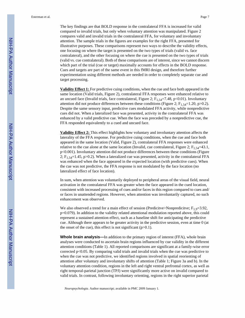
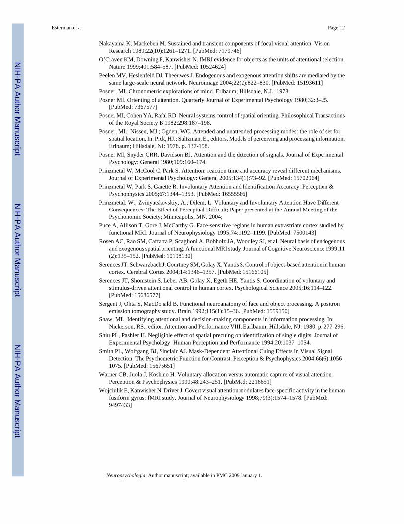
The key findings are that BOLD response in the contralateral FFA is increased for validcompared to invalid trials, but only when voluntary attention was manipulated. Figure 2compares valid and invalid trials in the contralateral FFA, for voluntary and involuntaryattention. The sample trials in the figures are examples for the right FFA, presented forillustrative purposes. These comparisons represent two ways to describe the validity effects,one focusing on where the target is presented on the two types of trials (valid vs. facecontralateral), and the other focusing on where the cue is presented on the two types of trials(valid vs. cue contralateral). Both of these comparisons are of interest, since we cannot discernwhich part of the trial (cue or target) maximally accounts for effects in the BOLD response.Cues and targets are part of the same event in this fMRI design, and therefore furtherexperimentation using different methods are needed in order to completely separate cue andtarget processing.
Validity Effect 1: For predictive cuing conditions, when the cue and face both appeared in thesame location (Valid trials, Figure 2), contralateral FFA responses were enhanced relative toan uncued face (Invalid trials, face contralateral, Figure 2; F1,54=7.48, p<0.01). Involuntaryattention did not produce differences between these conditions (Figure 2; F1,54=1.20, p>0.2).Despite the same sensory input, predictive cues modulated FFA activity, while nonpredictivecues did not. When a lateralized face was presented, activity in the contralateral FFA wasenhanced by a valid predictive cue. When the face was preceded by a nonpredictive cue, theFFA responded equivalently to a cued and uncued face.
Validity Effect 2: This effect highlights how voluntary and involuntary attention affects thelaterality of the FFA response. For predictive cuing conditions, when the cue and face bothappeared in the same location (Valid, Figure 2), contralateral FFA responses were enhancedrelative to the cue alone at the same location (Invalid, cue contralateral, Figure 2; F1,54=43.1,p<0.001). Involuntary attention did not produce differences between these conditions (Figure2; F1,54=1.45, p>0.2). When a lateralized cue was presented, activity in the contralateral FFAwas enhanced when the face appeared in the expected location (with predictive cues). Whenthe cue was not predictive, the FFA response is not modulated by the face location (nolateralized effect of face location).
In sum, when attention was voluntarily deployed to peripheral areas of the visual field, neuralactivation in the contralateral FFA was greater when the face appeared in the cued location,consistent with increased processing of cues and/or faces in this region compared to cues and/or faces in unattended regions. However, when attention was involuntarily captured, no suchenhancement was observed.
We also observed a trend for a main effect of session (Predicitve>Nonpredicitve; F1,9=3.92,p=0.079). In addition to the validity related attentional modulation reported above, this couldrepresent a sustained attention effect, such as a baseline shift for anticipating the predictivecue. Although there appears to be greater activity in the predictive session, even at time 0 (atthe onset of the cue), this effect is not significant (p>0.1).
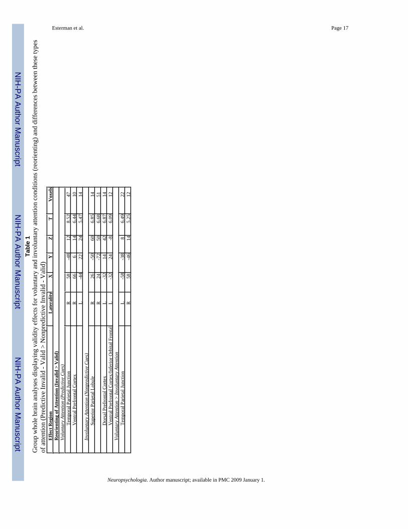
Whole brain analysis—In addition to the primary region of interest (FFA), whole brainanalyses were conducted to ascertain brain regions influenced by cue validity in the differentattention conditions (Table 1). All reported comparisons are significant at a family-wise errorcorrected p<0.05. By comparing valid trials and invalid trials when the cue was predictive towhen the cue was not predictive, we identified regions involved in spatial reorienting ofattention after voluntary and involuntary shifts of attention (Table 1; Figure 3a and b). In thevoluntary attention condition, regions in the left and right ventral prefrontal cortex, as well asright temporal-parietal junction (TPJ) were significantly more active on invalid compared tovalid trials. In contrast, following involuntary orienting, regions in the right superior parietal
Esterman et al. Page 7
Neuropsychologia. Author manuscript; available in PMC 2009 January 1.
NIH
-PA Author Manuscript
NIH
-PA Author Manuscript
NIH
-PA Author Manuscript
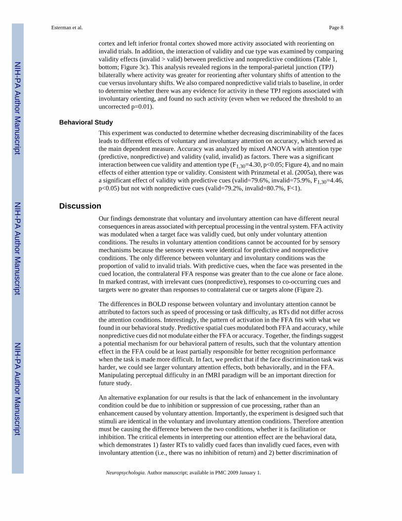
cortex and left inferior frontal cortex showed more activity associated with reorienting oninvalid trials. In addition, the interaction of validity and cue type was examined by comparingvalidity effects (invalid > valid) between predictive and nonpredictive conditions (Table 1,bottom; Figure 3c). This analysis revealed regions in the temporal-parietal junction (TPJ)bilaterally where activity was greater for reorienting after voluntary shifts of attention to thecue versus involuntary shifts. We also compared nonpredictive valid trials to baseline, in orderto determine whether there was any evidence for activity in these TPJ regions associated withinvoluntary orienting, and found no such activity (even when we reduced the threshold to anuncorrected p=0.01).
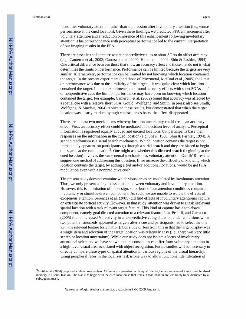
Behavioral StudyThis experiment was conducted to determine whether decreasing discriminability of the facesleads to different effects of voluntary and involuntary attention on accuracy, which served asthe main dependent measure. Accuracy was analyzed by mixed ANOVA with attention type(predictive, nonpredictive) and validity (valid, invalid) as factors. There was a significantinteraction between cue validity and attention type (F1,30=4.30, p<0.05; Figure 4), and no maineffects of either attention type or validity. Consistent with Prinzmetal et al. (2005a), there wasa significant effect of validity with predictive cues (valid=79.6%, invalid=75.9%, F1,30=4.46,p<0.05) but not with nonpredictive cues (valid=79.2%, invalid=80.7%, F<1).
DiscussionOur findings demonstrate that voluntary and involuntary attention can have different neuralconsequences in areas associated with perceptual processing in the ventral system. FFA activitywas modulated when a target face was validly cued, but only under voluntary attentionconditions. The results in voluntary attention conditions cannot be accounted for by sensorymechanisms because the sensory events were identical for predictive and nonpredictiveconditions. The only difference between voluntary and involuntary conditions was theproportion of valid to invalid trials. With predictive cues, when the face was presented in thecued location, the contralateral FFA response was greater than to the cue alone or face alone.In marked contrast, with irrelevant cues (nonpredictive), responses to co-occurring cues andtargets were no greater than responses to contralateral cue or targets alone (Figure 2).
The differences in BOLD response between voluntary and involuntary attention cannot beattributed to factors such as speed of processing or task difficulty, as RTs did not differ acrossthe attention conditions. Interestingly, the pattern of activation in the FFA fits with what wefound in our behavioral study. Predictive spatial cues modulated both FFA and accuracy, whilenonpredictive cues did not modulate either the FFA or accuracy. Together, the findings suggesta potential mechanism for our behavioral pattern of results, such that the voluntary attentioneffect in the FFA could be at least partially responsible for better recognition performancewhen the task is made more difficult. In fact, we predict that if the face discrimination task washarder, we could see larger voluntary attention effects, both behaviorally, and in the FFA.Manipulating perceptual difficulty in an fMRI paradigm will be an important direction forfuture study.
An alternative explanation for our results is that the lack of enhancement in the involuntarycondition could be due to inhibition or suppression of cue processing, rather than anenhancement caused by voluntary attention. Importantly, the experiment is designed such thatstimuli are identical in the voluntary and involuntary attention conditions. Therefore attentionmust be causing the difference between the two conditions, whether it is facilitation orinhibition. The critical elements in interpreting our attention effect are the behavioral data,which demonstrates 1) faster RTs to validly cued faces than invalidly cued faces, even withinvoluntary attention (i.e., there was no inhibition of return) and 2) better discrimination of
Esterman et al. Page 8
Neuropsychologia. Author manuscript; available in PMC 2009 January 1.
NIH
-PA Author Manuscript
NIH
-PA Author Manuscript
NIH
-PA Author Manuscript
faces after voluntary attention rather than suppression after involuntary attention (i.e., worseperformance at the cued location). Given these findings, we predicted FFA enhancement aftervoluntary attention and a reduction or absence of this enhancement following involuntaryattention. This correspondence with perceptual performance led to the current interpretationof our imaging results in the FFA.
There are cases in the literature where nonpredictive cues or short SOAs do affect accuracy(e.g., Cameron et al., 2002; Carrasco et al., 2000; Horstmann, 2002; Shiu & Pashler, 1994).One critical difference between those that show an accuracy effect and those that do not is whatdetermines the limits on performance. Performance can be limited because the targets are verysimilar. Alternatively, performance can be limited by not knowing which location containedthe target. In the present experiment (and those of Prinzmetal, McCool et al., 2005) the limiton performance was due to the similarity of the targets - it was quite clear which locationcontained the target. In other experiments, that found accuracy effects with short SOAs and/or nonpredictive cues the limit on performance may have been on knowing which locationcontained the target. For example, Cameron, et al. (2002) found that accuracy was affected bya spatial cue with a relative short SOA. Gould, Wolfgang, and Smith (in press; also see Smith,Wolfgang, & Sinclair, 2004) replicated these results, but demonstrated that when the targetlocation was clearly marked by high contrast cross hairs, the effect disappeared.
There are at least two mechanisms whereby location uncertainty could create an accuracyeffect. First, an accuracy effect could be mediated at a decision level of analysis. Perceptualinformation is registered equally at cued and uncued locations, but participants base theirresponses on the information in the cued location (e.g. Shaw, 1980; Shiu & Pashler, 1994). Asecond mechanism is a serial search mechanism. Which location contains the target is notimmediately apparent, so participants go through a serial search and they are biased to beginthis search at the cued location3. One might ask whether this directed search (beginning at thecued location) involves the same neural mechanism as voluntary attention. Our fMRI resultssuggest one method of addressing this question. If we increase the difficulty of knowing whichlocation contains the target, by adding a foil and/or additional locations, would be get FFAmodulation even with a nonpredictive cue?
The present study does not examine which visual areas are modulated by involuntary attention.Thus, we only present a single dissociation between voluntary and involuntary attention.However, this is a limitation of the design, since both of our attention conditions contain aninvoluntary or stimulus-driven component. As such, we are unable to isolate the effects ofexogenous attention. Serences et al. (2005) did find effects of involuntary attentional captureon extrastriate cortical activity. However, in that study, attention was drawn to a task irrelevantspatial location with a task relevant target feature. This kind of capture has a top-downcomponent, namely goal directed attention to a relevant feature. Liu, Pestilli, and Carrasco(2005) found increased V4 activity in a nonpredictive cuing situation under conditions whentwo potential sinusoids appeared as targets after a cue and participants had to select the onewith the relevant feature (orientation). Our study differs from this in that the target display wasa single item and selection of the target location was relatively easy (i.e., there was very littlesearch or location uncertainty). While our study does not isolate a locus of involuntaryattentional selection, we have shown that its consequences differ from voluntary attention ina high-level visual area associated with object recognition. Future studies will be necessary todirectly compare these types of spatial attention in various regions of the visual hierarchy.Using peripheral faces in the localizer task is one way to allow functional identification of
3Smith et al. (2004) proposed a related mechanism. All items are perceived with equal fidelity, but are transferred into a durable visualmemory in a serial fashion. The bias is to begin with the cued location so that items in that location are less likely to be disrupted by asubsequent mask.
Esterman et al. Page 9
Neuropsychologia. Author manuscript; available in PMC 2009 January 1.
NIH
-PA Author Manuscript
NIH
-PA Author Manuscript
NIH
-PA Author Manuscript
additional visual regions of interest in order to do similar comparison as presently done in theFFA.
While our study focused on faces and the FFA, we do not believe our voluntary attention effectsare unique to this region of cortex. Other studies of ‘pure’ voluntary attention have foundattentional modulations throughout the visual hierarchy, such as V2, VP, V4, and TEO(Hopfinger, Buonocore, & Mangun, 2000; Kastner, Pinsk, De Weerd, Desimone, &Ungerleider, 1999). Future studies using retinotopic mapping to identify such regions ofinterest may reveal similar dissociations between voluntary and involuntary attention, but thisremains an open question.
As previously noted, fMRI, in principal, does not have the temporal resolution to differentiatecue from target related activity, or even whether the current effects are early and perceptual,or occur after the task or decision process are completed. However, there are now indications,using EEG, that there are differences in gamma band response to voluntary and involuntaryspatial attention to faces (Landau, Esterman, Robertson, Bentin, & Prinzmetal, in press). EEGhas the temporal resolution to measure cue and face processing separately. Landau et al. findsdifferences in both cue and target processing between the two types of attention, suggestingthat our current findings do reflect differences in task-related stimulus processing.
Our whole brain analyses compared reorienting in voluntary and involuntary conditions andrevealed more temporal parietal junction activity (TPJ) associated with reorienting afterpredictive cues. This finding is partly consistent with other studies demonstrating that the TPJis associated with target processing when those targets appear at unexpected or unattendedlocations (Corbetta, Kincade, Ollinger, McAvoy, & Gordon, 2000; Corbetta & Shulman,2002; Kincade et al., 2005; Serences et al., 2005). While these studies find a right hemispherelateralized reorienting system, our study indicates that both hemispheres may participate inthis function. Importantly, this TPJ activity was not present following a shift of involuntaryattention. This finding is similar to of Kincade et al. (2005), which found that activity in theTPJ was associated with reorienting following an endogenous predicitive cue, but not anonpredicitive peripheral cue. That study also showed that TPJ did not respond to thenonpredictive peripheral cues above a neutral cue condition. Although we were not able toseparate cue from target related activity, we did not find any activity in these TPJ regions onnonpredicitive valid trials. Indovina & Macaluso (2007) also found that highly salient, but taskirrelevant stimuli did not engage the ventral attention network.
The present study demonstrates that voluntary and involuntary orienting of attention can havedifferent effects on a percept-related cortical area in the ventral stream and are consistent withperceptual enhancement when voluntary attention is employed. The interaction betweenvoluntary and involuntary attention and validity effects is also consistent with behavioraldifferences between errors and reaction times to valid and invalid cues reported previously inthe behavioral literature. Both voluntary and involuntary attention produced faster responsesat validly cued locations than invalidly cued locations. However, only voluntary attentionincreased accuracy in a situation where the limit on performance was not locating the target.Together, these results suggest that neural activity in the FFA reflects distinct changes inprocessing associated with the two types of attentional orienting.
While both the voluntary and involuntary systems recruit many overlapping regions of frontaland parietal cortex (Kim et al., 1999; Mayer et al., 2004; Peelen et al., 2004; Rosen et al.,1999), the present study indicates that their consequences on perceptual systems in the ventralcortex can in fact differ, both with regard to their effects on neural responses and onperformance.
Esterman et al. Page 10
Neuropsychologia. Author manuscript; available in PMC 2009 January 1.
NIH
-PA Author Manuscript
NIH
-PA Author Manuscript
NIH
-PA Author Manuscript
ReferencesAllison T, Ginter H, McCarthy G, Nobre AC, Puce A, Luby M, et al. Face recognition in human
extrastriate cortex. Journal of Neurophysiology 1994;71(2):821–825. [PubMed: 8176446]Cameron EL, Tai JC, Carrasco M. Covert attention affects the psychometric function of contrast
sensitivity. Vision Research 2002;42(8):949–967. [PubMed: 11934448]Carrasco M, Penpeci-Talgar C, Eckstein M. Spatial covert attention increases contrast sensitivity across
the CSF: Support for signal enhancement. Vision Research 2000;40:1203–1215. [PubMed: 10788636]Corbetta M, Kincade JM, Ollinger JM, McAvoy MP, Gordon SL. Voluntary orienting is dissocitated
from target detection in human posterior parietal cortex. Nature Neuroscience 2000;3:201–215.Corbetta M, Shulman GL. Control of goal-directed and stimulus-driven attention in the brain. Nature
Reviews Neuroscience 2002;3(3):201–215.Downing P, Liu J, Kanwisher N. Testing cognitive models of visual attention with fMRI and MEG.
Neuropsychologia 2001;39:1329–1342. [PubMed: 11566315]Friston KJ. Statistical parametric mapping: Ontology and current issues. Journal of Cerebral Blood Flow
& Metabolism 1995;15:361–370. [PubMed: 7713993]Gazzaniga, MS.; Ivry, RB.; Mangun, GR. In Cognitive neuroscience: The biology of the mind. W.W.
Norton; New York: 1998. p. 223Gould IC, Wolfgang BJ, L. SP. Spatial uncertainty explains exogenous and endogenous attentional cuing
effects in visual signal detection. Journal of Vision. in pressHopfinger JB, Buonocore MH, Mangun GR. The neural mechanism of top-down attentional control.
Nature Neuroscience 2000;3(3):284–291.Horstmann G. Evidence for attentional capture by a surprising color singleton in visual search.
Psychological Science 2002;13(6):499–505. [PubMed: 12430832]Indovina I, Macaluso E. Dissociation of stimulus relevance and saliency factors during shifts of
visuospatial attention. Cerebral Cortex 2007;17(7):1701–11. [PubMed: 17003078]Jonides J. Towards a model of the mind’s eye’s movement. Canadian Journal of Psychology
1980;34:103–112. [PubMed: 7448632]Jonides, J. Voluntary versus automatic control over the mind’s eye’s movement. In: Long, JB.; Baddeley,
AD., editors. Attention and performance IX. Erlbaum; Hillsdale, NJ: 1981.Kanwisher N, McDermott J, Chun MM. The fusiform face area: a module in human extrastriate cortex
specialized for face perception. Journal of Neuroscience 1997;17(11):4302–4311. [PubMed:9151747]
Kastner S, Pinsk MA, De Weerd P, Desimone R, Ungerleider LG. Increased activity in human visualcortex during directed attention in the absence of visual stimulation. Neuron 1999;22(4):751–761.[PubMed: 10230795]
Kim YH, Gitelman DR, Nobre AC, Parrish TB, LaBar KS, Mesulam MM. The large-scale neural networkfor spatial attention displays multifunctional overlap but differential asymmetry. Neuroimage 1999;9(3):269–277. [PubMed: 10075897]
Kincade JM, Abrams RA, Astafiev SV, Shulman GL, Corbetta M. An event-related functional magneticresonance imaging study of voluntary and stimulus-driven orienting of attention. Journal ofNeuroscience 2005;25(18):590–600.
Landau A, Esterman M, Robertson L, Bentin S, Prinzmetal W. Different Effects of Voluntary andInvoluntary Attention on EEG Activity in the Gamma Band. Journal of Neuroscience2007;27:11986–90. [PubMed: 17978039]
Liu T, Pestilli F, Carrasco M. Transient attention enhances perceptual performance and fMRI responsein human visual cortex. Neuron 2005;45:469–477. [PubMed: 15694332]
Lupianez J, Milan EG, Tornay FJ, Madrid E, Tudela P. Does IOR occur in discrimination tasks? Yes, itdoes, but later. Perception & Psychophysics 1997;59(8):1241–1254. [PubMed: 9401458]
Mayer AR, Dorflinger JM, Rao SM, Seidenberg M. Neural networks underlying endogenous andexogenous visual-spatial orienting. Neuroimage 2004;23(2):534–541. [PubMed: 15488402]
Miller E, Cohen J. An integrative theory of prefrontal cortex function. Annual Reviews Neurosicence2001;24:167–202.
Esterman et al. Page 11
Neuropsychologia. Author manuscript; available in PMC 2009 January 1.
NIH
-PA Author Manuscript
NIH
-PA Author Manuscript
NIH
-PA Author Manuscript
Nakayama K, Mackeben M. Sustained and transient components of focal visual attention. VisionResearch 1989;22(10):1261–1271. [PubMed: 7179746]
O’Craven KM, Downing P, Kanwisher N. fMRI evidence for objects as the units of attentional selection.Nature 1999;401:584–587. [PubMed: 10524624]
Peelen MV, Heslenfeld DJ, Theeuwes J. Endogenous and exogenous attention shifts are mediated by thesame large-scale neural network. Neuroimage 2004;22(2):822–830. [PubMed: 15193611]
Posner, MI. Chronometric explorations of mind. Erlbaum; Hillsdale, N.J.: 1978.Posner MI. Orienting of attention. Quarterly Journal of Experimental Psychology 1980;32:3–25.
[PubMed: 7367577]Posner MI, Cohen YA, Rafal RD. Neural systems control of spatial orienting. Philosophical Transactions
of the Royal Society B 1982;298:187–198.Posner, MI.; Nissen, MJ.; Ogden, WC. Attended and unattended processing modes: the role of set for
spatial location. In: Pick, HJ.; Saltzman, E., editors. Models of perceiving and processing information.Erlbaum; Hillsdale, NJ: 1978. p. 137-158.
Posner MI, Snyder CRR, Davidson BJ. Attention and the detection of signals. Journal of ExperimentalPsychology: General 1980;109:160–174.
Prinzmetal W, McCool C, Park S. Attention: reaction time and accuracy reveal different mechanisms.Journal of Experimental Psychology: General 2005;134(1):73–92. [PubMed: 15702964]
Prinzmetal W, Park S, Garette R. Involuntary Attention and Identification Accuracy. Perception &Psychophysics 2005;67:1344–1353. [PubMed: 16555586]
Prinzmetal, W.; Zvinyatskovskiy, A.; Dilem, L. Voluntary and Involuntary Attention Have DifferentConsequences: The Effect of Perceptual Difficult; Paper presented at the Annual Meeting of thePsychonomic Society; Minneapolis, MN. 2004;
Puce A, Allison T, Gore J, McCarthy G. Face-sensitive regions in human extrastriate cortex studied byfunctional MRI. Journal of Neurophysiology 1995;74:1192–1199. [PubMed: 7500143]
Rosen AC, Rao SM, Caffarra P, Scaglioni A, Bobholz JA, Woodley SJ, et al. Neural basis of endogenousand exogenous spatial orienting. A functional MRI study. Journal of Cognitive Neuroscience 1999;11(2):135–152. [PubMed: 10198130]
Serences JT, Schwarzbach J, Courtney SM, Golay X, Yantis S. Control of object-based attention in humancortex. Cerebral Cortex 2004;14:1346–1357. [PubMed: 15166105]
Serences JT, Shomstein S, Leber AB, Golay X, Egeth HE, Yantis S. Coordination of voluntary andstimulus-driven attentional control in human cortex. Psychological Science 2005;16:114–122.[PubMed: 15686577]
Sergent J, Ohta S, MacDonald B. Functional neuroanatomy of face and object processing. A positronemission tomography study. Brain 1992;115(1):15–36. [PubMed: 1559150]
Shaw, ML. Identifying attentional and decision-making components in information processing. In:Nickerson, RS., editor. Attention and Performance VIII. Earlbaum; Hillsdale, NJ: 1980. p. 277-296.
Shiu PL, Pashler H. Negligible effect of spatial precuing on identification of single digits. Journal ofExperimental Psychology: Human Perception and Performance 1994;20:1037–1054.
Smith PL, Wolfgang BJ, Sinclair AJ. Mask-Dependent Attentional Cuing Effects in Visual SignalDetection: The Psychometric Function for Contrast. Perception & Psychophysics 2004;66(6):1056–1075. [PubMed: 15675651]
Warner CB, Juola J, Koshino H. Voluntary allocation versus automatic capture of visual attention.Perception & Psychophysics 1990;48:243–251. [PubMed: 2216651]
Wojciulik E, Kanwisher N, Driver J. Covert visual attention modulates face-specific activity in the humanfusiform gyrus: fMRI study. Journal of Neurophysiology 1998;79(3):1574–1578. [PubMed:9497433]
Esterman et al. Page 12
Neuropsychologia. Author manuscript; available in PMC 2009 January 1.
NIH
-PA Author Manuscript
NIH
-PA Author Manuscript
NIH
-PA Author Manuscript
Figure 1.Spatial cueing procedure. Participants performed a face discrimination task. To examinevoluntary and involuntary attention, the cue was either predictive or nonpredictive of asubsequent target face location.
Esterman et al. Page 13
Neuropsychologia. Author manuscript; available in PMC 2009 January 1.
NIH
-PA Author Manuscript
NIH
-PA Author Manuscript
NIH
-PA Author Manuscript
Figure 2.Validity Effect in the FFA. ROI analysis of FFA, showing evoked blood oxygenation level-dependent (BOLD) time course in the contralateral FFA as a function of type of attention andcue validity. When the face is preceded by a valid spatial cue (valid trials), there is a greaterevoked response than to an uncued face (invalid trial, contralateral face), for voluntary attentionbut not for involuntary attention. Similarly, when the face appears in the cued location (validtrials), there is a greater evoked response than when the face does not appear in the cued location(invalid trials, contralateral cue), but only with voluntary attention and not with involuntaryattention. Example contrasts (top-left) are shown for right FFA, however the graphs representdata from both FFAs (for left FFA, the contrast is flipped left-right).
Esterman et al. Page 14
Neuropsychologia. Author manuscript; available in PMC 2009 January 1.
NIH
-PA Author Manuscript
NIH
-PA Author Manuscript
NIH
-PA Author Manuscript
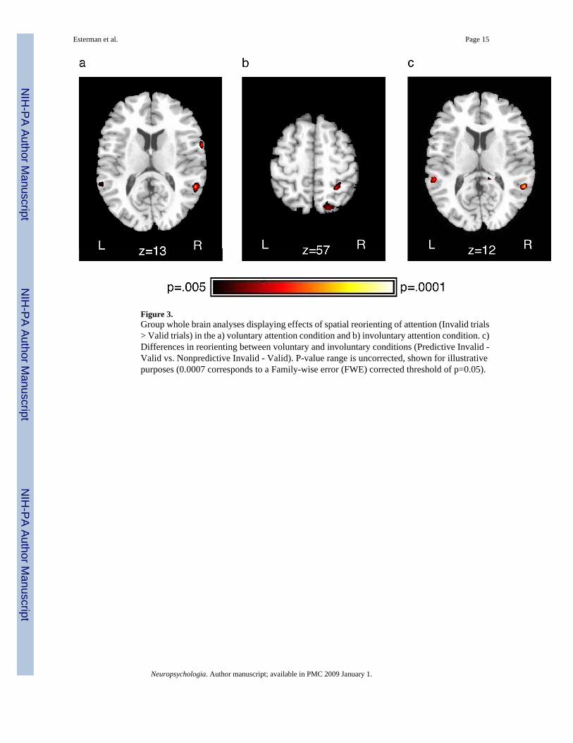
Figure 3.Group whole brain analyses displaying effects of spatial reorienting of attention (Invalid trials> Valid trials) in the a) voluntary attention condition and b) involuntary attention condition. c)Differences in reorienting between voluntary and involuntary conditions (Predictive Invalid -Valid vs. Nonpredictive Invalid - Valid). P-value range is uncorrected, shown for illustrativepurposes (0.0007 corresponds to a Family-wise error (FWE) corrected threshold of p=0.05).
Esterman et al. Page 15
Neuropsychologia. Author manuscript; available in PMC 2009 January 1.
NIH
-PA Author Manuscript
NIH
-PA Author Manuscript
NIH
-PA Author Manuscript
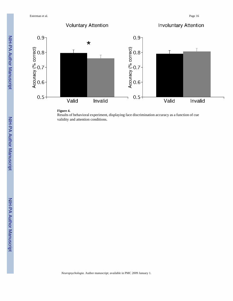
Figure 4.Results of behavioral experiment, displaying face discrimination accuracy as a function of cuevalidity and attention conditions.
Esterman et al. Page 16
Neuropsychologia. Author manuscript; available in PMC 2009 January 1.
NIH
-PA Author Manuscript
NIH
-PA Author Manuscript
NIH
-PA Author Manuscript
NIH
-PA Author Manuscript
NIH
-PA Author Manuscript
NIH
-PA Author Manuscript
Esterman et al. Page 17Ta
ble
1G
roup
who
le b
rain
ana
lyse
s dis
play
ing
valid
ity e
ffec
ts fo
r vol
unta
ry a
nd in
volu
ntar
y at
tent
ion
cond
ition
s (re
orie
ntin
g) a
nd d
iffer
ence
s bet
wee
n th
ese
type
sof
atte
ntio
n (P
redi
ctiv
e In
valid
- V
alid
> N
onpr
edic
tive
Inva
lid -
Val
id)
Effe
ct R
egio
nL
ater
ality
XY
ZT
Vox
els
Reo
rien
ting
of A
ttent
ion
(Inv
alid
> V
alid
)Vo
lunt
ary
Atte
ntio
n (P
redi
ctiv
e C
ues)
Tem
pora
l Par
ieta
l Jun
ctio
nR
58-4
812
8.52
47
Ven
tral P
refr
onta
l Cor
tex
R66
614
6.44
10L
-44
2224
5.47
14In
volu
ntar
y At
tent
ion
(Non
pred
ictiv
e C
ues)
Supe
rior P
arie
tal L
obul
eR
26-5
060
6.85
14R
24-7
250
6.68
51
Dor
sal P
refr
onta
l Cor
tex
L-3
214
426.
8714
Ven
tral P
refr
onta
l Cor
tex/
Infe
rior O
rbita
l Fro
ntal
L-3
224
-86.
0912
Volu
ntar
y At
tent
ion
> In
volu
ntar
y At
tent
ion
Tem
pora
l Par
ieta
l Jun
ctio
nL
-58
-38
86.
4922
R58
-46
145.
2512
Neuropsychologia. Author manuscript; available in PMC 2009 January 1.


![Demonstration of Uncued Surveillance of LEO Peter C ...€¦ · Ackermann & McGraw 2013 [2] for more information on streak detection). Thus for our one frame per second acquisition](https://img.pdfslide.us/doc/110x75/5f06cc397e708231d419c915/demonstration-of-uncued-surveillance-of-leo-peter-c-ackermann-mcgraw-2013.jpg)