Embed Size (px)
Citation preview
lable at ScienceDirect
Biomaterials 35 (2014) 71e82
Contents lists avai
Biomaterials
journal homepage: www.elsevier .com/locate/biomater ia ls
Augmentation of integrin-mediated mechanotransductionby hyaluronic acid
Anant Chopra a,c, Maria E. Murray c,d, Fitzroy J. Byfield d, Melissa G. Mendez d,Ran Halleluyan a, David J. Restle c,d, Dikla Raz-Ben Aroush d, Peter A. Galie d,Katarzyna Pogoda d,e, Robert Bucki d, Cezary Marcinkiewicz f, Glenn D. Prestwich i,Thomas I. Zarembinski g, Christopher S. Chen c,d, Ellen Puré h, J. Yasha Kresh a,b,**,Paul A. Janmey d,*
aDept. of Cardiothoracic Surgery, Drexel Univ. College of Med, Philadelphia, PA, USAbDept. of Medicine, Drexel Univ. College of Med, Philadelphia, PA, USAcDept. of Bioengineering, Univ. of Pennsylvania, Philadelphia, PA, USAd Institute for Medicine and Engineering, Univ. of Pennsylvania, Philadelphia, PA, USAe The Henryk Niewodnicza�nski Institute of Nuclear Physics, Kraków, PolandfDept. of Bioengineering, Temple University, Philadelphia, PA, USAgBioTime, Inc., Alameda, CA, USAhDept. of Animal Biology, School of Veterinary Medicine, University of Pennsylvania, Philadelphia, PA, USAiDepartment of Medicinal Chemistry, University of Utah, Salt Lake City, UT, USA
a r t i c l e i n f o
Article history:Received 20 August 2013Accepted 20 September 2013Available online 10 October 2013
Keywords:Hyaluronic acidMechanosensingYes associated protein (YAP)Traction stressesCell spreadingExtracellular matrix
* Corresponding author.** Corresponding author. Dept. of Cardiothoracic SuMed, Philadelphia, PA, USA.
E-mail address: [email protected] (P.A
0142-9612/$ e see front matter � 2013 Elsevier Ltd.http://dx.doi.org/10.1016/j.biomaterials.2013.09.066
a b s t r a c t
Changes in tissue and organ stiffness occur during development and are frequently symptoms of disease.Many cell types respond to the stiffness of substrates and neighboring cells in vitro and most cell typesincrease adherent area on stiffer substrates that are coated with ligands for integrins or cadherins. In vivocells engage their extracellular matrix (ECM) by multiple mechanosensitive adhesion complexes and othersurface receptors that potentially modify themechanical signals transduced at the cell/ECM interface. Herewe show that hyaluronic acid (also called hyaluronan or HA), a soft polymeric glycosaminoglycan matrixcomponent prominent in embryonic tissue and upregulated during multiple pathologic states, augmentsor overrides mechanical signaling by some classes of integrins to produce a cellular phenotype otherwiseobserved only on very rigid substrates. The spread morphology of cells on soft HA-fibronectin coatedsubstrates, characterized by formation of large actin bundles resembling stress fibers and large focaladhesions resembles that of cells on rigid substrates, but is activated by different signals and does notrequire or cause activation of the transcriptional regulator YAP. The fact that HA production is tightlyregulated during development and injury and frequently upregulated in cancers characterized by un-controlled growth and cell movement suggests that the interaction of signaling between HA receptors andspecific integrins might be an important element in mechanical control of development and homeostasis.
� 2013 Elsevier Ltd. All rights reserved.
1. Introduction
Changes in tissue and organ stiffness are frequently symptomsof diseases such as cancer [1], liver fibrosis [2], and atherosclerosis[3], and these physical changes have been suggested to contributeto and not only be symptoms of the disease. For example, liverstiffness, as quantified by its shear modulus, increases during
rgery, Drexel Univ. College of
. Janmey).
All rights reserved.
experimentally-triggered liver fibrosis prior to increased matrixdeposition or altered cell morphology [4] by amechanism involvinglysyl oxidase [5]. Similarly, the development of atherosclerotic le-sions in an apoE null mouse model can be reversed by inhibition ofabnormal lysyl oxidase activity and subsequent reversal of arterialstiffening [3]. Such results suggest that changes in tissue mechanicsthat can activate hepatic stellate cells [6], portal fibroblasts [7] orvascular smooth muscle cells [8] in the affected organs precede andtherefore might cause or at least contribute to development of thepathologic state. The response of cells to abnormal matrix stiffnesscan also render them resistant to chemotherapeutic agents,possibly because of the changes in the cytoskeleton-membrane
A. Chopra et al. / Biomaterials 35 (2014) 71e8272
interface [9]. Such effects in vivo have motivated studies in vitro todetermine how physical properties such as increased cellular ten-sion or adherence to substrates of differing stiffness affect cellfunction under conditions where physical stimuli can be isolatedfrom biochemical signals.
Many cell types alter their structure and function in vitrodepending on the mechanical properties of the materials to whichthey adhere [10] and on the type of adhesion receptor by whichthey bind [11e13]. Most studies of cellular mechanosensing haveused inert, non-adhesive, soft materials for which mechanicalproperties can be controlled, and coupled these substrates to celladhesion proteins or synthetic ligands that engage specific trans-membrane proteins. Independent control of mechanical and ad-hesive changes in the substrates has been essential to demonstratethat changes in substrate viscoelasticity per se, and not a coincidentchange in cell signaling caused by altered adhesion protein pre-sentation causes the change in phenotype. The large majority ofmechanosensing studies have used the integrin ligands fibronectin,collagen, laminin, or RGD-containing peptides as the adhesive an-chor, and often polyacrylamide or other hydrogels such as alginate,poly(ethylene glycol) or methacrylated hyaluronan to producesubstrates softer than 50 kPa. A smaller but growing number ofstudies have investigated mechanosensing mediated by cadherinsto mimic cellecell junctions [13,14].
Studies in vitro of cells anchored to substrates through integrinsor in some cases cadherins, show that a common, though notuniversal, response of cells to substrate stiffness is an increase inadherent area, increased traction forces applied to the substrate,assembly of large actin bundles called stress fibers, and activationof signaling intermediates such as small GTPases and tyrosine ki-nase pathways that regulate actin assembly and acto-myosincontractility [15,16]. The inference from such studies is that mostcell types actively probe the mechanics of their environment byacto-myosin dependent forces, which increase when the resistanceimposed by the substrate increases, and the feedback between celland substrate reorganizes the cytoskeleton to achieve a homeo-static state appropriate for each physical context [17]. Substratestiffness and the resulting increase in cell-generated forces can alsoincrease activity of matrix-bound growth factors such as TGF-beta,that further increase development of the phenotype associatedwith growth of stiff substrates [18].
Response to substrate stiffness is highly cell-type specific, andneurons for example, have a unique response to stiffness, in whichmatrix stiffness greater than that of the normal CNS tissue inhibitsneurite outgrowth and growth cone spreading [19e21]. Myocyteshave a particularly striking and well-documented response tomatrix stiffness, with a distinct optimum for development of sar-comeres and an elongated shape that depends on both matrixstiffness [22e24] and the type of adhesion receptor [13,14,25]. Onpolyacrylamide (PAA) gels that are laminated with ligands forintegrins, cardiac myocytes develop well organized sarcomeresonly when cultured on substrates with elastic moduli in the rangeof 10 kPae30 kPa, near those of the healthy tissue. On stiffer sub-strates (>60 kPa) approximating the damaged heart, myocytesform stress fiber-like filament bundles but lack organized sarco-meres or an elongated shape. On soft (<1 kPa) PAA gels myocytesexhibit disorganized actin networks and sarcomeres. On N-cad-herin-coated PAA gels, the response is similar but the optimum isshifted to slightly lower stiffness (5 kPa) [14].
In contrast to the simplified chemical composition of softsubstrates used for mechanosensing studies in vitro, cells engagetheir extracellular matrix (ECM) in vivo both by mechanosensi-tive adhesion complexes and by other surface receptors for ECMcomponents that cannot act as adhesive anchors, but thatpotentially modify the mechanical signals transduced at the cell/
ECM interface. Such ECM components include not only growthfactors such as TGF-beta but also proteoglycans and glycosami-noglycans such as hyaluronic acid that constitute a major fractionof the total ECM content, and that change in abundance duringdevelopment, wound healing, and disease. For example, duringdevelopment, cardiac myocytes assemble and organize their in-ternal structures within a complex mechanical tissue environ-ment bounded by an especially soft (E w 20e100 Pa) [26,27]hyaluronan-and fibronectin-containing cardiac jelly and aconsiderably stiffer (Ew10 kPa) [23] compacted myocardial tis-sue. How a sarcomere forms in such a soft matrix in vivo,whereas a substrate with the same low elastic modulus preventssarcomere formation in vitro is not known, but the transientexpression of hyaluronic acid during conditions where cellsmature within a very soft matrix suggests that it mightcontribute to the development of cell morphology in a mannerthat is not fully reproduced by integrin signaling alone.
Hyaluronic acid (HA) is a high molecular weight (6e7000 kDa),linear polysaccharide found in soft tissue and synovial fluid thatconsists of N-acetyl-D-glucosamine and D-glucuronic acid residuesthat give the molecule a highly negative charge. HA interacts withcells through its receptors CD44 [28], RHAMM [29], layilin [30] andICAM-1 [31]. HA can also bind fibronectin (Fn) [32] and collagen VI[33] in vitro, suggesting that HA might modify cell adhesion tothese integrin ligands. HA is synthesized by many cell types andeither retained on the cell surface as a pericellular coat or cleavedfrom the cell and released into the extracellular matrix (ECM) [34].HA and HA receptor syntheses are tightly regulated during devel-opment [35] and often activated during normal wound healing,especially during fetal wound repair that enables healing withoutscarring [36]. HA in either soluble or crosslinked forms is acommonly used simple and semi-synthetic soft material withnumerous current clinical applications [37], although usually in aform that is highly modified by methacrylation or other covalentlinkages that might affect its binding to HA receptors. The studies inthis report test the hypothesis that the presence of long unmodifiedhyaluronan polymers within a matrix that also contains integrinligands such as fibronectin alters the mechanosensing signalsmediated by the activated integrin to elicit a phenotype that cannotbe attained under the same mechanical conditions by integrinengagement alone.
2. Materials and methods
2.1. Cell line culture and/or isolation
Neonatal ventricular rat myocytes (NVRM) were harvested from the hearts of 1-to 3-day-old euthanized SpragueeDawley rat pups using a cell isolation kit (Cellu-tron Life Technology, Baltimore, MD) as described previously [14]. Isolated cardiacmyocytes were pre-plated for 1e2 h to purify the myocyte population. The cellswere cultured at a density of 7000 cells/cm2 in high serum (10% fetal bovine serum)medium (Cellutron) on the various gel substrates for 24 h. Themediumwas changedto low serum (2% fetal bovine serum) and maintained for another 24 h. This timeperiod proved sufficient to allow the cells to attach and spread completely after theisolation procedure. Human mesenchymal stem cells (hMSCs) (Lonza), 3T3 fibro-blasts and human umbilical vein endothelial cells (HUVECs) were cultured in theirrespective medium for a period of 24 h or greater.
2.2. Hydrogel substrate preparation
Polyacrylamide (PAA) and hyaluronan gels of desired stiffness were madeusing methods described elsewhere [10,12,14]. Briefly, the acrylamide solutions(Bio-Rad Laboratories, Hercules, CA) are polymerized using TEMED (Fisher Bio-Reagents, Fairlawn NJ) and 10% ammonium persulfate (Fisher BioReagents). Thesolution was deposited on a 20-mm square glass coverslip pretreated with 3-aminopropyltrimethoxysilane (SigmaeAldrich, St. Louis, MO) and 0.5% glutaral-dehyde (SigmaeAldrich). The gels were coated with 0.1 mg/ml of bovine fibro-nectin (Fn) (SigmaeAldrich) through the cross-linker N-sulfosuccinimidyl-6-(40-azido-20-nitrophenylamino) hexanoate (0.5 mg/ml in 50 mM HEPES buffer pH 8)(Thermo Fisher Scientific, Waltham, MA).
A. Chopra et al. / Biomaterials 35 (2014) 71e82 73
Semi-synthetic hyaluronan based hydrogels (Glycosil, BioTime, Inc.) were pre-pared by reacting the thiol-modified HA (Glycosil) with poly(ethyleneglycol) dia-crylate (PEGDAMw¼ 3400 Da, Extralink, Glycosan BioSystems). The hydrogels wereformed in combination with fibronectin or any other protein of choice. Modificationof these proteins to covalently link them throughout the hydrogel network wasperformed using maleimide-dPEG8-N-hydroxysuccinimide ester (M-dPEG-NHS,Quanta BioDesign). Briefly, Glycosil was dissolved with degassed water (DG Water,BioTime, Inc.) to result in a final gel concentration of 0.8 wt%. The protein wasactivated by reacting in 1:10 (wt/wt) ratiowith theM-dPEG-NHS in a sterile aqueoussolution for 30 min. This solution was then added to the previously dissolvedGlycosil, allowed to react for an additional 30 min, and finally crosslinked withExtralink using a 1vol:4vol Extralink to Glycosil ratio. Protein concentrations variedbetween 5 mg/ml up to 1 mg/ml and Extralink concentrations varied between 5 mg/ml up to 40 mg/ml. The stiffest hydrogels were prepared using poly(ethyleneglycol)tetra-acrylate (4-arm acrylated PEG, Mw 10,000 Da) at 40 mg/ml. Fibronectin hasbeen reported to bind directly to hyaluronan [32], for that reason unmodifiedfibronectin was added to the HA solution and crosslinked with PEGDA. Nodiscernible differences in cell spreading were observed. Ligand density on the sur-face of the hydrogels was quantified using a previously established method [14,38].Briefly, CY5-labeled fibronectin was incorporated in the HA gels and coated on thePAA gels. The gel surface was visualized using confocal microscopy (Olympus). Thefluorescence intensity on the surface of the hydrogels was quantified. Five randomimages of each gel surface were taken, analyzed, and averaged. The intensity of theunlabeled gels, which represents autofluorescence, was subtracted from the averageintensity of the labeled gels.
2.3. Quasi 3D and 3D culture system
Substrates were sandwiched in order to mimic a three-dimensional (3D) tissueculture system as described previously, with minor modification [38]. hMSCs wereplated on a 22-mm gel and allowed to spread overnight. Culture medium wasremoved and a second coverslip coated with a hyaluronic acid gel was inverted ontop of the cells. In order to bring the cells in close proximity with the secondcoverslip, a 30 g weight was placed on top of the sandwich for 30 s. Medium wasthen added around the weight, and then the weight was removed. Cells wereimaged with phase microscopy after 24 h.
Myocytes and support cells (fibroblasts) were isolated from neonatal rat cardiactissue as described previously. Cells were directly suspended in NaOH neutralizedsolution of 1 mg/ml fibrin, 0.25 mg/ml collagen-I, 1� medium and varying con-centrations of HA. The cell and ECM solution (0.5 ml) was allowed to polymerize at37 �C for 30 min in 24 well plates. Images of constructs at the specified time pointswere taken to calculate volume to compute compaction. To determine constructstiffness, constructs were tested in uniaxial, unconfined compression at a uniformstrain rate of 10%/sec. Elastic modulus was calculated by taking the slope of thestressestrain curve at a linear region from 5 to 10% strain to assure that the smallstrain assumption was valid.
2.4. Micropatterning of hyaluronan gels for traction force microscopy
Micropatterning of soft hyaluronan gels was done through an indirect micro-contact procedure developed in the laboratory. Polydimethylsiloxane (PDMS)stamps of 1 mm dot patterns for microcontact printing were generously provided byDr. ErdemTabdanov, Columbia University. These patterned stampswere inkedwith a5:1mixtureofDPEG8modifiedfibronectinandCY5conjugatedfibronectin. The inkedPDMS stamps were stamped onto a clean 20 mm glass coverslip. Hyaluronan andcrosslinker PEG-TA/DA were deposited onto a hydrophilic glass substrate. Thepatterned glass surfacewas thenplaced on topof theHA solution and theHA solutionwas allowed to polymerize for a period of 1 h. This method allowed the imprinting offibronectin micropatterns covalently to the HA gel. The top coverslip was removedcarefully and the patterned gel was washed thoroughly with 1XPBS before platingcells. The patterns were checked for uniformity using immunofluorescence.
2.5. Live cell imaging
Fn-HA or Fn-PAA gels were made on coverglasses in 60 mm petri dishes drilledwith an 18 mm hole in the center of the bottom to which a coverglass was glued(Sylgard 184; Dow Corning) to the underside. Mouse embryonic fibroblasts (MEFs)were collected following trypsinization and pipetted into a dish containing a gel andincubated in a humid chamber at 37 �C and 5% CO2 (Tokai Hit) such that the cellswould settle without contacting one another. Images were then collected everyminute. Image series were collected at multiple points using a motorized stage (ASI;MS-2000) mounted on a Leica widefield microscope at 10�. The cell perimeter wasthen traced (ImageJ) each minute and perimeter lengths used to ascertain the onsetand rate of cell spreading.
2.6. Immunofluorescence and morphological studies
Cells were fixed with 4% paraformaldehyde (Sigma) for 10 min at room tem-perature, permeabilized with 0.1% Triton X-100 in TBS for 10 min, and then
incubated for 1 h with primary antibodies directed against anti-alpha actinin(1:400; Sigma), paxillin (Sigma) or YAP (H-9, Santa Cruz). Secondary antibodiesincluded Alexa 488 or 568 (Invitrogen). For some experiments, cells were incubatedwith phalloidinetetramethylrhodamine B isothiocyanate (1 mg/ml; Sigma) or bis-benzimide (1 mg/ml; Sigma). Focal adhesions were visualized following transfectionwith GFP-paxillin.
The cells were imaged using a conventional microscope (Carl Zeiss, Thornwood,NY) at �20 and �63 using a proprietary software (Axiovision; Carl Zeiss). Cell areawas computed using a dedicated MATLAB program (The MathWorks, Natick, MA)that quantifies (in pixel intensity units) the contrast between the fluorescent cellsand the dark background. For each image, the degree of contrast at the cell borderwas defined and outlined by an unbiased observer.
2.7. Cell proliferation assay
hMSCs were cultured on various substrates at low densities and imaged at lowmagnification every several days. The number of cells per field of view was countedand averaged. 3T3 or HUVEC cells were sub-cultured onto various substrates at lowdensities. At least three times over the subsequent 24 h, the number of cells in manyfields of view was counted at low magnification. Population doubling times werecalculated by fitting an exponential growth equation (GraphPad Prism; GraphPadSoftware) to the average number of cells per field over time.
2.8. Atomic force microscopy
3T3 fibroblasts were plated on HA-Fn gels (0.8% HA, 0.1 mg/ml FN) preparedon top of CELLocate coverslips (square size 55 um) and allowed to spread for24 h. AFM measurements were conducted at room temperature using a BioscopeDAFMLN-AM head(Veeco, Woodbury, NY) mounted on an Axiovert 100 micro-scope (Zeiss, Thornwood, NY). A silicon nitride cantilever (196 mm long, 23 mmwide, 0.6 mm thick, 0.06 N/m spring constant) with a bead tip (3.5 mm indiameter) was used for indentation. Cells were indented at 3 areas between thenucleus and the cell edge (red dots, Fig. 6D) and 3 areas of the HA-Fn gel within30 mm of the cell edge (blue dots, Fig. 6D). Images were taken of each cellmeasured by AFM. Samples were then placed in Caþ/Mgþ free PBS at 37 �C for30 min then gently rinsed until cell detachment was observed. Areas of the HA-Fn gel to which cells were previously attached were located using grid co-ordinates from previously taken images and indented (blue dots, Fig. 6D). Tocalculate the Young’s modulus, the first 500e800 nm of tip deflection were fit tothe Hertz model for a sphere making contact with a homogenous elastic half-space
fbead ¼ k*dcantilever ¼ 43
E1� v2
ffiffiffiffiffiffiffiRd
32
q
where fbead is the force on the bead, dcantilever is the deflection of the cantilevermeasured by the AFM, E is the Young’s modulus, v is the Poisson ratio, R is the radiusof the bead, and d is the vertical displacement of the cantilever
2.9. Traction force microscopy
Traction forces were found as previously described [39,40]. Cells were culturedfor 24 h on fibronectin dot grid patterned 300 Pa HA gels. Traction stresses wereestimated by measuring the dot displacement vectors using a custom writtenMatlab software (The MathWorks, Natick, MA) used for detecting displacements ofPDMS posts [41]. A threshold displacement of 0.6 pixels was taken from a null imageof patterned dots (without cells), to account for any patterning error. The corre-sponding traction stress (T) vector for each dot was calculated assuming a uniformtangential traction stress distribution over a circular area on an isotropic elastic halfspace as described previously [42].
T ¼ 2pGau2� g
where G is shear modulus of the substrate, a is the area of the dot and u is thedisplacement vector.
The resting traction stresses exerted by myocytes on Fn coated PAA and HAsubstrates of 300 Pa rigidity were computed by measuring the displacement of0.2 mm fluorescent beads (Molecular Probes, Invitrogen) embedded within thegels as described previously [39,40]. Briefly, images of bead motion near thesubstrate surface, distributed in and around the contact region of a single cell(before and after cell detachment with 0.5% trypsin EDTA), were acquired (ZeissObserver Z1 Microscope), aligned using Image J (National Institutes of Health,Bethesda, MD) and converted into displacement vectors using the particle imagevelocimetry program implemented through the Image J plugin. An estimate ofcell traction stresses was computed from the substrate displacement fields usingthe Fourier transform traction cytometry (FTTC) method, the code for which wasobtained as an image J plugin (https://sites.google.com/site/qingzongtseng/piv)and is described elsewhere [39].
A. Chopra et al. / Biomaterials 35 (2014) 71e8274
2.10. Statistical analysis
Two-tailed t-test was used to determine statistical significance and analysis ofvariance was determined by ANOVA (a value of 0.05 was considered significant).
3. Results
3.1. Muscle and non-muscle cell spreading, stress fiber and focaladhesion assembly on soft HA-Fn gels
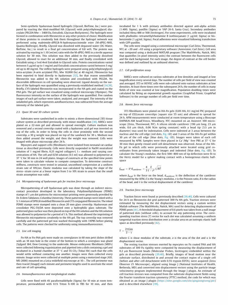
Fig. 1 shows human bone marrow-derived mesenchymal stemcells (hMSCs), rat cardiac myocytes, rat cardiac fibroblasts, humanumbilical vein endothelial cells (HUVECs), and NIH-3T3 fibroblastson soft gels with shear moduli between 200 and 300 Pa, formedby either crosslinked HA or polyacrylamide (PAA) and covalentlymodified with fibronectin (Fn). On PAA, an inert linearly elastichydrogel, cells attached through Fn-binding integrins, but they didnot spread or develop the large actin assemblies (Fig. 1b,e,h,k,n) asobserved for cells on rigid substrates (Fig. 1c,f,i,l,o). However,when HA rather than PAA forms the matrix, all five cell typesdevelop large adherent areas, actin bundles (Fig. 1a,d,g,j,m) andFAs (Fig. 1m inset) equivalent to those formed on rigid PAA orglass. Cells subcultured in serum-containing medium on micro-patterned Fn islands on HA gel surfaces adhere only to the Fnislands despite the availability of enough soluble Fn in the serumto saturate the gel by adsorption (Supplemental Fig. 1). ThereforeHA behaves similar to a non-adhesive inert substrate like PAA.Remarkably, cells on HA substrates were able to cluster integrinsto an extent similar to that observed on stiff 30 kPa PAA substrates(see arrows, Supplementary Fig. 1). These results correlate wellwith the observed focal adhesion size on HA, suggesting thatsignals mediated through HA receptors can enhance integrinclustering, which has largely been shown to be an adhesion andcytoskeleton force driven response [43].
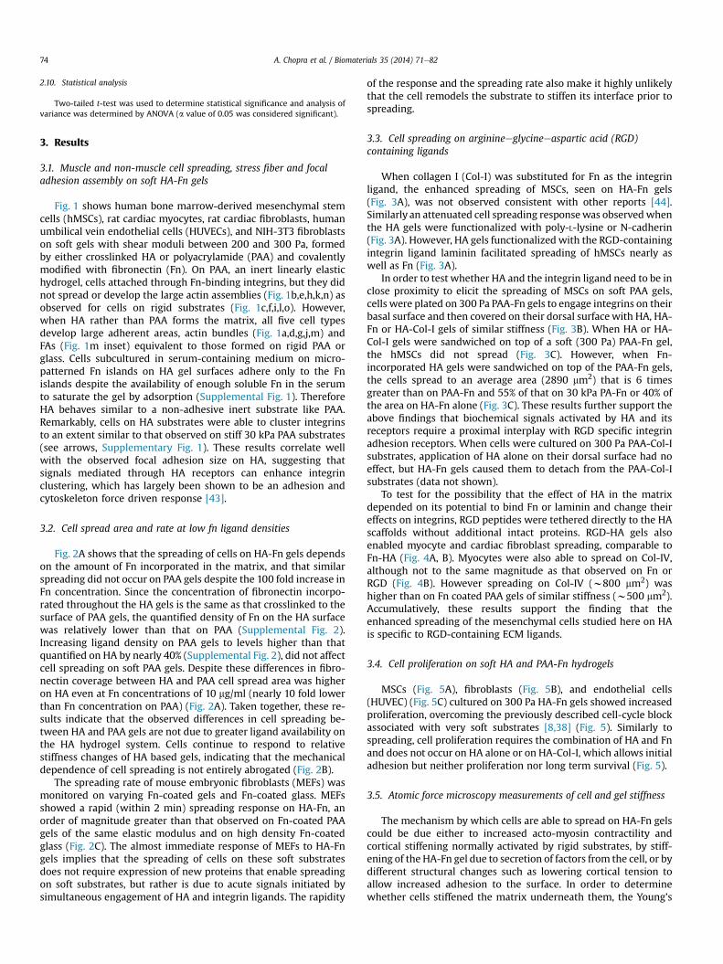
3.2. Cell spread area and rate at low fn ligand densities
Fig. 2A shows that the spreading of cells on HA-Fn gels dependson the amount of Fn incorporated in the matrix, and that similarspreading did not occur on PAA gels despite the 100 fold increase inFn concentration. Since the concentration of fibronectin incorpo-rated throughout the HA gels is the same as that crosslinked to thesurface of PAA gels, the quantified density of Fn on the HA surfacewas relatively lower than that on PAA (Supplemental Fig. 2).Increasing ligand density on PAA gels to levels higher than thatquantified on HA by nearly 40% (Supplemental Fig. 2), did not affectcell spreading on soft PAA gels. Despite these differences in fibro-nectin coverage between HA and PAA cell spread area was higheron HA even at Fn concentrations of 10 mg/ml (nearly 10 fold lowerthan Fn concentration on PAA) (Fig. 2A). Taken together, these re-sults indicate that the observed differences in cell spreading be-tween HA and PAA gels are not due to greater ligand availability onthe HA hydrogel system. Cells continue to respond to relativestiffness changes of HA based gels, indicating that the mechanicaldependence of cell spreading is not entirely abrogated (Fig. 2B).
The spreading rate of mouse embryonic fibroblasts (MEFs) wasmonitored on varying Fn-coated gels and Fn-coated glass. MEFsshowed a rapid (within 2 min) spreading response on HA-Fn, anorder of magnitude greater than that observed on Fn-coated PAAgels of the same elastic modulus and on high density Fn-coatedglass (Fig. 2C). The almost immediate response of MEFs to HA-Fngels implies that the spreading of cells on these soft substratesdoes not require expression of new proteins that enable spreadingon soft substrates, but rather is due to acute signals initiated bysimultaneous engagement of HA and integrin ligands. The rapidity
of the response and the spreading rate also make it highly unlikelythat the cell remodels the substrate to stiffen its interface prior tospreading.
3.3. Cell spreading on arginineeglycineeaspartic acid (RGD)containing ligands
When collagen I (Col-I) was substituted for Fn as the integrinligand, the enhanced spreading of MSCs, seen on HA-Fn gels(Fig. 3A), was not observed consistent with other reports [44].Similarly an attenuated cell spreading responsewas observedwhenthe HA gels were functionalized with poly-L-lysine or N-cadherin(Fig. 3A). However, HA gels functionalized with the RGD-containingintegrin ligand laminin facilitated spreading of hMSCs nearly aswell as Fn (Fig. 3A).
In order to test whether HA and the integrin ligand need to be inclose proximity to elicit the spreading of MSCs on soft PAA gels,cells were plated on 300 Pa PAA-Fn gels to engage integrins on theirbasal surface and then covered on their dorsal surface with HA, HA-Fn or HA-Col-I gels of similar stiffness (Fig. 3B). When HA or HA-Col-I gels were sandwiched on top of a soft (300 Pa) PAA-Fn gel,the hMSCs did not spread (Fig. 3C). However, when Fn-incorporated HA gels were sandwiched on top of the PAA-Fn gels,the cells spread to an average area (2890 mm2) that is 6 timesgreater than on PAA-Fn and 55% of that on 30 kPa PA-Fn or 40% ofthe area on HA-Fn alone (Fig. 3C). These results further support theabove findings that biochemical signals activated by HA and itsreceptors require a proximal interplay with RGD specific integrinadhesion receptors. When cells were cultured on 300 Pa PAA-Col-Isubstrates, application of HA alone on their dorsal surface had noeffect, but HA-Fn gels caused them to detach from the PAA-Col-Isubstrates (data not shown).
To test for the possibility that the effect of HA in the matrixdepended on its potential to bind Fn or laminin and change theireffects on integrins, RGD peptides were tethered directly to the HAscaffolds without additional intact proteins. RGD-HA gels alsoenabled myocyte and cardiac fibroblast spreading, comparable toFn-HA (Fig. 4A, B). Myocytes were also able to spread on Col-IV,although not to the same magnitude as that observed on Fn orRGD (Fig. 4B). However spreading on Col-IV (w800 mm2) washigher than on Fn coated PAA gels of similar stiffness (w500 mm2).Accumulatively, these results support the finding that theenhanced spreading of the mesenchymal cells studied here on HAis specific to RGD-containing ECM ligands.
3.4. Cell proliferation on soft HA and PAA-Fn hydrogels
MSCs (Fig. 5A), fibroblasts (Fig. 5B), and endothelial cells(HUVEC) (Fig. 5C) cultured on 300 Pa HA-Fn gels showed increasedproliferation, overcoming the previously described cell-cycle blockassociated with very soft substrates [8,38] (Fig. 5). Similarly tospreading, cell proliferation requires the combination of HA and Fnand does not occur on HA alone or on HA-Col-I, which allows initialadhesion but neither proliferation nor long term survival (Fig. 5).
3.5. Atomic force microscopy measurements of cell and gel stiffness
The mechanism by which cells are able to spread on HA-Fn gelscould be due either to increased acto-myosin contractility andcortical stiffening normally activated by rigid substrates, by stiff-ening of the HA-Fn gel due to secretion of factors from the cell, or bydifferent structural changes such as lowering cortical tension toallow increased adhesion to the surface. In order to determinewhether cells stiffened the matrix underneath them, the Young’s
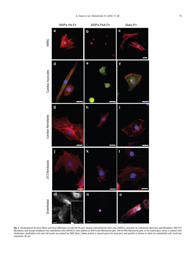
Fig. 1. Development of stress fibers and focal adhesions on soft HA-Fn gels. Human mesenchymal stem cells (hMSCs), neonatal rat ventricular myocytes and fibroblasts, NIH 3T3fibroblasts, and human umbilical vein endothelial cells (HUVECS) were plated on 300 Pa HA-fibronectin gels, 300 Pa PAA-fibronectin gels, or Fn-coated glass. Actin is stained withrhodamineephalloidin (red) and cell nuclei are stained by DAPI (blue). Alpha actinin is stained green for myocytes, and paxillin is shown in white for endothelial cells. Scale barrepresents 20 mm.
A. Chopra et al. / Biomaterials 35 (2014) 71e82 75
Fig. 2. Enhanced cell spreading response on HA-fibronectin hydrogels. (A) Myocyte spread area on HA and PAA gels with identical shear modulus coated with varying amounts of Fn(n > 100 cells). (B) MSC spread area on varying stiffnesses of polyacrylamide or HA gels coated with saturating amounts of Fn (n > 100 cells). (C) Quantification of early fibroblast cellspread area over time on varying substrates (n > 3). Error bars �1 S.E.
A. Chopra et al. / Biomaterials 35 (2014) 71e8276
modulus of the same spot of an HA-Fn gel was measured by atomicforce microscopy before and after 24 h of cell adhesion/spreading.
Storage moduli of hydrogels determined by AFM were consis-tent with our previous measurement by macroscopic rheological,methods [12], showing shear modulus values below G0w500 Pa
Fig. 3. Cell spreading response as a function of ligand type. (A) Mesenchymal stem cell (MSamounts of Fn, Collagen I, Poly-L-lysine (PLL), Laminin and N-cadherin. (B) Schematic represbottomwith different HA-ligand compositions at the top. (C) MSC spread areas for cells sandn ¼ 50 cells.
(Fig. 6A). Only a slight but not statistically significant increase inshear modulus of HA-Fn gels was observed when compared to HAalone or HA-collagen I gels (Fig. 6A). The elastic modulus of 3T3fibroblasts spread on HA-Fn, as determined by AFM, was less thanhalf than that of cells spread to the same extent on Fn-coated glass
C) spread area on different stiffness polyacrylamide or HA gels coated with saturatingentation of the sandwich construct, showing cells adhered to 300 Pa PAA-Fn gels at thewiched between 300 Pa PAA-Fn and HA-alone/Fn/Col-I. *p < 0.05, error bars �1 SEM for
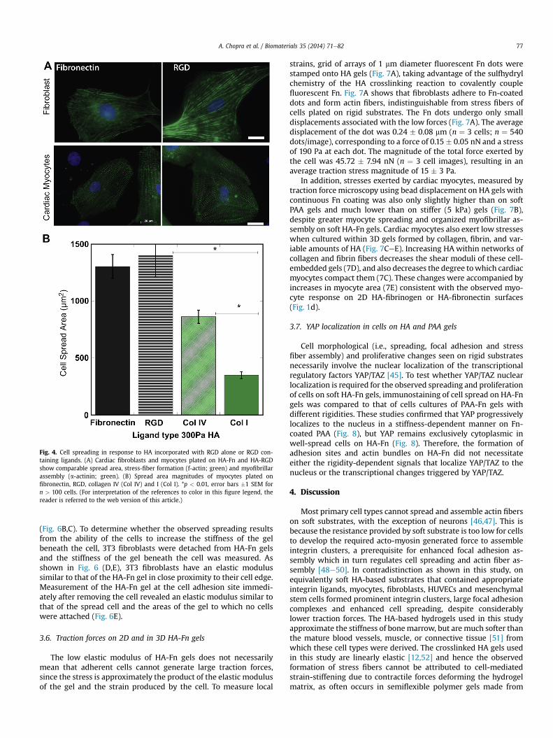
Fig. 4. Cell spreading in response to HA incorporated with RGD alone or RGD con-taining ligands. (A) Cardiac fibroblasts and myocytes plated on HA-Fn and HA-RGDshow comparable spread area, stress-fiber formation (f-actin; green) and myofibrillarassembly (a-actinin; green). (B) Spread area magnitudes of myocytes plated onfibronectin, RGD, collagen IV (Col IV) and I (Col I). *p < 0.01, error bars �1 SEM forn > 100 cells. (For interpretation of the references to color in this figure legend, thereader is referred to the web version of this article.)
A. Chopra et al. / Biomaterials 35 (2014) 71e82 77
(Fig. 6B,C). To determine whether the observed spreading resultsfrom the ability of the cells to increase the stiffness of the gelbeneath the cell, 3T3 fibroblasts were detached from HA-Fn gelsand the stiffness of the gel beneath the cell was measured. Asshown in Fig. 6 (D,E), 3T3 fibroblasts have an elastic modulussimilar to that of the HA-Fn gel in close proximity to their cell edge.Measurement of the HA-Fn gel at the cell adhesion site immedi-ately after removing the cell revealed an elastic modulus similar tothat of the spread cell and the areas of the gel to which no cellswere attached (Fig. 6E).
3.6. Traction forces on 2D and in 3D HA-Fn gels
The low elastic modulus of HA-Fn gels does not necessarilymean that adherent cells cannot generate large traction forces,since the stress is approximately the product of the elastic modulusof the gel and the strain produced by the cell. To measure local
strains, grid of arrays of 1 mm diameter fluorescent Fn dots werestamped onto HA gels (Fig. 7A), taking advantage of the sulfhydrylchemistry of the HA crosslinking reaction to covalently couplefluorescent Fn. Fig. 7A shows that fibroblasts adhere to Fn-coateddots and form actin fibers, indistinguishable from stress fibers ofcells plated on rigid substrates. The Fn dots undergo only smalldisplacements associated with the low forces (Fig. 7A). The averagedisplacement of the dot was 0.24 � 0.08 mm (n ¼ 3 cells; n ¼ 540dots/image), corresponding to a force of 0.15 � 0.05 nN and a stressof 190 Pa at each dot. The magnitude of the total force exerted bythe cell was 45.72 � 7.94 nN (n ¼ 3 cell images), resulting in anaverage traction stress magnitude of 15 � 3 Pa.
In addition, stresses exerted by cardiac myocytes, measured bytraction force microscopy using bead displacement on HA gels withcontinuous Fn coating was also only slightly higher than on softPAA gels and much lower than on stiffer (5 kPa) gels (Fig. 7B),despite greater myocyte spreading and organized myofibrillar as-sembly on soft HA-Fn gels. Cardiac myocytes also exert low stresseswhen cultured within 3D gels formed by collagen, fibrin, and var-iable amounts of HA (Fig. 7CeE). Increasing HA within networks ofcollagen and fibrin fibers decreases the shear moduli of these cell-embedded gels (7D), and also decreases the degree towhich cardiacmyocytes compact them (7C). These changes were accompanied byincreases in myocyte area (7E) consistent with the observed myo-cyte response on 2D HA-fibrinogen or HA-fibronectin surfaces(Fig. 1d).
3.7. YAP localization in cells on HA and PAA gels
Cell morphological (i.e., spreading, focal adhesion and stressfiber assembly) and proliferative changes seen on rigid substratesnecessarily involve the nuclear localization of the transcriptionalregulatory factors YAP/TAZ [45]. To test whether YAP/TAZ nuclearlocalization is required for the observed spreading and proliferationof cells on soft HA-Fn gels, immunostaining of cell spread on HA-Fngels was compared to that of cells cultures of PAA-Fn gels withdifferent rigidities. These studies confirmed that YAP progressivelylocalizes to the nucleus in a stiffness-dependent manner on Fn-coated PAA (Fig. 8), but YAP remains exclusively cytoplasmic inwell-spread cells on HA-Fn (Fig. 8). Therefore, the formation ofadhesion sites and actin bundles on HA-Fn did not necessitateeither the rigidity-dependent signals that localize YAP/TAZ to thenucleus or the transcriptional changes triggered by YAP/TAZ.
4. Discussion
Most primary cell types cannot spread and assemble actin fiberson soft substrates, with the exception of neurons [46,47]. This isbecause the resistance provided by soft substrate is too low for cellsto develop the required acto-myosin generated force to assembleintegrin clusters, a prerequisite for enhanced focal adhesion as-sembly which in turn regulates cell spreading and actin fiber as-sembly [48e50]. In contradistinction as shown in this study, onequivalently soft HA-based substrates that contained appropriateintegrin ligands, myocytes, fibroblasts, HUVECs and mesenchymalstem cells formed prominent integrin clusters, large focal adhesioncomplexes and enhanced cell spreading, despite considerablylower traction forces. The HA-based hydrogels used in this studyapproximate the stiffness of bonemarrow, but are much softer thanthe mature blood vessels, muscle, or connective tissue [51] fromwhich these cell types were derived. The crosslinked HA gels usedin this study are linearly elastic [12,52] and hence the observedformation of stress fibers cannot be attributed to cell-mediatedstrain-stiffening due to contractile forces deforming the hydrogelmatrix, as often occurs in semiflexible polymer gels made from
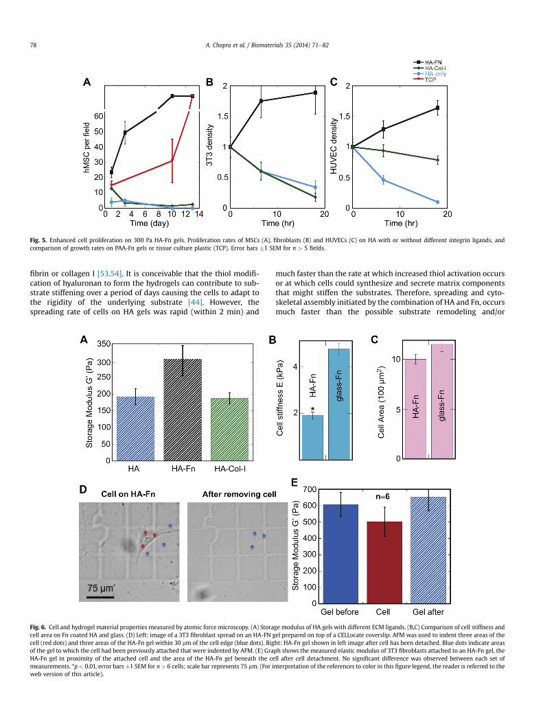
Fig. 5. Enhanced cell proliferation on 300 Pa HA-Fn gels. Proliferation rates of MSCs (A), fibroblasts (B) and HUVECs (C) on HA with or without different integrin ligands, andcomparison of growth rates on PAA-Fn gels or tissue culture plastic (TCP). Error bars �1 SEM for n > 5 fields.
A. Chopra et al. / Biomaterials 35 (2014) 71e8278
fibrin or collagen I [53,54]. It is conceivable that the thiol modifi-cation of hyaluronan to form the hydrogels can contribute to sub-strate stiffening over a period of days causing the cells to adapt tothe rigidity of the underlying substrate [44]. However, thespreading rate of cells on HA gels was rapid (within 2 min) and
Fig. 6. Cell and hydrogel material properties measured by atomic force microscopy. (A) Storacell area on Fn coated HA and glass. (D) Left: image of a 3T3 fibroblast spread on an HA-FN gcell (red dots) and three areas of the HA-Fn gel within 30 mm of the cell edge (blue dots). Rigof the gel to which the cell had been previously attached that were indented by AFM. (E) GraHA-Fn gel in proximity of the attached cell and the area of the HA-Fn gel beneath the cemeasurements. *p < 0.01, error bars �1 SEM for n > 6 cells; scale bar represents 75 mm. (For iweb version of this article).
much faster than the rate at which increased thiol activation occursor at which cells could synthesize and secrete matrix componentsthat might stiffen the substrates. Therefore, spreading and cyto-skeletal assembly initiated by the combination of HA and Fn, occursmuch faster than the possible substrate remodeling and/or
ge modulus of HA gels with different ECM ligands. (B,C) Comparison of cell stiffness andel prepared on top of a CELLocate coverslip. AFM was used to indent three areas of theht: HA-Fn gel shown in left image after cell has been detached. Blue dots indicate areasph shows the measured elastic modulus of 3T3 fibroblasts attached to an HA-Fn gel, thell after cell detachment. No significant difference was observed between each set ofnterpretation of the references to color in this figure legend, the reader is referred to the
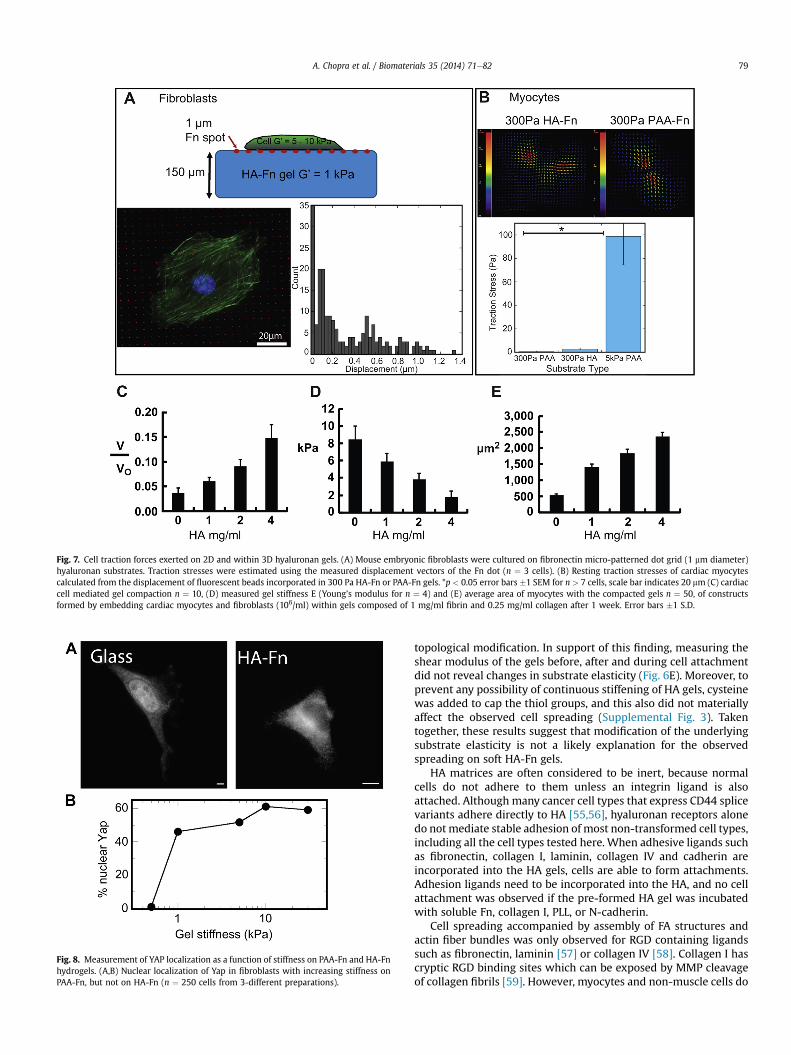
Fig. 7. Cell traction forces exerted on 2D and within 3D hyaluronan gels. (A) Mouse embryonic fibroblasts were cultured on fibronectin micro-patterned dot grid (1 mm diameter)hyaluronan substrates. Traction stresses were estimated using the measured displacement vectors of the Fn dot (n ¼ 3 cells). (B) Resting traction stresses of cardiac myocytescalculated from the displacement of fluorescent beads incorporated in 300 Pa HA-Fn or PAA-Fn gels. *p < 0.05 error bars �1 SEM for n > 7 cells, scale bar indicates 20 mm (C) cardiaccell mediated gel compaction n ¼ 10, (D) measured gel stiffness E (Young’s modulus for n ¼ 4) and (E) average area of myocytes with the compacted gels n ¼ 50, of constructsformed by embedding cardiac myocytes and fibroblasts (106/ml) within gels composed of 1 mg/ml fibrin and 0.25 mg/ml collagen after 1 week. Error bars �1 S.D.
Fig. 8. Measurement of YAP localization as a function of stiffness on PAA-Fn and HA-Fnhydrogels. (A,B) Nuclear localization of Yap in fibroblasts with increasing stiffness onPAA-Fn, but not on HA-Fn (n ¼ 250 cells from 3-different preparations).
A. Chopra et al. / Biomaterials 35 (2014) 71e82 79
topological modification. In support of this finding, measuring theshear modulus of the gels before, after and during cell attachmentdid not reveal changes in substrate elasticity (Fig. 6E). Moreover, toprevent any possibility of continuous stiffening of HA gels, cysteinewas added to cap the thiol groups, and this also did not materiallyaffect the observed cell spreading (Supplemental Fig. 3). Takentogether, these results suggest that modification of the underlyingsubstrate elasticity is not a likely explanation for the observedspreading on soft HA-Fn gels.
HA matrices are often considered to be inert, because normalcells do not adhere to them unless an integrin ligand is alsoattached. Although many cancer cell types that express CD44 splicevariants adhere directly to HA [55,56], hyaluronan receptors alonedo notmediate stable adhesion of most non-transformed cell types,including all the cell types tested here. When adhesive ligands suchas fibronectin, collagen I, laminin, collagen IV and cadherin areincorporated into the HA gels, cells are able to form attachments.Adhesion ligands need to be incorporated into the HA, and no cellattachment was observed if the pre-formed HA gel was incubatedwith soluble Fn, collagen I, PLL, or N-cadherin.
Cell spreading accompanied by assembly of FA structures andactin fiber bundles was only observed for RGD containing ligandssuch as fibronectin, laminin [57] or collagen IV [58]. Collagen I hascryptic RGD binding sites which can be exposed by MMP cleavageof collagen fibrils [59]. However, myocytes and non-muscle cells do
A. Chopra et al. / Biomaterials 35 (2014) 71e8280
not spread on collagen I incorporated HA gels, implying that thesecryptic RGD binding sites are not exposed when incorporated in HAgels. RGD peptide alone was sufficient for cardiac myocytes andfibroblasts to spread and assemble myofibrils or actin fibers. Thesefindings argue against the possibility that cell spreading on HAhydrogels are a result of changes in fibronectin conformation thatalter adhesion to integrins [60].
MSCs fibroblasts and endothelial cells (HUVEC) (Fig. 5) prolif-erated after being plated on 300 Pa HA-Fn, a stiffness at which theyare quiescent when cultured on PAA-Fn gels [38]. Remarkably, theproliferation was specific to fibronectin-containing HA hydrogels,whereas collagen I or HA alone were not capable to elicit a similarresponse. These results further support the findings that cellspreading and FA assembly are specific to RGD containing ligands incombination with HA.
Although the crosslinking chemistry for all incorporated ECMligands in HA hydrogels was similar, cell spreading, proliferationwas not universal and was specific to RGD alone or RGD containingligands. Importantly, incorporation of different ECM ligands did notsignificantly affect the mechanical properties of the substrate(Fig. 6A), ruling out the possibility of crosslinking chemistry being apossible explanation for the observed spreading on the soft HA gels.This result serves as added evidence (Fig. 6E) that the ability of cellsto spread on HA-Fn gels cannot be explained by increases in stiff-ness of the gel beneath the cell.
Numerous studies have established that the physical propertiesof cells such as contractility and cortical cell stiffness increase withstress fiber and FA assembly in spread cells as a function of sub-strate rigidity [14,24,61e64]. This study shows that despite similarspread areas of cells on HA-Fn gels and rigid substrates, the internalcell stiffness of cells on HA is nearly 50% lower than that measuredon glass. Traction stresses exerted by fibroblasts and muscle cellswere found to be low despite large spread areas consistent with alower degree of internal tension. The computed stress values of15 Pa are similar to that reported for poorly spread, muscle andnon-muscle cells on relatively soft (1 kPa) PAA gels [14,65]. Cardiacmyocytes and supporting cells also exerted low stresses and dis-played higher spreading when cultured within 3D gels formed bycollagen, fibrin, and variable amounts of high molecular weight HA,consistent with the spreading observed on the sandwich 3D likeconstructs. The observed increase in cell spreading and stress fiberassembly on soft HA gels are not accompanied by high internal cellstiffness.
Recent studies have shown that the effectors of the hippopathway, YAP/TAZ, are important transcriptional regulatorsinvolved in rigidity sensing and mechanotransduction [66]. Thelocalization of YAP/TAZ in the nucleus has been reported to bedirectly involved in the cell spreading [45] and proliferation [67e69] response. The results from this study show that, similar to theoriginal finding with epithelial cells [45], increasing the substraterigidity on PAA-Fn gels causes YAP to localize in the nucleus in fi-broblasts. Despite the enhanced cell proliferation and cell size onsoft HA-Fn hydrogels, YAP did not localize in the nucleus, implyingthat the signaling pathways mediating rigidity sensing andspreading on HA-Fn substrates are not the same.
The findings from this study suggest the likely involvement ofHA receptors alongwith RGD specific integrin adhesions, mediatingthe augmented cell phenotypic response as observed on soft HAhydrogels. The best characterized receptors of HA namely CD44 andCD168 or receptor for hyaluronan mediated motility (RHAMM)have been implicated in cellular processes such as motility associ-ated with dynamic cytoskeletal changes [70]. RHAMM has theability to bind and phosphorylate non-receptor tyrosine kinase Src[71], focal adhesion kinase [72] and erk kinase [73]. These signalingproteins are also engaged in the mechanosensory processes of cells
when plated on substrates of varying rigidity [15]. Accordingly, itcan be speculated that RHAMM activation by HA can lead to thephosphorylation of non-receptor tyrosine kinases, in a force-independent manner, capable of eliciting a cellular responsesimilar to what is observed on stiff substrates. In addition, CD44 candirectly interact with important cytoskeleton regulators like RhoA,a member of the Rho family of GTPases which control cellcontractility and in turn actin filament organization [74]. Thespecificity of cell proliferation and spreading to RGD ligands on softHA gels suggest that there is an interplay between HA receptors andRGD specific integrins such as a5b1 and avb3. In support of thisproposed mechanism, recent studies have suggested that there is adirect interplay between RGD specific integrins and RHAMM [75].Future studies will therefore be necessary to examine the interplaybetween these receptors and their downstream biochemical signalsthat may be intimately involved in overriding the rigidity sensingresponse of cells residing in a hyaluronan-enriched extracellularmilieu.
5. Conclusion
The magnitude of forces and stiffnesses that elicit specificcellular responses and the molecular mechanisms by which cellstransmit forces or transduce them into chemical or electrical sig-nals are incompletely known, and are likely to depend on simul-taneous chemical stimulation and other microenvironment inputs.The fact that HA production is tightly regulated during develop-ment and injury and frequently up-regulated in cancers charac-terized by uncontrolled growth and cell movement suggests thatthe interaction of signaling between HA receptors and specificintegrins might be an important element in mechanical control ofdevelopment and homeostasis. The results from this study showthat the mechanosensitivity of a cell depends on the biochemicalcomposition of its microenvironment even when the ligands thatalter mechanosensitivity, such as HA, are not the primary adhesionreceptors. These studies offer a new model for understanding thebiochemical and mechanical cooperative signaling emanating fromthe extracellular matrix. The integration of HA with integrin-specific ECM signaling proteins provides a rationale for engineer-ing a new class of soft hybrid hydrogels that can be used in thera-peutic/tissue engineering strategies.
Appendix A. Supplementary data
Supplementary data related to this article can be found at http://dx.doi.org/10.1016/j.biomaterials.2013.09.066.
References
[1] Levental I, Levental KR, Klein EA, Assoian R, Miller RT, Wells RG, et al. A simpleindentation device for measuring micrometer-scale tissue stiffness. J PhysCondens Matter 2010;22:194120.
[2] Wells RG, Discher DE. Matrix elasticity, cytoskeletal tension, and TGF-beta: theinsoluble and soluble meet. Sci Signal 2008;1:pe13.
[3] Kothapalli D, Liu SL, Bae YH, Monslow J, Xu T, Hawthorne EA, et al. Cardio-vascular protection by ApoE and ApoE-HDL linked to suppression of ECM geneexpression and arterial stiffening. Cell Rep 2012;2:1259e71.
[4] Georges PC, Hui JJ, Gombos Z, McCormick ME, Wang AY, Uemura M, et al.Increased stiffness of the rat liver precedes matrix deposition: implications forfibrosis. Am J Physiol Gastrointest Liver Physiol 2007;293:G1147e54.
[5] Perepelyuk M, Terajima M, Wang AY, Georges PC, Janmey PA, Yamauchi M,et al. Hepatic stellate cells and portal fibroblasts are the major cellular sourcesof collagens and lysyl oxidases in normal liver and early after injury. Am JPhysiol Gastrointest Liver Physiol 2013;304:G605e14.
[6] Olsen AL, Bloomer SA, Chan EP, Gaca MD, Georges PC, Sackey B, et al. Hepaticstellate cells require a stiff environment for myofibroblastic differentiation.Am J Physiol Gastrointest Liver Physiol 2011;301:G110e8.
[7] Li Z, Dranoff JA, Chan EP, Uemura M, Sevigny J, Wells RG. Transforming growthfactor-beta and substrate stiffness regulate portal fibroblast activation inculture. Hepatology 2007;46:1246e56.
A. Chopra et al. / Biomaterials 35 (2014) 71e82 81
[8] Klein EA, Yin L, Kothapalli D, Castagnino P, Byfield FJ, Xu T, et al. Cell-cyclecontrol by physiological matrix elasticity and in vivo tissue stiffening. CurrBiol 2009;19:1511e8.
[9] Schrader J, Gordon-WalkerTT,AucottRL, vanDeemterM,QuaasA,WalshS, et al.Matrix stiffness modulates proliferation, chemotherapeutic response, anddormancy in hepatocellular carcinoma cells. Hepatology 2011;53:1192e205.
[10] Pelham Jr RJ, Wang Y. Cell locomotion and focal adhesions are regulated bysubstrate flexibility. Proc Natl Acad Sci U S A 1997;94:13661e5.
[11] Byfield FJ, Wen Q, Levental I, Nordstrom K, Arratia PE, Miller RT, et al. Absenceof filamin A prevents cells from responding to stiffness gradients on gelscoated with collagen but not fibronectin. Biophys J 2009;96:5095e102.
[12] Chopra A, Lin V, McCollough A, Atzet S, Prestwich GD, Wechsler AS, et al.Reprogramming cardiomyocyte mechanosensing by crosstalk betweenintegrins and hyaluronic acid receptors. J Biomech 2012;45:824e31.
[13] Ganz A, Lambert M, Saez A, Silberzan P, Buguin A, Mege RM, et al. Tractionforces exerted through N-cadherin contacts. Biol Cell 2006;98:721e30.
[14] Chopra A, Tabdanov E, Patel H, Janmey PA, Kresh JY. Cardiac myocyteremodeling mediated by N-cadherin-dependent mechanosensing. Am JPhysiol Heart Circ Physiol 2011;300:H1252e66.
[15] Discher DE, Janmey P, Wang YL. Tissue cells feel and respond to the stiffness oftheir substrate. Science 2005;310:1139e43.
[16] Giannone G, Sheetz MP. Substrate rigidity and force define form throughtyrosine phosphatase and kinase pathways. Trends Cell Biol 2006;16:213e23.
[17] Bischofs IB, Safran SA, Schwarz US. Elastic interactions of active cells with softmaterials. Phys Rev E Stat Nonlin Soft Matter Phys 2004;69:021911.
[18] Wipff PJ, Rifkin DB, Meister JJ, Hinz B. Myofibroblast contraction activateslatent TGF-beta1 from the extracellular matrix. J Cell Biol 2007;179:1311e23.
[19] Ju YE, Janmey PA, McCormick ME, Sawyer ES, Flanagan LA. Enhanced neuritegrowth from mammalian neurons in three-dimensional salmon fibrin gels.Biomaterials 2007;28:2097e108.
[20] Georges PC, Miller WJ, Meaney DF, Sawyer ES, Janmey PA. Matrices withcompliance comparable to that of brain tissue select neuronal over glialgrowth in mixed cortical cultures. Biophys J 2006;90:3012e8.
[21] Kostic A, Sap J, Sheetz MP. RPTPalpha is required for rigidity-dependent in-hibition of extension and differentiation of hippocampal neurons. J Cell Sci2007;120:3895e904.
[22] Zhang S, Sun A, Liang Y, Chen Q, Zhang C, Wang K, et al. A role of myocardialstiffness in cell-based cardiac repair: a hypothesis. J Cell Mol Med 2009;13:660e3.
[23] Berry MF, Engler AJ, Woo YJ, Pirolli TJ, Bish LT, Jayasankar V, et al. Mesen-chymal stem cell injection after myocardial infarction improves myocardialcompliance. Am J Physiol Heart Circ Physiol 2006;290:H2196e203.
[24] Engler AJ, Carag-Krieger C, Johnson CP, Raab M, Tang HY, Speicher DW, et al.Embryonic cardiomyocytes beat best on a matrix with heart-like elasticity:scar-like rigidity inhibits beating. J Cell Sci 2008;121:3794e802.
[25] Kresh JY, Chopra A. Intercellular and extracellular mechanotransduction incardiac myocytes. Pflugers Arch 2011;462:75e87.
[26] Zamir EA, Taber LA. Material properties and residual stress in the stage 12chick heart during cardiac looping. J Biomech Eng 2004;126:823e30.
[27] Yao J, Varner VD, Brilli LL, Young JM, Taber LA, Perucchio R. Viscoelastic ma-terial properties of the myocardium and cardiac jelly in the looping chickheart. J Biomech Eng 2012;134:024502.
[28] Lesley J, Hyman R, Kincade PW, Frank JD. CD44 and its interaction withextracellular matrix. Adv Immunol 1993:271e335.
[29] Turley EA, Austen L, Vandeligt K, Clary C. Hyaluronan and a cell-associatedhyaluronan binding protein regulate the locomotion of ras-transformedcells. J Cell Biol 1991;112:1041e7.
[30] Borowsky ML, Hynes RO. Layilin, a novel talin-binding transmembrane pro-tein homologous with C-type lectins, is localized in membrane ruffles. J CellBiol 1998;143:429e42.
[31] McCourt PA, Ek B, Forsberg N, Gustafson S. Intercellular adhesion molecule-1is a cell surface receptor for hyaluronan. J Biol Chem 1994;269:30081e4.
[32] Isemura M, Yosizawa Z, Koide T, Ono T. Interaction of fibronectin and itsproteolytic fragments with hyaluronic acid. J Biochem 1982;91:731e4.
[33] McDevitt CA, Marcelino J, Tucker L. Interaction of intact type VI collagen withhyaluronan. FEBS Lett 1991;294:167e70.
[34] Heldin P, Pertoft H. Synthesis and assembly of the hyaluronan-containingcoats around normal human mesothelial cells. Exp Cell Res 1993;208:422e9.
[35] Rooney P, Kumar S. Inverse relationship between hyaluronan and collagens indevelopment and angiogenesis. Differentiation 1993;54:1e9.
[36] Longaker MT, Chiu ES, Adzick NS, Stern M, Harrison MR, Stern R. Studies infetal wound healing. V. A prolonged presence of hyaluronic acid characterizesfetal wound fluid. Ann Surg 1991;213:292e6.
[37] Burdick JA, Prestwich GD. Hyaluronic acid hydrogels for biomedical applica-tions. Adv Mater 2011;23:H41e56.
[38] Winer JP, Janmey PA, McCormick ME, Funaki M. Bone marrow-derived humanmesenchymal stem cells become quiescent on soft substrates but remainresponsive to chemical or mechanical stimuli. Tissue Eng Part A 2009;15:147e54.
[39] Tseng Q, Duchemin-Pelletier E, Deshiere A, Balland M, Guillou H, Filhol O,et al. Spatial organization of the extracellular matrix regulates cell-cell junc-tion positioning. Proc Natl Acad Sci U S A 2012;109:1506e11.
[40] Tseng Q, Wang I, Duchemin-Pelletier E, Azioune A, Carpi N, Gao J, et al. A newmicropatterning method of soft substrates reveals that different tumorigenicsignals can promote or reduce cell contraction levels. Lab Chip 2011;11:2231e40.
[41] Yang MT, Fu J, Wang YK, Desai RA, Chen CS. Assaying stem cell mechanobi-ology on microfabricated elastomeric substrates with geometrically modu-lated rigidity. Nat Protoc 2011;6:187e213.
[42] Polio SR, Rothenberg KE, Stamenovic D, Smith ML. A micropatterning andimage processing approach to simplify measurement of cellular tractionforces. Acta Biomater 2012;8:82e8.
[43] Friedland JC, Lee MH, Boettiger D. Mechanically activated integrin switchcontrols alpha5beta1 function. Science 2009;323:642e4.
[44] Rehfeldt F, Brown AE, Raab M, Cai S, Zajac AL, Zemel A, et al. Hyaluronicacid matrices show matrix stiffness in 2D and 3D dictates cytoskeletalorder and myosin-II phosphorylation within stem cells. Integr Biol (Camb)2012;4:422e30.
[45] Dupont S, Morsut L, Aragona M, Enzo E, Giulitti S, Cordenonsi M, et al. Role ofYAP/TAZ in mechanotransduction. Nature 2011;474:179e83.
[46] Yeung T, Georges PC, Flanagan LA, Marg B, Ortiz M, Funaki M, et al. Effects ofsubstrate stiffness on cell morphology, cytoskeletal structure, and adhesion.Cell Motil Cytoskelet 2005;60:24e34.
[47] Georges PC, Janmey PA. Cell type-specific response to growth on soft mate-rials. J Appl Physiol 2005;98:1547e53.
[48] Rossier OM, Gauthier N, Biais N, Vonnegut W, Fardin MA, Avigan P, et al. Forcegenerated by actomyosin contraction builds bridges between adhesive con-tacts. EMBO J 2010;29:1055e68.
[49] Galbraith CG, Yamada KM, Sheetz MP. The relationship between force andfocal complex development. J Cell Biol 2002;159:695e705.
[50] Yu CH, Law JBK, Suryana M, Low HY, Sheetz MP. Early integrin binding to Arg-Gly-Asp peptide activates actin polymerization and contractile movementthat stimulates outward translocation. Proc Natl Acad Sci U S A 2011;108:20585e90.
[51] Levental I, Georges PC, Janmey PA. Soft biological materials and their impacton cell function. Soft Matter 2007;1:299e306.
[52] Vanderhooft JL, Alcoutlabi M, Magda JJ, Prestwich GD. Rheological propertiesof cross-linked hyaluronan-gelatin hydrogels for tissue engineering. Macro-mol Biosci 2009;9:20e8.
[53] Storm C, Pastore JJ, MacKintosh FC, Lubensky TC, Janmey PA. Nonlinear elas-ticity in biological gels. Nature 2005;435:191e4.
[54] Winer JP, Oake S, Janmey PA. Non-linear elasticity of extracellular matricesenables contractile cells to communicate local position and orientation. PLoSOne 2009;4:e6382.
[55] Welsh CF, Zhu D, Bourguignon LY. Interaction of CD44 variant isoforms withhyaluronic acid and the cytoskeleton in human prostate cancer cells. J CellPhysiol 1995;164:605e12.
[56] Ananthanarayanan B, Kim Y, Kumar S. Elucidating the mechanobiology ofmalignant brain tumors using a brain matrix-mimetic hyaluronic acidhydrogel platform. Biomaterials 32:7913e23.
[57] Tashiro K, Sephel GC, Greatorex D, Sasaki M, Shirashi N, Martin GR, et al. TheRGD containing site of the mouse laminin A chain is active for cell attachment,spreading, migration and neurite outgrowth. J Cell Physiol 1991;146:451e9.
[58] Pedchenko V, Zent R, Hudson BG. alpha(v)beta(3) and alpha(v)beta(5) integ-rins bind both the proximal RGD site and non-RGD motifs within non-collagenous (NC1) domain of the alpha 3 chain of type IV collagen eimplication for the mechanism of endothelial cell adhesion. J Biol Chem2004;279:2772e80.
[59] Davis GE. Affinity of integrins for damaged extracellular-matrix e alpha-V-beta-3 binds to denatured collagen type-I through Rgd sites. Biochem Bio-phys Res Commun 1992;182:1025e31.
[60] Keselowsky BG, Collard DM, Garcia AJ. Surface chemistry modulates fibro-nectin conformation and directs integrin binding and specificity to control celladhesion. J Biomed Mater Res A 2003;66:247e59.
[61] Solon J, Levental I, Sengupta K, Georges PC, Janmey PA. Fibroblast adaptationand stiffness matching to soft elastic substrates. Biophys J 2007;93:4453e61.
[62] Tee SY, Fu J, Chen CS, Janmey PA. Cell shape and substrate rigidity bothregulate cell stiffness. Biophys J 2011;100:L25e7.
[63] Kasza KE, Nakamura F, Hu S, Kollmannsberger P, Bonakdar N, Fabry B, et al.Filamin A is essential for active cell stiffening but not passive stiffening underexternal force. Biophys J 2009;96:4326e35.
[64] Trichet L, Le Digabel J, Hawkins RJ, Vedula SR, Gupta M, Ribrault C, et al. Ev-idence of a large-scale mechanosensing mechanism for cellular adaptation tosubstrate stiffness. Proc Natl Acad Sci U S A 2012;109:6933e8.
[65] Califano JP, Reinhart-King CA. Substrate stiffness and cell area predict cellulartraction stresses in single cells and cells in contact. Cell Mol Bioeng 2010;3:68e75.
[66] Halder G, Dupont S, Piccolo S. Transduction of mechanical and cytoskeletalcues by YAP and TAZ. Nat Rev Mol Cell Biol 2012;13:591e600.
[67] Xin M, Kim Y, Sutherland LB, Murakami M, Qi X, McAnally J, et al. Hippopathway effector Yap promotes cardiac regeneration. Proc Natl Acad Sci U S A2013;110:13839e44.
[68] Mendez MG, Janmey PA. Transcription factor regulation by mechanical stress.Int J Biochem Cell Biol 2012;44:728e32.
[69] Zhao B, Tumaneng K, Guan KL. The Hippo pathway in organ size control, tissueregeneration and stem cell self-renewal. Nat Cell Biol 2011;13:877e83.
[70] Turley EA, Noble PW, Bourguignon LY. Signaling properties of hyaluronanreceptors. J Biol Chem 2002;277:4589e92.
[71] Hall CL, Lange LA, Prober DA, Zhang S, Turley EA. pp60(c-src) is required forcell locomotion regulated by the hyaluronan receptor RHAMM. Oncogene1996;13:2213e24.

A. Chopra et al. / Biomaterials 35 (2014) 71e8282
[72] Hall CL, Wang C, Lange LA, Turley EA. Hyaluronan and the hyaluronan re-ceptor RHAMM promote focal adhesion turnover and transient tyrosine ki-nase activity. J Cell Biol 1994;126:575e88.
[73] Hall CL, Yang B, Yang X, Zhang S, Turley M, Samuel S, et al. Overexpression ofthe hyaluronan receptor RHAMM is transforming and is also required for H-ras transformation. Cell 1995;82:19e26.
[74] Nobes CD, Hall A. Rho, rac, and cdc42 GTPases regulate the assembly ofmultimolecular focal complexes associated with actin stress fibers, lamelli-podia, and filopodia. Cell 1995;81:53e62.
[75] Gares SL, Pilarski LM. Balancing thymocyte adhesion and motility: a functionallinkage between beta1 integrins and the motility receptor RHAMM. DevImmunol 2000;7:209e25.