Embed Size (px)
Citation preview
1 http://www.tutis.ca/NeuroMD/index.htm 20 February 2013
Auditory Physiology
2 http://www.tutis.ca/NeuroMD/index.htm 20 February 2013
Contents
Auditory Physiology ........................................................................................................... 1
Introduction ..................................................................................................................... 3
What is sound? ................................................................................................................ 3
The three parts of the ear ................................................................................................ 3
The function of the middle ear ........................................................................................ 4
Impedance matching: ...................................................................................................... 4
Gating:............................................................................................................................. 4
Conduction deafness ....................................................................................................... 5
Sensorineural hearing loss .............................................................................................. 5
What is the function of the round window? .................................................................... 6
How is fluid motion transduced into neural firing? ........................................................ 6
Different parts of the basilar membrane are sensitive to different frequencies. ............. 7
How does the basilar membrane code the frequency of a sound? .................................. 8
Four major causes of hearing loss ................................................................................... 8
What are the cues to sound direction? ............................................................................ 9
The role of the superior olive in sound localization ..................................................... 10
The columnar structure in primary auditory cortex ................................................... 10
Where are the higher auditory language regions? ......................................................... 11
Language development ................................................................................................. 11
Wernicke-Geschwind model ......................................................................................... 12
Compare the language disorders (aphasias) caused by lesions in Broca's and Wernicke's areas. ...................................................................................................................... 12
Practice problems .......................................................................................................... 13
see also .......................................................................................................................... 14
3 http://www.tutis.ca/NeuroMD/index.htm 20 February 2013
Introduction Our sense of hearing is often under appreciated. If you had to choose, which would you
give up, hearing or vision? People who lose hearing feel very isolated from the world, and most importantly, the company of people. Imagine what life would be like if at dinner you were unable to participate in conversation with your friends and family. . Children who are hard of hearing are often misdiagnosed as cognitively impaired. The sound of approaching cars can save your life. A ringing in your ears, tinnitus, can drive you crazy.
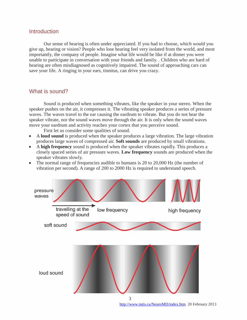
What is sound? Sound is produced when something vibrates, like the speaker in your stereo. When the
speaker pushes on the air, it compresses it. The vibrating speaker produces a series of pressure waves. The waves travel to the ear causing the eardrum to vibrate. But you do not hear the speaker vibrate, nor the sound waves move through the air. It is only when the sound waves move your eardrum and activity reaches your cortex that you perceive sound.
First let us consider some qualities of sound. A loud sound is produced when the speaker produces a large vibration. The large vibration
produces large waves of compressed air. Soft sounds are produced by small vibrations. A high frequency sound is produced when the speaker vibrates rapidly. This produces a
closely spaced series of air pressure waves. Low frequency sounds are produced when the speaker vibrates slowly.
The normal range of frequencies audible to humans is 20 to 20,000 Hz (the number of vibration per second). A range of 200 to 2000 Hz is required to understand speech.
4 http://www.tutis.ca/NeuroMD/index.htm 20 February 2013
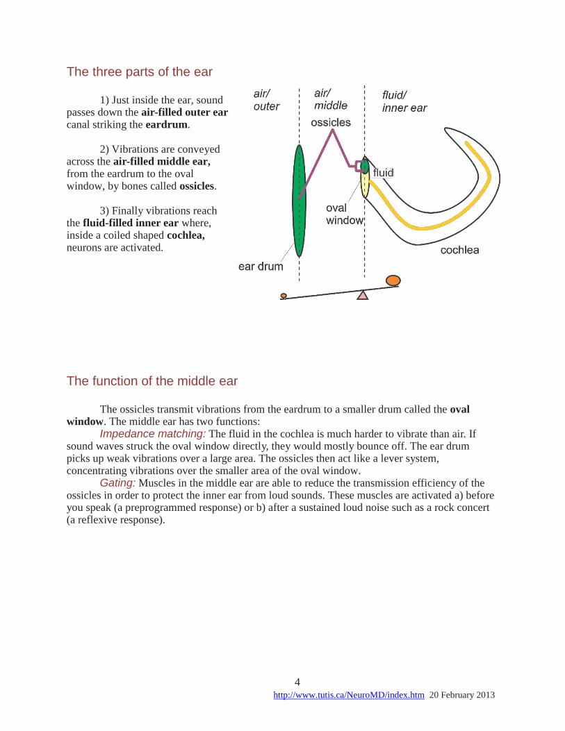
The three parts of the ear 1) Just inside the ear, sound
passes down the air-filled outer ear canal striking the eardrum.
2) Vibrations are conveyed
across the air-filled middle ear, from the eardrum to the oval window, by bones called ossicles.
3) Finally vibrations reach
the fluid-filled inner ear where, inside a coiled shaped cochlea, neurons are activated.
The function of the middle ear The ossicles transmit vibrations from the eardrum to a smaller drum called the oval
window. The middle ear has two functions: Impedance matching: The fluid in the cochlea is much harder to vibrate than air. If
sound waves struck the oval window directly, they would mostly bounce off. The ear drum picks up weak vibrations over a large area. The ossicles then act like a lever system, concentrating vibrations over the smaller area of the oval window.
Gating: Muscles in the middle ear are able to reduce the transmission efficiency of the ossicles in order to protect the inner ear from loud sounds. These muscles are activated a) before you speak (a preprogrammed response) or b) after a sustained loud noise such as a rock concert (a reflexive response).
5 http://www.tutis.ca/NeuroMD/index.htm 20 February 2013
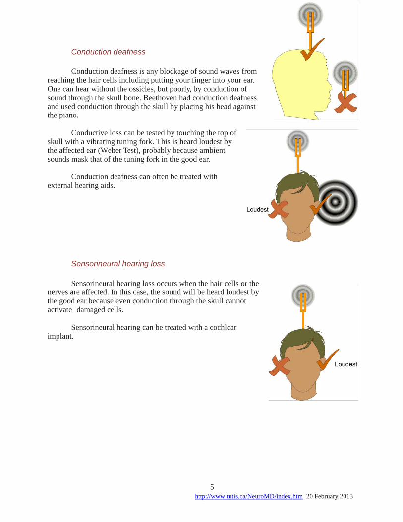
Conduction deafness
Conduction deafness is any blockage of sound waves from
reaching the hair cells including putting your finger into your ear. One can hear without the ossicles, but poorly, by conduction of sound through the skull bone. Beethoven had conduction deafness and used conduction through the skull by placing his head against the piano.
Conductive loss can be tested by touching the top of
skull with a vibrating tuning fork. This is heard loudest by the affected ear (Weber Test), probably because ambient sounds mask that of the tuning fork in the good ear.
Conduction deafness can often be treated with
external hearing aids.
Sensorineural hearing loss
Sensorineural hearing loss occurs when the hair cells or the
nerves are affected. In this case, the sound will be heard loudest by the good ear because even conduction through the skull cannot activate damaged cells.
Sensorineural hearing can be treated with a cochlear
implant.
6 http://www.tutis.ca/NeuroMD/index.htm 20 February 2013
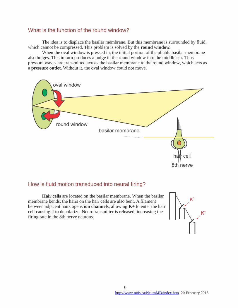
What is the function of the round window? The idea is to displace the basilar membrane. But this membrane is surrounded by fluid,
which cannot be compressed. This problem is solved by the round window. When the oval window is pressed in, the initial portion of the pliable basilar membrane
also bulges. This in turn produces a bulge in the round window into the middle ear. Thus pressure waves are transmitted across the basilar membrane to the round window, which acts as a pressure outlet. Without it, the oval window could not move.
How is fluid motion transduced into neural firing? Hair cells are located on the basilar membrane. When the basilar
membrane bends, the hairs on the hair cells are also bent. A filament between adjacent hairs opens ion channels, allowing K+ to enter the hair cell causing it to depolarize. Neurotransmitter is released, increasing the firing rate in the 8th nerve neurons.
7 http://www.tutis.ca/NeuroMD/index.htm 20 February 2013
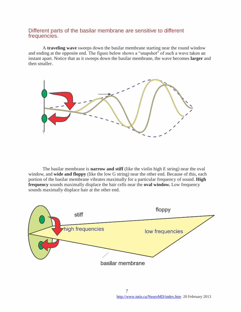
Different parts of the basilar membrane are sensitive to different frequencies.
A traveling wave sweeps down the basilar membrane starting near the round window
and ending at the opposite end. The figure below shows a “snapshot" of such a wave taken an instant apart. Notice that as it sweeps down the basilar membrane, the wave becomes larger and then smaller.
\ The basilar membrane is narrow and stiff (like the violin high E string) near the oval
window, and wide and floppy (like the low G string) near the other end. Because of this, each portion of the basilar membrane vibrates maximally for a particular frequency of sound. High frequency sounds maximally displace the hair cells near the oval window. Low frequency sounds maximally displace hair at the other end.
8 http://www.tutis.ca/NeuroMD/index.htm 20 February 2013
How does the basilar membrane code the frequency of a sound?
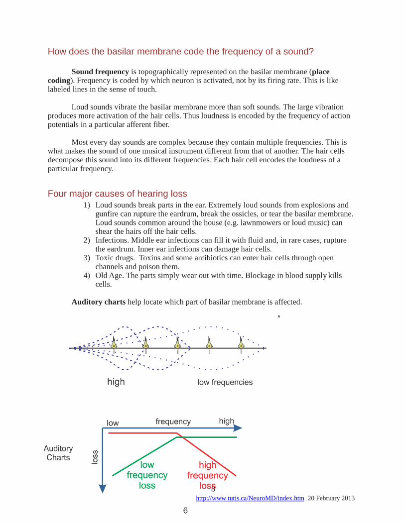
Sound frequency is topographically represented on the basilar membrane (place
coding). Frequency is coded by which neuron is activated, not by its firing rate. This is like labeled lines in the sense of touch.
Loud sounds vibrate the basilar membrane more than soft sounds. The large vibration
produces more activation of the hair cells. Thus loudness is encoded by the frequency of action potentials in a particular afferent fiber.
Most every day sounds are complex because they contain multiple frequencies. This is
what makes the sound of one musical instrument different from that of another. The hair cells decompose this sound into its different frequencies. Each hair cell encodes the loudness of a particular frequency.
Four major causes of hearing loss 1) Loud sounds break parts in the ear. Extremely loud sounds from explosions and
gunfire can rupture the eardrum, break the ossicles, or tear the basilar membrane. Loud sounds common around the house (e.g. lawnmowers or loud music) can shear the hairs off the hair cells.
2) Infections. Middle ear infections can fill it with fluid and, in rare cases, rupture the eardrum. Inner ear infections can damage hair cells.
3) Toxic drugs. Toxins and some antibiotics can enter hair cells through open channels and poison them.
4) Old Age. The parts simply wear out with time. Blockage in blood supply kills cells.
Auditory charts help locate which part of basilar membrane is affected.
9 http://www.tutis.ca/NeuroMD/index.htm 20 February 2013
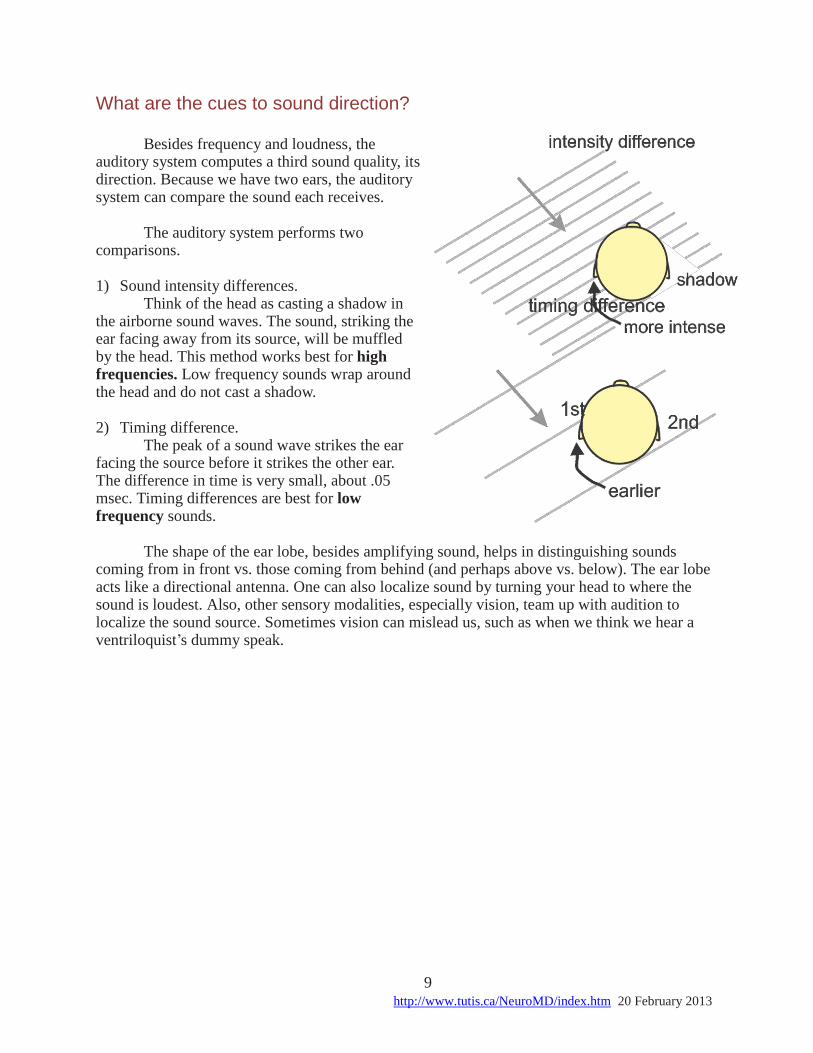
What are the cues to sound direction? Besides frequency and loudness, the
auditory system computes a third sound quality, its direction. Because we have two ears, the auditory system can compare the sound each receives.
The auditory system performs two
comparisons.
1) Sound intensity differences. Think of the head as casting a shadow in
the airborne sound waves. The sound, striking the ear facing away from its source, will be muffled by the head. This method works best for high frequencies. Low frequency sounds wrap around the head and do not cast a shadow.
2) Timing difference.
The peak of a sound wave strikes the ear facing the source before it strikes the other ear. The difference in time is very small, about .05 msec. Timing differences are best for low frequency sounds.
The shape of the ear lobe, besides amplifying sound, helps in distinguishing sounds
coming from in front vs. those coming from behind (and perhaps above vs. below). The ear lobe acts like a directional antenna. One can also localize sound by turning your head to where the sound is loudest. Also, other sensory modalities, especially vision, team up with audition to localize the sound source. Sometimes vision can mislead us, such as when we think we hear a ventriloquist’s dummy speak.
10 http://www.tutis.ca/NeuroMD/index.htm 20 February 2013
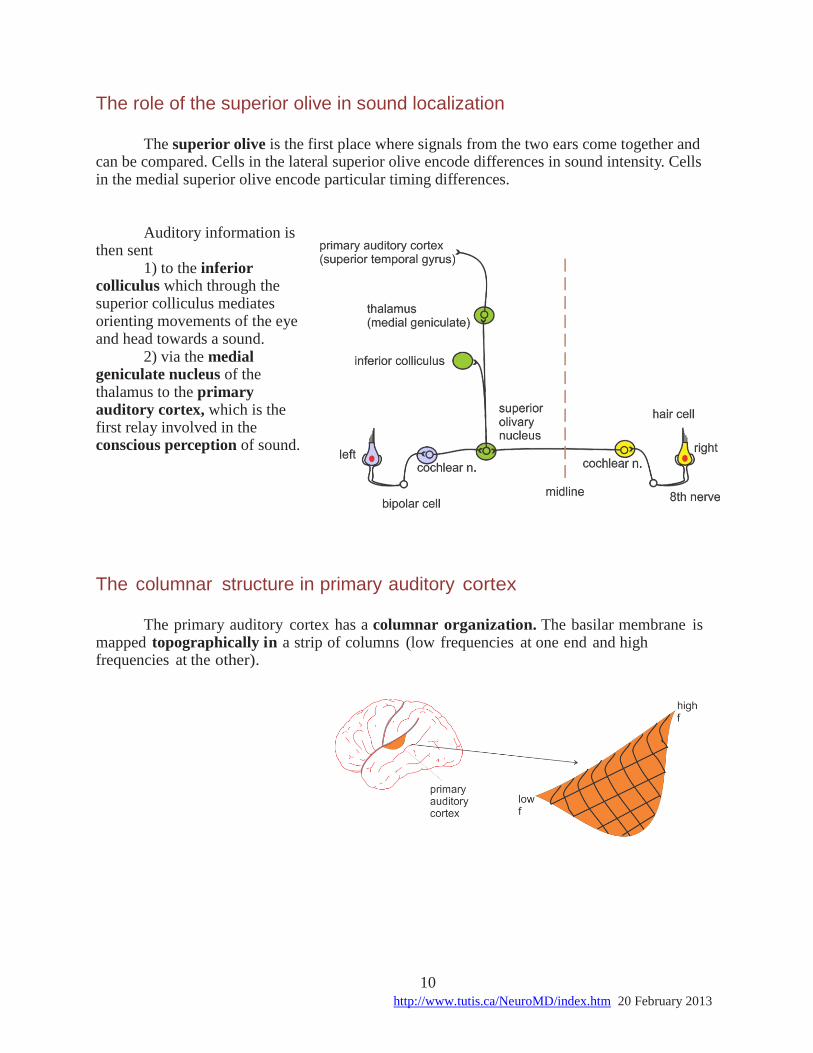
The role of the superior olive in sound localization The superior olive is the first place where signals from the two ears come together and
can be compared. Cells in the lateral superior olive encode differences in sound intensity. Cells in the medial superior olive encode particular timing differences.
Auditory information is
then sent 1) to the inferior
colliculus which through the superior colliculus mediates orienting movements of the eye and head towards a sound.
2) via the medial geniculate nucleus of the thalamus to the primary auditory cortex, which is the first relay involved in the conscious perception of sound.
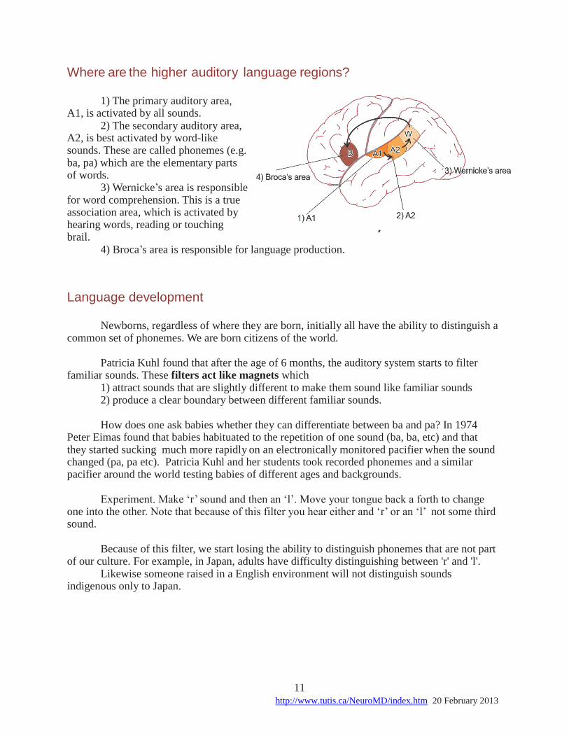
The columnar structure in primary auditory cortex The primary auditory cortex has a columnar organization. The basilar membrane is
mapped topographically in a strip of columns (low frequencies at one end and high frequencies at the other).
11 http://www.tutis.ca/NeuroMD/index.htm 20 February 2013
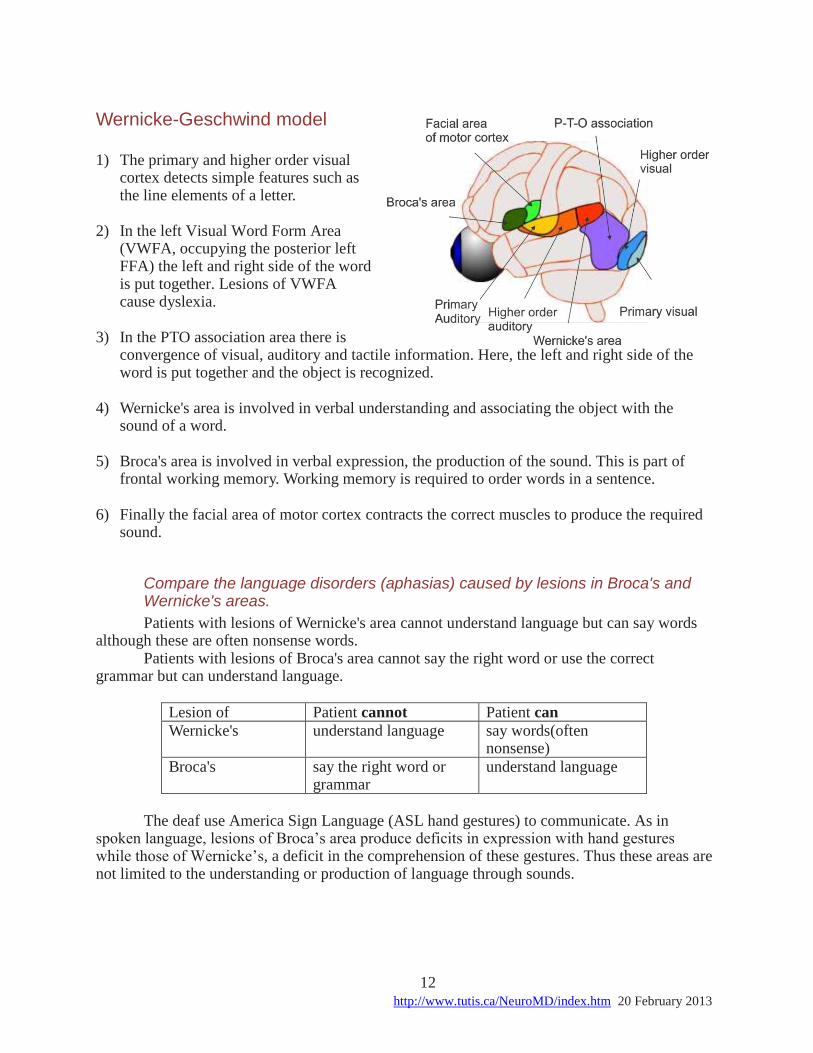
Where are the higher auditory language regions?
1) The primary auditory area,
A1, is activated by all sounds. 2) The secondary auditory area,
A2, is best activated by word-like sounds. These are called phonemes (e.g. ba, pa) which are the elementary parts of words.
3) Wernicke’s area is responsible for word comprehension. This is a true association area, which is activated by hearing words, reading or touching brail.
4) Broca’s area is responsible for language production.
Language development Newborns, regardless of where they are born, initially all have the ability to distinguish a
common set of phonemes. We are born citizens of the world. Patricia Kuhl found that after the age of 6 months, the auditory system starts to filter
familiar sounds. These filters act like magnets which 1) attract sounds that are slightly different to make them sound like familiar sounds 2) produce a clear boundary between different familiar sounds. How does one ask babies whether they can differentiate between ba and pa? In 1974
Peter Eimas found that babies habituated to the repetition of one sound (ba, ba, etc) and that they started sucking much more rapidly on an electronically monitored pacifier when the sound changed (pa, pa etc). Patricia Kuhl and her students took recorded phonemes and a similar pacifier around the world testing babies of different ages and backgrounds.
Experiment. Make ‘r’ sound and then an ‘l’. Move your tongue back a forth to change
one into the other. Note that because of this filter you hear either and ‘r’ or an ‘l’ not some third sound.
Because of this filter, we start losing the ability to distinguish phonemes that are not part
of our culture. For example, in Japan, adults have difficulty distinguishing between 'r' and 'l'. Likewise someone raised in a English environment will not distinguish sounds
indigenous only to Japan.
12 http://www.tutis.ca/NeuroMD/index.htm 20 February 2013
Wernicke-Geschwind model
1) The primary and higher order visual cortex detects simple features such as the line elements of a letter.
2) In the left Visual Word Form Area
(VWFA, occupying the posterior left FFA) the left and right side of the word is put together. Lesions of VWFA cause dyslexia.
3) In the PTO association area there is
convergence of visual, auditory and tactile information. Here, the left and right side of the word is put together and the object is recognized.
4) Wernicke's area is involved in verbal understanding and associating the object with the
sound of a word.
5) Broca's area is involved in verbal expression, the production of the sound. This is part of frontal working memory. Working memory is required to order words in a sentence.
6) Finally the facial area of motor cortex contracts the correct muscles to produce the required
sound.
Compare the language disorders (aphasias) caused by lesions in Broca's and Wernicke's areas.
Patients with lesions of Wernicke's area cannot understand language but can say words although these are often nonsense words.
Patients with lesions of Broca's area cannot say the right word or use the correct grammar but can understand language.
Lesion of Patient cannot Patient can
Wernicke's understand language say words(often nonsense)
Broca's say the right word or grammar
understand language
The deaf use America Sign Language (ASL hand gestures) to communicate. As in
spoken language, lesions of Broca’s area produce deficits in expression with hand gestures while those of Wernicke’s, a deficit in the comprehension of these gestures. Thus these areas are not limited to the understanding or production of language through sounds.
13 http://www.tutis.ca/NeuroMD/index.htm 20 February 2013
Practice problems
1. Hearing is impaired if fluid builds up in the middle ear. Hearing is most impaired
because
a) most of the sound energy bounces off the ear drum and is lost.
b) the middle ear muscle can no longer contract effectively.
c) the hair cells can no longer be bent.
d) the fluid impairs the movement of the basilar membrane.
e) the ionic concentration around the hair cells is changed.
2. The direction of a sound source is best determined by
a) the inter-aural intensity differences of low frequency sounds.
b) the inter-aural timing differences of high frequency sounds.
c) a comparison of inter-aural differences by the cochlear nucleus.
d) a comparison of inter-aural differences by the superior olivary nucleus.
e) a comparison of inter-aural differences by the superior colliculus.
14 http://www.tutis.ca/NeuroMD/index.htm 20 February 2013
Answers
1. a)
2. d)
see also: http://www.tutis.ca/NeuroML7Aud/AudProb.swf